HELENA CHRISTELL
COSTS AND BENEFITS OF
CONE BEAM COMPUTED
TOMOGRAPHY
For maxillary canines with eruption disturbance
HELEN A C HRIS TELL MALMÖ UNIVERSIT C OS T S AND BENEFIT S OF C ONE BEAM C OMPUTED T OMOGR APHY DOCT OR AL DISSERT A TION IN ODONT OL OG Y
C O S T S A N D B E N E F I T S O F C O N E B E A M C O M P U T E D T O M O G R A P H Y
Malmö University, Faculty of Odontology
Doctoral Dissertations 2017
© Helena Christell, 2017 Illustrations: Helena Christell
The image on the front page illustrates costs being outweighed by the benefits to the patient, created with inspiration from “Takeover” by Keith Haring.
ISBN 978-91-7104-724-3 (print) ISBN 978-91-7104-725-0 (pdf) Holmbergs, Malmö 2017
HELENA CHRISTELL
COSTS AND BENEFITS OF
CONE BEAM COMPUTED
TOMOGRAPHY
For maxillary canines with eruption disturbance
Malmö University, 2017
Department of Oral and Maxillofacial Radiology
Faculty of Odontology
Malmö, Sweden
This publication is also available in electronic format at: http://dspace.mah.se/handle/2043/22147
CONTENTS
LIST OF ARTICLES ... 9
ABBREVIATIONS AND GLOSSARY... 11
ABSTRACT ... 13
Clinical implications and importance ...15
POPULÄRVETENSKAPLIG SAMMANFATTNING ... 16 Klinisk betydelse ...17 BACKGROUND ... 18 GENERAL AIM ... 28 SPECIFIC AIMS ... 29 METHODS ... 30
Systematic review (Study I)...30
Cost-analysis (Study II, III and IV) ...32
Web-based survey (Study IV) ...38
RESULTS ... 44 Systematic review ...44 Cost-analysis ...46 Web-based survey ...50 DISCUSSION ... 53 Main results ...53
Systematic literature review ...54
Cost-analysis ...55
Web-based survey ...58
Outcome of imaging method in orthodontics ...60
Societal cost ...64
FUTURE RESEARCH ... 70
CONCLUSIONS ... 72
REFERENCES ... 73
ACKNOWLEDGEMENTS ... 82
LIST OF ARTICLES
This thesis is based on the following articles, referred to in the text by their Roman numerals. All articles are reprinted with permission from the copyright holders and appended to the end of the thesis. I. Economic evaluation of diagnostic methods used in dentistry. A systematic review. Christell H, Birch S, Horner K, Lindh C, Rohlin M, The SEDENTEXCT consortium. Journal of Dentistry. 2014;42: 1361-71.
II. A framework for costing diagnostic methods in oral health care: An application comparing a new imaging technology with the conventional approach for maxillary canines with eruption disturbance. Christell H, Birch S, Horner K, Rohlin M, Lindh C, The SEDENTEXCT consortium. Community Dentistry and Oral Epidemiology. 2012;40:351-61.
III. Variations in costs of cone beam CT examinations among health care systems. Christell H, Birch S, Hedesiu M, Horner K, Ivanauskaité D, Nackaerts O, Rohlin M, Lindh C, The SEDENTEXCT consortium. Dentomaxillofacial Radiology. 2012;41:571-7.
IV. The impact of Cone Beam CT on financial costs and orthodontists’ treatment decisions in the management of maxillary canines with eruption disturbance. Christell H, Birch S, Bondemark L, Horner K, Lindh C, The SEDENTEXCT consortium.
Submitted for publication in European Journal of Orthodontics January 7, 2017.
ABBREVIATIONS AND GLOSSARY
CA Cost Analysis
CBA Cost Benefit Analysis
CEA Cost Effectiveneness Analysis
CUA Cost Utility Analysis
CT Computed Tomography
GDP Gross Domestic Product
HTA Health Technology Assessment
OHRQoL Oral Health Related Quality of Life
QALY Quality Adjusted Life Time
WTP Willingness To Pay
RCT Randomised Controlled Trial
ALARA (As Low As Reasonably Achievable): This phrase, deriving
from the radiation protection principle of optimisation, means that (individual) doses should be as low as reasonably achievable, taking into account economic or social factors.
CBCT (Cone beam Computed Tomography): An X-ray imaging
technique in which a volumetric image dataset is obtained by acquisition of multiple projectional sets of X-ray attenuation data by rotation around the object of interest of a divergent X-ray beam (conical or pyramidal), with subsequent computed reconstruction.
Maxillary canines with eruption disturbance: Clinical conditions
where the maxillary canine has any eruption disturbance, also termed impaction, retention, ectopic eruption or displacement of maxillary canines.
Patients’ preferences: The extent to which individuals appreciate
different phenomena. The more something is appreciated by an individual, the higher the utility-level when the person receives the product or service. Patients’ preferences represent the choices patients would make faced with alternative options. In order to compare between options that involve different costs (and hence different investments of scarce resources) the strength of preferences, not just choice, is required.
ABSTRACT
With a continuous influx of new imaging technologies, the question must be raised whether the use of new methods provides benefits for the patients justifying any additional costs incurred. There is a growing awareness of the need for economic evaluations in dentistry and further understanding in this field is needed. The use of Cone Beam Computed Tomography (CBCT) in dentistry has emerged during the last decades and it is important to investigate if it fulfils its purpose of forming a basis for treatment planning in a cost-effective way.
Evidence for the costs and benefits of CBCT is still scarce.
In Study I, a systematic review of the literature regarding economic
evaluations of diagnostic methods in dentistry was conducted according to international guidelines. Four databases were searched and relevant publications were retrieved and selected according to predetermined criteria for inclusion and exclusion. The methodological quality of the studies was assessed combining a protocol for diagnostic studies and a checklist for economic evaluations. Key findings: Of the 160 economic evaluations in dentistry, 12 concerned diagnostic methods. In general, the perspective of the study was not stated and the methods for costing varied. A need for improved quality of future economic evaluations was identified.
In Study II, a framework for analysing costs of diagnostic methods in
dentistry was constructed. The framework was tested for costing of examination using intraoral and panoramic radiography compared with CBCT and panoramic radiography regarding patients with maxillary canines with eruption disturbance Key findings: The
framework demonstrated a feasibility of analysing relevant costs for capital, consumables, labour and patient-related costs. Examination with CBCT and panoramic radiography was associated with a
significantly higher cost of 128.38€ compared with that of intraoral
and panoramic radiography 81.80€ resulting in an incremental cost
for CBCT and panoramic radiography of 46.58€.
In Study III the costs of CBCT-examinations were analysed comparing
four different clinical conditions in four different settings in Leuven -Belgium, Cluj -Romania, Malmö -Sweden and Vilnius -Lithuania.
Key findings: The estimates for clinic-related costs varied among the
health care systems, being highest in Malmö and lowest in Cluj. This variation was mainly due to different purchase costs for the CBCT
equipment (range 148 000–227 000€). The variation in examination
fees (range 0–102.02€) was the principle reason for the differences
in patient-related costs. Costing of a dental radiographic method cannot be generalised from one health care system to another, without considering their specific circumstances.
In Study IV, a web-based survey was constructed in order to measure
the proportion of orthodontists’ treatment decisions that were different when comparing CBCT and panoramic radiography with intraoral and panoramic radiography and to analyse the costs of producing different treatment plans, for patients with maxillary canines with eruption disturbance. The orthodontists were randomly assigned to denote treatment decisions for four patient cases presented with either intraoral and panoramic radiography or CBCT and panoramic radiography, respectively, at two occasions for the same patient case.
Key-findings: Twenty-four percent of the treatment decisions were
different when the orthodontists had access to CBCT and panoramic radiography instead of intraoral and panoramic radiography. The
total diagnostic cost per examination was 99.84€ using intraoral
and panoramic radiography and 134.37 using CBCT and panoramic radiography, resulting in an incremental cost per examination of
34.53€ for CBCT and panoramic radiography. This can be expressed
in terms of an incremental cost-effectiveness ratio (ICER) which is a measure of the average additional cost per treatment decision that is
Clinical implications and importance
This thesis will add to the knowledge in an area where technical development rather than patients’ benefits tends to dominate the discussion. Currently there are no clear selection criteria for examination with CBCT regarding patients with a suspected eruption disturbance of maxillary canines. This condition is mostly detected and treated in younger patients to whom radiation exposure poses a greater risk than it does to adults. This makes the justification of radiographic examination particularly important.
The systematic review in Study I highlighted a need for standardised methods of economic evaluations in dentistry and confirmed that there is a knowledge gap regarding economic evaluations, in particular concerning diagnostic methods. To the best of our knowledge, the cost-analyses in Study II and III are the first performed in this field. Applications of the framework will support the development of a standardised methodology for conducting economic evaluations in dentistry and thereby promote the generalisability of future studies. Study IV is one of the first to compare the costs of dental radiographic examinations with the possible benefits for the patients. For a majority of the patients, CBCT is not beneficial in terms of a different treatment decision. For patients with severe space deficiency in the maxilla, indicating a need for tooth extractions, CBCT can provide additional information which may prevent the extraction of healthy and undamaged teeth.
This thesis provides no evidence to support routine use of CBCT for patients with maxillary canines with eruption disturbance. A sub-group of patients was identified for whom CBCT examination may be beneficial. The results of costs and effects for different imaging methods provided in this thesis can inform the development of clinical practice guidelines and support decision makers regarding investment decisions and prioritisation.
POPULÄRVETENSKAPLIG
SAMMANFATTNING
I tandvården finns allt fler metoder för prevention, diagnostik och behandling. Eftersom resurserna är begränsade bör den metod väljas där nyttan uppväger kostnaden. Idag saknas dock vetenskapliga
belägg gällande kostnadseffektivitet för undersökning med Cone Beam Computed Tomography (CBCT).
En systematisk översikt i Studie I identifierade 160 hälsoekonomiska utvärderingar inom tandvård varav 12 gällde diagnostiska metoder. Översikten kunde inte identifiera någon kostnadsanalys på patientnivå för diagnostiska metoder. Därför konstruerades i Studie II ett ramverk för analys av kostnader förknippade med diagnostiska metoder i tandvården. Applicering av ramverket visade att CBCT
och panoramaröntgen hade en signifikant högre kostnad (128,38€)
jämfört med intraoral och panoramaröntgen (81,80€). I Studie III
jämfördes kostnader för CBCT undersökning av patienter med olika kliniska tillstånd på kliniker i Cluj -Rumänien, Leuven -Belgien, Malmö -Sverige och Vilnius -Litauen. Klinikrelaterade kostnader varierade mellan sjukvårdssystemen vilket främst berodde på olika höga inköpskostnader för CBCT-apparaterna medan skillnader i patientrelaterade kostnader främst berodde på variation i patientavgifter. I enkätundersökningen i Studie IV ombads specialister i tandreglering att ta behandlingsbeslut gällande patienter med eruptionsstörning av överkäkshörntanden. Majoriteten av behandlingsvalen var samma oavsett om specialisterna hade tillgång till röntgenbilder från CBCT och panoramaröntgenundersökning eller till bilder från intraoral- och panoramaröntgenundersökning. CBCT kan vara till nytta för en del av patienterna genom att i högre grad än intraoral röntgen och panoramaröntgen identifiera en resorptionsskada på granntänder till
hörntanden med eruptionsstörning, vilket kan göra att en skadad tand tas bort istället för en frisk tand.
Sammanfattningsvis kan sägas att det finns ett stort behov av hälso ekonomiska studier med god kvalitet gällande diagnostiska metoder inom tandvård. Undersökning med CBCT medför en högre kostnad än konventionella röntgenmetoder och för majoriteten av patienter med misstanke om eruptionsstörning av överkäkshörntänderna medför den ingen extra nytta avseende behandlingsbeslut.
Klinisk betydelse
Denna avhandling har ökat kunskapen inom ett område där den tekniska utvecklingen av diagnostiska metoder inom tandvård stundtals dominerar framför patientnyttan. För närvarande är det vetenskapliga underlaget bristfälligt för indikationer då det gäller CBCT undersökning av patienter där det finns misstanke om felaktig eruption av överkäkshörntänderna. Detta tillstånd upptäcks och behandlas oftast på unga patienter vilka är mer känsliga för röntgenstrålning än vuxna och det är därför särskilt viktigt att röntgenundersökningar av denna patientgrupp är berättigade.
Den systematiska översikten i Studie I belyste behovet av standardiserade metoder för hälsoekonomiska analyser inom tandvård, speciellt gällande diagnostiska metoder. Enligt vår kännedom är kostnadsanalysen i Studie II den första inom detta område. Det ramverk för kostnadsanalys som konstruerades i Studie II, kan bidra till utveckling av standardiserade metoder för hälsoekonomiska analyser inom tandvård. Studie III visar svårigheterna med att jämföra kostnader mellan olika länder om man inte beaktar specifika omständigheter i respektive land. Studie IV är en av de första studier som jämför kostnader för dentala röntgenmetoder med den associerade patientnyttan. Studien visar att CBCT undersökning i de flesta fall inte ändrar behandlingsbeslutet. För majoriteten av patienter där överkäkshörntänderna har en eruptionsstörning tillför CBCT undersökning sannolikt ingen extra nytta jämfört med undersökning med intraoral röntgen och panoramaröntgen. CBCT kan emellertid bidra med ökad information gällande identifiering av resorptionsskador på granntänder till överkäkshörntänderna vilket gör att man kan undvika borttagning av friska oskadade tänder.
Resultaten från denna avhandling visar att rutinmässigt användande av CBCT för patienter med eruptionsstörning av överkäkshörntänder inte kan tillrådas. De visar också att CBCT kan vara till nytta för en liten del av dessa patienter. Framtida studier bör formulera indikationer för CBCT undersökning som skiljer ut patienter för vilka denna undersökning kan medföra någon nytta. Resultaten kan bidra till utveckling av riktlinjer för klinisk praxis och ligga till grund för beslut om investering och prioritering inom tandvård.
BACKGROUND
General background
The rapid development of health technology in recent decades has provided exceptional possibilities for diagnosis and treatment, which has resulted in improvements in clinical outcomes for patients. The plethora of available methods in health care has consequently increased the demands on health care. Providers of health care now have to choose between an array of different health care methods for diagnosis, treatment and prognosis to ensure that resources are used in the best alternative manner. Thus, that the health benefits to the patients are maximised using available health care resources. There is a growing interest in the results of health economic evaluations among decision makers who are required to optimise resource allocation within health care as well as in dental care.
Dental Cone Beam Computed Tomography (CBCT) is an emerging technique for diagnostic imaging. It offers images in three planes with a high level of detail which makes it an attractive method for many dental applications. CBCT is currently requested for a number of dental conditions, one of which is to identify maxillary canines with a suspected eruption disturbance and any root resorption on the neighbouring teeth. CBCT-examination may however, be associated with a greater radiation dose as well as with higher costs compared with conventional dental imaging methods (1).
SedentexCT 2008-2011, was a project financed by the European Union (EU) aiming to improve the safety and efficacy of CBCT examinations (2). Within the project, methods for quality assurance and safety testing were developed, and studies on technical, diagnostic and therapeutic efficacy were performed by collaborators in Manchester
and Leeds (United Kingdom (UK)), Leuven (Belgium), Athens (Greece), Cluj (Romania), Vilnius (Lithuania) and Malmö (Sweden).
The aim of the collaborators in Malmö was to analyse the cost effectiveness of CBCT compared with conventional imaging methods and these studies formed the basis of this thesis.
Health technology assessment
Health technology assessment (HTA) originated in the United States (US) in the early 1970s as a consequence of an increased influx of new medical technology. Among providers of health care and decision makers this raised the question of how to choose the most beneficial method, also with regard to the cost. Costly medical technology like computed tomography (CT) developed throughout the 1970s and 1980s. This resulted in an increased share of the state budget being devoted to health care, which might have provided a powerful incentive for a broadened technology assessment (3). This development led to a discussion in the US and in Europe of the effectiveness of the interventions used and of the inequity of health care (4).
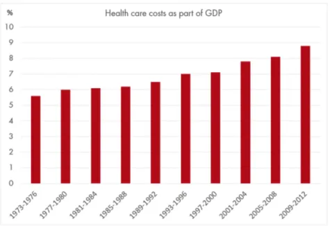
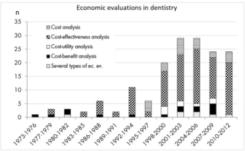
In the last decades, the cost of health care, including public and private expenditures, has continued to increase and now constitutes a growing share of the gross domestic product (GDP) of European member countries in the Organisation for Economic Co-operation and Development (OECD) (5) as seen in Figure 1. The resources spent on health care differs however fundamentally between countries reflecting inequalities in the access to health care due to varying political systems. Also a substantial part of the health budget is spent on dental care in the OECD countries (6).
Figure 1. The increase of health care costs in percentage of the Gross Domestic Product (GDP) in 27 OECD countries from 1973 to 2012. The figure was constructed based on data from OECD health statistics (5).
Economic evaluation is an essential part of HTA forming a framework for assessment of evidence for clinical effects and the associated resource use. Economic evaluation always implies a comparison of alternative interventions often with a new intervention being compared with the one currently used. This evaluation can be performed using different perspectives i.e. that of the patient, the provider, or society. The perspective is important to consider when planning an economic evaluation, as an intervention can look appealing from one perspective but less appealing from another perspective (7). Furthermore, the perspective determines which effects and costs that are relevant to include.
There are four types of economic evaluations whereof effectiveness analysis (CEA), utility analysis (CUA) and cost-benefit analysis (CBA) are considered to be full economic evaluations as they assess both costs and effects of an intervention. The difference between these analyses is mainly the way the effects are measured and expressed. The fourth type of economic evaluation is cost-analysis
(CA) which analyses the additional cost of the different interventions being compared and does not include analysis of effects.
CA is useful when comparing interventions where the effects are considered equal. Drummond et al. (2005)(7) recommend that the analysis of costs be performed in three subsequent steps:
• Identification of relevant costs i.e. costs that are assumed to affect the outcome of the analysis. Costs can be defined as “resources denied other uses” (7).
• Measuring of the number of resources consumed and cost per unit. This step in the analysis answers the question of how many and how much.
• Valuation of each resource consumed to get a monetary cost which is summarised to a total cost of the intervention. Com-parison of the total cost for the interventions being assessed will give the incremental cost i.e. the additional cost for the costlier intervention.
CEA is useful for comparing the costs and effects of different interventions aimed at serving the same patient group and/or producing the same type of outcome. It values effects measured in physical or natural units like additional life-years or clinical outcome measures like number of cases correctly diagnosed, or avoided decayed tooth-surfaces. CEA enables comparison of interventions with one single effect in common. The result is presented as the mean additional cost per additional unit of effect or an incremental cost-effectiveness ratio (ICER). ICER is a way to summarise the cost-effectiveness. It can be defined as the difference in cost between the methods being compared divided by the difference in their effect.
CUA is useful for comparison of interventions with multiple effects. CUA extends CEA to use preference-based units of outcome that support incorporating several different outcomes into a single outcome measure using e.g. quality adjusted life years (QALY) to measure the effects (7). QALY is based on utility i.e. patients’ preferences which is a measure that weights duration of a change (or difference) in health states by the relative preference of an individual between those health states. The weight is derived by comparing the particular health state with states of perfect health and death. The weighting of
a health state can be estimated using direct methods like Time Trade Off (TTO), Standard Gamble (SG), Visible Analogue Scale (VAS) or indirect methods like EQ-5D (8). QALY is a generic measure that allows comparison of different interventions of the same or different clinical conditions in terms of changes in the health related quality of life related to life-years. The result of CUA is presented as the mean additional cost per additional QALY or expressed as an incremental cost utility ratio (ICUR).
Finally, cost-benefit analysis (CBA) measures the effects and costs of interventions in the same units i.e. in monetary terms. CBA often uses willingness to pay (WTP) as outcome measure. WTP means that an intervention is evaluated by asking patients and/ or individuals in a population what they would be willing to pay for the intervention. Both costs and effects are expressed in monetary terms and the intervention for which the willingness to pay exceeds the costs should be chosen. The result is presented as the net gain to society.
The National Board of Health and Welfare was clear in their national guidelines that there is a knowledge gap regarding costeffectiveness for the interventions used in dentistry and that the study quality of health economic evaluations had to be improved (9).
Diagnostic methods
The purpose of a diagnostic method is to distinguish the healthy from the diseased based on positive or negative findings. It can also predict a disease or be used to screen an entire population in order to prevent and/ or to diagnose disease. Diagnostic methods in dental care include clinical examinations, imaging methods and laboratory tests.
As HTA gained an increasingly important role in healthcare and with an amplified use of methods for diagnostic imaging, decision makers within health care demanded to see value for the cost involved. Consequently, there was a need for conceptual thinking regarding assessment of the outcome of diagnostic methods. In 1991 Fryback and Thornbury therefore presented a model for organising assessments of diagnostic methods in a six-level hierarchy (10). Efficacy can be defined as the probability of a test benefiting patients when applied under ideal conditions of use. An acceptable level of efficacy for a diagnostic method must exist at lower levels to assure efficacy at higher
levels. Diagnostic methods are often assessed regarding their efficacy concerning technical aspects or diagnostic accuracy. The choice to use an imaging method should however, be based on an understanding of its efficacy also on higher levels. Evidence of the aggregate benefits of a diagnostic method to society may constitute a more powerful influence on the allocation of resources in health care. An imaging method with no impact on treatment-planning cannot be expected to benefit the patient, except by means of ‘clinician’s reassurance’.
The different levels of assessment for efficacy of diagnostic methods correspond to costeffectiveness analysis with the highest level concerning the consequences for society in terms of the cost to society and general health. Higherlevel benefits with impact on the treatment decision regarding CBCT examination of maxillary canines in relation to the associated costs are still unknown.
Maxillary canines with eruption disturbance
With a prevalence of 0.9%-3% the maxillary canine is, after the third molar, the tooth most frequently found with impaction (11-13). A majority of impacted canines are unilateral and palatally positioned and occurs twice as frequently in females (14,15). The normal eruption time for permanent maxillary canines may vary from nine to 13 years (13). Clinical examination of patients aged eight to ten years showing a lack of a buccal bulge on palpation in the region of the maxillary canine in the age eight to ten years may indicate an eruption disturbance. In 7-10% of children the clinical findings must be supplemented with radiographic examination to determine the position of the canine (13,16).
The position of unerupted maxillary canines can be buccal, normal (i.e. central in the alveolar process and distal or bucco-distal to the lateral incisive) or palatal. Canines that are palatally displaced have the least tendency for eruption (12). The etiology of eruption disturbance of maxillary canines is still not clear (17).
Early treatment often involves extraction of primary canines, sometimes followed by limited appliance therapy (18,19). If no improvement occurs within 12 months after extraction of the primary canine, it can be assumed that the permanent canine will not self-correct. In cases without self-correction, an overall orthodontic
treatment plan is required which may include treatment aiming to creating space between the canine and the incisor root in order to prevent further resorption (20,21). Consideration of any extraction of the permanent teeth and whether any root resorption is present on the teeth neighboring the impacted maxillary canine, will be essential for the initiation of the treatment. Without diagnosis of resorbed incisor roots close to the impacted canine, there is a high risk that severely resorbed incisors are left in place and that healthy premolars are extracted (22).
Early extraction of the deciduous maxillary canine has been recommended in order to facilitate the eruption of permanent palatally displaced canines but a Cochrane review from 2009 showed no robust evidence to support this practice (23). Recent randomised controlled trials have however showed significantly higher success-rate for eruption of the maxillary canine after extraction of the primary canine compared with a control-group (24,25). If left undetected and untreated, maxillary canines could move further medially and thereby increasing the risk of causing root resorption on neighbouring teeth, notably lateral incisors (18). The prevalence of root resorption on teeth neighbouring the maxillary impacted canine is between 27-67% (26-28).
The position of palatally impacted maxillary canines on radiographs has been associated with a variation in prognosis for spontaneous eruption and with the success-rate of orthodontic treatment aiming to move the canine to the correct position in the dental arch (18). Less angulation to the midline, shorter distance of the canine cusp-tip to the dental arch plane and longer distance between the canine cusp-tip and the midline) is crucial to a good prognosis for spontaneous eruption (29). In addition, the canine position subsequently affects the duration of the treatment and the cost associated with the treatment (30) as well as the prevalence of root-resorption on teeth neighboring the canine. The best predictor of root resorption seems to be the location of the canine mesial in relation to the midline of the lateral incisor root (31,32).
Many patients with maxillary canines with eruption disturbance are referred for CBCTexamination but the question is whether CBCT examination has impact on the treatment decision.
Imaging methods for localisation of maxillary canines
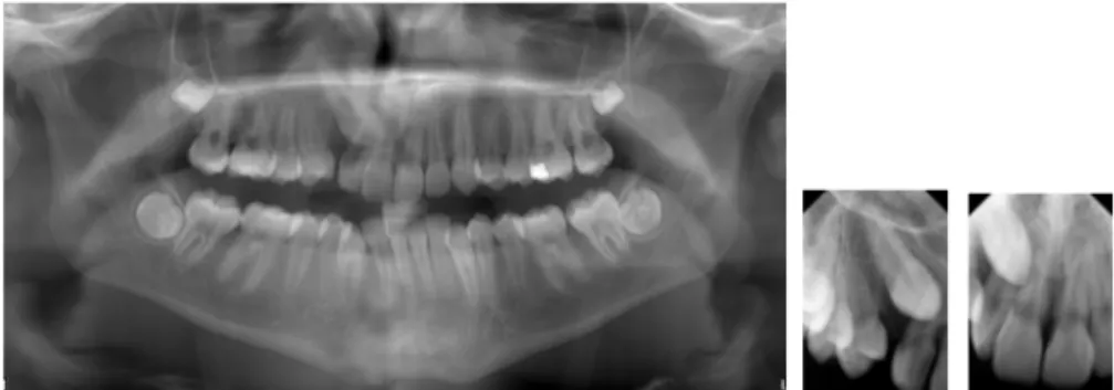
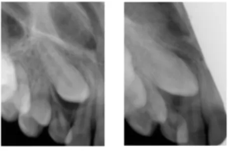
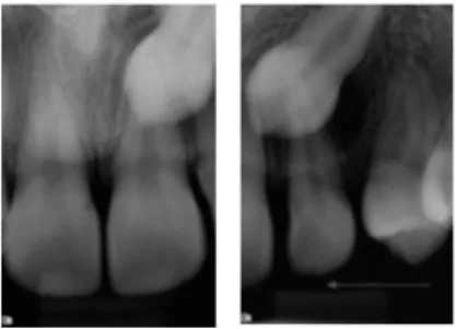
Already in 1910 Clark described intraoral radiographic examination of teeth “buried” in the incisor and canine region and the difficulties in deciding which tooth is in front of the other when depicted superimposed (33). He described a solution to the problem by taking periapical radiographs using different angles in the horisontal plane. This method was later called Clarks rule, parallax technique, or stereo-roentgenography (Figure 2).
Figure 2. Panoramic and two periapical intraoral radiographs (Patient
case 2 in Study IV). The two intraoral radiographs illustrate the parallax technique identifying the right maxillary canine palatal to the lateral and central incisor.
Paatero presented the first dental application of panoramic radiology in 1949 as “a new tomographical method for radiographing curved outer surfaces” (34). Panoramic radiographs provide an overview of the maxillary and mandibular dental arches with the teeth and their supporting structures as well as the temporomandibular joints and maxillary sinus. Ericson and Kurol (1988) proposed an index based on the canine position in a panoramic radiograph; the angulation of the canine to the midline, the distance of the canine crown-tip to the occlusal line and the medial position of the crown in different degrees of overlap of the lateral and central incisors (18).
According to the magnification technique, impacted teeth located palatally are more enlarged in the panoramic radiograph than the adjacent normally located teeth. Palatally impacted maxillary canines was possible to correcty identify in 80-90% of the cases using the magnification technique (14,35). Compared with postero-anterior skull radiographs, there was a tendency for the canine angulation to
be increased and appearing to be further away from the midline in the panoramic radiograph (36). Intraoral and panoramic radiography are commonly used imaging methods for identification of maxillary canines with eruption disturbance (17). Only about 50% of the resorptions on the palatal or buccal aspects of incisor roots however, can be detected by intraoral or panoramic radiographs (37).
Computed Tomography (CT) provides images in three planes. The
basic principle for CT is that the x-ray tube is connected to a detector on the opposite side synchronously rotate around the patient. CT uses a fan-shaped beam that is projected through the patient from several different angles. The radiation is attenuated (i.e. weakens) differently depending on the type of tissue that it passes through with high attenuation in high-density tissue such as bone tissue. A curved detector registers the intensity of the radiation and the linear attenuation-coefficient for each pixel in the detector is measured and re-calculated to Hounsfield values. With information of the angle for each measured data, a two-dimensional sectional image of the object is reconstructed. The two-dimensional images are combined in the computer to produce a volume of data that can be manipulated in order to demonstrate various anatomic structures with good detail reproduction. Modern scanners allow the volume of data to be reformatted in various planes or even as 3D volumetric representations of structures (38).
Cone beam computed tomography was introduced in the early 1980s
for angiography, but has since then become the most significant advance in dental- and maxillofacial imaging. The development of specialised CBCT scanners for use in dentistry begun in the late 1990s (39). CBCT uses a cone-shaped source of ionising radiation, similar in shape to the divergent beam of conventional 2D imaging. The cone-shaped beam is directed through the centre of the depicted object i.e. centre of rotation, and the attenuated radiation is projected on a detector on the opposite side. The x-ray source and detector rotate around the centre of rotation where the image volume is acquired. Most CBCT systems use a fixed source-to-object distance and object-to-detector distance and a flat panel detector with a layer of scintillator material used to convert the x-ray photon into light photons. The light is absorbed allowing formation of electric charges that are converted to
a digital signal. During one rotation of 180° to 360° (in some cases two rotations of 360°), multiple sequential planar projection images are acquired covering the field of view (FOV). The single projection transmission images constitute raw data that are reconstructed by computer algorithm to generate cross-sectional images (38).
The image quality characteristics can be described using four fundamental parameters, spatial resolution and contrast, noise and artefacts. Increased spatial resolution (sharpness) and contrast provides improved reproduction of details but also results in more noise. Hence, image quality should be assessed relative to the imaging task detection of bone fracture or visualisation of a soft-tissue abnormality (38). CBCT imaging has been suggested for localisation of maxillary canines with eruption disturbance as well as for detection of root resorption on adjacent teeth (40).
The diagnostic accuracy of CBCT regarding identification of maxillary canines with eruption disturbance as well as identification of any rootresorption on adjacent teeth seems to be higher compared with conventional 2D technique (27). The question is however, whether the information from CBCTexamination will change treatment planning compared with conventional imaging methods.
Radiation dose
The effective dose (sievert (µSv)) of CT for maxillofacial imaging has been reported to have a range of 280-1 210 µSv (41) which is usually higher than that of CBCT. The effective dose for CBCT varied for different machines and different field of views (FOV) with a range of 9.7-197 µSv for a small FOV (≤5 cm height), a range of 3.9-674 µSv for medium FOV (5.1-10 cm height), whereas the range was 8.8-1 073 µSv for a large FOV (>10 cm height) (41). The effective dose for CBCT is however considerably higher than that of conventional imaging methods with <1.5 µSv for one single intraoral radiograph, and range of 2.7-24.3 µSv for one panoramic radiograph (42).
For medical CT there are diagnostic reference levels with recommendations of the maximum dose for different procedures (43) but for CBCT there are no such reference levels. The SEDENTEXCT guidelines recommend an ‘Achievable Dose’ level of 250 mGy.cm2 for CBCT appropriate for examination prior to implant treatment
GENERAL AIM
A general aim of the SEDENTEXCT project was to acquire key information necessary for scientifically based clinical use of CBCT in dental- and maxillofacial imaging. Economic evaluation, weighing costs and benefits of alternative interventions, provides information that is necessary to achieve complete evaluation of a new imaging method and its contingent benefits in terms of patients’ health. The general aims of the present thesis, based on four studies referred to by their roman numerals (I-IV), were to assess any benefits of CBCT as well as the costs for producing these benefits in the context of maxillary canines with eruption disturbance.
SPECIFIC AIMS
The specific aims of the studies on which the present thesis are based were to:
• Elucidate scientific information on economic evaluations in oral health care with emphasis on diagnostic methods (Study I).
• Propose a framework for costing diagnostic methods in oral health care and to illustrate the application of the framework to the radiographic examination of maxillary canines with eruption disturbance (Study II).
• Analyse the costs of Cone Beam Computed Tomography (CBCT) in different health care systems for patients with dif-ferent clinical conditions (Study III).
• Measure the proportion of orthodontists’ treatment decisions that were different based on CBCT and panoramic radio-graphy compared with intraoral and panoramic radioradio-graphy and the costs of producing different treatment plans, regarding patients with maxillary canines with eruption disturbance (Study IV).
METHODS
Systematic review (Study I)
The literature review was performed according to the PRISMA Statement (44) and CRD’s guidance for undertaking reviews in health care (45). The subsequent steps that were defined in order to conduct a systematic review are presented in Table 1.
Table 1. The subsequent four steps defined to conduct a systematic review; (i) specification of problem, (ii) formulation of a plan for the literature search, (iii) literature search and retrieval of records, and (iv) extraction and quality assessment of data.
Specification of problem
Which diagnostic methods in dentistry have been analysed?
What types of economic evaluations have been conducted?
Formulation of a plan for the literature search
Consulting librarian for guidance. Construction of search strategies.
Literature search and retrieval of records
Searches for literature in four electronic data-bases; MEDLINE (PubMed), the Web of Science, the Cochrane Library and the NHS Economic Evaluation Database. Retrieval of records and subtraction of duplicate publications.
Identification of relevant studies according to abstract and/ or title by independent assessment of two reviewers.
independently using protocol with criteria for inclusion and exclusion.
Selection of original studies reporting economic evaluations in dentistry with abstract in English. Summary of the selection process in a PRISMA Flowdiagram (44). The flow diagram can be seen in Study I, Figure I.
Extraction and quality assessment of data
Critical appraisal of economic evaluations of diagnostic methods in dentistry using a second protocol.** Data was captured of the type of economic evaluations, the perspective, time horizon, discounting and outcome. Study design of included studies was “decision model (simulation)-based analyses and empirical economic evaluations, which collect patient-level data on costs and outcomes” as defined by Anderson (2010) (46).
**Protocol constructed based on the QUADAS tool (47), for assessment of the diagnostic methods and the checklist by Drummond et al. (2005) (7) for assessing economic evaluations.
Cost-analysis (Study II, III and IV)
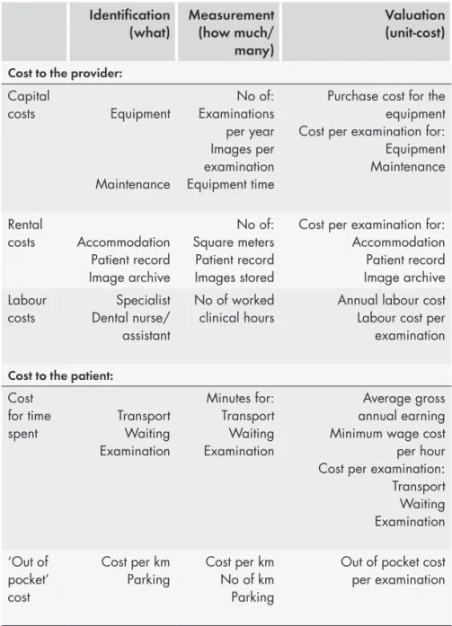
Framework for costing
The framework for costing diagnostic methods in dentistry incorporated three consecutive steps as presented in Table 2; i) identification of resources believed to have any impact on the results, ii) measuring the amount of resources consumed and finally iii) valuation of the used in monetary terms to get a cost per intervention. The framework included costs for the health care system in delivering the service from a providers’ perspective. Patient-related costs for using the service were included to enable a societal perspective according to Drummond et al. (2005) (7).
Data collection was aided using four forms constructed for i) apportioning the cost of capital equipment for each radiographic examination and ii) to measure consumable items to report the time spent on the intervention by health care providers and iv) to measure the cost for the patients and accompanying persons.
Table 2. The three consecutive steps of: (i) identification (ii) measurement and (iii) valuation of costs comprised in the framework for costing of diagnostic methods in dentistry based on recommendations by Drummond et al. (2005) (7). The example is described for diagnostic imaging methods.
Identification
(what) Measurement(how much/ many)
Valuation (unit-cost) Cost to the provider:
Capital costs Equipment Maintenance No of: Examinations per year Images per examination Equipment time
Purchase cost for the equipment Cost per examination for: Equipment Maintenance Rental costs Accommodation Patient record Image archive No of: Square meters Patient record Images stored
Cost per examination for: Accommodation Patient record Image archive Labour
costs Dental nurse/Specialist assistant
No of worked
clinical hours Annual labour costLabour cost per examination Cost to the patient:
Cost for time
spent TransportWaiting Examination Minutes for: Transport Waiting Examination Average gross annual earning Minimum wage cost
per hour Cost per examination: Transport Waiting Examination ‘Out of pocket’ cost Cost per km
Parking Cost per kmNo of km Parking
Out of pocket cost per examination
Direct costs included costs for capital equipment i.e. radiographic machines, computers, maintenance and repair of the equipment, costs for accommodation i.e. clinic and office space as well as cost for labour. The annual cost for the radiographic apparatus was calculated by dividing the purchase costs with the expected lifetime. The cost for maintenance was based on the average annual cost for a defined period of time. Cost for accommodation for the clinic and office space was calculated as cost per square metre and included overhead costs (e.g. cleaning, lighting and heating).
The clinic and office space was estimated based on the construction map of the clinic. The costs for capital, maintenance, and accommodation for one year was divided by the total number of images/volumes taken during one year to get the mean cost per image/volume. The cost per image/volume was then multiplied by the mean number of images/volumes recorded per examination to get the mean cost per examination. The total number of images/volumes per year was retrieved from the billing codes for a radiographic examination. The calculations were based on an observed average number of radiographs per examination. Scrap value or scrap cost of old equipment was not included as it was assumed not to differ between the radiographic machines. Hence it would not affect the incremental cost. Cost for consumables was based on the actual units consumed and the unit price.
Labour cost included valuation of the actual time spent on
administrative work, patient contact, and for interpretation of images and report writing. The quantity of time was valued according to the full employment costs i.e. salary plus employment ‘overheads’ or benefits. See example of calculation of labour cost in Table 3. Other aspects of overhead costs (e.g. management and administration) were not included as these are assumed not to differ between the interventions being compared. Hence, they would not affect the incremental cost.
Table 3. Example of calculation of labour cost per hour for a specialist and a dental assistant.
Calculation of labour cost
Specialist
Annual gross labour cost for a specialist including on-costs (51% in Sweden 2008). Based on the valuation of wage and salary structures (general
practice dentist) according to official statistics in Sweden 2008 (48). 74 340€
Annual labour cost for worked clinic hours (20% of annual
gross labour cost) 14 870€
Worked clinic hours – hours worked in clinic according to schedule (350h)
minus time for sick and parental leave (7h), and ‘downtime’ (43h) 300h Labour cost per hour = Labour cost for worked clinic hours (14 870€)
Total annual worked clinic hours (300 h) 49.60€
Dental assistant
Annual gross labour cost for a dental assistant including on-costs (51% in Sweden 2008). Based on the valuation of wage and salary structures (dental nurse) according to official statistics in Sweden 2008 (48).
(Full-time employment in Sweden is 1980h) 39 115€
Labour cost for worked clinic hours (100% of annual gross employment
cost) 39 115€
Worked clinic hours – hours worked in clinic according to schedule (1265h) minus time for sick and parental leave (114h) and ‘downtime’ (125h)
1 026h
Labour cost per hour = Labour cost for worked clinic hours (39 115€)
Total annual worked clinic hours (1 026h) 38.10€
Patientrelated costs included valuation of the time spent by patients
and accompanying persons on transport to and from the clinic and the time spent on the actual examination as well as outlays for travelling and parking. Time spent by the patient and accompanying persons over 18 years of age was valued according to the national average wage cost per hour and the time spent by patients below 18 years is valued according to minimum wage. “Out-of-pocket” expenses implied cost for driving a private car and parking or fares for taxi, train, or bus. An analysis from a users’ perspective would also include fees or co-payments for the examinations.
Empirical test of the framework
The framework for costing was tested in Study II and III. Cost data from Study II (cost year 2008) was updated in Study IV (cost year 2014). Study II estimated the costs associated with examination using intraoral and panoramic radiography compared with CBCT and panoramic radiography regarding maxillary canines with eruption disturbance in one clinic (Malmö). In Study III the framework was applied for calculation of the costs associated with CBCT examinations comparing four different health care systems (clinics in Cluj -Romania, Leuven -Belgium, Malmö -Sweden and Vilnius -Lithuania) regarding three different clinical conditions i) maxillary canine with eruption disturbance, ii) tooth loss prior to implant treatment and iii) lower wisdom tooth planned for removal.
Calculation of capital costs was based on data regarding the expected working life of the equipment. IIn the clinic in Malmö this was estimated to be 10 years for the three radiological apparatus. In Study IV, the working life of the panoramic machine was adjusted to 12 years corresponding to its actual working life, and the working life of the equipment for intraoral radiography was changed to 20 years. Study IV also included the opportunity cost and depreciation of the radiographic equipment using a rate of 3% and 5% according to recommendations by Drummond et al. (2005) (7).
Cost data for maintenance was based on either the actual average cost for repair and service during a 3-year period or on the pre-paid maintenance contracts with the costs for 2008. In Malmö, the cost for
accommodation was valued in accordance with local market rental
prices whereas for the other centres included in Study III the cost
per m2 by hospital finance was used. Valuation of the cost for access
to the digital system for patient records and the image archive was based on the total amount of radiographs according to billing codes and for image storing, the cost per digital space expressed in terabyte. Calculation of labour cost (Table 3) regarding the clinic in Malmö was based on data on wage and salary structures of a dentist and a dental assistant according to national statistics in 2008 (48)(Study II, III) and 2014 (49) (Study IV). For the centres in Cluj, Leuven and Vilnius involved in Study III, labour cost was based on the reported annual salary of the radiologists and radiographers. The clinic hours worked for a dental assistant, dental assistant or a radiographer included the imaging procedure in the radiographic examinations as
well as the administrative time spent on taking care of the patients when booking appointments. The working time for a specialist in oral and maxillofacial radiology included time for quality control of images, review and interpretation of CBCT-images as well as writing the radiological report. The estimation was based on observation of one radiologist performing these tasks for a subsample of 20 patients in Malmö.
Patientrelated costs were retrieved using self-reported data for each
patient. For several patients in the clinics in Cluj, Leuven and Vilnius, the appointment at the radiological clinic was combined with a visit at a nearby clinic and therefore no cost for travelling were recorded for these visits. The value of time for adult patients and accompanying persons in Study II and III was based on the average gross annual earnings for 2007 from Eurostat Statistics (50) for Romania, Belgium and Sweden and for Lithuania on the gross average monthly earnings for 2007 according to Statistics Lithuania (51). In Study IV it was based on official statistics from Sweden (52). The figures for annual earnings were divided by the number of working days (holidays and vacations excluded) in 2008 (Study II and III) and in 2014 (Study IV) to determine the average earnings per worked day. The average earning per day was then divided by eight (the number of working hours per day) to get an estimate of the average wage rate per hour. The data were used as a measure of the value of adults’ time in each system.
The value of the time for patients under 18 years of age was based on the minimum wage rates for Belgium, Lithuania and Romania according to Eurostat statistics of gross earnings (53). For 2008 there was no national minimum wage recorded for Sweden. Instead the last reported national minimum wage for 2004 according to a report from The Institute for Labour Market Policy Evaluation 2004 (IFAU) (54) was used. Transport cost per kilometer for patients using private transport was based on rates used by Rotary International for 2008 (55) Study I and II) and the European Commission for 2014 (56) (Study IV). When the patients walked or cycled, the cost was limited to the valuation of their time.
All costs were adjusted to 2008 (Study II, III) or to 2014 (Study IV) values. In Study II and III at the 2008 average rate of one Euro = SEK 9.60, and at the 2014 average rate of one Euro = SEK 9.00. See Study II, III and IV for presentation of the radiographic equipment used.
Web-based survey (Study IV)
A web-based survey was created in association with a web developer. All members of the Swedish Orthodontic Society (n= 314) were invited by e-mail, in an attempt to find specialists in orthodontics with at least one year of experience in treating orthodontic patients.
Twelve patient cases were constructed for the survey. For further
information of patient characteristics see Study IV, Table 1 and 2.
Parameters common to all cases: • A subjective treatment need
• One maxillary canine with eruption disturbance and no likelihood for spontaneous eruption
• A complete permanent dentition including the second molars • Normal sagittal molar relation (Class I)
• Normal skeletal relations between the maxilla and the mandible • Cephalometric analysis showed normal maxillary incisor inclination • No subjective need for correction of the maxillary dental midline • Good oral hygiene
Parameters varying between cases: • Age and gender
• Left or right maxillary canine with eruption disturbance • Any maxillary centreline displacement
• Any space deficiency in the maxilla
For each case there were two intraoral periapical radiographs at different horisontal angles, one panoramic radiograph and CBCT images (Figure 3,4). There was also a radiological report describing the position of the maxillary canine, the distance of the canine crown in relation to the marginal bone, root morphology and any resorption of teeth neighbouring the maxillary canine. Intraoral and panoramic radiographs as well as CBCT-images from patients already examined in Study II, were reviewed, and all patients with unilateral eruption disturbance of the maxillary canine with otherwise no, or minor, need for orthodontic treatment were included in Study IV. A specialist i oral radiology interpreted the images and wrote the report.
The orthodontists were randomly assigned to four of the 12 patient cases. In part one of the survey two cases were presented with intraoral and panoramic radiography and two cases with CBCT and panoramic radiography. In part two the cases presented with intraoral and panoramic radiography in the first round were instead presented with CBCT and panoramic radiography, whereas cases presented with CBCT and panoramic radiography were presented with intraoral and panoramic radiography.
In the survey, the participants were asked to denote; • their age, gender and years of experience,
• a treatment for four cases choosing from the following alternatives; 1) no treatment, 2) nonextraction treatment, 3) orthodontic treatment and extraction of the first premolar, 4) orthodontic treatment and extraction of the maxillary lateral incisor, 5) orthodontic treatment and extraction of the maxillary canine with eruption disturbance.
• the level of confidence in their decision on a five-point scale; 1) very confident, 2) fairly confident, 3) neither confident – nor unconfident, 4) fairly unconfident or 5) very unconfident.
Figure 3. An example showing the radiographs presented in the survey for one (case 8) of the 12 patient cases.
CBCT images Intraoral radiographs
Figure 4. An example showing the radiographs presented in the survey for one (case 10) of the 12 patient cases.
Intraoral radiographs
Panoramic radiograph
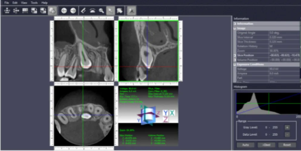
In the survey a viewer program for the CBCT images (i-Dixel One Data Viewer Plus software, J. Morita®, Kyoto, Japan) was available enabling the orthodontists to view the CBCT images in all planes (the axial, coronal and sagittal) dynamically by scrolling through the consecutive orthogonal image stack (Figure 5).
The survey was open from May 2016 to November 2016. The original e-mail invitation to participate was followed by four reminder e-mails sent via MailChimp® (The Rocket Science Group, Atlanta, US). Finally, an attempt was made to contact all non-respondents by telephone. A group of three orthodontists, one specialist in periodontology, one trainee in oral radiology and one external web-developer evaluated the web-based survey before implementation. The orthodontists in the evaluation group were not included as subjects in the study.
Figure 5. Image of the viewer program for the CBCT images (i-Dixel One
Data Viewer Plus software, J. Morita®, Kyoto, Japan) available in the survey.
Study II, III and IV were conducted in accordance with the ethical principles of the World Medical Association Declaration of Helsinki (2008 version) and approved by the Iuliu Hatieganu University of Medicine and Pharmacy in Cluj-Napoca Ethics Committee, Romania (18514/4.09.2008), the Commission for Medical Ethics of the Faculty of Medicine of the Katholieke Universiteit Leuven, Belgium (B32220083749), the Regional Ethical Review Board, Lund,
Sweden (H15606/2008) and Vilnius regional committee for biomedical research ethics, Lithuania (2010-05-05 No. 158200-01-138-39).
Statistical analyses
Intrarater agreement for treatment decisions based on observations of the same patient case using the same imaging method was estimated in terms of percent agreement per patient case. Kappa statistics and percent agreement was used to analyse the treatment decision of the different imaging methods. Fisher’s Exact Test was used to compare treatment decisions and associated confidence levels when using the different imaging methods. IBM SPSS Statistics (Version 23, Chicago, Illinois, USA) was used for all calculations. Statistical significance was set at a p-value less than 0.01.
RESULTS
Systematic review
A total of 3 708 records were retrieved with 3 380 remaining after subtraction of duplicates whereof 159 were considered to be economic evaluations in the field of dental care (Figure 6). Full-text publications were excluded if they were; not comparing two alternative methods in terms of the costs, non-systematic reviews or concering other topics than economic evaluations of methods in dental care. For further information of the characteristics of the 12 included economic evaluations on diagnostic methods see Study I, Table 2.
Figure 6. The distribution of all studies identified as economic evaluations
The 12 included economic evaluations of diagnostic methods:
Empirical economic evaluations:
Perry et al. (1994) (57) Scaf et al. (1997) (58) Xia et al. (2006) (59)
Subramanian et al. (2009) (60) Christell et al. (2012) (61)
Decision model-based analyses:
Taylor-Weetman et al. (2002) (62) Gray et al. (2000) (63)
Zavras et al. (2000) (64) Van der Meij et al. (2002) (65) Speight et al. (2006) (66) Norlund et al. (2009) (67) Dedhia et al. (2011) (68)
The different types of diagnostic methods being analysed were tests for diagnosis described by five studies using imaging methods (58,59,61,62,67) and one using probes for examination of periodontal health (57). Tests for screening a population were describing five studies regarding oral cancer (60,63,65,66,68) and risk of caries (64). The outcome measures of the diagnostic methods were described as values of sensitivity and specificity by six studies whereof one regarding imaging methods (67) and five regarding tests for screening (63-66,68). One study described the correlation of agreement between probing instruments (57) and one presented difference in radiation dosages (58).
All but two studies presented a rationale for the diagnostic methods and the methods were described in a transparent way permitting replication. One of the studies with a non-transparent description of the rationale was based on a pre-doctoral thesis not possible to retrieve (64) and in the other study the data was based on expert opinion (59).
The type of economic evaluation performed was cost-effectiveness analysis in three studies (60,63,67), CUA was performed by three studies (65,66,68) and CA was performed by six studies ((57-59,61,62,64). In three studies the type of economic evaluation was
not stated (57,58,67). The study perspectives stated in four studies, wereof a societal perspective was stated in three studies (60,61,68) and patients’ perspective was stated in one study (66).
Presentation of costs based on identification and measurements of the resources used and valuation of these resources in monetary terms were presented in four studies (57,60,61,64). Six studies presented cost-estimations based on fees, expenditures and/or other estimations of payments made or received (58,62,65-68). Incremental cost was presented by two studies (61,64) and ICER was presented in five studies (60,65-68). For further information of the characteristics of the 12 included economic evaluations on diagnostic methods see Study I, Table 2.
Cost-analysis
The framework for cost-analysis facilitated the costing with its three subsequent steps of identification, measurement and validation with the different costs separately described in detail.
Empirical test of the framework
In the clinic in Malmö, for maxillary canines with eruption disturbance, CBCT and panoramic radiography had a mean total cost per examination
of 128.38€ and for intraoral and panoramic radiography the cost was
81.80€, resulting in an incremental cost for CBCT and panoramic
radiography of 46.58€. In the up-dated cost-analysis (in 2014) the mean
total cost per examination for using CBCT and panoramic radiography
was 134.37€ and for using intraoral and panoramic radiography it
was 99.84€ (Table 4). Hence, the resulting incremental cost of 34.53€
was 26% lower compared with that of 2008. When also including equipment depreciation and opportunity cost of funds using a rate of
3% the incremental cost per examination increased by 2.79€ (7%)
whereas a rate of 5% resulted in an increase of 4.75€ (12%).
The comparison between different health care systems showed that the cost for CBCT-examinations varied, being the highest in Malmö irrespective of the clinical condition examined (maxillary canine with eruption disturbance, tooth loss prior to implant treatment and lower wisdom tooth planned for removal). The lowest provider-related costs were seen in Cluj whereas the lowest patient-related costs were seen in Leuven. Variation in direct costs was mainly owing to different
capital costs for the CBCT equipment arising from differences in
purchase prices (range 148 000–227 000€).Compared with the cost
of 147 880€ in Cluj (year 2006), the purchase cost was 37% higher
in Malmö (year 2003), 52% higher in Vilnius (year 2002) and 8% higher in Leuven (year 2008) (Table 5).
Capital costs for equipment per examination for CBCT per
examination varied between 10.67€ in Leuven to 52.14€ in Vilnius.
This was due to the number of images taken during 2008 that in Malmö was 4 009 intraoral radiographs, 1 765 panoramic radiographs and 948 CBCT volumes. The average number of CBCT volumes per examination was 450 in Cluj, 1500 in Leuven and 435 in Vilnius.
There was a variation of the cost for accommodation per
examination across systems (1.29€ in Cluj, 1.60€ in Leuven, 6.14€
in Malmö and 1.82€ in Vilnius) which was due to differences in the
space for accommodation (19m2 in Cluj, 6m2 in Leuven, 16m2 in
Malmö, 28m2 in Vilnius) and the cost per m2 (30.49€ in Cluj, 400.00€
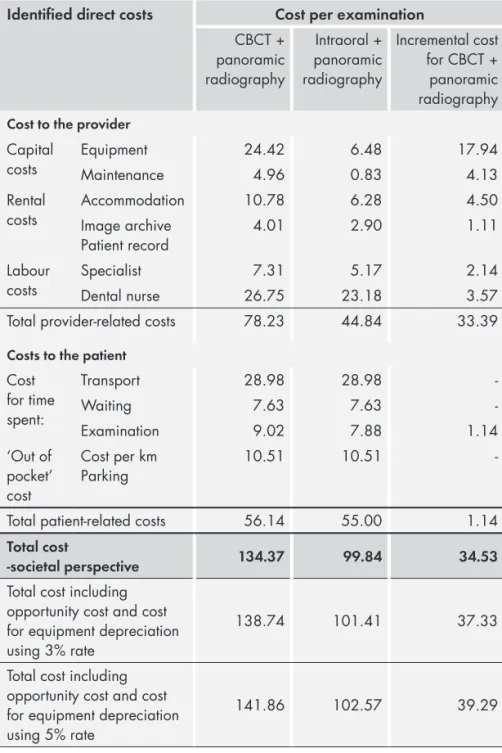
Table 4. Distribution of direct costs per examination based on CBCT and panoramic radiography compared with intraoral and panoramic radiography. Costs were estimated using 2014 prices (Euro).
Identified direct costs Cost per examination
CBCT + panoramic radiography Intraoral + panoramic radiography Incremental cost for CBCT + panoramic radiography Cost to the provider
Capital
costs EquipmentMaintenance 24.424.96 6.480.83 17.944.13 Rental
costs AccommodationImage archive 10.78 6.28 4.50
Patient record 4.01 2.90 1.11
Labour
costs SpecialistDental nurse 26.757.31 23.185.17 2.143.57
Total provider-related costs 78.23 44.84 33.39
Costs to the patient Cost for time spent: Transport 28.98 28.98 -Waiting 7.63 7.63 -Examination 9.02 7.88 1.14 ‘Out of pocket’ cost Cost per km Parking 10.51 10.51
-Total patient-related costs 56.14 55.00 1.14
Total cost
-societal perspective 134.37 99.84 34.53
Total cost including opportunity cost and cost for equipment depreciation using 3% rate
138.74 101.41 37.33
Total cost including opportunity cost and cost for equipment depreciation using 5% rate
Labour cost per examination in Malmö for a specialist was 5.37€
for CBCT and panoramic radiography and 3.80€ for intraoral and
panoramic radiography. The average gross labour cost per worked clinic hour for a specialist was 49.60€ and for a dental assistant it was 38.10 (Table 3). In the up-dated costing (for 2014) the corresponding
cost for a specialist was 67.44€ whereas for a dental assistant is was
51.12€.
Patientrelated costs per CBCT-examination (not including
patient-fee) varied between health care systems and between different clinical
situations with a range of 16.15€-59.44€. The cost was the highest
in Malmö irrespective of clinical condition. Also the average patient-related cost per examination including patient-fee, was the highest
in Malmö where it varied between 59.44€ and 147.48€ depending
on clinical condition. It was the second highest in Vilnius
(21.73-100.09€), the third highest in Cluj (90.43–96.30€) and the lowest
in Leuven (21.76-41.96€).
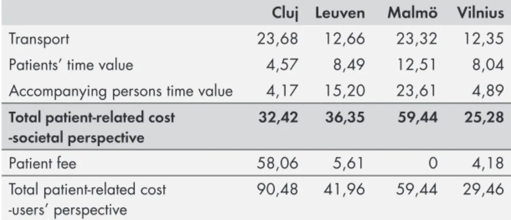
Table 5. The patient-related costs from a societal perspective and from a
users’ perspective (including patient fee) in different settings regarding CBCT-examination of maxillary canines with eruption disturbance (Costs in Euro).
Cluj Leuven Malmö Vilnius
Transport 23,68 12,66 23,32 12,35
Patients’ time value 4,57 8,49 12,51 8,04
Accompanying persons time value 4,17 15,20 23,61 4,89
Total patient-related cost -societal perspective
32,42 36,35 59,44 25,28
Patient fee 58,06 5,61 0 4,18
Total patient-related cost
-users’ perspective 90,48 41,96 59,44 29,46
The valuation of the time spent by the patients and, where applicable, by the accompanying person resulted in the highest cost in Malmö, second highest in Leuven, third highest in Vilnius and lowest in Cluj. In Malmö all visits to the radiolgical clinic represented separate trips for the patients. In the other settings however, the visit to the radiolgical clinic was added to the visit to another clinic in 30–50% of the
patients in Cluj, 23–61% in Leuven and 34–66% of the patients in Vilnius. The average travel distance to and from the radiology clinic for the three clinical conditions was shortest in Malmö (23–28 km), followed by Leuven (21–30 km) and Vilnius (15–47 km), and longest in Cluj (72–126 km).
Web-based survey
A total of 314 members of the Swedish Orthodontic Society were invited to participate in the study.
Exclusion was made of orthodontists (n=93); • not active within orthodontics
• being orthodontic resident • not working in Sweden
• with insufficient contact information • on parental or sick leave
• not working with this patient group
(n=30) (n=20) (n=17) (n=10) (n=9) (n=7)
Of the 221 remaining orthodontists, 154 accepted to participate.
Responses were not used when orthodontists had not (n=42);
• answered the second part of the survey • responded after four reminders
• been able to open the CBCT files/survey • been able to save the answers
• approved of the study design • other reasons (n=17) (n=12) (n=7) (n=3) (n=2) (n=1)
When the survey closed, 112 orthodontists had answered both parts of the survey with 445 assessments based on CBCT and panoramic radiography and intraoral and panoramic radiography respectively.
Intrarater agreement
Intrarater agreement based on 30 orthodontist’s assessment regarding the treatment alternative varied from 50% to 100% using intraoral and panoramic radiography. Full agreement was observed for eight cases. Based on CBCT and panoramic radiography, the agreement
varied between 0% for one case and 100% for seven cases. The number of assessments varied between 3 to 7 per case.
Comparison between imaging methods
The number of assessments varied between 31 to 44 per case. Agreement between imaging methods, intraoral and panoramic radiography compared with CBCT and panoramic radiography regarding the treatment alternatives chosen, varied between 100%
and 12.8% for different cases κ-values varied from 1.0 to 0.018.
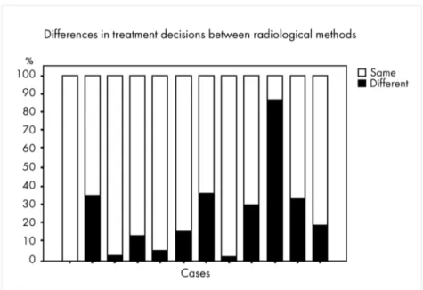
Figure 7. Treatment decisions (%) per patient case by 112 orthodontists
based on intraoral and panoramic radiography or CBCT and panoramic radiography at two occasions for the same patient cases.The difference between case 10 and all other cases was significant (p < 0.01).
Orthodontic treatment not including tooth-extraction was the most frequently denoted treatment alternative, chosen in 66% of the assessments based on intraoral and panoramic radiography and in 64% of the assessments based on CBCT and panoramic radiography.
Overall 24% (107 of 445) of the treatment decisions were different when based on CBCT and panoramic radiography compared with intraoral and panoramic radiography. One case (number 10) differed