The use of imaging in necrotising enterocolitis - results from
a survey
Poster No.: C-2872
Congress: ECR 2017
Type: Scientific Exhibit
Authors: M. K. E. Ahle1
, E. Rubesova2, H. Ringertz2; 1Linköping/SE,
2
Stanford, CA/US
Keywords: Pediatric, Gastrointestinal tract, Conventional radiography, Ultrasound, Diagnostic procedure
DOI: 10.1594/ecr2017/C-2872
Any information contained in this pdf file is automatically generated from digital material submitted to EPOS by third parties in the form of scientific presentations. References to any names, marks, products, or services of third parties or hypertext links to third-party sites or information are provided solely as a convenience to you and do not in any way constitute or imply ECR's endorsement, sponsorship or recommendation of the third party, information, product or service. ECR is not responsible for the content of these pages and does not make any representations regarding the content or accuracy of material in this file.
As per copyright regulations, any unauthorised use of the material or parts thereof as well as commercial reproduction or multiple distribution by any traditional or electronically based reproduction/publication method ist strictly prohibited.
You agree to defend, indemnify, and hold ECR harmless from and against any and all claims, damages, costs, and expenses, including attorneys' fees, arising from or related to your use of these pages.
Please note: Links to movies, ppt slideshows and any other multimedia files are not available in the pdf version of presentations.
Aims and objectives
Necrotising Enterocolitis (NEC) remains a potentially devastating emergency in neonates, predominantly the premature. Ever since it was first described in the 60's, imaging has played a great role in definition, staging, and monitoring of the disease. The radiographic image can change before the clinical condition, but typical signs are often transient and may be missing even in severe NEC [1-4]. These circumstances have led to the recommendation of frequent imaging and to the insight that the clinical decisions cannot rely solely on radiological signs [5-7]. Ultrasound (US) as a possibility to enhance sensitivity and diagnostic accuracy was first described in the mid 80's [8, 9] and was included in a diagnostic algorithm suggested by in 1994 [6], but despite great effort to develop and validate the method, its role in the management of NEC has not yet been established [7, 10, 11]. Meanwhile, in order to improve interobserver agreement and diagnostic accuracy of AR, the radiographic signs of NEC have also been systematized into the DAAS scale [12].
Imaging, as an adjunct to clinical assessment [11], is crucial in the diagnosis and management of NEC. The purpose of this survey was to investigate current views and routines, as described by involved specialists, and identify areas in need of further study and discussion.
Methods and materials
A web-based questionnaire on differential diagnosis, modality use, impact of findings, decision making etc. in the management of NEC was created in two versions, one for clinicians, i.e. neonatologists and paediatric surgeons, and one for radiologists. The questionnaires were distributed through e-mailing to members of specialist organizations for paediatric radiology, neonatology, and paediatric surgery in Europe and the USA, as well as through personal contacts.
70 neonatologists, 58 paediatric surgeons and 74 radiologists from various countries around the world responded. The results were analysed with proportion estimation with 95% confidence intervals (CI). Detected differences between subgroups were regarded as significant if CI did not overlap. Although this was not the main objective of the study, significant differences were reanalysed with the dominant groups of American radiologists and/or neonatologists from the British Islands excluded, attempting to discern whether the findings were most likely due to differences in perceptions between specialties or rather to variations in clinical traditions between countries.
Results
How is imaging used in the management of suspected or known NEC?
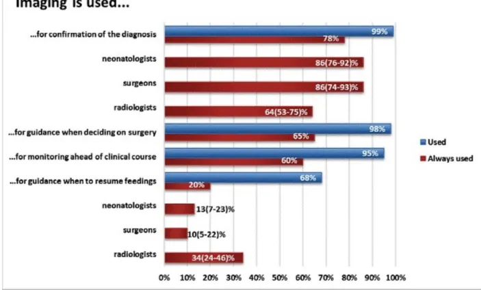
As shown in fig. 1, there was generally great consensus among the respondents on what imaging should be used for.
The reported differences in the use of imaging for guidance on when to resume feedings seems to be a result of variations in clinical traditions, but the difference in perception between neonatologists and radiologists regarding the importance of imaging for confirmation of the diagnosis is probably not.
The clinical picture was clearly thought to be the most important for differential diagnosis, but radiographic findings were of at least some importance according to 98% of all responding clinicians, and of great importance according to 84%. Corresponding numbers for ultrasonographic findings were 75 and 29% respectively.
Choice of modality
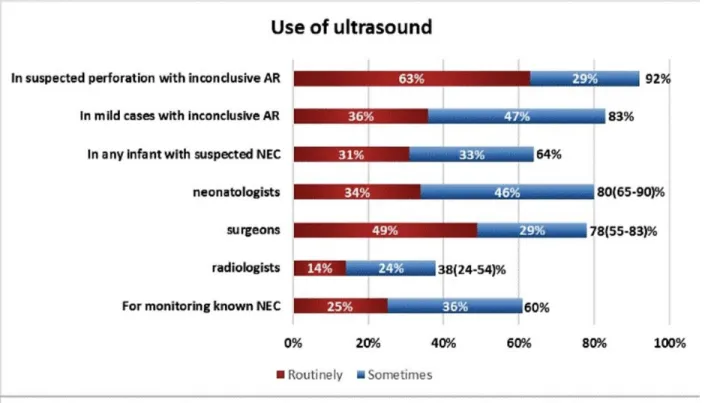
Responses are summarized in fig. 2. AR is generally first choice modality, but there is a considerable interest in US as well.
Projections used in AR
Results are presented in fig. 3
85% of the respondents stated that AR with a vertical beam would be used in all patients with known or suspected NEC. Out of 16 respondents who did not chose a positive response alternative ("all patients" or "selected patients"), 13 stated that they did not know or left an empty response.
Adherence to the praxis to obtain images with a horizontal beam in all patients at diagnosis and during the first 24-48 hours [7], is reported by only 20% of the respondents. Horizontal beam images obtained with the patient supine or in the left decubitus position are almost equally common.
When is ultrasound done?
As seen in fig. 2, however, US was indicated as a possible part of the work-up by a greater proportion than those 58%.
Among respondents from hospitals where US is used, 93% reported that AR and US were, at least sometimes, used in combination, 24% stating that they always were. 52% might even use US as first choice, 7% always.
Fig. 4 shows that US, although sometime a part of the routine work-up, is more often used
for additional information when AR is inconclusive or, as suggested by Silva et.al, "as a problem-solving modality only in those neonates suspected or known to have NEC and in whom it is thought that more information provided by AUS might facilitate management decisions"[13]. The differences between radiologists and clinicians regarding the use of US in any infant with suspected NEC can probably be assumed to mirror the clinical traditions at different hospitals.
Repeated imaging
As shown in the following table, there was considerable variation in examination frequency:
Examination frequency More than rarely Often More than once every 24 h 76% 42%
About every 24 h 78% 33%
Less than every 24 h 56% 13%
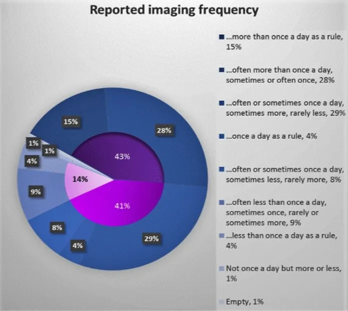
For the three suggested frequencies in the table, respondents could choose response alternatives "Often", "Sometimes", or "Rarely". In this summary, "More than rarely" includes response alternatives "Often" and "Sometimes", with "Often" presented separately as well. Combinations of response alternatives are summarized at greater detail in fig. 5.
Imaging about every 24 hours appear to be as widespread as routines including more frequent imaging, whereas longer intervals between examinations were less common. The corresponding questions in the survey of Zani et al [14], were differently put, but keeping that in mind, the results may be regarded as similar.
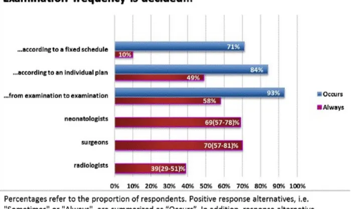
Responses on what determines the examination frequency are shown in fig. 6. Although fixed schedules were used by some respondents, the examination frequency was usually decided individually. The differences between radiologists and clinicians regarding decisions from one examination to another seem to be partly, but not entirely, explained
by differences in local traditions. The diversity in the resulting examination frequencies suggests that there are no generally accepted guidelines to support the decision.
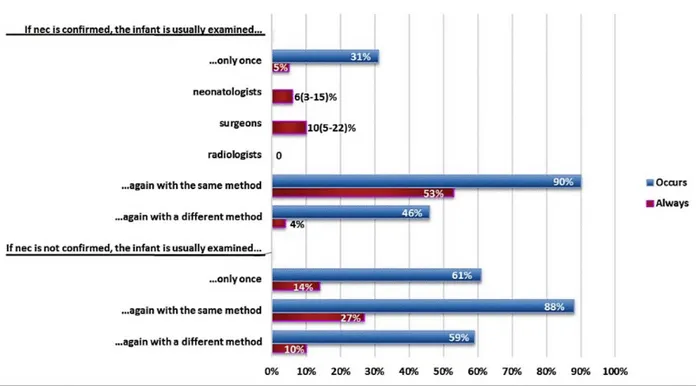
As shown in fig. 7, whether NEC was confirmed or not, the same method as before was most commonly used for repeated imaging, but it was not uncommon to use a different method or even abstain from further imaging.
63% of the respondents stated occasional concerns about radiation in connection with repeated imaging.
Images for this section:
Fig. 1: Reported use of imaging in NEC
© Medical and Health Sciences, division of Radiology, Linköping University, Faculty of
Fig. 2: Choice of modality
© Medical and Health Sciences, division of Radiology, Linköping University, Faculty of
Fig. 3: Projections used in AR
© Medical and Health Sciences, division of Radiology, Linköping University, Faculty of
Health Sciences - Linköping/SE
Fig. 4: Use of ultrasound
© Medical and Health Sciences, division of Radiology, Linköping University, Faculty of
Fig. 5: Imaging frequency
© Medical and Health Sciences, division of Radiology, Linköping University, Faculty of
Fig. 6: Decisions on imaging frequency
© Medical and Health Sciences, division of Radiology, Linköping University, Faculty of
Fig. 7: Choice of modality for repeated imaging
© Medical and Health Sciences, division of Radiology, Linköping University, Faculty of
Conclusion
• Imaging remains an important tool in the management of NEC. • Despite great consensus on the purposes of imaging in NEC, there
is considerable diversity in routines, especially regarding examination frequency and the use of ultrasound.
• A growing interest in ultrasound is suggested by the fact that respondents from hospitals, where it is not yet used for NEC, attribute significance to it. • Decisions in imaging are often based on individualized assessments rather
than fixed algorithms. This should be taken into account, when working on future guidelines.
Personal information
References
1. Rabinowitz JG, Siegle RL. Changing clinical and roentgenographic patterns of necrotizing enterocolitis. AJR Am J Roentgenol. 1976;126(3):560-566. doi:10.2214/ ajr.126.3.560.
2. Siegle RL, Rabinowitz JG, Korones SB, Eyal FG. Early diagnosis of necrotizing enterocolitis. AJR Am J Roentgenol. 1976;127(4):629-632. doi:10.2214/ajr.127.4.629.
3. Tam AL, Camberos A, Applebaum H. Surgical decision making in necrotizing enterocolitis and focal intestinal perforation: predictive value of radiologic findings. J
Pediatr Surg. 2002;37(12):1688-1691. doi:10.1053/jpsu.2002.36696.
4. McBride WJ, Roy S, Brudnicki A, Stringel G. Correlation of complex ascites with intestinal gangrene and perforation in neonates with necrotizing enterocolitis. J Pediatr
Surg. 2010;45(5):887-889. doi:10.1016/j.jpedsurg.2010.02.011.
5. Leonard T, Johnson JF, Pettett PG. Critical evaluation of the persistent loop sign in necrotizing enterocolitis. Radiology. 1982;142(2):385-386. doi:10.1148/ radiology.142.2.7054826.
6. Fotter R, Sorantin E. Diagnostic imaging in necrotizing enterocolitis. Acta Paediatr
7. Epelman M, Daneman A, Navarro OM, et al. Necrotizing enterocolitis: review of state-of-the-art imaging findings with pathologic correlation. Radiographics. 2007;27(2):285-305. doi:10.1148/rg.272055098.
8. Merritt CR, Goldsmith JP, Sharp MJ. Sonographic detection of portal venous gas in infants with necrotizing enterocolitis. AJR Am J Roentgenol. 1984;143(5):1059-1062. doi:10.2214/ajr.143.5.1059.
9. Lindley S, Mollitt DL, Seibert JJ, Golladay ES. Portal vein ultrasonography in the early diagnosis of necrotizing enterocolitis. J Pediatr Surg. 1986;21(6):530-532.
10. Muchantef K, Epelman M, Darge K, Kirpalani H, Laje P, Anupindi SA. Sonographic and radiographic imaging features of the neonate with necrotizing enterocolitis: correlating findings with outcomes. Pediatr Radiol. 2013;43(11):1444-1452. doi:10.1007/ s00247-013-2725-y.
11. Bohnhorst B. Usefulness of abdominal ultrasound in diagnosing necrotising enterocolitis. Arch Child Fetal Neonatal Ed. 2013;98(5):F445-50. doi:10.1136/ archdischild-2012-302848.
12. Coursey CA, Hollingsworth CL, Gaca AM, Maxfield C, Delong D, Bisset G. Radiologists' agreement when using a 10-point scale to report abdominal radiographic findings of necrotizing enterocolitis in neonates and infants. AJR Am J Roentgenol. 2008;191(1):190-197. doi:10.2214/AJR.08.1090.
13. Silva CT, Daneman A, Navarro OM, et al. Correlation of sonographic findings and outcome in necrotizing enterocolitis. Pediatr Radiol. 2007;37(3):274-282. doi:10.1007/ s00247-006-0393-x.
14. Zani A, Eaton S, Puri P, et al. International Survey on the Management of Necrotizing Enterocolitis. Eur J Pediatr Surg. 2014;25(01):27-33. doi:10.1055/s-0034-1387942.