Faculty of Natural Resources and Agricultural Sciences
Mechanical properties of films made of faba
bean protein nanofiber and non-fibrillated
protein.
Mechanical properties of films made of faba bean protein
nanofiber and non-fibrillated protein.
Alice Dunge
Supervisor: Anja Herneke, Swedish University of Agricultural Sciences, Department of Molecular Sciences
Examiner: Maud Langton, Swedish University of Agricultural Sciences, Department of Molecular Sciences
Credits: 30 hp
Level: Second cycle, A2E
Course title: Independent Project//degree project in Food Science - Master's thesis
Course code: EX0877
Program/education: Agriculture Programme – Food Science
Place of publication: Uppsala
Year of publication: 2019
Title of series: Molecular Sciences
Part number: 2019:12
Online publication: https://stud.epsilon.slu.se
Abstract
The production of livestock puts a huge strain on our ecosystem and the planet. It is the largest source of greenhouse gases and it causes loss in biodi-versity and pollution of water. One way to reduce these environmental dam-ages is to decrease the intake of meat and increase the intake of plant-based proteins. However, the sensory feeling of meat is something many people prefer, creating the need to find a plant-based substitute that still fulfills the sensory need the consumers have. The faba bean is a legume with high levels of proteins that has similar properties to the soybean but is able to grow in colder climates countries. In recent years the creation of amyloid nanofibrils from proteins, for example from faba beans, and their unique mechanical properties has become an area of high interest. Potentially, they can be used to create a meat-like substitute and in that way fulfill that sensory need the consumers have. This study therefore aims to conform nanofibrils from the extracted faba beans proteins that can be used as raw materials to create a texturized vegetal-based food.
Films as systems models were developed from different concentrations of protein nanofibrils and the mechanical properties of each film were evaluated in terms of young modulus (modulus of elasticity), maximum strain and breaking strength. The influence of the concentration of nanofibrils in the modulus of elasticity of the films were assessed to establish a correlation be-tween the film structure and the mechanical properties (i.e., stiffness of the film). To achieve this faba bean proteins were isolated by alkaline precipita-tion and fibrillated by an acid treatment at high temperature.
Films were made with a combination of glycerol plasticizer, faba bean pro-teins, and faba bean protein nanofibrils with an increasing concentration ratio of the nanofibrils. The mechanical properties of films were tested with help of a tensile tested to determine their stress-strain curve and therefore, their stiffness and elasticity. The result showed a successful isolation of protein from the faba bean. The film casting, and tensile test were however inconclu-sive, showing irregular films and variating data with no correlation between concentrations of nanofibrils and the modulus of elasticity. There are multiple theories why this may be, such as bacteria growth, bubbles trapped within the film, lack of nanofibrils or fibril break down during process.
Keywords: Faba bean, amyloid, fibrils, meat substitutes, elasticity, stiffness,
Produktionen av boskap är en stor påfrestning på vårt ekosystem och därmed vår planet. Den är den största källan till växthusgaser, och orsakar förluster i biodiversiteten och även förorening av vatten. En möjlighet till att lösa dessa miljöproblem är att minska intaget av kött och öka intaget av växtbaserade livsmedel. Det finns dock en del hinder för denna möjlighet, framför allt att många konsumenter föredrar den speciella sensoriska egenskaper som kött har vilket skapar behovet av ett köttliknande vegetabiliskt livsmedel. Faba-bönan är en proteinrik baljväxt med liknande egenskaper som sojaFaba-bönan, men som kan växa i kallare klimat. Forskare har på senare år fått upp intresset för att använda extraherat protein från bl.a. fababönor för att skapa nanofibriller då dessa har visat sig ha intressanta mekaniska egenskaper. Potentiellt skulle dessa fibriller kunna användas för att skapa köttliknande substitut och därmed fylla det sensoriska behovet hos vissa konsumenter. I den här studien var må-let att skapa fibriller ifrån extraherat protein från fababönor och undersöka deras mekaniska egenskaper i ett filmbaserat modellsystem. Filmer bildades med en kombination fababön-fibriller, protein och glycerol, med en gradvis ökning av fibrillfraktionen. Filmer testade med hjälp av en dragcell och deras mekaniska egenskaper undersökes med Young’s moduls, maximal belastning och styrkan vid brytpunkten. Resultaten visade på en lyckad isolation av pro-teiner från fababönor. Däremot var resultaten från filmformationen och tensil-testerna ofullständiga med ojämna filmer och varierande data utan någon tyd-lig korrelation mellan koncentrationen av fibriller och filmernas mekaniska egenskaper. Det finns flera förklaringar till hur detta kan komma sig, så som bakterietillväxt, luftbubblor som fastnat i filmerna, brist på fibriller eller att fibrillerna bröts ned under processens gång.
Nyckelord: Faba bönor, amyloid, fibriller, köttsubstituts, elasticitet, proteiner
1 Introduction 7
1.1 Aim 8
1.2 Legumes 8
1.3 Faba bean 8
1.4 Faba bean cultivation 9
1.5 Nutritional content of Faba bean 10
1.6 Faba bean proteins and extraction 11
1.7 Nanofibrils 13
1.7.1 Environmental factors affecting the creation of nanofibrils 14 1.7.2 The effect of protein concentration in the creation of nanofibrils 14 1.7.3 The effect of the protein source on the created nanofibrils 15 1.7.4 The effect of pH and ionic strength on the created nanofibrils 15 1.7.5 The effect of stirring and incubation time on the created nanofibrils 16 1.7.6 The effect of other substances on the formation of nanofibrils 17
1.7.7 The mechanics of fibril formation 17
1.7.8 Nanofibril morphology and protein source 18
1.8 Mechanical properties definitions 18
1.9 Properties and applications of nanofibrils 19
2 Material and method 22
2.1 Material 22
2.2 Method 22
2.2.1 Protein extraction and isolation 22
2.2.2 Fibrillation 23
2.2.3 Th-T assay 23
2.2.4 Dumas method 23
2.2.5 Film formation 24
2.2.6 Tensile test 24
3 Result and discussion 27
3.1 Protein extraction and isolation 27
3.2 Fibrillation 28
3.3 Film formation 31
3.4 Tensile test 34
4 Conclusion and future perspectives 41
5 References 42
Acknowledgements 45
Appendix 1: raw data from tensile test of first set of films 46 Appendix 2: raw data from tensile test of second set of films 48
Appendix 3: flow chart of batches and sets 50
Appendix 4: Regression curve of elasticity for the films 51 Appendix 5: Can nanofibrils made from plant proteins become the future
The production of livestock puts a huge strain on our ecosystem and the planet. It is the largest source of greenhouse gases and a big factor when it comes to losses in biodiversity. In developing countries, it plays a big role in pollution of water. Other negative impacts that livestock has on the environment are odor, land degradation and erosion. One way to avoid these environmental problems is to decrease the in-take of meat and increase the inin-take of plant-based proteins, mainly with legumes due to their protein content (Multari et al., 2015; Pimentel and Pimentel, 2003). Animal protein has a reputation of being a better protein source than plants, espe-cially in high-economic countries. This is partly due it being known as a full-worthy protein source, meaning that it contains all the essential amino acids (Multari et al., 2015). Meat is moreover the biggest source of the vitamins A, B1, B2, B12 and the minerals calcium, iron, phosphorus, selenium and zinc. Meat is the major source of proteins in high-economic countries partly due to these factors (Mj et al., 2012). There is a barrier of preferences, since consumers that normally did not eat meat substitutes or ate only a small amount considered the substitute to be less sensory satisfying. This group of people has a tendency to avoid new foods in general. When consumers have a low habit of consuming meat substitutes they have a tendency to want the substitutes to have a more meat-like texture (Hoek et al., 2011). In another study concerning the acceptance of meat substitutes among consumers the major issue was found to be that the consumer felt bored eating the same food over and over even if, with time, they came to like the substitute. Meaning that it is not enough to just substitute the protein source of meat but also the texture of the meat and with that the eating experience of the food (Hoek et al., 2013).
Meat-like substitutes are relatively new in the western world and it is a challenge to create products mimicking the complexity of meat structure and its flavors (Hoek et
al., 2013). Therefore, it is of big interest to search for fulfilling meat substitutes and other substitutes for animal-based products.
1.1 Aim
This study aims to: a) isolate proteins from faba beans; b) fibrillate the isolated pro-teins; c) develop different films with different concentrations of fibrillated and iso-lated proteins as model systems; d) characterize the mechanical properties of the developed films, and e) correlate the structural properties of the films with their mechanical properties (such as stiffness and elasticity) to be able to gain in knowledge to create new texturized plant-based food.
1.2 Legumes
All legumes have a similar amino acid composition and only vary slightly. Soy bean protein mostly consists of 7S (conglutin) and 11S (glycinin) while other legumes mostly consist of 7S (vicilin) and 11S(legumin) (Makri et al., 2005). So most pulses from legumes contain some essential amino acids, such as lysine, leucine, aspartic acid, glutamic acid and arginine but still miss other essential amino acids (Boye et al., 2010).
Legumes plays a big role in human diets, either directly or when used as feedfor animals. Legumes have a positive effect on human health such as preventing obesity and diabetes. They are especially important in developing countries where animal protein might be limited or not an option due to religious or cultural habits, they are even considered to be the “poor man’s meat” (Sparvoli et al., 2015).
Additionally, due to the legumes’ protein content and already existing production, they could potentially be a replacement for meat (Multari et al., 2015). Legumes have good functional properties such as water holding, fat binding, foaming and gelation, making them useful in various food applications (Boye et al., 2010).
1.3 Faba bean
Even if the soybean is the major provider of plant protein around the world it is not possible to grow it in some countries due to e.g. the climate. Therefore, another plant with similar properties is needed. In wet or cold climates where soy cannot grow faba bean could be a potential replacement (McCurdy and Knipfel, 1990).
Even if the faba bean were not to be used as human food there would still be a positive effect of substituting soybean with faba bean as feed for animals (Holmberg, n.d.). Faba bean as a protein source comes with other advantages, since it contains components that are health promoting (Multari et al., 2015). Faba bean can fixate nitrogen under vastly different environments and can work as a good break crop in cereal dominated crop rotation (Köpke and Nemecek, 2010).
The faba bean (Vicia faba) is a temperate crop that originates from the Mediterra-nean region (Vaughan et al., 2009; Duc et al., 2015). It is today cultivated in 50 countries wherea China stands for 65% of the produced faba (Vaughan et al., 2009). More common names for faba beans are broad bean, horse bean, and field bean (Multari et al., 2015). In this study we are focusing on field bean, the variety named Gloria. Faba beans can be split up in different types: long pods with 8 seeds and Windsors with 4 seeds per pod (Vaughan et al., 2009).
Different variations are popular in different areas. For example, small seeded forms are used in southwestern Asia while large-seeded variations are common in the west (Fouad et al., 2013).
1.4 Faba bean cultivation
In 2011 faba beans were grown on 1.2% of the worlds 200 million ha dedicated to legume cultivation. This makes faba bean the seventh most produced legume, the top being soy, groundnut and common bean (Duc et al., 2015).
The faba bean is grown under different cropping systems around the world. They support a sustainable cropping system by fixating nitrogen to the soil and contrib-uting with diversity in the rotation system (Fouad et al., 2013). When used in a cereal dominated crop system it is a good break crop, it reduces the need for fossil energy (Fouad et al., 2013; Köpke and Nemecek, 2010).
As mentioned, faba bean is a nitrogen fixating plant, it can fixate more nitrogen than other legumes in the same soil conditions (Multari et al., 2015). Due to this they do not need nitrogen added with fertilizers (Holmberg, 2013). This means that faba bean can be good solution for future agronomic issues (Multari et al., 2015). How-ever, for the nitrogen fixation to properly function it is important that the soil is not too acid and even if it can fixate nitrogen it still needs addition of phosphorus, po-tassium and micro nutrients (Holmberg, 2013).
Drought is the main limiting factor for the plant (Multari et al., 2015).
The faba bean is well adapted to various climates existing in Europe (Crépon et al., 2010), partly because it is the leguminous plant that is the best adapted to colder climates (Liene and Sandra, 2016).
In Sweden, faba bean crops are planted at different times in different regions. In a colder climate, it is planted during spring while in a warmer climate it is planted during winter. (Multari et al., 2015).
In Sweden, the areal for faba bean has increasing in the few past years (Holmberg, 2013). In 2018 it was grown on 29400 ha in Sweden. However, due to the extreme weather during the summer of 2018 the harvest became 63% lower than usual (34500 tons (74 900-ton less)). Meaning that the areal increased but the harvest de-creased (Jordbruksverket, 2017).
1.5 Nutritional content of Faba bean
The dried seeds contain approximately 25% protein, 1,5% fat and 49% carbohy-drates. (Vaughan et al., 2009)
Because legumes contain such a high amount of proteins they are a good protein source when producing high protein foods. Specifically, faba bean is predicted to be a good replacement to soybean but to accomplish this the faba beans’ properties as a plant and as a food need to be more thoroughly examined (Arogundade et al., 2006). Even if faba beans have a good protein content it is low on the essential amino acids methionine, cysteine and tryptophan meaning that it is crucial to include other protein sources in the diet to ensure that the protein quota is met (Crépon et al., 2010).
However, faba beans contain some antinutritional components e.g. asphytic acid, saponins and lectins (Multari et al., 2015). These are only a problem for monogastric animals, like humans, and not for non-monogastric animals such as cows (Crépon et al., 2010). Derivatives from vicine and convicine can for individuals with a spe-cific genetic variation cause a hemolytic anemia, called favism, that can be lethal (Crépon et al., 2010). These compounds can be reduced or completely inhibited during food processing such as cooking (Multari et al., 2015) and there are varieties of the plant that have a lower tannin content (Holmberg, 2013).
Even if there are ways to avoid the problem of tannins they do not inhibit the same issue for non-monogastric animals and the bean is therefore commonly used as feed for animals (Holmberg, 2013). Moreover, the highest amount of the tannins is found in the hull of the seed, which is commonly removed (Multari et al., 2015).
Even if faba beans are of good nutritional value there are possibilities to improve it even further by solving the issue of antinutritional compounds by breeding or refin-ing the beans (Crépon et al., 2010; McCurdy and Knipfel, 1990).
1.6 Faba bean proteins and extraction
To be able to use the proteins in textural foods they must first be extracted, this is affected by many different parameters.
Proteins are one of the major components in biological processes and in structures (Phillips and Williams, 2011). All proteins are a mixture of the 20 amino acids and they all have different structures and properties (Vaclavik and Christian, 2014). The amino acid composition affects the proteins function and properties. For example, whether an amino acid is hydrophobic or hydrophilic will decide how the protein folds (Phillips and Williams, 2011).
This means that depending on the amino acid composition of a protein, and their charges, a protein has an isoelectric point that is characteristic for just that specific protein group. The isoelectric point is the pH where the net charge of the protein is zero, it must be noted that the protein might still have positive and negative charges, but the overall charge of the protein is zero. Having a pH above this makes the proteins mostly negatively charged and will therefore repulse each other. The same happens at a pH below the pI were the proteins are mostly positively charged. How-ever using pI as a way of extracting protein leads to irrHow-eversible denaturation that might under certain circumstances not be wanted. (Novák and Havlíček, 2016) Another factor that affects the solubility of proteins is the salt concentration in the protein solution. By salting in (having a low salt concentration) the solubility of the protein increases and by salting out (adding salt) the solubility decreases. This is because when salting in the charged groups in the proteins will bind to the salt that in turns binds to the water making the solution more stable. When salting out the salt binds up the majority of the water resulting in too little water available for the proteins to bind to causing the protein to precipitate. (Vaclavik and Christian, 2014)
In the study of Arogundade et al (2006), the maximum proteins from faba were obtained at pH 4, at this pH the faba bean proteins solubility was also shown to increase with addition of salt. This has the effect that the protein dissociated from the protein aggregates. This is an especially good characteristic to have when using the protein as additive in foods. It must however be noted that bipolar and albumin proteins still behave differently, at pH 4 the bipolar proteins create insoluble aggre-gates and therefore precipitated whereas the albumins were still soluble.
The major storage protein of faba beans is globulins, accounting for about 69 to 78% of the total protein content. Globulins in faba consist of two proteins that both have a high molecular weight: legumin, 11S (40-45%) and vicilin, 7S (20-25%). These are insoluble in pure water but precipitate at extreme pH values. These proteins have high concentrations of the amino acids; aspartic acid, glutamic acids, leucine, and arginine. When using legume protein for making gel the higher the 11s fraction, the better the hardness, springiness, and cohesiveness (Multari et al., 2015). The other major protein in faba bean is albumin which consists of tryosin inhibitor and phyto-lectin. Albumins have a higher amount of amino acids containing sulfur than glob-ulin (Liu et al., 2017).
Since human diets mostly consist of grains, one way to increase the intake of plant protein is to include legumes with cereals and therefore get a more complete amino acid profile. For example, faba bean proteins have high levels of lysine that contrib-ute with the cereals amino acid composition. (Multari et al., 2015; (Liene and Sandra, 2016)
Faba bean protein isolates have been well investigated and there is a growing inter-est from the food industry to use this bean as raw material, due to it having a lower cost than animal proteins and their potential positive abilities in food systems. How-ever, consumer acceptance is still a challenge to face (Multari et al., 2015).
Faba bean protein isolates have previously been examined for their emulsifying and emulsion properties (Krause et al., 1996). Due to this there are potential unexploited usages of faba bean protein (Multari et al., 2015).
Another factor that affects the solubility of proteins is the temperature together with flour particle size, quality of the flour and solvent to flour ratio. Especially when it comes to protein quality, one of the most important parameters is the solubility (Arogundade et al., 2006).
In this study the proteins were extracted to separate them from other compounds, such as carbohydrates and fat, that might interfere with the results.
1.7 Nanofibrils
Proteins can aggregate in different ways; examples are fibrils, flexible strands, branched structures and “random” aggregates (Bolder et al., 2006). The most com-mon aggregated stage is for the proteins to form a random aggregate of denatured proteins (Konno et al., 1999).
When it comes to nanofibrils, mainly amyloid nanofibrils, they mostly consist of β -sheets (88%) (Paul et al., 2016). These β-sheets consist of β-strands. All β-strands have the same long, mostly unbranched, ribbon-like structure even if the protein source varies (Biancalana and Koide, 2010). The folding process of β-strands into β -sheets is driven by non-covalent interactions between themselves, creating a sort of stacking that in turn makes up the nanofibrils (Schleeger et al., 2013).
When looking more closely at the structure of the amyloid nanofibrils they consist of segments that all have tightly packed side chains. This phenomenon results in the β-strands lying in straight lines at an angle of 90° along the side of the nanofibrils. This packing is stabilized by intermolecular hydrogen bonds called cross-β struc-tures. This is what gives the nanofibrils their rigid structure and mechanical strength (Jansens et al., 2018). The molecules involved in forming these hydrogen bonds is the nitrogen and oxygen from the peptide bond of the native protein chain. In addi-tion to this any existing cysteine from the native protein can create disulfide bonds that increase the stability of the protein further (Vaclavik and Christian, 2014). The amyloids then folds into β -sheets that create non-covalent interactions between themselves to create a stacking (Schleeger et al., 2013). Somewhere between 2 and 4 of these chains, or protofibrils, are twisted together to create the fibril (Schleeger et al., 2013).
Looking at the overall structure of these β -sheets that make up the nanofibrils, they have a zigzag formation that differs from the corkscrew-like structure that the alfa helix has (Vaclavik and Christian, 2014).
The amyloid nanofibrils have a diameter of around 3 to 15 nm and a length of 0.1 to 10 μm (Jansens et al., 2018).
It has been shown that straight nanofibrils have a slightly higher concentration of β -sheets and a lower content of alfa-helixes and disordered regions compared to the curly ones. Meaning that the core building blocks were different between the differ-ent types of nanofibrils (Ye et al., 2018a).
Amyloid nanofibrils have similar features as silk: both are created from unstructured proteins and are defined by their structure and abilities instead of their chemical composition. When examining the hydrogen bond pattern and the packaging de-scribed previously nanofibrils might be the most stable structure for polypeptides (Jansens et al., 2018; Schleeger et al., 2013).
Even if today these amyloid nanofibrils are shown as a resource this has not always been the case. Previously they have been associated with multiple diseases known as amyloidoses. These are for example Alzheimer’s and type II diabetes.(Rambaran and Serpell, 2008). However, even if amyloid nanofibrils have this connection to diseases they have been shown to have minimal impact on human health when in-cluded in the diet (Jansens et al., 2018).
1.7.1 Environmental factors affecting the creation of nanofibrils
There are various factors that affects the morphology and the abilities of the fibrils.
1.7.2 The effect of protein concentration in the creation of nanofibrils When creating pea protein nanofibrils around 50% of the hydrolyzed proteins came to assemble into nanofibrils in the study of Munialo et al. (2014) while in the study of Ye et al (2018b), where whey was used, 75% of the starting material was turned into the nanofibrils.
The protein concentration in the solution affects the morphology of the created nan-ofibrils. Having a lower concentration of protein in the sample (3%) results in long straight nanofibrils compared to a high concentration of proteins (7.5%) resulting in short curly nanofibrils (Ye et al., 2018a).
In the study of Loveday et al (2012) it was found that the concentration of the solu-tion was critical when trying to create highly flexible lactoglobulin nanofibrils at a pH of 3.35. If the solution had a concentration over 6.9% the nanofibrils just entan-gled themselves into clumps (microgels).
In addition to this the equilibrium between the protein concentration and the hydrol-ysis rate is the main factor that determines the structure of the nanofibrils (Ye et al., 2018a)
1.7.3 The effect of the protein source on the created nanofibrils
The impact that the original proteins amino acid sequences has on the resulting film properties, such as strength, sturdiness, and elasticity, is still under evaluation. (Paul et al., 2016).
However, since different studies have been focused on different protein sources some variations in the nanofibrils have been found. Soy protein isolated has suc-cessfully created semiflexible aggregates of nanofibrils where the majority of the properties that soy glycine nanofibrils showed were similar to those of β-lactoglo-bulin nanofibrils (Tang et al., 2010).
It has been shown that it is unlikely that different fractions of globular proteins in the same solution would interact with each other to create fibrils but more likely to create their own different nanofibrils (Bolder et al., 2006). The best results when creating nanofibrils is when both the matrix and nanofibrils come from the same source since they are then more compatible with each other (Ye et al., 2018b).
1.7.4 The effect of pH and ionic strength on the created nanofibrils
When making nanofibrils one needs to work at pH values that differ from the pI of the protein. This is due to pH affecting the ionic strength that in turn affects the proteins’ conformation and their function (Arogundade et al., 2006).
For example, whey proteins can come to assemble into long and semi-flexible, smooth looking nanofibrils at pH 2.0 while they assembled into shorter, thinner and rough surfaced nanofibrils at a pH of 6.7. On the other hand, when heating intact ovalbumin at neutral pH straight nanofibrils are formed (Jansens et al., 2018). As for plant proteins, it has been discovered that when heating kidney proteins above its denaturation temperature while having a low pH curly fibril are created. This reaction was shown to be strongly dependent on the applied ionic strength (Tang and Wang, 2010).
As mentioned before the pH of the solution affects the structure of the nanofibrils and with that the ability of the nanofibrils. This could be seen in films created with long semi-flexible nanofibrils, they had a higher complex modulus then films made with short rigid nanofibrils (Jansens et al., 2018).
When having an over representation of proteins compared to the concentration of H+ pH gradually increases during the fibrillation, making this even more compli-cated. This pH change during fibrillation has however not been linked to the differ-ence in morphology for the nanofibrils. It might however be explained by crowding effect within the sample, creating a lack of space for the molecules (Ye et al., 2018a). Since fibril formation is dependent on the ionic strength of the medium the addition of salts naturally affects the morphology of the nanofibril. When having a high ionic strength short, curvy and highly branched nanofibrils are created while at low ionic strength long and straight nanofibrils are created. Not only the ionic strength of the medium affects the conformation, but also the charge of the amino acids from the protein (Jansens et al., 2018).
Another aspect of this is the produced nanofibrils’ abilities. Higher ionic strength leads to nanofibrils that are more flexible while low iconic strength creates semi-flexible nanofibrils. Connecting it to the different classes that has already been dis-cussed. Moreover, there was a loss of elastic modulus when increasing the pH dur-ing the incubation (Jansens et al., 2018; Bolder et al., 2006).
1.7.5 The effect of stirring and incubation time on the created nanofibrils There can be a slight decrease in fluorescent light (absorbance) during incubation of the nanofibrils. This can be explained by a part breakdown of the structure taking place through polypeptide hydrolysis (Munialo et al., 2014).
Mixing while heating the solution has been shown to assist the formation of longer nanofibrils. This may be due to stirring making it possible to for the monomers to reach the fibril tips. This however has a limit, with too much stirring the created nanofibrils will break apart creating shorter nanofibrils (Akkermans et al., 2008; Jansens et al., 2018).
1.7.6 The effect of other substances on the formation of nanofibrils
Nanofibrils can be added into different medias such as gels or films. In this study we have been examining their effect on the mechanical properties of films
When creating films with nanofibrils it has been shown that addition of glycerol did not interfere with the films microstructure. (Ye et al., 2018b; Paul et al., 2016) As a conclusion of all these factors the importance of ionic strength, heating and movement are the most crucial. Therefore, the most common condition to have when creating proteins nanofibrils is a combination of heating, strongly acidic con-ditions and stirring. It is however unclear if the environment, described above, is the same as if using other conditions (Jansens et al., 2018).
1.7.7 The mechanics of fibril formation
Since forming nanofibrils at a low pH and a higher temperature has shown to be the most common way to create fibril this procedure has been carried out on different sources, such as soy protein, green pea protein, kidney bean proteins, cottonseed and bovine caseins (Ye et al., 2018a).
Amyloid nanofibrils can be created from 3 different protein types. Firstly, from na-tive protein that has partially unfolded, secondly; from proteins that have been bro-ken down into e.g. small peptides and thirdly from proteins that show conforma-tional changes that can start the assembly of nanofibrils (van der Linden and Venema, 2007).
The self-assembly molecule by molecule that occurs during fibril formation is due to the intermolecular forces. These are, for example, hydrogen bonds, electrostatic and hydrophobic interactions, and π−π stacking (Paul et al., 2016). This means that the more the proteins have unfolded before starting to fibrillate the closer the align-ment of polypeptide chains can become and with that stronger hydrogen bonds can be formed (Jansens et al., 2018).
When growing nanofibrils there is first a lag phase. This is because the initial bonds within the nanofibrils are weak and need time to mature to get stronger. The first bonds in nanofibrils are mainly hydrophobic and electrostatic and later on hydrogen bonds between the strands and sheets are created resulting in a strong network that holds the β-strands in place (Akkermans et al., 2008).
After the fibrillation is finished there are different methods to identify if the creation of protein nanofibrils was successful. Amongst these ThT is most commonly used. It has been used for 50 years and is considered to be a sensitive and easy method for detection (Biancalana and Koide, 2010).
1.7.8 Nanofibril morphology and protein source
As already touched on different proteins behave differently on the same treatment. Lysozyme and ovalbumin create peptides that are amyloid-promoting when incu-bated at moderate temperature and low pH. While ovalbumin, bovine serum albu-min can form amyloid-like aggregates already at neutral pH (Tang and Wang, 2010; Jansens et al., 2018).
It has been shown that plant protein nanofibrils are similar to each other, for example pea and soy nanofibrils, while whey nanofibrils differ. Whey proteins are less branched and less curly then the nanofibrils of plants. Whey nanofibrils have shown to be able to create networks at a higher efficiency than pea protein fibrils (Munialo et al., 2014).
This can cause issues since most of the research has been made on nanofibrils made from whey proteins (Tang and Wang, 2010). The whey protein β-lactoglobulin is even used as a model system when it comes to nanofibrils both in the sense of mech-anism but also for different applications of the material (Ye et al., 2018a).
This means that the properties of the nanofibrils, such as foaming and emulsifying properties, are dependent on the origin of the native protein. (Jansens et al., 2018). The higher amount of different morphology possibilities possible due to the active environment also affects the stiffness of the β -sheets. A possible explanation to this is the implication the sidechains of the origin protein have on the morphology. But this is yet to be examined (Schleeger et al., 2013).
1.8 Mechanical properties definitions
Rheology experiments help to find the correlation between stress and strain. Rheol-ogy knowledge about the material is important for the end product quality, engi-neering calculations, and development of food processing (Masood and Trujillo, 2016). It is important to examine the mechanical properties of food to be able to
estimate their behavior during processing, storage, distribution, and consumption (Roos, 1995).
When describing rheology of food, the terms of stress and strain are commonly used. Stress is defined as force per unit area and has the unit of Pa (N/m^2). Stress is normally divided into two groups: normal stress and shear stress. Normal stress is defined as force applied perpendicular to the plane of unit area, e.g. tensile (stretch) or compressive (pressure). Shear stress on the other hand is defined as force applied parallel to the plane per unit area. Using these concepts one can calculate the strain and stress. Strain is defined as unit change in size, or shape, of a material using the initial length or shape as staring point (Sahin and Sumnu, 2007).
𝑆𝑆𝑆𝑆𝑆𝑆𝑆𝑆𝑆𝑆𝑆𝑆 = 𝑒𝑒𝑒𝑒𝑆𝑆𝑒𝑒𝑆𝑆𝑒𝑒𝑆𝑆𝑒𝑒𝑆𝑆/𝑆𝑆𝑆𝑆𝑆𝑆𝑆𝑆𝑆𝑆𝑆𝑆𝑖𝑖 𝑖𝑖𝑒𝑒𝑆𝑆𝑙𝑙𝑆𝑆ℎ
Strains can also be categorized into two different groups, normal strain and shear strain. Shear strain is, the same concept as stress, change in an angle formed between two planes that are orthogonal prior to deformation as a result of the applied force (Sahin and Sumnu, 2007).
Normal strain is change in length per unit length in the direction of the applied nor-mal stress (Sahin and Sumnu, 2007).
A material that doesn’t recover after the applied force is removed is plastic. Material that can recover to its original form after the applied force is removed is elastic. The ratio of this stress is called a modulus. Young’s modulus or modulus of elastic-ity is defined as the ratio of normal stress to normal tensile or compressive strain: (Sahin and Sumnu, 2007)
𝑀𝑀𝑒𝑒𝑀𝑀𝑀𝑀𝑖𝑖𝑀𝑀𝑒𝑒 𝑒𝑒𝑜𝑜 𝑒𝑒𝑖𝑖𝑆𝑆𝑒𝑒𝑆𝑆𝑆𝑆𝑒𝑒𝑆𝑆𝑆𝑆𝑒𝑒 = 𝑆𝑆𝑒𝑒𝑆𝑆𝑛𝑛𝑆𝑆𝑖𝑖 𝑒𝑒𝑆𝑆𝑆𝑆𝑒𝑒𝑒𝑒𝑒𝑒/𝑆𝑆𝑒𝑒𝑆𝑆𝑒𝑒𝑆𝑆𝑖𝑖𝑒𝑒 𝑒𝑒𝑆𝑆𝑆𝑆𝑆𝑆𝑆𝑆𝑆𝑆
1.9 Properties and applications of nanofibrils
Fibrils created from proteins have the potential to be used in various applications within food systems. It has been shown that even a small amount of nanofibrils are able to create a network that can increase the bulk viscosity. These networks can also be used to get a good structure in different food matrixes (Munialo et al., 2014; Akkermans et al., 2008).
In previous work films have been made with separately grown whey protein nano-fibrils together with a whey protein matrix. These tests showed an increased modu-lus of elasticity and a decreased extensibility with increasing content of nanofibrils. When examined the nanofibrils morphology showed to have strongly hydrogen-bonded linked β-sheets. A single fibril has a tensile strength that is similar to steel. However, the modulus of elasticity varies a lot depending on the structure of the nanofibrils. This variation can also be seen in films made with nanofibrils. (Ye et al., 2018b)
When using a self-assembly approach when creating films of protein nanofibrils, the results showed ordered and highly stiff films with a Young’s modulus between 5–7 GPa. Moreover, the results showed that the casting and drying of the films did not affect the core structure of the nanofibrils. The addition of plasticizing molecules caused a further ordering of the structure on the solid phase. (Knowles et al., 2010) Nanofibrils have shown to be able to have a Young’s modulus similar to silk and a strength similar to steel. These abilities have been linked to the cross – β sheet struc-ture of amyloid nanofibrils. This β -helix strucstruc-ture can be extended up to 800% of its original length without breaking. (Schleeger et al., 2013)
Amyloid nanofibrils seem to be resistant to degradation, making it possible for them to withstand vastly different conditions. (Rambaran and Serpell, 2008; Schleeger et al., 2013)
The strength that nanofibrils have shown is remarkable since their main bonding is non-covalent interactions. It must however be noted that non-covalent bonding can appear on the sidechains and they can in turn help stabilize the fibril even further and in that way increases the Young’s modulus (Schleeger et al., 2013).
When rehydrating, straight nanofibrils can break down into shorter parts while curly nanofibrils can withstand the change with only a little damage (Loveday et al., 2012).
Nanofibrils can, beyond food applications, be used in many different areas. They can for example be used for functioning coating, artificial bones and hormone stor-age (Knowles and Mezzenga, 2016).
There have been tests made that confirm that protein nanofibrils can improve the mechanical properties of foods. But there is still the question if they are useful in more complex food systems and it is not known how they react to food processing
(Jansens et al., 2018). Additionally, even if the nanofibrils have become an increas-ingly interesting topic, the mechanical properties of these nanofibrils are yet not completely understood (Schleeger et al., 2013).
2.1 Material
Faba beans (Vicia faba) type Gloria (RISE)
2.2 Method
2.2.1 Protein extraction and isolation
The faba bean protein isolate (FPI) from the milled faba beans was extracted by using alkaline extraction and isolated by acid precipitation.
Faba beans were milled into a fine flour using an Ultra-Centrifugal Mill at 18000 rmp (ZM-1, Retsch GmbH, Germany). The flour was mixed with milliQ water with a ratio of 1:10 and the pH was adjusted to 9.0 with 2 M NaOH. The slurry was incubated at room temperature (20 ± 2 ℃) for 2 h while stirring. The slurry was centrifuged at 3700G for 30 min and the supernatant was collected. The pH of the supernatant was adjusted to 4 with HCl and incubated at room temperature for 1.5 h while stirring. The slurry was once again centrifuged at 3700G for 30 min. The pellet was collected and redispersed in Milli-Q with a ratio of approximately 1:10 and pH readjusted to 4 for washing. The slurry was once more centrifuged at 3700 G for 30 min. The faba bean protein isolate samples (FPI) were stored in -50 degrees and then freeze-dried. Freeze-drying is essentially removing all water from the product after freezing by using vacuum. This is made in three different phases: freezing, primary drying (turning the ice directly into gas) and secondary drying (removing bound water in the sample). Freeze drying is a gentle way to increase the
keeping of the product and makes it possible to store it in room temperature (Sven-ska Labex AB, 2019)
2.2.2 Fibrillation
A solution with a concentration of 20 mg/ml of protein (concentration of protein in the FPI was assumed to be 80%) was made by dissolving FPI in Milli-Q and stirred for 1h. Then, pH was adjusted to 2 using 6 M HCl and left for stirring for 2 h. The slurry was centrifuged at 3700G for 30 min to remove insoluble materials. The supernatant was filtered through a 0.45 μm nylon syringe filter.
The dry weight of the protein solutions was examined by investigating the weight loss after 4h incubation in 105℃ for 4h.
Fibrillation was carried out by incubating the filtrated protein solution at 85 ℃ for 24 h. The fibrillated faba bean isolate (FFPI) samples were then cooled and stored in fridge (4℃).
2.2.3 Th-T assay
Th-T assay was performed to examine if fibrillation was successful. A working so-lution was prepared by diluting 0.2 μm filtered Th T soso-lution into phosphate buffer (10 mM phosphate, 150 mM NaCl, pH 7.0). 100μl fibrillated faba bean isolate (FFPI) was mixed with 900μl Th-T working solution and incubated in the dark for 20 min at room temperature.
The Fluorescence intensity was measured using a multi-mode microplate reader (Polarstar Omega, BMG Labtech, Germany) at the wavelength of 440 nm and an emission wavelength of 480 nm (slit=10.0nm). For blank Th-T working solution was used (Biancalana and Koide, 2010).
2.2.4 Dumas method
Dumas method was performed to determine the protein content of the isolate. This is done by determining the total nitrogen content in the sample by release at a high temperature into a gas that is then detected using a thermal conductivity detector. This was performed by an external resource at Alnarp (Mæhre et al., 2018).
2.2.5 Film formation
FPI was used to make a 40 mg/ml protein solution with a pH of 7. The solution was left to stir for 1h to ensure that the most of the FPI was dissolved. The solution was centrifuged at 3700 G for 15 min to remove undissolved substances. Supernatant was collected, and dry weight was examined. The supernatant was mixed with 40% glycerol (based on the wanted protein content in each film). Fibrillated protein was added in different concentrations: 0 (control), 2.5% 5%, 10% and 20% FFPI (based on dry weight of the FFPI). pH was adjusted to 7 by NaOH.
The protein solutions prepared as indicated above were heated in a water bath at 85℃ for 30 min while stirring. The solutions were then left to cool at room temper-ature (20 ± 2 ℃) and then poured into Petri dishes so that each Petri dish contained 1g of protein (FPI and FFPI). The Petri dishes were dried in an oven at 30℃ for 48h. The films were then stored at room temperature and humidity of 50% for 48h before further testing.
2.2.6 Tensile test
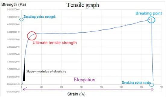
Tensile testing was used to investigate the stress-strain curve of the samples, and thereby the elasticity and stiffness of the films. The films were cut 0.4cm wide using parallel knives and approximately 4cm long. The thickness of each strip was deter-mined by measuring the thickness of the strip at 3 different parts of the strip and calculating the average. Each strip clamped at the ends between two clamps that each had a rubber band between them to prevent slipping. The measurement was performed using a Linkam ST-350 tensile stage with temperature and humidity con-trol. The machine ran for 5min before elongation started to ensure a humidity of 50% and then ran with a speed of elongation at 1 mm.s-1. The stress-strain curve of each sample was plotted, and the elastic modulus, ultimate tensile strength, breaking strength and elongation was calculated from each graph (Figure 1). Below is the correlation between strength, strain and elastic modulus
Strength =
𝐶𝐶𝑆𝑆𝑒𝑒𝑒𝑒𝑒𝑒 𝑒𝑒𝑒𝑒𝑒𝑒𝑆𝑆𝑆𝑆𝑒𝑒𝑆𝑆 𝑆𝑆𝑆𝑆𝑒𝑒𝑆𝑆 =
𝐹𝐹𝑒𝑒𝑆𝑆𝑒𝑒𝑒𝑒
𝑊𝑊𝑊𝑊
𝐹𝐹
Strain =
𝐼𝐼𝑆𝑆𝑆𝑆𝑆𝑆𝑆𝑆𝑆𝑆𝑖𝑖 𝑖𝑖𝑒𝑒𝑆𝑆𝑙𝑙𝑆𝑆ℎ =
𝐸𝐸𝑖𝑖𝑒𝑒𝑆𝑆𝑙𝑙𝑆𝑆𝑆𝑆𝑆𝑆𝑒𝑒𝑆𝑆
Δ𝐿𝐿
𝐿𝐿
Elastic modulus =
𝑆𝑆𝑆𝑆𝑆𝑆𝑒𝑒𝑆𝑆𝑙𝑙𝑆𝑆ℎ
𝑆𝑆𝑆𝑆𝑆𝑆𝑆𝑆𝑆𝑆𝑆𝑆
Where F: Force, W: width of the film, T: thickness of the film, ∆L: elongation, L0: initial length.
By plotting the result span between 10% of the breaking point strength and 35% of the breaking strength a lineal correlation can be obtained where the slope of the curve is the elastic modulus. Using values before the point where the film starts to stretch and using values after can result in values where the line starts to flatten out creating an unreliable result. The r value of the line is obtained to decide if the sam-ple is to be used or not, with an r value above 0.9 being desirable.
Figure 1 below shows where these numbers can be seen on the tensile graph
Figure 1: Stress-Strain curve with explanation about how to calculate the mechanical parameters
Two different batches of freeze-dried faba bean protein isolate (FPI) were produced. From them, three different of fibrillated samples (FFPI) were created (1 and 2 from the first extraction batch and sample 3 from the second extraction batch). FFPI 1
and 2 was used for the first set of film were FFPI 3 was used for the second set of films. This can be seen as a flow chart in appendix 3
3.1 Protein extraction and isolation
The two different batches of protein fractions (FPI) were isolated from faba
beans following the same procedure. 300 g milled faba bean flour was used
as starting material and 70.2 g was recovered after the alkaline and acidic
treatment, retrieving a yield of 23.4 %. For the second batch this number was
lost due to loss of solution during preparation.
Dumas test was performed on the different FPI bathes by duplicate. The
con-tent of the proteins in the isolated fractions was calculated and the results are
summarized in the table 1. The results show all batches contained a high
amount of protein.
Table 1: Protein content in faba bean protein isolate
Sample Sample weight (mg) % N N(mg) Protein (mg) Protein (%) Batch 1 (1) 2.95 14.49 0.43 2.67 90.56 Batch 1 (2) 3.00 12.07 0.36 2.27 75.46 Batch 2 (1) 3.04 14.71 0.45 2.80 91.96 Batch 2 (2) 2.87 14.60 0.42 2.61 91.22
The acquired solid yield after alkaline and acidic precipitation was around 23% of the original mass and the percentage of protein in faba bean, according to literature, is around 25%. This fact together with the result that the isolated samples contained around 90% of protein in their composition, test two on batch 1 considered a failed test, one can assume that the extraction of proteins was successful and therefore it will be assumed here forward that the FPI samples contained high yields of proteins.
3.2 Fibrillation
Three different protein slurries were made from the FPI batches with a concentration of 20 mg/ml. After pH adjustment, stirring and centrifugation the solution was fil-tered. The concentration of the filtrate was calculated (Table 2). The FPI batch 1 was used to obtain the sample 1 and 2, whereas the batch 2 was used to obtain the sample 3.
Table 2: Concentration of FPI filtrate
Samples Concentration (mg/ml)
Sample 1 13.71 Sample 2 13.56 Sample 3 14.11
The protein concentration of the filtrate before fibrillation was between 13.7-14.1 mg/ml. Meaning that 70% of the FPI sample was successfully dissolved in the so-lution before fibrillation. Meaning that both batches showed similar solubility. In previous experiments such a high percentage of proteins ended up producing curly nanofibrils and if the sample also contained an amount above 6.9mg/ml the nano-fibrils entangled themselves into clumps within the solution. This could be seen in the fibrillated solution made in this study as clumps in the solution.
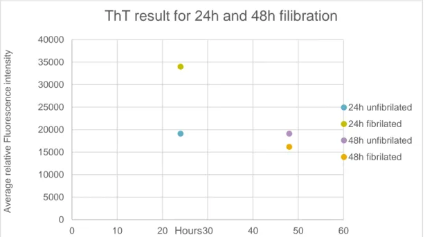
The three different samples were then incubated in a heating block during 24h for fibrillation. Th-T assay was performed on these samples before and after the heat treatment (Figure 2) to verify the formation of nanofibrils in their composition. Sam-ples 1 and 3 were also incubated during 48h and a Th-T assay was also performed on them to evaluate the effect of the incubation time on the formation of nanofibrils (Figure 3).
Figure 2: Th-T result for samples 1, 2 and 3 before and after of incubation during 24h
Samples 1 and 3 showed a similar increase in the absorbance (around 6000 units) when they were heat treated for 24h. However, sample 2 showed a lightly larger difference between fibrillated and fibrillated samples.
This shows that there were nanofibrils in the sample, but the quantity could not be determined. 0 2000 4000 6000 8000 10000 12000 14000 16000 18000 20000 0 1 2 3 4 A v er age r el at iv e F luor es c enc e int ens it y Batch
ThT result for batch 1, 2 and 3 of fibrils
Sample 1 - unfibrillated FFPI sample 1 - fibrillated Sample 2 - unfibrillated FFPI sample 2 - fibrillated Sample 3 - unfibrillated FFPI sample 3 - fibrillated
Figure 3: Th-T result for 24h and 48h fibrillation
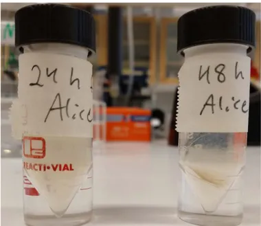
Th-T test performed on the same sample at different incubation times showed that the samples incubated for 24h reached higher absorbance values than at 48 hours. These results proved 24 hours of incubation will form higher concentration of nan-ofibrils. Figure 4 shows the different appearance between the 24h sample and the 48h sample, where 24h look more clouded and more even while the 48h sample has a big pellet in its solution
0 5000 10000 15000 20000 25000 30000 35000 40000 0 10 20 30 40 50 60 A v er age r el at iv e F luor es c enc e int ens it y Hours
ThT result for 24h and 48h filibration
24h unfibrilated 24h fibrilated 48h unfibrilated 48h fibrilated
Figure 4: FPI samples after 24h incubation and 48h incubation
3.3 Film formation
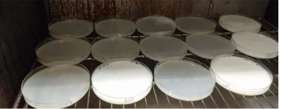
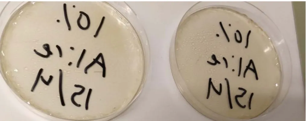
A 40mg/ml protein solution with a pH of 7 was made. After stirring and centrifuga-tion the solucentrifuga-tion was mixed with 40% glycerol and nanofibrils added in the concen-trations of 0, 2.5% 5%, 10% and 20%. The different solutions were heated, cooled and then poured into Petri dishes with each containing 1g of protein in total. These were then dried in an oven. Two set of films were created so this was done twice The two different sets of films can be viewed in figure 5 and 6. The first set has a whiter color then the second set. The whiter color also got more intense with in-creased fibril content.
Figure 5: First set of films while drying
Figure 6: Second set of films while drying
Irregularities in the films were found in both sets of films (figure 7-9). Figure 7 shows of a nerve looking elevation throughout the sample. Figure 8 shows an une-ven surface with small dots covering the surface. Figure 9 shows the difference in the dots between the films dried in the front of the oven or at the back, where the back film ended up having more dots.
Figure 8: Irregularities found in 5% 10 and 20% films in the second set of films
Figure 9: to the left is the film lying further out in the oven and to the right is the film laying further in in the oven while drying
3.4 Tensile test
Tensile tests were performed on both sets of films using a tensile machine. From these tests the modulus of elasticity (Young´s modulus), ultimate tensile strength, strength at break and strain at break was calculated. (figure 10-17). Figure 10 shows the changes in modulus of elasticity in the first set of films, showing an increase in the modulus of elasticity as a function of the increase of fibril concentration in the films with 2.5 and 5% of nanofibrils. However, the films with higher fibril concen-trations (10% and 20%) showed a drastic drop in the modulus of elasticity. Figure 10: modulus of elasticity of first set of films
0 0,005 0,01 0,015 0,02 0,025 E la c s tic it y ( P a )
Modulus of elasticity for first set of
films
0% 2.5% 5% 10% 20%Figure 11 shows the ultimate tensile strength of the films from the first set, where 20% had the lowest value and 5% had again the highest one.
Figure 11: Ultimate tensile strength for first set of films
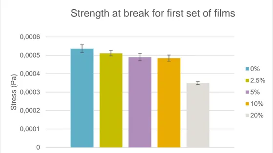
Figure 12 shows the strength at break for the first set of films where the first four concentrations all had similar values while 20% had a lower value.
Figure 12: strength at break for first set of films
0 0,0001 0,0002 0,0003 0,0004 0,0005 0,0006 St re s s ( Pa )
Ultimate tensile strength for first set
of films
0% 2.5% 5% 10% 20% 0 0,0001 0,0002 0,0003 0,0004 0,0005 0,0006 St re s s ( Pa )Strength at break for first set of films
0% 2.5% 5% 10% 20%
Figure 13 shows the strain at break where it goes down for the first concentrations to then increase for 10% and the become lower again at 20%
Figure 13: strain at break for first set of films
Figure 14 shows the modulus of elasticity for the second set of films. Despite the big deviation in the samples the average values show that the 5% film is the stiffest and the one showing less strain, meaning that this was the most brittle of the films. If looking at separate samples, it can be seen that the stiffest and the one with the lowest strain is one of the 20% samples meaning that it was very fragile. Surpris-ingly the sample without any fibrils at all showed a quite good Young’s modulus of the samples but still being quite stiff. This might indicate a poor interaction of the fibrillated proteins and the unfibrillated proteins.
Figure 14:Modulus of elasticity for second set of films
0 0,2 0,4 0,6 0,8 1 1,2 1,4 S tr ai n ( % )
Strain at break for first set of films
0% 2.5% 5% 10% 20% 0 0,005 0,01 0,015 0,02 E la c s tic it y ( Pa )
Modulus of elasticity for second set
of films
0% 2.5% 5% 10% 20%Figure 15 shows the ultimate tensile strength of films. There was a decrease in val-ues with increased concentration of fibrils, except for 10% that showed a slight in-crease in ultimate tensile strength.
Figure 15: Ultimate tensile strength for second set of films
Figure 16 shows the strength at break for the second set of films where the strength decreases with increased concentration.
Figure 16: Strength at break for second set of films
0 0,00005 0,0001 0,00015 0,0002 0,00025 0,0003 0,00035 0,0004 0,00045 0,0005 S tr es s peak ( P a)
Ultimate tensile strength for
second set of films
0% 2.5% 5% 10% 20% 0 0,0001 0,0002 0,0003 0,0004 0,0005 0,0006 S tr es s peak ( P a)
Strength at break for second set of films
0% 2.5% 5% 10% 20%
Figure 17 shows the strain at break for the first set of films where there was a de-crease of strain at break for the first 3 concentrations and then an inde-crease for con-centration 10% and 20%
Figure 17: Strain at break for second set of films
To further examine this a linear regression was created for each set of films (see appendix 4) which showed how well the sample points fitted the created regression. This was 36% for the first set of films and 9% for the second set, showing a corre-lation too low to be statistically valid.
So sadly, the tensile test showed no connection between added nanofibrils and their effect on the mechanics of the films. This was surprising and there are some possible explanations why this is.
Firstly, there might have been few nanofibrils in the samples to begin with. There are some speculations why this might be.
It might be that the nanofibrils created mainly existed in these gel clumps within the solution and since they were not dissolved they did not end up in the pipetted amount of solution, therefore the main solution contained nanofibrils, but they were not in-dicated. In previous reports this has happened if the solution used for fibrillation had a high concentration of fibrils, with the high protein concentration of the isolate and the good solubility one can argue that this might also been the case in this report (Ye et al., 2018a).
0 0,2 0,4 0,6 0,8 1 1,2 1,4 1,6 s tr ai n ( % )
Strain at break for second set of films
0% 2.5% 5% 10% 20%
Another explanation is that since there was such a high amount of proteins in the sample it might be assumed that the produced nanofibrils ended up curly. Curly nanofibrils have less β-sheets then straight nanofibrils and since Th-T indicates the presence of β-sheets the curly nanofibrils would show less fluorescence light then straight ones (Ye et al., 2018a). This is however unlikely since the difference would probably be small.
Third explanation for this is simply that the fibrillation failed and that there were few nanofibrils in the solution and instead other aggregates were formed and lastly that there had been nanofibrils in the solution but that they for some reason broken down.
The last explanation for this can be more valid if looking at the Th-T test made for sample 2 and 24h test. Both these samples were made from batch 1 isolate and the same pre-fibrillation solution. However, batch 2 was stored in the fridge for some weeks before the Th-T test (as were batch 1 and 3) was performed while for the 24h sample the Th-T test was performed the same week. Looking at the result from these Th-T test one can easily see that the 24h test had a much higher fluorescence light than the batch 2 sample, indicating that somehow the nanofibrils might have broken down during storage. However, it must be noted that Th-T tests can only show if there are nanofibrils in the sample and not estimate the concentration of the nano-fibrils.
So, if there were nanofibrils in the sample but they got broken down there are some speculations on how that happened. One of the most likely explanation is the sudden pH changes that the proteins and nanofibrils went through and that they are not as sturdy as assumed.
Secondly is the same as with the Th-T test, that the past of the solution that ended up in the films contained little nanofibrils due to the nanofibrils becoming clumps within the solution. Moreover, if these clumps were moved into the films there might been an uneven distribution of them within the film due to their assumed resilience.
Another explanation is the fact that the filtrate in this experiment had a high con-centration of proteins. It has previously been shown that this high concon-centration created curly fibrils (Ye et al., 2018a). Curly fibrils have a lower percentage of β -sheets then straight fibrils, and it is the β --sheets that gives fibrils their wanted
me-chanical abilities (Jansens et al., 2018, Ye et al., 2018a). So assuming that curly fi-brils were crated instead of straight fifi-brils it would partly explain the unexpected low modulus of elasticity in this experiment.
There are also some more loosely based speculations.
Firstly there might have been bacteria growth in the films while drying, breaking down the structure in the films. The arguments for this are that the first set of films had a very distinct smell and were extreme fragile and did not produce any result in the tensile test. These films were quickly discarded, and the dying oven disinfected. The second set of films however had a hint of the same smell, but only those that were dried closest to the fan, these were the ones with the most bubbles in them. That leads us to the second speculation: bubbles. Even if the speculated bacteria did not break down the structure of the films they might be the reason for the bubbles in the films. The bubbles might have ended up in the films with the nanofibrils, which have shown to have a good foaming and network abilities, and therefore could be able to trap microscopic air bubbles in the film and with that made the films uneven.
The films with the bubbles or bumps were not used in the testing but, as stated, there might have been microscopic bubbles in the tested samples.
There is also the simple explanation that the faba bean protein profile does poorly when creating nanofibrils or that the faba bean nanofibrils are less resilient then other protein-based nanofibrils.
There is also a wide variation in each concentration. This can be explained with the small number of test points. However, it can be observed that with increased amount of fibrils the more variation there was, and this might be due to the nanofibrils some-how making the films uneven, perhaps by trapping air or any of the other options previously discussed.
4
Conclusion and future perspectives
In conclusion, faba bean proteins were successfully isolated and extracted from the faba bean flour, although the fibrillation and film casting were inconclusive. There was no correlation between the concentrations of nanofibrils in the films and the resulting modulus of elasticity. There are multiple theories why this may be, such as bacteria growth, bubbles trapped within the film, lack of nanofibrils, fibril break down during process or that too high concentration of proteins in the filtrate inhib-ited the creation of fibrils, but none of these can be certainly confirmed. The aim of the study was not completely reached.
In the end, more tests are needed to reach the aim, such as examinations of how to remove microscopical air bubbles from the samples, and the effect of the pH change on the nanofibrils and their conformation.
It might also be a good idea to produce some films in a more sterile environment to ensure that they do not contain any bacteria if it is shown that specifically faba bean isolate is susceptible to them.
It would be preferable to quantify the concentration of fibrils to be able to control the process more.
It must be noted that there have been successful tests performed on nanofibrils be-fore and they have shown promising mechanical abilities. Since these test results are inconclusive and multiple possible errors have been discovered it might still be possible to use faba bean protein nanofibrils for meat-like substitutes once these errors are addressed.
Akkermans, C., van der Goot, A.J., Venema, P., van der Linden, E., Boom, R.M., 2008. Formation of fibrillar whey protein aggregates: Influence of heat and shear treatment, and resulting rheol-ogy. Food Hydrocoll. 22, 1315–1325. https://doi.org/10.1016/j.foodhyd.2007.07.001 Arogundade, L., Tshay, M., Shumey, D., Manazie, S., 2006. Effect of ionic strength and/or pH on
Extractability and physico-functional characterization of broad bean (Vicia faba L.) Protein concentrate. Food Hydrocoll. 20, 1124–1134. https://doi.org/10.1016/j.foodhyd.2005.12.010
Biancalana, M., Koide, S., 2010. Molecular Mechanism of Thioflavin-T Binding to Amyloid Fibrils. Biochim. Biophys. Acta 1804, 1405–1412. https://doi.org/10.1016/j.bbapap.2010.04.001 Bolder, S.G., Hendrickx, H., Sagis, L.M.C., van der Linden, E., 2006. Fibril assemblies in aqueous
whey protein mixtures. J. Agric. Food Chem. 54, 4229–4234.
https://doi.org/10.1021/jf060606s
Boye, J., Zare, F., Pletch, A., 2010. Pulse proteins: Processing, characterization, functional properties and applications in food and feed. Food Res. Int., Molecular, Functional and Processing Characteristics of Whole Pulses and Pulse Fractions and their Emerging Food and Nutraceu-tical Applications 43, 414–431. https://doi.org/10.1016/j.foodres.2009.09.003
Crépon, K., Marget, P., Peyronnet, C., Carrouée, B., Arese, P., Duc, G., 2010. Nutritional value of faba bean (Vicia faba L.) seeds for feed and food. Field Crops Res., Faba Beans in Sustainable Agriculture 115, 329–339. https://doi.org/10.1016/j.fcr.2009.09.016
Duc, G., Aleksić, J.M., Marget, P., Mikic, A., Paull, J., Redden, R.J., Sass, O., Stoddard, F.L., Vand-enberg, A., Vishnyakova, M., Torres, A.M., 2015. Faba Bean, in: De Ron, A.M. (Ed.), Grain Legumes, Handbook of Plant Breeding. Springer New York, New York, NY, pp. 141–178. https://doi.org/10.1007/978-1-4939-2797-5_5
Fouad, M., Mohammed, N., Aladdin, H., Ahmed, A., Xuxiao, Z., Shiying, B., Tao, Y., 2013. 5 - Faba Bean, in: Singh, M., Upadhyaya, H.D., Bisht, I.S. (Eds.), Genetic and Genomic Resources of Grain Legume Improvement. Elsevier, Oxford, pp. 113–136. https://doi.org/10.1016/B978-0-12-397935-3.00005-0
Hoek, A.C., Elzerman, J.E., Hageman, R., Kok, F.J., Luning, P.A., Graaf, C. de, 2013. Are meat sub-stitutes liked better over time? A repeated in-home use test with meat subsub-stitutes or meat in meals. Food Qual. Prefer. 28, 253–263. https://doi.org/10.1016/j.foodqual.2012.07.002 Hoek, A.C., Luning, P.A., Weijzen, P., Engels, W., Kok, F.J., de Graaf, C., 2011. Replacement of meat
by meat substitutes. A survey on person- and product-related factors in consumer acceptance. Appetite 56, 662–673. https://doi.org/10.1016/j.appet.2011.02.001
Holmberg, I., 2013. Åkerböna - i ekologiska odlingssystem [WWW Document]. URL https://stud.ep-silon.slu.se/6025/ (accessed 5.23.19).
Hopp, T.P., Hemperly, J.J., Cunningham, B.A., 1982. Amino acid sequence and variant forms of favin, a lectin from Vicia faba. J. Biol. Chem. 257, 4473–4483.
Jansens, K., Rombouts, I., Grootaert, C., Brijs, K., Van Camp, J., Van der Meeren, P., Rousseau, F., Schymkowitz, J., Delcour, J., 2018. Rational Design of Amyloid-Like Fibrillary Structures for Tailoring Food Protein Techno-Functionality and Their Potential Health Implications.