From patient to specimen
and back again
Radical surgeries and pelvic pathologies
in the Museum Obstetricum
HELENA FRANZÉN*
In late November 1862, Anna Dahlström, a thirty-four-year-old unwed maid who was in labour, entered the lying-in unit at Uppsala University Hos-pital, Sweden.1 She was examined by the professor of surgery and
obstet-rics, Carl Benedict Mesterton (1826–1889), who concluded that her pelvic canal was too narrow for the fetus to pass through. After consulting with a colleague, “it was decided that a caesarean section was to be suggested to the patient, as the only way to save the mother without sacrificing the still living fetus”.2 In the course of that same afternoon, her body was
monitored while additional colleagues from the medical faculty, students, and practicing physicians from the city of Uppsala gathered in anticipation of the impending surgery. Dahlström herself was briefed in the evening:
At 9 pm I notified the patient about her situation and about the interven-tion, which was considered to be necessary for saving her and the fetus. The dangers of the operation for her were presented, and she was left with the choice of cutting up the living fetus and retracting it the natural way, to save her alone, or, with greater danger for her, also trying to save her child through the suggested surgery. Without hesitation and with a ready bravery the woman preferred the latter option.3
An hour later, Dahlström was put on the operating table and rendered unconscious by chloroform, closely surrounded by Mesterton and his seven assistants. Items such as ice and new, boiled swabs had been placed on a nearby table. The incision was made, and layer after layer of her abdomen was unfolded. Blood spurts were curbed by an assistant, who
*PhD student Helena Franzén, Department of History of Science and Ideas, Uppsala Univer-sity, helena.franzen@idehist.uu.se
pressed them with a swab dipped in iced water. When the uterus was breached, hands reached down and perforated the amniotic sac, hauling out the fetus. Dahlström woke up while the innards of her abdomen were still exposed, and the work to close it up started. There were, however, complications. Dahlström perished thirty hours after her belly had been opened, yet her daughter survived. The mother was autopsied the follow-ing day, her pelvis prepared into a specimen and collection object—by severing it from the rest of her body, skeletonizing, and labelling it—and, Mesterton expressed, “kept as no. 38 in the University’s beautiful and valuable collection of pathological pelves”.4
In the early 1860s, this collection was merged with the recently pur-chased obstetrical collection of Magnus Retzius (1795–1871), professor of obstetrics at the Karolinska Institute—at the time called the Carolinska Medico-Kirurgiska Institutet—in Stockholm. The two collections became a new unit called the Museum Obstetricum. For several decades it was housed within the anatomical department, alongside the collections tied to the chairs of anatomy and pathology.5 In 1903, the objects in the
Museum Obstetricum were separated. The pelvis specimens, together with another category of collection objects—obstetrical instruments— were moved to the Uppsala University Hospital, and in the mid-1990s, a few pelves and the instruments were relocated to the Museum of Medical History in Uppsala. In 2006, the pelves remaining at the hospital were transferred to the museum. They remain there today, while some of the other objects initially left behind at the anatomical department are now managed by the Gustavianum, Uppsala University Museum.6
This article is situated within the wider field of the history of collec-tions, but more specifically within the branch of medical collections.7 In
the context of research within this field, human remains in the form of specimens are—in line with the material turn—framed as historical arte-facts. By approaching them as tangible remnants of the past, enquiries into their contexts can unravel various themes.8 Scholars have examined
aspects of knowledge production regarding medical collections, such as their role in establishing new fields or disciplines, and in training stu-dents.9 Moreover, the importance of personal, professional, and
institu-tional networks for collecting specimens has been investigated.10
How-ever, obstetrical collections—a specific kind of medical collection which intersects with the fields of history of medicine and reproduction—have received little attention.11 Furthermore, the connections between
obstet-rical collections and surgical interventions on labouring women remain largely uninvestigated.
The emergence of the Museum Obstetricum at Uppsala University, in the mid-nineteenth century, is explored in this article. The aim is to
examine its formation, the motivations of the doctor-collectors who as-sembled it, and ways in which they articulated the intended usage of the collection objects. The acquisition of the pelvis specimens and casts there-of was shaped by the dynamic between collecting and obstetrical practice, specifically radical surgeries—such as the caesarean section—on women in labour. By investigating this interaction, I analyse how knowledge was produced through the Museum Obstetricum. By situating this obstetrical collection in the intersection of the contemporary medical interest in pathological conditions and the infrastructure of maternity care, I show that the two collections—from Stockholm and Uppsala—were merged in order to create a selection of pathological pelvis specimens and obstetrical instruments, which could be used in medical education as preparation for obstetrical practice. The collection emerged from complex moral and cultural contexts, which involved questions such as framings of the con-sent of female patients and their relatives, doctors’ conflicting interests in saving lives or obtaining new reference material, and decisions about whether to save the woman or the fetus when the pelvic canal was too narrow for the fetus to pass through.
Surviving historical medical collections might appear as merely a static way of storing knowledge, in the form of material objects. However, earlier studies have shown that they had a multifaceted role. They were actively used as a hands-on resource for teaching and scientific enquiry in the nine-teenth century, an era which has been referred to as the age of museum medicine.12 Moreover, scholars have conceptualized this use in the
meta-phor of collections as libraries.13 Accordingly, the Museum Obstetricum
fuelled obstetrical knowledge production at Uppsala University, under-pinning education and research as well as symbolizing Uppsala Univer-sity’s prestige in the medical arena. However, the formation of knowledge was not a linear process; there was a mutual exchange on several levels— between medicine and society, as well as between the collection and its objects, social networks of physicians, and obstetrical practice. Previous research has stressed that co-production of knowledge takes place in the relations between people, artefacts, and the uses thereof—an approach which this study adopts in order to discuss the dynamic of collection objects’ way from patient to specimen and back again.14
The concept of co-production enables close examination of these rela-tions and their outcomes. Sheila Jasanoff has specified that “work in the co-productionist idiom stresses the constant intertwining of the cogni-tive, the material, the social and the normative”. Hence, this approach does not favour one of these aspects over the others; it rather focuses the analytical lens on the interplay between them.15 Co-production draws
separate spheres, are intertwined, have effects on each other, and are con-nected by people and things.16 Furthermore, it sheds light on how social
relations are materialized in technology, and how the design of such tech-nologies steers future interactions between people.17 In order to employ
this idiom, I frame collection objects—pelvis specimens, casts, and instru-ments—as medical technologies that were used to teach, diagnose, and treat medical conditions, as well as demonstrating medical power and professionality.18
To conduct this investigation, a broad spectrum of sources is required. I tap into three categories: collection objects, archival texts, and medical publications. This combination enables me to trace the processes of co-production, since each type of source sheds light on different points in the loop, as well as on diverse aspects. By examining collection objects, it is possible to gain clues to their origin. Archival texts enable me to further examine the collection’s contents and uses. And medical publications offer insights into how the formation of the Museum Obstetricum was driven by doctors and their collecting networks, as well as revealing tensions within obstetrical practice, which also was a context of obtaining speci-mens. By considering these sources together, it is possible to analyse how technologies and practices were mutually shaped. A serious limitation is the absence of the voices of the female patients. Their agency is only in-directly acknowledged through the descriptions and statements of the physicians who performed the surgical interventions and gathered pelvis specimens from women who died of caesarean sections.
This article consists of four sections. Firstly, I situate the Museum Obstetricum in the wider context of obstetrical collections and doctors’ interest in pathological conditions. Secondly, I discuss the infrastructure of maternity care and radical surgeries on labouring women in Sweden during the first half of the nineteenth century. Subsequently, I analyse the dynamic of collecting pelvis specimens in the context of performing the caesarean section, a radical surgery. In the last section, I examine the specificities of pelvic pathologies in the Museum Obstetricum. In the conclusion, I discuss the mechanisms and effects of co-production in the interplay between the formation of the Museum Obstetricum and obstet-rical practice.
Setting the stage for the Museum Obstetricum
Early modern cabinets of curiosities contained rarities and wonders, and the period’s natural history collections were intended to represent the whole world in miniature.19 In the nineteenth century, collections
was gradual, and different ways of organizing collections overlapped.21
Collections could be either privately owned or institutionalized. Physi-cians obtained body parts from deceased patients during autopsy, or from corpses during dissection, and prepared them into specimens by different means of conservation.22 Previously, individual physicians networked by
sending each other specimens, and in the nineteenth century institutions also participated in networking through this practice.23
In contrast to other specialized medical collections, obstetrical collec-tions had been around since the seventeenth century. While midwifery has older roots, obstetrics—the management of obstructed labours—was staked out as a specialist subject by physicians in early modern Europe. But there was no clear-cut shift from midwifery to obstetrics, and the process differed between countries.24 Obstetrics and obstetrical collections
were introduced in Sweden, more specifically Stockholm, around 1700.25
Objects in obstetrical collections, such as phantoms—models which rep-resented the labouring woman’s body and that of her fetus—were used as visual, as well as tactile, pedagogical aids to instruct midwives in delivery.26
When the Public Lying-In Hospital in Stockholm was founded in the lat-ter part of the eighteenth century, a collection containing women’s pelves, fetal crania, and phantoms was established there, in order to teach mid-wifery and medical students the tactile skills of managing parturition.27
The reasons for establishing this lying-in hospital were twofold. Beyond generating training opportunities for students, it was also intended to provide maternity care for pregnant women.28
During the first half of the nineteenth century, several collections con-nected to medical institutions or professionals emerged in Stockholm. One such institution was the newly founded Karolinska Institute, where Anders Retzius (1796–1860), an influential professor of anatomy, built up collections of anatomical specimens.29 While Anders Retzius collected for
the institution he represented, his brother Magnus Retzius established a private collection of obstetrical objects. It is likely that he commenced assembling it in the 1820s, after starting his obstetrical career. Magnus Retzius became assistant physician at the Pro Patria Lying-In Hospital in Stockholm in 1819, and chief physician there in 1830. In 1849 he assumed the chair of obstetrics at the Karolinska Institute and became the director of the Public Lying-In Hospital.30 In this capacity, Magnus Retzius’s
col-lection was tallied as a resource for the obstetrical education of medical students at the Karolinska Institute. Beyond obtaining pelvis specimens from his deceased patients, he had a vast national and international net-work of colleagues with whom he exchanged specimens.31
When Magnus Retzius sold his collection to Uppsala University in 1862, it contained pelvis specimens described according to pathological
and racial categories, casts of pelvis specimens in plaster and papier mâché, crania and whole mounted skeletons of fetuses, obstetrical instruments, and models of the pregnant body.32 It was merged with the collection of
pathological pelves in Uppsala, which together constituted the foundation of a new unit—the Museum Obstetricum. The creation of this collection was encouraged by representatives of the medical faculty as a way to strengthen Uppsala University’s position as an actor in the arena of med-ical education in Sweden, especially in relation to the Karolinska Institute, their competitor.33 By extension, the formation of the Museum Obstetricum
was also considered as a means to provide maternity care to the population. The year before obtaining Magnus Retzius’s collection, the medical faculty had purchased seventeen casts of “deformed pelves” from the Fleischmann papier-mâché factory in Nuremberg, purportedly for the teaching of obstetrics. This purchase was highlighted as an important acquisition in the Uppsala University yearbook.34 The pathological was
emphasized in the Museum Obstetricum, which marked a division be-tween obstetrics and anatomy in general, as well as the content of the disciplines’ collections.35 While the general aim of anatomical coursework
at the time was to give students an understanding of the body as a whole, and how the parts related to each other, obstetrical education was focused on teaching students to handle obstructed labours—that is, situations when the fetus could not pass through the pelvic canal without interven-tion.36 While contracted pelves were noticed in the eighteenth century, a
wider array of specific forms emerged in the first half of the nineteenth century.37 This development tied in with the general trend of
specializa-tion within medicine, and the overarching interest in pathological condi-tions, which permeated many disciplines.38
Obstetricians—or in Sweden, physicians with training in obstetrics— were primarily interested in pathological pelves because obstructed labour was their specialist subject. The collections were used for scientific inves-tigations and training students, and were seen as means to understand different kinds of pathologies, such as obstructed labours. Hence, their contents, like those of other medical collections, mirrored the collectors’ field of expertise. The Museum Obstetricum was positioned as a resource, meant to be used like a library, for learning about a wide array of pelves with pathological conditions which could be encountered in obstetrical practice. Like books, which were printed in multiple copies, pelvis speci-mens could be reproduced by casting. This enabled the experiences of individual physicians to be transferred to students, who could see and touch the pelvis pathology. The students were thus taught to read the shape of bones, by handling the materialization of previous patient cases, in order to learn to identify pelvic pathologies in future patients.
Maternity care and radical surgeries
in nineteenth-century Sweden
Obstetrical collections were enmeshed with obstetrical practice. There-fore, to understand the formation of the Museum Obstetricum, the infra-structure of maternity care in Sweden during the first half of the nine-teenth century has to be taken into account. Radical surgeries within obstetrics at the time—the dismemberment of the fetus and the caesarean section—took place within this infrastructure and were conducted by physicians and midwives as responses to obstructed labour, which was a matter of life or death. These surgeries were conducted in a limited num-ber during the eighteenth century, but were performed to an increasing degree in the nineteenth century. This shift tied in with the tightening state control of maternity care, a grip rooted in mercantilism, for which a growing population was considered a resource.39
The majority of the Swedish population lived in the countryside in the nineteenth century, most childbirths took place at home, and many women preferred to be attended by local women. However, the Swedish state had stipulated that labours taking place at home should be supervised by a midwife or a physician.40 Moreover, while previously there had been just
a few lying-in hospitals located in the bigger cities, there was a surge in the establishment of lying-in units at county hospitals in the mid-nine-teenth century—such as in Uppsala, where the city’s first lying-in unit was founded in 1859. Doctors viewed these institutions as a way to provide maternity care for poor women, as well as to provide students with clini-cal experience of managing labours.41
This shift is equivocal. On the one hand, the tightened state control of maternity care enabled physicians to access women’s bodies; on the other, it resonated with the sometimes desperate situations of giving birth.42 For
example, some patients at the Public Lying-In Hospital were described by physicians as “poor unfortunate creatures from the countryside who have been transferred several miles on jolting carriages during the labour, and not earlier than after several days of the most painful and strenuous at-tempts of delivering at home”.43 It is likely that this example refers to a
woman in obstructed labour. If no interventions were made in cases like this, the woman and fetus both would die—which, in line with the mer-cantilist approach, was framed as a problem by the state. This problem was expected to be solved by the practitioners of maternity care, that is, midwives and physicians.
The pathology of obstructed labour was most often understood as situ-ated in the woman’s pelvis, in the sense that it was too narrow for the fetus to pass through. However, the pathology was only actualized when
women who had a contracted pelvis were pregnant or in labour. The rela-tion between the bodies of the woman and the fetus was rooted in a mechanistic understanding of labour. As Emily Martin has observed, physicians imagined their obstetrical practice as mechanistic in the sense that “the woman’s body is the machine and the doctor is the mechanic or technician who ‘fixes’ it”.44
To solve the problem of the woman and fetus both dying because of obstructed labour, obstetrical instruments were applied. There were two categories of these for retrieving the fetus through the pelvic channel— blunt and sharp. These instruments were designed to be used for different kinds of fetal retrieval. While the forceps (blunt) was intended to get the fetus out alive, tools such as perforators and hooks (sharp) were used to dismember it. With the former, holes could be drilled in the fetus’s cra-nium, and the latter could be inserted into the perforated skull, to drag the fetus out. The designs of the different categories of instruments were based on the intended results, and at the same time the material properties of the instruments steered what kind of action could be taken. Hence, the instruments and the practices of managing obstructed labours were mutu-ally shaped, co-produced. The majority, around two hundred, of the ob-jects in Magnus Retzius’s collection when sold to Uppsala University were blunt and sharp obstetrical instruments.45
Different criteria governed the choice of instruments, when attempting to retrieve a fetus through the pelvic canal. Blunt ones were primarily used, if there appeared to be a possibility of retrieving the fetus alive, that is, if the woman’s pelvic canal was thought to be wide enough. If that endeavour was unsuccessful, the next step could be to attempt dismem-bering it.46 To dismember a fetus, especially a living one, was framed by
physicians as a horrible experience for everyone involved.47 However,
sharp instruments were sometimes repeatedly applied to deliver women who had too narrow a pelvis for the fetus to pass through. These women were encouraged by their provincial physicians to travel to the nearest lying-in hospital in the latter part of their next pregnancy, to be induced before the fetus was full term, but considered viable.48 This advice
illus-trates attempts to avoid interventions considered to be extreme.
The practitioners of maternity care had different realms of authority: midwives were expected to tend to the uncomplicated labours, and physi-cians the complicated. However, in this period there were more midwives than physicians, and the likelihood was often that there was no physician in close enough proximity to be called in when a labour was deemed complicated. Because of these circumstances, midwives had an unusually strong position in Sweden throughout the nineteenth century, in the sense that they were allowed to use blunt and sharp obstetrical instruments.49
However, they were not allowed to use sharp instruments on a fetus which was deemed to be alive, but were instead encouraged to wait until it was sure to be dead. Midwives were warned by physicians that to use sharp instruments on a living fetus was murder.50 Physicians, on the other hand,
considered themselves entitled to dismember a living fetus; the practice did occur, although rarely, and it was viewed as a way to possibly save the woman before she perished from exhaustion.51
Furthermore, operations which targeted the woman’s body, such as the caesarean section, were considered the domain of physicians.52 The
major-ity of caesarean sections performed in Sweden were carried out from 1850 onwards, and most of them after the 1880s. It is likely that this was due in part to the general trend of not performing abdominal surgery before mid-century. When inhalation anaesthesia was introduced in 1847, physi-cians were enabled to venture into the abdominal cavity of living patients, even though it resulted in a high mortality rate.53 Some of the caesarean
sections took place at lying-in hospitals, and others in farmer’s cottages.54
Some physicians who carried out caesarean sections in Europe during the first half of the nineteenth century reported that approximately 50 per cent of patients who underwent this operation lived. Physicians in Sweden were aware of these reports, and the caesarean section was accordingly framed as an intervention which women had a chance to survive.55
How-ever, in the Swedish cases, all the women died in the aftermath, while about half of the fetuses lived.56 Hence, in Sweden, this operation
gener-ated the opposite results compared with the dismemberment of the fetus, in terms of who was intended, or expected, to survive.
The ambiguities of the caesarean section
The main dilemma of whether to conduct a caesarean section was the question of whom the physician should attempt to save in cases of ob-structed labour, when the fetus was deemed to be alive but the woman’s pelvic canal too narrow for retrieval with blunt instruments. While the dismemberment of the fetus generated results in absolute terms—that is, its death—the caesarean section opened up a scale of possibilities and expectations. Even though this operation was known to result in a high mortality rate for the women, it was hailed by several physicians as an intervention which both mother and fetus had a chance to survive. The motivations for conducting this surgery differed between countries on an overarching scale, but also depended on factors such as the attitude of local communities and individual physicians.57
Religion played into these calculations as well. In general terms, within Catholicism, it was forbidden to take innocent life. What this meant in
practice could be framed in various ways, and the Catholic Church and its representatives struggled with this issue in relation to the surgical techniques—and expectations thereof—which emerged in the nineteenth century.58 Within Protestantism, by default, there was no consensus
re-garding how to frame the life of the woman and the fetus. Furthermore, there were not uniform approaches within countries in which neither Catholicism nor Protestantism was predominant.59 For example, in France
and England, the dismemberment of the fetus was the more common intervention in cases of obstructed labour in the nineteenth century.60 In
the United States, where the Catholic Church staked out a strong pres-ence, the caesarean section was conducted with greater frequency.61
Some comparative features can be teased out, concerning how the di-lemma of saving the woman or the fetus played out in different countries. The entanglement of religion in obstetric practice can be traced through factors such as how involved the state and the church—or Protestant con-gregations—were in maternity care in individual countries, as well as the relation between religious organizations and specific states. Moreover, the personal conviction of individual physicians could come into play, but to some degree decision-making depended on how regulated their practice was, and by whom. Another feature was where the woman in obstructed labour was located, and who were present.
One prevalent aspect in the cases when a caesarean section was con-ducted in Protestant Sweden is that several of the physicians, such as Mesterton, made a point of stressing that they had informed the woman of the risks for herself, and asked for her permission to go through with the operation.62 However, what consent meant, or how it was constructed,
is not a given. Previous research has shown that consent is culturally and historically contingent, with factors—beyond religion—such as gender, class, and race influencing how it is constructed, and in whom it is situat-ed.63 Moreover, being informed and consenting did not necessarily
over-lap.64
Nevertheless, not all physicians in Sweden who discussed the caesarean section approached the issue of consent in the same way. For example, Magnus Retzius aligned himself with the view of the German obstetrician Hermann Friedrich Kilian (1800–1863), who argued that the caesarean section should be performed if the life of the fetus possibly could be saved by it. Retzius further stated that even though the woman had a say, she should listen to the physician’s recommendation.65 This indicates that, in
Retzius’s opinion, there was not really a choice for her. In contrast to Retzius, his predecessor as professor of obstetrics at the Karolinska Insti-tute, Pehr Gustaf Cederschiöld (1782–1848), had instead been adamant that a caesarean section could be conducted only if the woman agreed.66
One reason for the physicians to recount that they had asked for the patient’s permission—regardless of whether they actually did or not—was most likely a fear of accusations of misconduct. Physicians could be brought to trial. For example, in the 1850s, Magnus Retzius was charged with causing a recently delivered woman to die from lack of treatment.67
To avoid this, Cederschiöld had stated that one should “demonstrate the situation for other educated and credible persons” before proceeding with a caesarean section, in order to “protect oneself against slander”.68 In this
light, to consult with one or several colleagues was not only a way to as-certain diagnosis. In Sweden this was a strategy for the physician who wielded the scalpel to have peer witnesses, who could attest to the diag-nosis which formed a reason for conducting the operation. Since the caesarean section was such a dangerous surgery for the woman, it is like-ly that asking for her consent—or stating that this had been done—was a way to remove liability from the physician and transfer it to the woman.
Another aspect is that some physicians also stressed the female patients’ desire to obtain a child. Beyond Mesterton’s report, this is present in the case description of the caesarean section conducted in 1860 by Gustaf Fredrik Hjort (1818–1876), midwifery teacher at the Sahlgrenska hospital in Gothenburg. The woman’s previous pregnancy had ended in the dis-memberment of the fetus, and when asked whether she was willing to submit to the caesarean section, Hjort stated, “she told me that she was ready to submit to whichever risk, as long as the fetus would live”. He further argued that when asked whether she wanted to consult with her husband, “she said, ‘he probably only wants me to live, regardless of how it will end for the fetus’”. Hjort declared himself to have heeded her wishes and not contacted her husband.69 Also Mesterton stressed that the
decision should be made by the woman, and that no relatives had the right to oppose her if she wanted to go through with the operation, even though this prerogative was not regulated by law. He also stated that it was equally her right to opt for dismembering the fetus, because the fatal risk was much higher for her.70 In this study it is not possible to gauge whether the
women actually had a say, but these examples shed light on some of the ambiguities connected with the caesarean section in Sweden during this time.
While radical surgeries saved, or were intended to save, lives, they often did not; body parts from deceased patients, in turn, were considered as means to generate knowledge for saving the lives of future patients. Pelvis specimens obtained during autopsies on some of the women who died in the aftermath of radical surgeries were incorporated into obstetrical col-lections, such as the Museum Obstetricum.71 All of these stemmed from
seem-ingly no pelvis specimens were obtained from women who perished during surgeries conducted in their homes.72 This ties in with the general trend
that physicians conducted autopsies of patients who died at hospitals, even though it sometimes occurred that deceased patients were autopsied in their homes.73 Hospitals were framed by physicians as sites where women
could receive better care than at home, but at the same time they were a central site for collecting specimens for medical collections, and by exten-sion facilitating physicians’ claim to expertise.
While women who underwent the caesarean section in Sweden during the period of study allegedly consented to the surgery and the mortal risks involved, one predominant factor is that all of them belonged to lower social classes, and found themselves in an acute and desperate situation. By entering lying-in hospitals, the physicians’ realm of expertise, they could have been seen as basically subjecting themselves to that very expertise.74
Previous historically oriented research has shown that in a situation in which poor and/or lower-class patients actively sought out treatment, it was likely that they deferred to the physician’s recommendation—which might have been motivated by a desire to test new interventions. Furthermore, this type of situation does not necessarily exclude degrees of coercion.75
In Magnus Retzius’s collection, there were two pelvis specimens which derived from women who had perished in relation to caesarean sections and subsequently became part of the Museum Obstetricum.76 One was
from the operation conducted by Retzius himself, and one from an opera-tion conducted by Johan August Liborius (1802–1870), a chief physician at the Sahlgrenska hospital. Liborius was part of Retzius’s vast network, and they both partook in the practice of exchanging specimens with col-leagues.77 This practice enabled physicians to add specimens that they
themselves had not obtained first-hand, thereby expanding the selection in their own collections, or those of the institutions they represented. The desire for a wider selection of pathological pelves drove physicians to obtain specimens, and at the same time, knowledge gained from experi-ences and specimens of past surgeries steered obstetrical practice. This mutual shaping, co-production, was an ongoing loop.
The caesarean section conducted by Retzius took place in September 1851, when the twenty-four-year old Hedda Sofia Ersdotter entered the Public Lying-In Hospital after already having been in labour for three days. A large tumour in her vagina obstructed the fetus’s path. Even though this tumour had been present a few years earlier, when she last gave birth, it had then been small enough to let the previous child pass. But the tumour had since grown. Retzius enquired about her health and examined her body. He deemed it unlikely that the fetus was still alive, but it had to be removed from Ersdotter’s womb.78
The verdict was that the fetus could not pass through the pelvic channel without being dismembered, but that there was not enough space for manoeuvring the sharp instruments. Magnus Retzius, together with the midwifery teacher Joseph Elliot (1799–1855) and Hjort—who was the former’s adjunct at the time—decided that the only solution to possibly save Ersdotter—and the fetus if it was still alive—was to perform a caesar-ean section. In this case, there was deemed to be no collision between the lives of the woman and the fetus. It was rather an issue of trying not to lose both. However, the fetus turned out to be putrefied, and Ersdotter did not survive the ordeal. The autopsy was conducted by Magnus Retz-ius the following day, attended by a handful of colleagues, along with the medical students who interned at the lying-in hospital at the time.79
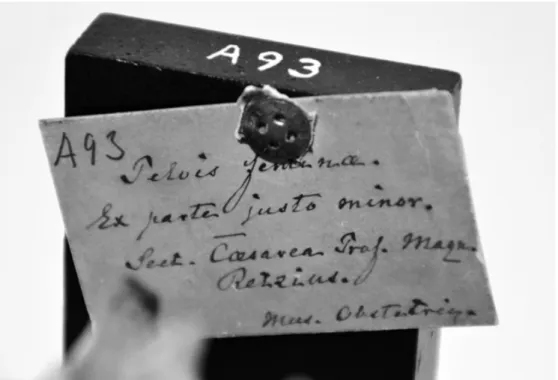
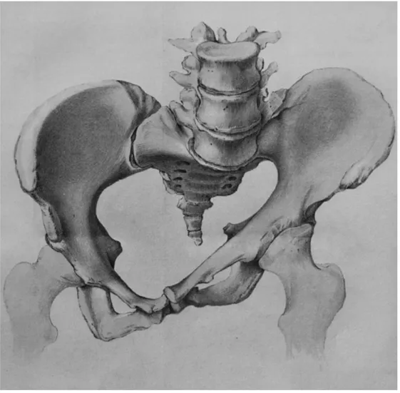
After the autopsy, Ersdotter’s pelvis was prepared as a collection object (fig. 1), and a few years later, it was featured in Museum Anatomicum Hol-miense (1855)—a printed catalogue of selected specimens from the collec-tions of the Karolinska Institute—in which Ersdotter’s case was described, and her pelvis illustrated (fig. 2).80 The information that the pelvis
speci-men derived from the caesarean section performed by Magnus Retzius was noted in the inventory that formed part of the sales contract between
Fig. 1. Ersdotter’s pelvis—skeletonized, mounted, and labelled; hence, made into a collection object and intended as a reference to be stored and con-sulted. Source: Uppsala Museum of Medical History, Object A93, UMM 408. Photograph by author.
Fig. 2. Print illustrating the pelvis specimen from Ersdotter, with the tumour featured. The print was, together with the textual description, linked to the patient case. Source: Museum Antomicum Holmiense (1855). Courtesy of the Hagströmer Medico-Historical Library, Karolinska Institute.
him and Uppsala University, as well as on the specimen’s label (fig. 3).81
This record-keeping was done in order to connect the information—the patient case—to the specimen, which was necessary for the latter’s use as reference material. While the pathological condition in this case was not situated in the woman’s actual pelvic bones, the process followed the script of diagnosing obstruction and attempting treatment. And when the patient died, her pelvis was turned into a collection object and medical technology intended to be used for learning how to identify the specific pathology in other, living, patients.
Pelvic pathologies in the Museum Obstetricum
Beyond constituting a foundation for scientific enquiry, obtaining speci-mens from deceased patients was part of the training of students. By being taught to conduct autopsy, students were trained to identify pathological lesions and causes of death, making connections between dead bodies and living patients. This process was enmeshed with the clinic, that is, teach-ing hospitals, which emerged to a greater degree durteach-ing this time.82
Obstructed labour was mostly understood as situated in the width and form of the woman’s pelvic bones. Hence, the pelvis was the focus of
Fig. 3. The label for Ersdotter’s pelvis, with the text “A93. Pelvis femina. Ex parte justo minor. Sect. Caesarea Prof. Magnus Retzius. Mus. Obstetric.” Source: Uppsala Museum of Medical History, Object A93, UMM 408. Photograph by author.
investigation when figuring out the extent of obstruction. The pelvis was defined as pathological if it was too narrow for the fetus to pass through. This condition was therefore constituted in contrast with being wide enough, relative to the size of the fetus’s head.83 In effect, the pathological
was defined in terms of ideas about function in the context of giving birth; hence, it was founded on cultural norms and actualized in pregnant and labouring women.84 Establishing the minimum width of a pelvis for a
full-term fetus to pass through allowed pathological pelves to be identified according to measurements.
The practice of measuring pelves was called pelvimetry.85 The
instru-ments used for taking pelvic measureinstru-ments were called pelvimeters, and there were three different kinds depending on which width was being quantified: external, internal, and both. There were six of these instruments in Magnus Retzius’s collection when it was sold to Uppsala University, and it is likely that they were used in his obstetric practice.86 Since the
pathology of obstructed labours was understood as situated in the pelvic bones, the pelvis specimens obtained from deceased patients were skele-tonized in order to make the pathology visible, tangible, and measurable. Magnus Retzius took part in debates both within Sweden and interna-tionally, concerning where the category of the normal pelvis ended and the pathological started. When he applied for the position of professor of obstetrics at the Karolinska Institute, he did so with the dissertation Af-handling om bäckenförträngning (1848), dealing with various kinds of con-tracted pelves and their role in obstructed labour. Moreover, he discussed different interventions in relation to the specific kinds of contracted pelves, and the measurements of the pelvic canal. This research corre-sponded with his collection, which when sold to Uppsala University was reported to “contain specimens of the majority of kinds of deformed pelves”, along with the instruments for measuring the obstruction, as well as managing it.87 This collection, like a library, was seen as a resource that
could be consulted. But its contents were also meant to be read together with patient cases documented in text.
The pelvimeters in the Museum Obstetricum were used in obstetrical practice, as when Mesterton examined Dahlström. The measurements obtained by this action contributed to his diagnosis of contracted pelvis.88
Hence, a collection instrument was used for deciding that a surgical inter-vention should be conducted on a labouring woman. Moreover, medical students at Uppsala University were present at the autopsy of Dahlström, and the professor of pathology demonstrated the cause of death.89 Her
corpse was linked to how she had been diagnosed while still alive, and the subsequent operation. As shown in the introduction, her pelvis was ob-tained during the autopsy and incorporated into the collection, so that it
could be used as reference material. These were acts understood as crucial for equipping students with the necessary tools to save lives in the future.
A selection of different kinds of pathological pelves for the collection could, beyond the practices of conducting autopsies of the collectors’ own patients and collegial exchange, be obtained by purchasing them. Not only specimens of deceased patients but also casts were considered as reference material. Several of the pelvis casts in the Museum Obstetricum were bought from the anatomist Guy Ainé’s workshop in Paris and the Fleisch-mann papier-mâché factory in Nuremberg.90 These traders were part of
an industry in the first half of the nineteenth century; there were several workshops which manufactured anatomical models and casts for com-mercial purposes.91
Ainé merchandised casts, as well as specimens, in printed trade cata-logues in the 1840s and early 1850s. He sold his goods to medical muse-ums and schools in France, as well as to foreign universities. In Ainé’s catalogue of 1850, there is an appendix titled “Collection of Artificial Pelves”.92 Next to each of these items, the name of an individual or a
museum is specified within brackets, such as Ainé himself, or Museum Dupuytren. The latter housed the collections tied to the chair of pathol-ogy at the medical faculty of Paris University.93 The items in this appendix
seem to have been casts of specific specimens, which were considered representative of certain types of pelvic pathologies. It is likely that the names within brackets indicated who possessed the original specimen or had been the first to describe it.
That the “artificial pelves” were cast after specific specimens is further indicated by how the selection at the Fleischmann factory was described. In 1859, a British physician visited the factory and in his travel report mentioned that he, for example, had seen a cast of a pelvis with a skeletal deformation “which is copied from the preparation of Dr Martin of Jena.” He further wrote that “models of this kind possess many advantages over those made in wax, for the uses of medical lectures. They are equally faith-ful and beautifaith-ful; while, at the same time, they are not only very cheap, but almost indestructible”.94 The manufacturing and merchandising of
them were hence aimed at teaching students how to identify and manage pelvic pathologies. The emphasis put on their material properties in terms of durability ties in with the tradition of hands-on learning in obstetrics. These different types of pelvic pathologies and their management emerged in tandem. One example of the co-production of a specific kind of pelvic pathology and interventions to treat it is pelvis oblique ovata, which is represented by several pelvis casts in the Museum Obstetricum.95
The obstetrician who is acknowledged as the first to describe this form of contraction of the pelvis was the German Franz Karl Naegele (1778–1851).
Magnus Retzius paid attention to Naegele’s instructions concerning how this condition could be detected “on still living persons” by pelvimetric investigations.96 This condition was reported to be difficult to diagnose in
living patients, since women’s bodies did not show any visible signs of this particular pelvic deformity, and most often it was diagnosed during autopsy. Magnus Retzius had visited Naegele in Heidelberg in 1845 and had seen several specimens with the form oblique ovata in his collection.97
This experience had an impact on Magnus Retzius’s own practice.
In April 1846, one of the women at the Pro Patria Lying-In Hospital was brought to Magnus Retzius’s attention. It turned out that the thirty-one-year-old married woman had a contracted pelvis, and he claimed that he was able to diagnose her with pelvis oblique ovata. Since the fetus was
Fig. 4. Print illustrating the pelvis specimen from the woman who was diag-nosed with the pathological pelvic form oblique ovata at the Pro Patria Lying-In Hospital. Severing the pelvis from the rest of the body and skeletonizing it exposed and highlighted the pathology of the pelvic form. Source: Magnus Retzius, Afhandling om bäckenförträngning (1848). Photograph by author.
deemed to be dead, it was dismembered.98 The woman died in the
after-math. She was then autopsied, and her pelvis made into a specimen, which was depicted in the only drawing included in Magnus Retzius’s disserta-tion (fig. 4). Her individual case was connected to the pathology pelvis oblique ovata, and when depicting the specimen, he wrote,
I instructed the artist to give the drawn pelvis the same position as Naegele chose for the figures he attached to his abovementioned work, so that com-parison between them could be conducted more easily, and the surprising similarity, which, according to Naegele’s report, the one example of a pelvis so constituted has with the other, can be detected clearly.99
This example illustrates the dynamic of how physicians drew on their inter action with colleagues’ experiences and existing specimens, in order to intervene in obstructed labours they themselves encountered. In turn, this sometimes generated new specimens, references, and medical tech-nologies, for their own collections.
Conclusion
In this article, I have explored the formation of the Museum Obstetricum at Uppsala University in the mid-nineteenth century, and the knowledge produced through it—by situating it at the intersection of the medical interest in pathological conditions and maternity care. This study draws attention to how the obstetrical collection and surgical interventions on labouring women, such as the caesarean section, co-produced hands-on medical knowledge. Underpinning such materializations were the social relations between on a general level medicine and society, and on a con-crete level physicians and patients. I have shed light on that in the period of study, the caesarean section was hailed by physicians as a new solution to an old problem; obstructed labours in which woman and fetus both perished if the latter was not dismembered. This new intervention opened up for the possibility to also save the fetus, but involved mortal risk for the mother. From the carried out investigation we can conclude that the decision to carry out the procedure was anchored in physicians’ authority. Consequently, this article demonstrates that the Museum Obstetricum manifested Uppsala University’s expert knowledge, gained from the ex-periences of individual physicians in their treatment of previous patient cases. This collective professional expertise was stored in the Museum Obstetricum, but also transferred to colleagues and medical students through interaction. The collection objects such as skeletonized pelves could be seen, touched, and measured, which steered how physicians con-ceptualized their own expertise through the management of the (dead and
alive) bodies of their patients, with the medical technologies in the col-lection. By extension, this knowledge had an impact on how future pa-tients were treated, which in turn generated new specimens. The training they received as medical students was a point of reference for physicians when encountering obstructed labours. The training was normative, in the sense that it shaped physicians’ comprehensions of pathological con-ditions and how to intervene.
By exchanging pelvis specimens with colleagues and purchasing pelvis casts, doctor-collectors could expand the selection of various kinds of pathological pelves in their collections. I have highlighted that beyond certain types they themselves had encountered, they could draw on col-leagues’ experiences, and hence social networks were paramount for the formation of obstetrical collections and practices. Hence, the Museum Obstetricum was framed as a resource to consult, like a library, regarding the treatment of current and future patients. At the same time, the col-lection was built on the deaths of women who had once been in the care of these same collectors. This dynamic was central to the materiality of obstetrical collections and their objects, which in turn shaped practices such as obstetrical interventions. The physicians themselves linked to-gether past and future labouring women, by moving back and forth be-tween their obstetrical practice and obstetrical collections.
Notes
1. The research for this article was conducted within the project Medicine at the
borders of life. Fetal research and the emergence of ethical controversy in Sweden, financed by
the Swedish Research Council (Dnr 2014–1749).
2. “beslöts att kejsarsnitt skulle föreslås patienten, såsom det enda sätt att rädda modren utan utan det lefvande fostrets uppoffring.” All translations are mine. Carl Benedict Mesterton: Ett kejsarsnitt. Inbjudningsskrift till medicine doktorspromotionen i
Upsala den 6 september 1877 (Uppsala, 1877), 3. This case has been noticed in previous
research; see Barbro Holmdahl: Människovård och människosyn. Om omvårdnad i Upp sala
före år 1900 (Uppsala, 1988), 122–124; Bo Lindberg: “Kejsarsnitt. Då och nu” in Bo
Lindberg (ed.): Kvinnosjukvård under 100 år. En jubileumsbok från Kvinnokliniken,
Aka-demiska sjukhuset (Uppsala, 1994), 68.
3. “Kl. 9 e. m. underrättade jag patienten om hennes tillstånd och om den åtgärd, som ansågs till hennes och fostrets räddning böra företagas, dervid operationens faror för henne tydligen framhölls, och åt henne lemnades att fritt välja emellan försök att på den naturliga vägen, efter styckning af det lefvande fostret, uttaga detsamma och derigenom söka att rädda henne ensam, eller att, med större fara för henne sjelf, genom den föreslagna operationen söka att äfven rädda hennes barn. Utan tvekan och med gladt mod föredrog qvinnan den senare utvägen.”Mesterton: Ett kejsarsnitt, 3–4.
4. “förvaras under n:o 38 i universitetets vackra och dyrbara samling af patholo-giska bäcken”. Ibid., 25. On ideas about the aesthetics of specimens, see Eva Åhrén:
“Figuring things out. Visualizations in the work of Swedish anatomists Anders and Gustaf Retzius, 1829–1921” in Nuncius 32:1 (2017), 166–211.
5. Carl Benedict Mesterton: Om nosocomium academicum och den kliniska undervis ningen
i Upsala (Uppsala, 1870), 35.
6. “Kungl. Universitetet i Uppsala redogörelse för det akademiska året 1903–1904” in Uppsala universitets årsskrift 44 (1904), 80. The pelvis specimens which are now lo-cated at the Museum of Medical History in Uppsala are currently all registered under one identifier: UMM 408. Henceforth, when referring to a specific pelvis specimen, I will state the object no. according to the old labels, followed by UMM 408.
7. For recent work on such collections, see Elizabeth Hallam & Samuel JMM Al-berti: “Bodies in museums” in Elizabeth Hallam & Samuel JMM Alberti (eds.):
Medical museums. Past, present, future (London, 2013), 1–15.
8. Rina Knoeff & Robert Zwijnenberg (eds.): The fate of anatomical collections (Farn-ham, 2015); Anita Guerrini: “The material turn in the history of life sciences” in
Literature Compass 13:7 (2016), 469–480; Kaat Wils, Raf de Bont & Sokhieng Au (eds.): Bodies beyond borders. Moving anatomies, 1750–1950 (Leuven, 2017).
9. Eva Åhrén: “Museerna. Vetenskap i tre dimensioner” in Karin Johannisson, Ingemar Nilsson & Roger Qvarsell (eds.): Medicinen blir till vetenskap. Karolinska
Insti-tutet under två århundraden (Stockholm, 2010), 127–169; Tony Gustafsson: “Anatomi.
Praktik och pedagogik” in Johannisson, Nilsson & Qvarsell (eds.): Medicinen blir till
vetenskap, 170–199.
10. Joris Vandendriessche: “Anatomy and sociability in nineteenth-century Belgium” in Wils, Bont & Au (eds.): Bodies beyond borders, 51–72.
11. The presence of pelvis specimens in obstetric collections in the eighteenth cen-tury has been analyzed by Pam Lieske: “Deformity of the maternal pelvis in late eighteenth-century Britain” in Raymond Stephanson & Darren N. Wagner (eds.):
The secrets of generation. Reproduction in the long eighteenth-century (Toronto, 2015),
319–343; Tove Paulsson Holmberg: Onaturlig födelse. Johan von Hoorn och det obstetriska
dilemma 1680–1730 (Lund, 2017), chapter four.
12. Jonathan Reinarz: “The age of museum medicine. The rise and fall of the medical museum at Birmingham’s school of medicine” in Social History of Medicine 18:3 (2005), 419–437.
13. For this library metaphor, see Erin Hunter McLeary: Science in a bottle. The
medical museum in North America, 1860–1940 (Philadelphia PA, 2001), 1; Åhrén:
“Mu-seerna”, 127, 141.
14. Sven Widmalm: “Artefakter i nätverk” in Sven Widmalm (ed.): Artefakter.
In-dustrin, vetenskapen och de tekniska nätverken (Hedemora, 2004), 9–24.
15. Sheila Jasanoff: “The idiom of co-production” in Sheila Jasanoff (ed.): States of
knowledge. The co-production of science and social order (London, 2004), 6.
16. Anna Tunlid: “Den nya biologin. Forskning och politik i tidigt 1960-tal” in Sven Widmalm (ed.): Vetenskapens sociala strukturer. Sju historiska fallstudier om konflikt,
samverkan och makt (Lund, 2008), 99–135.
17. Judy Wajcman: “From women and technology to gendered technoscience” in
Information, Community, and Society 10:3 (2007), 293–295.
18. This framing of collection objects has previously been used by Anna Maerker:
“Anatomizing the trade. Designing and marketing anatomical models as medical technologies, ca. 1700–1900” in Technology and Culture 54:3 (2013), 531–562.
tradi-tion (London, 1995); Helen Anne Curry, Nick Jardine, James Andrew Secord & Emma
C. Spary (eds.): Worlds of natural history (Cambridge, 2018).
20. Eva Åhrén: “Making space for specimens. The museums of the Karolinska Institute, Stockholm” in Hallam & Alberti (eds.): Medical museums, 102–115.
21. John V. Pickstone: Ways of knowing. A new history of science, technology, and medicine (Chicago IL, 2001).
22. Eva Åhrén: Death, modernity, and the body. Sweden 1870–1940 (Rochester NY, 2009), chapter two.
23. Simon Chaplin: “Anatomy and the ‘museum oeconomy’. William and John Hunter as collectors” in E. Geoffrey Hancock, Nick Pearce & Mungo Campbell (eds.):
William Hunter’s world. The art and science of eighteenth-century collecting (Farnham, 2015),
29–41.
24. Jean Donnison: Midwives and medical men. A history of inter-professional rivalries
and women’s rights (London, 1977).
25. Paulsson Holmberg: Onaturlig födelse, 156–163.
26. Ibid., 164–170. On phantoms, see Pam Lieske: “‘Made in imitation of real women and children’. Obstetrical machines in eighteenth-century Britain” in Andred Mangham & Greta Depledge (eds.): The female body in medicine and literature (Liverpool, 2011), 69–88.
27. This collection is mentioned in Stockholm city archive: Allmänna barnbördshu-set. F6: 1 Övriga ämnesordnade handlingar: 1773–1854, “Kongl majts. Nådiga instruc-tion, hvarefter artis obstetriae Professorens adjunct i Stockholm hafver sig i dess syssla att rätta. Gifven Stockholm å Rådkammaren d. 28 Juni 1778.”
28. Yvonne Hirdman: “Allmänna barnbördshusets 200-åriga utveckling i ett spe-cialhistoriskt perspektiv” in Lennart Lindgren (ed.): Allmänna Barnbördshuset 200 år.
1775 23/6 1975 (Stockholm, 1975), 57–58.
29. Åhrén: “Museerna”, 135–142.
30. Roger Qvarsell: “Patriotism, paternalism och offentlig vård. Pro Patrias barn-bördshus 1774–1951” in Erik Amnå: För det allmänna bästa. Ett kungligt sällskap mellan
stat och marknad under 250 år. Kungl. sällskapet Pro Patria 1766–2016 (Stockholm, 2016), 88.
31. Helena Franzén: Bodies in transformation. Obstetrical and embryological collections at
Uppsala University (diss. manuscript in preparation).
32. The contents of Magnus Retzius’s collection when sold to Uppsala University are listed in an inventory accompanying the sales contract. Uppsala University Archive (UUA): Medicinska fakulteten. FIV:1 Övriga handlingar: 1740–slutet av 1800-talet. “Professor CM Retzius salubrev”. On the pelvis specimens described according to racial categories, see Helena Franzén: “‘Pelves of various nations’. Race and sex in a mid-nineteenth-century obstetric collection” in Solveig Jülich (ed.): Medicine at the
borders of life(manuscript in preparation). 33. Franzén: Bodies in transformation.
34. “Felaktiga bäcken”. “Program för rectors-ombytet 1861” in Uppsala universitets
årsskrift 1 (1861), 9.
35. Pelvis specimens from women were incorporated into the anatomical collection as well. The majority of those were not described beyond the fact that they were taken from women’s bodies, and the year in which they had been added to the col-lection. UUA: Museum Anatomicum Upsaliense Catalogus Specialis. I. Osteologia. They were most likely obtained during dissections, like many of the other specimens in that collection. For dissections as sources of specimens in the anatomical collection, see
Fredrik Svanberg: Människosamlarna. Anatomiska museer och rasvetenskap i Sverige ca
1850–1950 (Stockholm, 2015), 106–108.
36. For contemporary anatomical coursework, see Gustafsson: “Anatomi”, 184–185. 37. For the eighteenth century, see Lieske: “Deformity of the maternal pelvis”. 38. Samuel JMM Alberti: Morbid curiosities. Medical museums in nineteenth-century
Britain (Oxford, 2011).
39. Hirdman: “Allmänna barnbördshusets”, 52–54.
40. Lisa Öberg: Barnmorskan och läkaren. Kompetens och konflikt i svensk förlossningsvård
1870–1920 (Uppsala, 1996), 44–46.
41. Mesterton: Om nosocomium, 34–35.
42. There was a much higher maternal mortality in nineteenth-century Sweden than today; see Ulf Högberg: Maternal mortality in Sweden (Umeå, 1985).
43. “arma olyckliga varelser från landet hvilka under själva förlossningsarbetet blifvit förda flera mil på skakande åkdon, unsterstundom icke förr än man uti hemmet flera dagar förut gjort de smärtsammaste och mest ansträngande försök till deras förlösande.” Joseph Elliot quoted in Hirdman: “Allmänna Barnbördshusets”, 71.
44. Emily Martin: The woman in the body. A cultural analysis of reproduction (Boston MA, 1987), 54.
45. “Professor CM Retzius salubrev”; Bo S. Lindberg: Obstetriska instrument. En
historia om gamla förlossningsinstrument i Medicinhistoriska museet i Uppsala (Uppsala,
2019).
46. Christina Romlid: “Swedish midwives and their instruments in the eighteenth and nineteenth centuries” in Hilary Marland & Anne Marie Rafferty (eds.): Midwives,
society and childbirth. Debates and controversies in the modern period (London, 1997), 43–46.
47. Pehr Gustaf Cederschiöld: Lärobok i vården om qvinnans slägtlif part 2 (Stockholm, 1837), 80.
48. This was called partus praematurus. Magnus Retzius: Afhandling om
bäckenförträng-ning (Stockholm, 1848), 191–192.
49. Romlid: “Swedish midwives and their instruments”.
50. Pehr Gustaf Cederschiöld: Utkast till handbok i den instrumentala
barnförlossnings-konsten (Stockholm, 1830), 42.
51. Cederschiöld: Lärobok, 79–83. 52. Ibid., 96–105.
53. G. Liljestrand: “The introduction of surgical anesthesia in Sweden” in Journal
of the History of Medicine and Allied Sciences 1:4 (1946), 635–640; Stephanie Snow: Blessed days of anaesthesia. How anaesthetics changed the world (Basingstoke, 2004), 76–77, 80–82.
54. For an overview of the caesarean sections conducted in Sweden in the nineteenth century, see Otto Lindfors: “Om kejsarsnitt i Sverige” in Upsala läkareförenings
förhand-lingar 5 (1899–1900), 352–355.
55. Retzius: Afhandling, 149.
56. Lindfors: “Om kejsarsnitt i Sverige”, 352–355.
57. Jacqueline Wolf: “Risk and reputation. Obstetricians, caesareans, and consent” in Journal of the History of Medicine and Allied Sciences 73:1 (2017), 7–11.
58. Terrence Reynolds: “Moral absolutism and abortion. Alan Donagan on the hysterectomy and craniotomy cases” in Ethics 95:4 (1985), 866–873; Joseph G. Ryan: “The chapel and the operating room. The struggle of Roman Catholic clergy, physi-cians, and believers with the dilemmas of obstetric surgery, 1800–1900” in Bulletin of
59. For attitudes towards interventions in cases of obstructed labour in eighteenth-century Sweden, where Protestantism was the official religion, see Paulsson Holmberg:
Onaturlig födelse, especially chapter seven.
60. Retzius: Afhandling, 148–149.
61. Ryan: “The chapel and the operating room”.
62. See also the case description of a caesarean section conducted at the Sahlgren-ska hospital in 1860. Gustaf Fredrik Hjort: “Kejsarsnitt för bäckenträngsel” in Hygiea 23 (1861), 148.
63. Andreas-Holger Maehle: “‘God’s ethicist’. Albert Moll and his medical ethics in theory and practice” in Medical History 56:2 (2012), 225–226; Jacqueline Wolf:
Cesarean section. An American history of risk, technology, and consequence (Baltimore MD,
2018), 23–27.
64. Cay-Rüdiger Prüll & Marianne Sinn: “Problems of consent to surgical proce-dures and autopsies in twentieth century Germany” in Andreas-Holger Maehle & Johanna Geyer-Kordesh (eds.): Historical and philosophical perspectives on biomedical
ethics. From paternalism to autonomy? (Aldershot, 2002), 73–91.
65. Retzius: Afhandling, 148–149. 66. Cederschiöld: Lärobok, 100.
67. Qvarsell: “Patriotism, paternalism och offentlig vård”, 93–96.
68. “framställa förhållandet för några andra upplysta och vittnesgilla personer” and “till skydd för sig sjelf mot eftertal”. Cederschiöld: Lärobok, 100–101.
69. “förklarade hon mig, att hon vore beredd underkasta sig hvilken risk som helst, blott fostret blefve vid lif” and ”hon yttrade sig, ’han nog endast vill att jag skall lefva, hurusomhelst det än kan komma att gå med fostret’”. Hjort: “Kejsarsnitt för bäckenträngsel”, 147.
70. Mesterton: Ett kejsarsnitt, 24.
71. Pelvis specimens were also incorporated into an obstetrical collection at the Sahlgrenska hospital. Carl Magnus Ullman: Sahlgrenska sjukhusets förlossningsanstalt
från 1782 till och med 1855. Ett bidrag till de svenska förlossningsanstalternas historia. Festskrift med anledning af det nya barnbördshusets öppnande (Göteborg, 1900), 20–23.
72. For example, objects A93 & A60, UMM 408 are the pelves of women who perished in the aftermath of caesarean sections.
73. Åhrén: Death, modernity, and the body, 24–28.
74. There was a kind of underpinning social contract in that the bodies of poor people who received care and died at state-funded hospitals were made available for anatomical studies; see Annika Berg: Döda kroppar i vetenskapens tjänst. Om anatomiska
dissektioner och utsatta människors rättigheter i 1920-talets Sverige (Uppsala, 2003).
75. Ulrich Tröhler: “Human research. From ethos to law, from national to interna-tional regulations” in Maehle & Geyer-Kordesh (eds.): Historical and philosophical
perspectives on biomedical ethics, 95–117; Sally Wilde: “Truth, trust, and confidence in
surgery, 1890–1910. Patient autonomy, communication, and consent” in Bulletin of
the History of Medicine 83:2 (2009), 308.
76. Objects A93 & A60, UMM 408.
77. A shared youth in Lund with Liborius is mentioned in a letter by Anders Retzius, Magnus Retzius’s brother. Gustaf Retzius (ed.): Skrifter i skilda ämnen jämte några bref
af Anders Retzius (Stockholm, 1902), 25.
78. Magnus Retzius: “Fall af operatio caesarea” in Hygiea 14 (1852), 157–159. 79. Ibid.
80. Museum Anatomicum Holmiense (Stockholm, 1855). 81. “Professor CM Retzius salubrev”.
82. Michel Foucault: The birth of the clinic. An archaeology of medical perception, trans. A. M. Sheridan (London, 1989 [1963]); Karin Johannisson: “Kliniken. Medicinens praktik” in Johannisson, Nilsson & Qvarsell (eds.): Medicinen blir till vetenskap, 44–48.
83. Anja Hiddinga & Stuart S. Blume: “Technology, science, and obstetric practice. The origins and transformation of cephalopelvimetry” in Science, Technology, & Human
Values 17:2 (1992), 158.
84. Laura Cházaro: “Mexican women’s pelves and obstetrical procedures. Interven-tions with forceps in late 19th-century medicine”, trans. Paul Kersey, in Feminist Review 79 (2005), 100–115.
85. Joanna Grant: “‘Those parts peculiar to her organization’. Some observations on the history of pelvimetry, a nearly forgotten obstetric sub-speciality” in Mangham & Depledge (eds.): The female body in medicine and literature, 135–147
86. “Professor CM Retzius salubrev”.
87. “innehållande specimina af de flesta arter af felaktiga bäcken”. “Program för
rectors-ombytet 1863” in Uppsala universitets årsskrift 3 (1863), 8–9. 88. Mesterton: Ett kejsarsnitt, 2.
89. Swedish National Archives (RA): Kanslerämbetet för Uppsala universitet 1570–1894. FI: 44 Föreläsningsdiarier och studentförteckningar Uppsala universitet: 1862–1863. “Diarium öfver undertecknadts [P. Hedenius] föreläsningar under novem-ber månad 1862”.
90. See for example objects A145 & A152, UMM 408.
91. Anna Maerker: “Papier-mâche anatomical models. The making of reform and empire in nineteenth-century France and beyond” in Carla Bittel et al. (eds.):
Work-ing with paper. Gendered practices in the history of knowledge (Pittsburgh PA, 2019), 177–192.
92. “Collection de Bassins artificiels”. British library: Printed catalogue. Guy Ainé:
Anatomie en cire. Anatomie humaine et comparée, phrénologie, histoire naturelle (Paris, 1850),
5–6.
93. Paul P. de Saint-Maur: “The birth of the clinicopathological method in France. The rise of morbid anatomy in France during the first half of the nineteenth century” in Virchows Archive 460:1 (2012), 115
94. Andrew Mercer Adam: “Medical notes from the continent. Or, sketches of the Universities, hospitals, lunatic asylums, and mineral baths of Holland, Belgium, Germany, and Austria” in The Edinburgh Medical and Surgical Journal 5 (1860), 333–334.
95. Objects A136, A137 & A138, UMM 408.
96. “ännu lefvande personer”. Retzius: Afhandling, 85. Retzius probably refers to Naegele’s Das schräg verengte Becken (1839).
97. Retzius: Afhandling, 84–87. 98. Ibid., 87–89.
99. “Jag har låtit ritaren gifva åt det avtecknade bäckenet samma ställning, som Naegele valt för de figurer, han bifogat sitt ofvanförda arbete, så att jemförelse dem emellan så mycket lättare kan anställas, och den förvånande likhet, som, enligt Nae-geles uppgift, det ena exemplaret af så beskaffade bäcken har med det andra, så mycket tydligare kan skönjas.” Ibid., 86.
Abstract
From patient to specimen and back again. Radical surgeries and pelvic pathologies in the Museum Obstetricum. Helena Franzén, PhD student, Department of History of Science
and Ideas, Uppsala University, helena.franzen@idehist.uu.se
Pelvis specimens, casts thereof, and obstetrical instruments are dispersed among museums connected to Uppsala University. These collection objects were once part of a specialized unit at the medical faculty—the Museum Obstetricum. Contributing to the growing scholarship on medical collections, this article deals with the museum’s formation in the mid-nineteenth century. It examines the motivations of the doctor-collectors who acquired the objects and how they framed their intended usage, as well as uncovering the circumstances of acquiring pelvis specimens. The collection is situ-ated at the intersection of the contemporary medical interest in pathological condi-tions and the infrastructure of maternity care. To analyse the knowledge produced through the Museum Obstetricum, the concept of co-production is employed, which stresses that society and medicine are interlaced in the making of medical knowledge. Moreover, it focuses the analytical lens on the interplay between the collection and obstetrical practices of managing obstructed labours, such as the caesarean section. In addition, the metaphor of collections as libraries assists the analysis of how knowl-edge is stored, transferred, and used. Drawing on collection objects, archival texts, and medical publications, this article sheds light on aspects such as framings of consent and tensions between saving lives and acquiring new pelvis specimens, as well as the question of whom to save: the woman or the fetus.
Keywords: obstetrical collections, caesarean section, nineteenth century, co-produc-tion, Sweden