Cariogenic potential of dental
biofilm bacteria
- support for caries as a polymicrobial disease
Carolina Robertsson
Soha Abdul Rahim
Supervisors: Gunnel Svensäter, Madeleine Blomqvist and Bertil Kinnby
Thesis in Odontology (30 ECTS)
Malmö University
Dental program
Faculty of Odontology
Cariogenic potential of dental
biofilm bacteria
- support for caries as a polymicrobial disease
Carolina Robertsson
Soha Abdul Rahim
Handledare: Gunnel Svensäter, Madeleine Blomqvist and Bertil Kinnby
Examensarbete (30 hp)
Malmö högskola
Tandläkarprogrammet
Odontologiska fakulteten
Table of Contents
Abstract ... 1 Sammanfattning ... 1 Introduction ... 2 Background ... 2 Aim ... 5 Research questions ... 5 Hypotheses ... 5Materials and methods ... 6
Bacterial strains ... 6
Growth media ... 6
Bacterial growth in different environments ... 7
Growth measurement ... 7
pH measurement ... 7
Acid production ... 7
Alkali production in artificial consortium ... 7
Alkali production in oral consortium ... 8
Statistical analysis ... 8
Results ... 9
Comparison of growth at neutral and acidic pH ... 9
Acid production at low pH ... 11
Growth in complex media with and without glucose ... 12
Acid production in different growth media with and without glucose ... 13
Control media ... 14 Alkali production ... 15 Discussion ... 17 Project meaning ... 21 Conclusion ... 21 References ... 23
1
Abstract
Aim: To investigate dental biofilm bacteria for growth and acid production in different environments. The occurrence of inherent buffering capacity by alkali production in an artificial consortium and oral consortium was also examined.
Materials and method: Fresh isolates of Actinomyces, Lactobacillus paracasei,
Streptococcus mutans, Streptococcus intermedius and Streptococcus oralis were incubated in
growth media with neutral or acidic initial pH and with and without glucose and/ or urea. Growth was monitored by measuring optical density at 600nm. Acid and alkali production was monitored using a pH meter.
Results: L. paracasei and S. intermedius showed growth independent of pH and a more acidic final pH at acidic than neutral initial pH. S. oralis and Actinomyces showed a greater growth at neutral pH and had an acid production insensitive to an acidic environment. All species reached a final pH below the critical pH for enamel (5.5) independent of initial pH. Growth was unaffected by glucose. In oral consortium, the final pH was less acidic in the medium with glucose and urea compared to in the medium with only glucose.
Conclusion: All species showed acidic and aciduric properties and may contribute to tooth demineralization. Further studies with a larger number of measurements are needed to assess the cariogenicity of these species with a higher reliability. Alkali production in saccharolytic species may affect the plaque pH. Further studies are needed to assess the occurrence of alkali production in saccharolytic species related to caries and its effects on the pH in the biofilm.
Sammanfattning
Syfte: Tillväxt och syraproduktion undersöktes i olika suspensioner hos bakterier som är vanligt förekommande i den dentala biofilmen. Förekomst av buffringskapacitet till följd av proteolytisk aktivitet i artificiellt konsortium och oralt konsortium undersöktes också. Material och metod: Färska isolat av Actinomyces, Lactobacillus paracasei, Streptococcus
mutans, Streptococcus intermedius och Streptococcus oralis inkuberades i tillväxtmedium
med neutralt eller surt initialt pH, med eller utan glukos och/ eller urea. Tillväxten
undersöktes genom mätning av optisk densitet vid 600nm. Syra- och alkaliproduktion mättes med pH-meter.
Resultat: L. paracasei och S. intermedius uppvisade en tillväxt oberoende av pH, och ett surare slut-pH i mediet med surt initialt pH jämfört med neutralt initialt pH. S. oralis och
Actinomyces uppvisade större tillväxt vid neutralt initialt pH och hade en syraproduktion som
var oberoende av initiala pH. Alla arter nådde ett slut-pH under kritiska pH för emaljen (5,5) oberoende av initialt pH. Tillväxten påverkades inte av glukos. I oralt konsortium var slut-pH högre i mediet med glukos och urea jämfört med i mediet med bara glukos.
Konklusion: Alla arter uppvisade syraproduktion och syratolerans och kan sannolikt bidra till demineralisering av tandens hårdvävnad. Ytterligare studier med fler mätvärden behövs för att utreda kariogeniciteten hos dessa arter med större tillförlitlighet. Alkaliproduktion hos
sackarolytiska arter kan eventuellt påverka plackets pH. Ytterligare studier behövs för att undersöka förekomsten av alkaliproduktion hos sackarolytiska arter i förhållande till karies och dess effekt på pH i biofilmen.
2
Introduction
Background
The intraoral milieu is thriving with microlife, with a wide diversity of bacterial species that co-exist and interact with one another in different ways (1,2). The microbial plaque that occupies all surfaces inside the mouth harbours a variety of microenvironments. These microenvironments influence and are influenced by the microbes that inhabit that particular local sphere. Different bacteria prosper in different ecological niches. The colonization of a surface takes place through an array of events and is ever changing. (1,3,4)
The features of each surface that influence the conditions for initial colonization vary
according to the type of tissue, local oral clearance, type and availability of nutritional supply and pH as well as capacity and accessibility for the host immune mechanisms. (2,5-7) Oral clearance interferes with static adhesion mechanically. Saliva also has qualities that affect microbial capacity for colonizing and metabolizing, such as pH-buffering HCO3- ions
and a variety of proteins. The proteins in saliva have a broad spectrum of functional mechanisms. They can both inhibit bacterial colonization and growth, like lysozyme and lactoferrin, but also favor bacterial metabolism like mucins. (2,6)
Nutritional supply greatly affects what species will be able to reside at a specific location more permanently (1,5,8). Different bacteria tend to be drawn to local niches and if they get the opportunity, settle in the preferred location and contribute to a growing biofilm that organizes over time. Bacterial metabolism will in turn start affecting the habitat, and lead to alterations in the characteristics of the microenvironment, such as changes in pH and oxygen supply. (3,8) This specialization of the local milieu will then allow for succession, previously less common microorganisms may become more competitive and colonize the surface under the newly established environmental circumstances (1,3,8). This dynamic process is what is described in the ecological plaque hypothesis (3,8).
The ecological plaque hypothesis suggests that caries is caused by opportunistic pathogens that become competitive and it is a sequence of events that leads to disease (3-5,8) . Different bacteria may fill similar functions in a multibacterial biofilm and may be able to contribute to disease development (3,5,8). There does not seem to exist any single pathogen and therefore there is no point in searching for a treatment to kill a specific pathogen. A broader approach seems to be necessary. (1,3,8)
In caries initiation frequent carbohydrate intake, especially in combination with other risk factors such as reduced flow of saliva, affects the composition of biofilms accumulated on tooth surfaces. As the acid production and oxygen consumption are raised in plaque as a result of the shift in nutritional supply, the proportion of saccharolytic acidogenic, aciduric species are increased and a gradual change in the local milieu follows. (1,2,9) Species such as streptococci, lactobacilli and actinomyces are given an advantage to establish themselves in
3
increasing numbers since they have the ability to adjust to a more acidic environment (1,2). The pH of the biofilm and plaque fluid declines gradually due to the accumulation of acidic waste products. The mean pH in saliva is 6.75-7.25 under normal conditions. When the local pH in the biofilm decreases below the “critical pH” of 5.5 for enamel, demineralization of the tooth surface takes place and a lesion is established. (2) Oral streptococci and lactobacilli have been shown to continue to grow and metabolize at pH values well below the critical pH (1). Bacteria obtain energy in the form of ATP from glycolysis. Metabolite residues are acids such as formic acid, acetic acid and ethanol. At higher sugar concentrations lactic acid is produced, which gives the most deleterious effects on the tooth surfaces because of its very low pH.
Veillonella species utilizes lactic acid and other metabolites from saccharolytic fermentation
for its own metabolism. Earlier colonizers enable attachment and their metabolism creates more suitable environments for later colonizers. (2)
The specific characteristics of the bacteria associated with caries are:
1. Sugar transport; a more effective sugar uptake in comparison to other microorganisms present in the plaque.
2. Acidogenicity; rapid convertion of carbohydrates through the glycolytic pathway. 3. Aciduricity; adaptation to the altered environment with lowered pH.
4. Production of extracellular polysaccharides (EPS); increasing the plaque volume and thereby promoting bacterial colonization by enabling attachment, and altering the diffusion properties of the dental plaque. (2,9)
5. Production of intracellular polysaccharides (IPS); storing nutrition (2)
It is known that some oral bacteria, mainly described for S. mutans, have an ability to induce an acid tolerance response (ATR) to keep a functional metabolism when subjected to acidic environments (1). The response is due to changes in the cellular physiology (10). ATR is attributed to a change in gene expression, but the full mechanism of action is not yet known (1,11). Lactobacilli are speculated to have an inherent ATR while the ATR of S. mutans is suggested to be adaptive (10). This means that different bacteria have different prerequisites to survive in an acidic environment. A previous study (12) showed that S. mutans in
established biofilms have a higher ATR than planktonic bacterial cells.
Some of the most commonly found resident microbes in oral plaque are the four main groups of Streptococci, namely the Mutans-, Salivarius-, Anginosus- and Mitis-groups. Other
commonly found species are Actinomyces and Lactobacillus. Neisseria- and Veillonella-groups can normally also be found. S. mutans has been considered the main pathogen for caries since the 1960s. This study will focus on three species of Streptococci (S. intermedius,
S. oralis, S. mutans), one strain of Actinomyces and one strain of Lactobacillus (L. paracasei).
These strains constitute a large proportion of common oral bacteria found in supragingival plaque. (2)
A study made in vitro showed that mutans streptococci have the ability to produce acid at the highest rate and that they produce the highest amount of acid, in comparison to other oral streptococci (13). S. mutans has been seen to produce a final pH in the range between pH
4
3.95- 4.10 (2). The acid that is converted from carbohydrates is mainly lactic acid (2,14). S.
mutans also has the cariogenic ability to produce IPS and EPS (2,15).
Another streptococcus that frequently occurs in the resident microflora is S. intermedius, which belongs to the Anginosus-group. This strain is acidogenic and aciduric, but does not produce EPS. S. oralis is a strain of the bigger Mitis-group and is one of the most common aciduric species in the mouth. (2,16) Some strains produce extracellular glucan (2,9). S. oralis is commonly recovered from the lips, cheek, dorsum of the tongue and supragingival plaque in health, but also from carious sites. It has been shown that S. oralis can produce a final pH in the range between pH 4.05- 4.5. (2) An extensive variation in biochemical and
physiological features in different strains of S. oralis has been found, which may indicate a great adaptability to different environmental stresses of the species (13).
Lactobacilli have recently been associated with advanced caries, because of their high acid tolerance and acidogenicity. A high prevalence of Lactobacilli in root caries has been seen. The L. paracasei is a hetero-fermentative species which produces lactic acid and acetate. (2)
Lactobacillus has been associated with caries progression to a higher extent rather than the
initiation of caries lesions (2,9).
Actinomyces species are heterotrophic rods, they are mainly short rods but can also be
filamentous. They are common in oral plaque, especially on approximal sites
and in the gingival crevice. Actinomyces are saccharolytic and metabolic end products are succinate, acetate and lactic acid. Urease production has been seen in some species. (2) During cariogenic progression, bacteria from the resident microflora with proteolytic abilities that produce alkali products are partly outrivaled when the plaque becomes dominated by acid producing, aciduric species (17,18). However, cariogenic species such as S. mutans and
Lactobacillus are also known to be able to excrete proteases. The breakdown of peptides from
the saliva and collagen leads to accumulation of alkaline waste products that may help buffer the continuously decreasing pH from the saccharolytic metabolism. (2,18,19) Bacterial urease hydrolyzes urea to ammonium and CO2 (18). It has been shown that some oral Streptococci
such as S. mutans can induce urease synthesis at low pH, which may contribute to the buffering system and protect them against harmful acidification (1).
A previous study (13) investigated the acid production per time unit (Vap) at different pH of
nine different species of Streptococci. Among these were S. mutans, S. oralis and S.
intermedius. In this study, some strains of S. oralis, and also S. mitis, were found to have a
higher Vap than some strains of S. mutans, especially at pH values around 6-7. This suggests
that these strains may play important roles in initiating cariogenic lesions. When the pH value decreased, some strains of S. mutans and S. mitis maintained a relatively high Vap. This
suggests that these strains may also play important roles in maintaining caries. To our knowledge, this study was the only one that that included non-mutans Streptococci. No studies were found that investigated other species than Streptococci, or where consortia of multiple species were used.
5
Previous studies (17,20) showed that the urease-activity was higher in oral plaque from caries free subjects compared to caries active subjects. The aim of our study was not to conduct a comparison, but to examine the alkali production of certain bacteria associated with caries. A pilot study (21) showed no statistically significant effect when measuring pH before and after an adaptation period of rinsing with urea. However, a slightly larger increase in pH was noted in caries active subjects 5-10 minutes after rinsing, compared to caries free subjects. A
suggested explanation for this was that the cariogenic plaque contained microorganisms that were already adapted to a more acidic environment, and therefore had a greater capacity for urease-activity as a protecting mechanism.
Aim
The aim was to investigate dental biofilm bacteria for growth and acid production in different environments. The occurrence of inherent buffering capacity by alkali production in an artificial consortium and oral consortium was also examined.
Research questions
How does the growth and acid production of Lactobacillus paracasei, Streptococcus
intermedius, Streptococcus oralis, Streptococcus mutans and Actinomyces vary in different
growth media with and without glucose and with an initial pH of 7.2 vs 5.5? Can production of NH3 from breakdown of urea counteract pH drops in plaque or
consortium?
Hypotheses
1. All tested strains will be able to adapt to an initial pH of 5.5 due to aciduric properties, but since an acidic environment constitutes a stress factor, their growth will not be as great as at neutral initial pH. Their acidurity will also enable a continued acid production at acidic initial pH. However, because of differences in metabolic products between species the final pH may vary.
2. Because of their saccharolytic nature, all tested strains will have an increased growth in suspensions with glucose compared to suspensions with only 25% or 10% Todd Hewitt medium with an initial pH of 7.2. Due to accumulation of acidic waste products from their saccharolytic metabolism, all strains will also reach a final pH below the critical pH of enamel (5.5) in all media with glucose.
3. In the experiments with the consortium with added urea, an increase in pH will be seen after some time due to bacterial expression of urease and thereby breakdown of urea which will reverse the fall in pH.
6
Materials and methods
Bacterial strains
The bacterial strains used in this study were fresh isolates collected from patients.
Lactobacillus paracasei, Streptococcus intermedius, Streptococcus oralis, Streptococcus mutans and Actinomyces were used. Species were identified and classified by sequencing the
16S rRNA gene at the department for Clinical microbiology in Lund and with enzyme- and fermentation test at the department for Oral Biology, Faculty of Odontology, Malmö. The strain of Actinomyces used in this study is currently being identified by genetic sequencing by the Faculty of Odontology, in Malmö. The fresh isolates were stored in cryotubes with
skimmed milk at -80oC. The bacteria were thawed for 10 minutes at room temperature, and
then spread on blood agar with an inoculation loop. The bacterial isolates were then grown overnight in 37oC in an atmosphere of 5% CO2.
Growth media
For bacterial growth monitoring and pH measurement for acid production, six different growth media based on Todd Hewitt broth (Becton Dickinson, Sparks, MD 21152, USA) were used. Todd Hewitt medium is a broth containing Beef heart infusion 3.1g/L, Neopeptone 20g/L, Dextrose 2g/L, Sodium Chloride 2g/L, Disodium Phosphate 0.4g/L, Sodium
Carbonate 2.5g/L. Todd Hewitt with different concentrations was used to model conditions in the oral cavity.
The growth media were (a) 100% Todd Hewitt medium pH 7.2, (b) 100% Todd Hewitt medium pH 5.5, (c) 25% Todd Hewitt medium pH 7.2, (d) 25% Todd Hewitt medium pH 7.2+ 10mM glucose, (e) 10% Todd Hewitt medium pH 7.2 and (f) 10% Todd Hewitt medium pH 7.2+ 10mM glucose. Distilled water was used for dilution.
For pH measurements investigating alkali production in an artificial consortium, the growth media were (1) 25% Todd Hewitt medium pH 7.2, (2) 25% Todd Hewitt medium pH 7.2 + 10mM glucose (3) 25% Todd Hewitt medium pH 7.2 + 10mM glucose + 500mM urea. Distilled water was used for dilution.
For pH measurements investigating alkali production in oral consortium, the growth media were (α) 10mM PBS pH 7.2, (β) 10 mM pH 7.2 + 10mM glucose (γ) 10 mM PBS pH 7.2 + 10mM glucose + 500mM urea. Distilled water was used for dilution.
pH 7.2 was chosen as an initial pH because it is close to salivary pH and favors growth of most of the oral bacteria (2). Streptococci and Lactobacilli are known to be aciduric and therefore a pH of 5.5 was also tested to see how well they managed to adapt to this lower pH. pH in plaque has been measured to be even lower than this during cariogenic activity. 5.5 is known to be the critical pH at which demineralization of enamel starts. (22)
7
Bacterial growth in different environments
Growth of single species was investigated in different environments as follows. 2 µl of each bacteria was inoculated from blood agar with inoculation loops separately into 2.5 ml growth media a-f in sterile tubes. Each suspension was vortexed for 30 seconds. The OD600 was
measured at 0 hours at 600 nm with a spectrophotometer, using 800 µl of bacterial suspension. 1ml of each suspension was added to polystyrene twelve-well plates for measuring OD600 after 24 hours. The twelve-well plate was placed in a humid chamber to
reduce the evaporation and was incubated between measurements in 37oC with an atmosphere of 5% CO2. The OD600 was measured in 800 µl of each suspension after 24 hrs of incubation.
Growth measurement
In the current study, a spectrophotometer was used for estimating the number of bacteria present in a liquid medium by measuring the optical density (OD600). This method is
commonly used for monitoring bacterial growth over time. Light is transmitted through a sample of the bacterial suspension to a photometric cell that registers the amount of light that passes through the medium. More cells give a higher turbidity which leads to scattering of the light and less light is able to pass through the medium. The amount of light that passes
through is measured in percent transmission, %T. Thus fewer cells will give a higher value of %T. The OD600 is the opposite of %T as it describes the amount of light scattered. This
enables a more straightforward visualization since fewer cells will instead give a lower value of OD600. To get a reading on the spectrophotometer, a minimum of 10-100 million cells per
ml suspension is needed. (23) Fold change was calculated by dividing the mean growth in the neutral medium with the mean growth in the acidic medium.
pH measurement
All pH measurements were carried out using a calibrated pH meter. The pH meter was rinsed with distilled water between measurements.
Acid production
Investigation of acid production was carried out using single species. 2 µl of each bacteria were inoculated from blood agar with inoculation loops to 2.5 ml of growth media a-f separately. Each suspension was vortexed for 30 seconds. 100 µl of each suspension was added in duplicates to two 96-well-plates. As a control, duplicates of 100 µl of growth media was also added to the well plates. The initial pH was measured at 0 hours in one of the 96-well plates. The other 96-96-well plate was placed in a humid chamber to reduce evaporation and incubated for 24 hrs in 37oC with an atmosphere of 5% CO2. After 24 hours the pH was
measured in the incubated 96-well plate. Alkali production in artificial consortium
These experiments were carried out with the five bacterial species in a consortium. 2µl of each bacterial strain was inoculated from blood agar with inoculation loops to 5 ml of growth media 1-3 separately. Each suspension was vortexed for 30 seconds. 100µl of each bacterial suspension was added to 96-well-plates in duplicates for each point of measurement, in total
8
14 wells. As a control, duplicates of 100 µl of growth media was also added to the 96-well plates. The two 96-well plates were placed in a humid chamber to reduce evaporation and incubated between measurements in 37oC with an atmosphere of 5% CO2. After 0, 30, 60 and
120 minutes and 24, 48, 72 hours the pH was measured in the incubated 96-well. Alkali production in oral consortium
These experiments were carried out with dental plaque. Plaque was collected with sterile Quick-sticks from approximal and gingival surfaces from one subject before breakfast. The plaque was suspended in 1.3 ml of growth media α-γ separately. Each suspension was
vortexed for 30 seconds. 100µl of each bacterial suspension was added to 96-well-plates in duplicates for each point of measurement, in total 10 wells. As a control, duplicates of 100 µl of growth media was also added to the 96-well plates. The 96-well plates were placed in a moist container to reduce evaporation and incubated between measurements in 37oC with an atmosphere of 5% CO2. After 0, 30, 60 and 120 minutes and 72 hours the pH was measured in
the incubated 96-well.
Statistical analysis
Growth from 0h to 24h within species was tested for significance by paired t-test. Comparison in growth between species was tested for significance by a one-way ANOVA (analysis of variance); Kruskal-Wallis test with Dunn’s multiple comparison as post test. Differences in pH measurements for acid production were tested for significance by paired t-test. p<0.05 was selected as significance level for all tests.
9
Results
Comparison of growth at neutral and acidic pH
The purpose of this experiment was to compare growth of different species of oral bacteria in Todd Hewitt broth at neutral and acidic initial pH. This was done by measuring OD600 at 0
and 24 hours. A mean value (0.174) was calculated for the initial OD600 of all species. The
main results are presented in fig. 1-3.
In the medium with a neutral initial pH, S. mutans reached an OD600 of 1.2–1.3 in all three
experiments, while the other species reached a mean OD600 of 0.4-1.0 and showed a larger
diversity between experiments, see fig. 1. However, the differences were not statistically significant (ANOVA p=0.09). In the medium with an acidic initial pH, all species reached a mean OD600 of between 0.3-0.8, see fig. 2. The differences were not statistically significant
(ANOVA p=0.09).
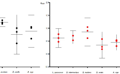
Figure 1. Growth at neutral initial pH. Depicting bacterial growth in 100% Todd Hewitt broth with an initial pH of 7,2, measured with spectrophotometer at 0 and 24 hours.
Figure 2. Growth at acidic initial pH. Depicting bacterial growth in 100% Todd Hewitt broth with an initial pH of 5,5, measured with spectrophotometer at 0 and 24 hours.
Fold change was calculated by dividing the mean growth in the neutral medium with the mean growth in the acidic medium L. paracasei, S. mutans, S. oralis and Actinomyces showed a more extensive growth in the medium with a neutral initial pH compared to an acidic initial pH (fold change 1.2, 1.5, 2.2 and 2.0 respectively), see fig. 3. On the contrary, growth for S.
intermedius was higher in the medium with an acidic initial pH (fold change 0.8). None of these differences were significant at the 5% level. All species showed growth at both neutral and acidic initial pH (p= 0.0046 and 0.0054 respectively).
10
Figure 3. Comparison of growth at neutral and acidic pH. Depicting bacterial growth in 100% Todd Hewitt broth with an initial pH of 7.2 and 5.5 respectively, measured with spectrophotometer at 0 and 24 hours. Dashed lines connect experiments inoculated at the same time to ensure congruity.
11
Acid production at low pH
The purpose of this experiment was to compare acid production in different species of oral bacteria in Todd Hewitt broth at neutral and acidic initial pH. This was done by measuring pH at 0 and 24 hours. The main results are presented in Fig. 4.
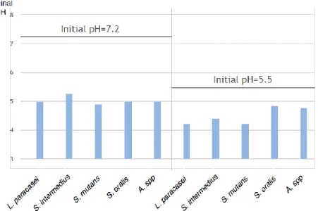
Figure 4. Final pH in neutral and acidic media. Depicting pH change in 100% Todd Hewitt broth with a initial pH of 7.2 and 5.5 respectively, measured with a pH meter at 0 and 24 hours.
All species reached a final pH below pH 5.5 in both media, see fig. 4.
All species showed a decrease in pH in the medium with a neutral initial pH (p=0.0001). L.
paracasei, S. intermedius, S. mutans and Actinomyces showed a decrease in pH in the medium
with acidic initial pH (p=0.0001). S. oralis also showed a decrease in pH in the medium with an initial acidic pH (p=0.0017).
Of all species, S. mutans showed the lowest mean pH in the medium with a neutral initial pH after 24 hours, pH 4.89. L. paracasei and S. mutans both showed the lowest mean pH in the medium with an acidic initial pH after 24 hours, pH 4.21.
12
Growth in complex media with and without glucose
The purpose of this experiment was to compare growth in different species of oral bacteria in Todd Hewitt broth with and without 10 mM glucose at pH 7.2. This was done by measuring OD600 at 0 and 24 hours. A mean value was calculated for the initial OD600 of all species
(0.197 in 25% TH and 0.212 in 10% TH). The main results are presented in Table 1.
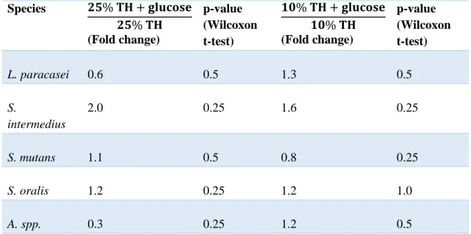
Table 1. Effects of glucose on growth. Depicting fold change in growth from 0 to 24 hours in 25% Todd Hewitt with glucose compared to 25% Todd Hewitt without glucose, and 10% Todd Hewitt with glucose compared to 10% Todd Hewitt without glucose.
Species 𝟐𝟓% 𝐓𝐇 + 𝐠𝐥𝐮𝐜𝐨𝐬𝐞 𝟐𝟓% 𝐓𝐇 (Fold change) p-value (Wilcoxon t-test) 𝟏𝟎% 𝐓𝐇 + 𝐠𝐥𝐮𝐜𝐨𝐬𝐞 𝟏𝟎% 𝐓𝐇 (Fold change) p-value (Wilcoxon t-test) L. paracasei 0.6 0.5 1.3 0.5 S. intermedius 2.0 0.25 1.6 0.25 S. mutans 1.1 0.5 0.8 0.25 S. oralis 1.2 0.25 1.2 1.0 A. spp. 0.3 0.25 1.2 0.5
Fold change <1= Growth was more extensive in the medium without glucose. Fold change 1= The growth did not differ between the media.
Fold change>1=Growth was more extensive in the medium with glucose.
L. paracasei and Actinomyces showed a pattern of more extensive growth in the growth media
with 25% TH without glucose compared to in a growth media with glucose. On the contrary, the pattern for S. intermedius, S. mutans and S. oralis showed a more extensive growth in the media with glucose. All species showed growth in 25% TH both with and without glucose (p= 0.007 and 0.03 respectively).
L. paracasei, S. intermedius, S. oralis and Actinomyces showed a more extensive growth in
the growth media with 10% TH + glucose compared to in a growth media without glucose. On the contrary, growth for S. mutans was more extensive in the media without glucose. All species in the media with 10% TH with and without glucose showed growth (p= 0.111 and 0.172 respectively).
In the media with 25% and 10% TH with glucose, S. intermedius showed a fold change of 2.0 and 1.6 respectively, see table 1. In the medium with 25% TH + glucose, the fold change was
13
statistically significantly higher for S. intermedius compared to Actinomyces (ANOVA p=0.03).
Acid production in different growth media with and without glucose
The purpose of this experiment was to compare acid production in different species of oral bacteria in growth media with and without 10 mM glucose at pH 7.2. This was done by measuring pH at 0 and 24 hours. The main results are presented in table 2 and 3.
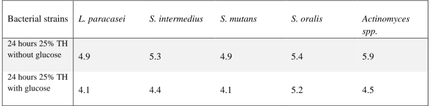
Table 2. Final pH after 24 hrs of incubation in 25 % Todd Hewitt (TH), with and without 10 mM glucose.
Bacterial strains L. paracasei S. intermedius S. mutans S. oralis Actinomyces spp.
24 hours 25% TH
without glucose 4.9 5.3 4.9 5.4 5.9
24 hours 25% TH
with glucose 4.1 4.4 4.1 5.2 4.5
Table 3. Final pH after 24 hrs of incubation in 10 % Todd Hewitt (TH), with and without 10 mM glucose.
Bacterial strains L. paracasei S. intermedius S. mutans S. oralis Actinomyces spp.
24 hours 10% TH
without glucose 5.1 5.7 5.0 5.5 6.7
24 hours 10% TH
with glucose 3.9 4.5 4.1 5.3 4.6
L. paracasei, S. intermedius, S. mutans and Actinomyces showed a decrease in pH in both
media with 25% TH (p=0.0001). S. oralis also showed a decrease in pH in the medium without glucose (p=0.0026) and with glucose (p=0.003). The general pattern of acid
production in 10% TH was the same as in 25% TH for these experiments, see table 2 and 3. All species showed a decrease in pH in the media with 10% TH with and without glucose (p=0.0001).
L. paracasei, S. intermedius, S.mutans and S. oralis reached a final pH below pH 5.5, in both
media with 25% TH. Actinomyces reached a final pH of 5.9 in the medium without glucose, but the final pH in the medium with glucose was below 5.5. In the experiments with 10% TH,
S. intermedius and Actinomyces were the only species that did not reach a final pH below 5.5
in the medium without glucose. All species reached a final pH below 5.5 in the medium with 10% TH with glucose.
Of all species, S. mutans showed the lowest mean pH in the medium with 25% TH without glucose after 24 hours, pH 4.9. S. mutans also showed the lowest mean pH in the medium
14
with 25% TH with glucose after 24 hours, pH 4.08. In the experiments with 10% TH, S.
mutans showed the lowest mean pH in the medium without glucose after 24 hours, pH 5.03.
Contrary to in the medium with 25% TH with glucose, L. paracasei showed the lowest mean pH in the medium with 10% TH with glucose after 24 hours, pH 3.91.
Control media
The mean pH in all the neutral control media at 0 hours was 7.36 with a standard deviation of 0.09. The mean pH in all the acidic control media at 0 hours was 5.79 with a standard
deviation of 0.05. After 24 hours the mean pH in all the neutral control media was 7.06 with a standard deviation of 0.19. After 24 hours the mean pH in all the acidic control media was 5.75 with a standard deviation of 0.075.
15
Alkali production
The purpose of this experiment was to investigate if breakdown of urea can counteract pH drops in artificial or oral consortium in growth media. This was done by measuring pH. The main results are presented in Fig. 6 and 7.
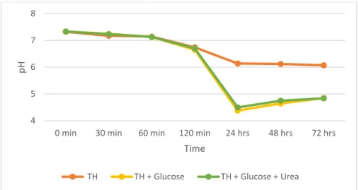
Figure 6. Alkali production in an artificial consortium. Depicting pH change in 25% Todd Hewitt broth with and without glucose and urea with an initial pH of 7.2 measured with a pH meter at 0, 30, 60, 120 minutes and 24, 48 and 72 hours for an artificial consortium.
Figure 7. Alkali production in an oral consortium. Depicting pH change in 10 mM PBS medium with and without glucose and urea with an initial pH of 7.2 measured with a pH meter at 0, 30, 60, 120 and 180 minutes for an oral consortium.
In the artificial consortium the pH dropped to 4.41 after 24 hours in the medium with glucose, see fig. 6. In the medium with glucose and urea the pH dropped to 4.47 after 24 hours. At 72
4 5 6 7 8
0 min 30 min 60 min 120 min 24 hrs 48 hrs 72 hrs
pH
Time
TH TH + Glucose TH + Glucose + Urea
4 5 6 7 8
0 min 30 min 60 min 120 min 180 min
pH
Time
16
hours the pH had risen and the final pH was 4.85 in both media. In the oral consortium the pH dropped to 6.78 after 30 minutes in the medium with glucose, see fig. 7. In the medium with glucose and urea the pH dropped to 6.96 after 30 minutes. At 180 minutes the final pH was 6.42 in the medium with glucose. In the medium with glucose and urea the pH had risen and the final pH was 7.44 at 180 minutes.
17
Discussion
To better understand the etiology of dental caries, it is important to assess the acidogenic and aciduric properties of bacteria present in the supragingival biofilm. S. mutans and L.
paracasei have traditionally been considered the main pathogens of caries. (24) In recent
time, a more polymicrobial etiology for caries has been suggested (25) and it is therefore of interest to investigate the aciduric profile of other saccharolytic bacteria that might contribute to the development of caries. The aim of this study was to examine the acid production and growth in an acidic environment in bacterial species commonly found in supragingival biofilms. The species selected were S. intermedius, S. mutans, S. oralis, L. paracasei and
Actinomyces. All these microbes are considered saccharolytic and can be found in
supragingival dental biofilms on intact as well as carious tooth surfaces. S. oralis and
Actinomyces are early colonizers and generally constitute a large proportion, up to 40%, of
supragingival biofilms. Both species have also been associated with the early stages of enamel demineralization. S. intermedius, S. mutans and L. paracasei are normally present in
supragingival plaque only in small numbers, up to 2%. However, S. mutans is clearly associated with early as well as more advanced caries lesions while L. paracasei is more related to advanced caries. Non-mutans streptococci and Actinomyces have been found to be more prevalent in initial caries lesions compared to S. mutans. Of interest is that no single bacterial species can be uniquely related to caries in the sense that demineralization would occur only in the presence of a specific species that would never be found at sites without evidence of demineralization. Overall, this brings up the polymicrobial etiology of caries in which the contribution by different microbes is not yet fully understood. (2,25,26)
As a first step, the ability to grow at pH 5.5 was examined in order to determine the aciduric properties of the strains selected for this investigation. One clinical study (27) showed that the proportion of dental plaque that exhibited acid tolerance was constituted of a large variety of cell morphologies. This suggests that a number of bacterial species may contribute to the acid tolerance of the microbiota in dental plaque. A larger proportion of acid tolerant flora in the dental plaque may lead to an increased risk of developing caries. A previous study (28) reported that Lactobacillus and S. mutans exhibited slightly greater growth at acidic initial pH compared to non-mutans streptococci (S. oralis and S. intermedius) and Actinomyces. In accordance with our Hypothesis 1, growth was seen for all test strains at pH 5.5 in the current study. Contrary to the hypothesis however, L. paracasei and S. intermedius showed a pattern of approximately the same growth independent of neutral or acidic initial pH. This leads to the assumption that the growth of these species may be less affected by an acidic environment than the other species. Contrary to previous findings, no statistically significant differences in growth between neutral and acidic initial pH was shown for any species. This may be due to a wide dispersion of values, and no conclusion can be drawn in comparing the species. It is also worth noting that in the current study, only fresh isolates of clinical strains were used since they would more properly reflect the real life situation than type collection strains. The latter have the disadvantage of being in storage for a long period of time and exposed to multiple
18
growth conditions in the laboratory that may lead to alterations in their properties, such as acid tolerance.
Many previous studies have investigated the cariogenicity of mutans streptococci and
lactobacilli, but few studies have focused on other oral bacteria, that are likely to have similar acidogenic and aciduric properties. In the current study, the tested strains of S. intermedius, S.
mutans, S. oralis, L. paracasei and Actinomyces showed growth and acid production in TH
broth at pH 7.2, giving rise to final pH values below 5.5, the critical pH below which demineralization of enamel occurs. Also in highly diluted TH broth, 25% and 10% respectively, L. paracasei, S. mutans and S. oralis exhibited a significant acid production resulting in final pH-values below 5.5. It is important to note that the pH drops occurred even though no additional glucose support was given to the growth medium. TH is traditionally used when cultivating oral bacteria and has a high nutritional content consisting of different proteins, peptides, carbohydrates and minerals, which makes it difficult to discern what molecules constitute the source of nutrition and take part in the metabolic processes that results in acid production and lower pH-values. Actinomyces and S. intermedius did not reach a final pH below 5.5 in diluted TH indicating that these strains are more dependent on the surrounding environment and nutritional supply compared to the other species tested and may therefore be considered less acidogenic when nutrient supply is limited. Contrary to our Hypothesis 2, no effect of glucose on the growth was seen in this study. All species showed growth in both 25% and 10% TH with and without glucose. Furthermore, no relation was seen between the growth and acid production. Further studies are required to investigate the
correlation between these mechanisms more thoroughly.
The expression ‘feast and famine’ have been used to describe the general nutrient supply available to bacteria in supragingival biofilms. ‘Famine’ is the presumed prevailing condition during much of the day while the ‘feast’ occurs during intake of rapidly degraded
carbohydrates. (11) The pH in dental biofilms can change up to four units, depending on the age of the biofilm and concentrations of the sugar (29). In the current study, the addition of glucose to TH resulted in pronounced pH drops to a final pH ranging from 3.9 to 5.3, a finding that was in accordance with our Hypothesis 2. One previous study (30) compared the extent of the lesions formed on root and enamel surfaces on 192 molars in vitro after being exposed to different carbohydrate solutions with S. mutans and Actinomyces viscosus respectively over three weeks. In some cases, presence of S. mutans led to statistically significantly deeper lesions in root as well as enamel surfaces compared to A. viscosus. In other cases however, presence of A. viscosus resulted in similar or more extensive lesions on root and enamel surfaces. This finding suggests that Actinomyces actually possesses a cariogenic capacity, even if it is not clear how this compares to that of the S. mutans or other potentially cariogenic species. Collectively, our results combined with those of others suggest that if exposed to a frequent carbohydrate intake in vivo an array of bacterial species can contribute to the acid production and create an acidic environment that give rise to demineralization of enamel.
19
The rationale behind investigating acid production at pH 5.5 was that individuals with a frequent intake of carbohydrates, or a low buffering capacity, are expected to experience a lower pH in dental biofilms both between meals and over longer periods of time in relation to carbohydrate intake. Moderately low pH, such as 5.5, can elicit an acid stress response in bacteria (ATR) that allows survival and carbohydrate metabolism in acidic environments. In the present study, all bacterial strains tested were able to decrease pH from 5.5 to levels between 4.2 and 4.8 demonstrating their aciduric properties. This coincides with our
Hypothesis 1. Even if the differences in final pH were very small, S. oralis and Actinomyces showed a tendency of not reaching as low values as the other species. This might be attributed to a weak ATR in these strains. In a previousstudy, A. naeslundii showed no ATR when exposed to an acidic environment, S. oralis a weak or no ATR while L. casei and S. mutans showed a strong ATR (10). However, it is important to keep in mind that aciduric properties vary between strains of the same species. For example, S. mutans isolated from biofilms on intact teeth showed a less extensive ATR as compared to isolates recovered from biofilms on carious tooth surfaces (31). It is therefore difficult to draw general conclusions when using only one strain for each species. However, all strains tested here sustained an acidic environment with continued metabolism, further lowering the pH below the critical pH for enamel and may therefore collectively contribute to the development of caries.
Since 1944, it is a well-known fact that pH drops after exposure to glucose are more extensive and last longer in dental plaque collected from caries-active than from caries-free individuals (32,33). It is therefore evident that caries-active individuals possess highly aciduric biofilms comprised of microorganisms that keep a functional metabolism with continued acid
production also in acidic environments. A continued saccharolytic metabolism under acidic conditions contributes to a larger and longer pH drop, which in turn contributes to the development of caries as a consequence of the breakdown of enamel. In the current study, S.
mutans and L. paracasei reached the most acidic final pH in all media. This coincides with a
previous study (13) where mutans streptococci were shown to produce acid at the highest rate and in the largest quantity in comparison to some non-mutans streptococci. The extensive acid producing ability of L. paracasei and S. mutans in the media with a relatively low content of nutrients supports the perception that these species are key pathogens, but does not rule out the significance of other species in the caries process. The caries lesion arises through a dynamic process as a consequence of the tooth surface being exposed to a large variety of microbial acids. All acidic products are added together in a pool and give a collective effect on the continued progression of the lesion, where the system should be viewed as a whole and not as an assemblage of parts. Therefore, all tested species may have the ability to effectively establish themselves in a biofilm promoting development of caries.
Many environmental factors influence the pH of multi-species supragingival biofilms. For example, Veillonella utilizes lactic acid for its metabolism and produces acetic acid as its end product, and thereby contributes somewhat to neutralizing the plaque pH. Alkali production by acidogenic species may also have an effect in counteracting the pH drop in dental biofilms. Some bacteria produce ureases that can break down the urea in saliva to ammonium, which also may have a neutralizing effect in the plaque. This ability has been seen in species such as
20
S. mutans (1) and A. naeslundii (2). In this study we investigated ammonium production in an
artificial consortium comprised of dental biofilm bacteria. When the artificial consortium was challenged with glucose alone or glucose with urea, similar pH profiles were observed (Fig. 6) indicating that no significant alkali production took place in the presence of urea. Another plausible explanation is that the presence of TH masked the actual pH changes due to
nutritional components, both carbohydrates and sources of protein, which may have
influenced the final pH in unknown directions. Therefore, another experiment was performed using an oral consortium suspended in PBS. This consortium had a higher microbial diversity and showed much less saccharolytic activity than the artifical consortium. In fact, the pH drop was minimal compared to the pH drop seen in the current single species acid production experiments, see fig.5, and did not reach below the critical pH of enamel. However, the pH profile differed for glucose alone and glucose with urea (see fig. 7). This indicates an alkali production from urea in this oral consortium, which supports our Hypothesis 3.
Based on previous experiments (13,21) a greater pH drop was expected for the oral
consortium. In one previous in vivostudy (21) where the subjects rinsed with 10% sucrose, pH measurements after 30 minutes showed a final pH in oral consortium in vivo of below 5.5. Because of the environmental differences, an oral consortium may behave differently in vitro compared to in vivo. This may be one reason for the minimal pH drop in the current study. The oral consortium was also collected from only one subject, with low caries activity, which may have affected the results since this sample probably contained a minor proportion of acidogenic bacteria. In addition, the amount of urea used does not correspond to the urea level in the saliva, which may vary between individuals and occasions. Future studies on buffer capacity of dental biofilms are needed since it may reveal some new aspects on how different microorganisms interact to counteract environmental change and maintain homeostasis in the biofilm that in turn may contribute to a more thorough understanding of caries etiology.
To our knowledge, few studies have investigated alkali production in single or multi-species biofilms. Previous studies (17,20) have shown that the alkali production was higher in oral plaque from caries-free compared to caries active individuals. This is not surprising since a healthy plaque is composed of a more mixed flora containing a higher proportion of bacteria with proteolytic metabolism and abilities, compared to the more specialized cariogenic acidic plaque dominated by saccharolytic species. However, even if it is lower in cariogenic plaque, an alkali production has been seen in acidogenic species which may contribute to the plaque homeostasis. Further studies on single species, saccharolytic consortia and cariogenic plaque are needed to assess the occurrence of alkali production in saccharolytic species related to caries and it is effects on the pH in cariogenic plaque. In conclusion, breakdown of urea in oral plaque may affect the pH in the biofilm.
Limitations
This study was made in vitro. It is important to keep in mind that the growth environments in
vitro differ from the environment in vivo. In the current experiments, the bacteria grew in
21
were added at one time, whereas in the mouth there are early and late colonizers that affect the local niche and attachment and enables the establishment of subsequent species. In the cases were no statistical significance was found, the values were widely dispersed between experiments. Diverging values affect the results greatly, due to the fact that there were few values. In the experiment with growth at neutral initial pH, one value for S.
intermedius is clearly divergent. In repeating these experiments, a higher quantity of
measurements would be of importance.
The term “acid production” is used loosely in this study. The final pH is of the most clinical relevance since it is related to tooth demineralization. Different bacteria produce different kinds of acids. For example, Actinomyces produces succinate (2) which is a weaker acid than the lactic acid produced by L. paracasei and the streptocooci.
One limitation with the current study was that it did not include a non-saccharolytic species in the experiments as a control, which would have been of interest.
In order to avoid artefactual effects, no buffer was used when preparing the suspensions and controls. Because of this, the initial pH values vary somewhat. The mean value of controls was calculated and considered acceptable. At pH measurements, the temperature also differed in some cases, which may affect the values. In future attempts, the temperature should be consistent at all measurements.
Project meaning
To predict caries it is interesting to look at shared properties of cariogenic bacteria, which is enlightened in this study. Since the 1960’s, the prevailing opinion has been that S. mutans is the primary etiologic agent for caries due to its acidic and aciduric properties. Estimation of salivary counts of mutans streptococci and Lactobacilli is a clinical tool to identify individuals with increased caries risk. In this study, non-mutans streptococci and Actinomyces are also being investigated for their aciduric and acidic properties. A comparison is interesting to examine whether the traditionally considered cariogenic pathogens showed statistically significantly more extensive acid production and acidurity compared to other saccharolytic species present in oral plaque. This is to give additional information for and to provide evidence for new microbiological risk assessment of caries. To better understand the etiology of dental caries, it is important to assess the roles of the microorganisms present in the plaque and the course of caries progression as thoroughly as possible. The results can be used to support the suggestion that caries is a multibacterial disease, and thus that the present multifactorial treatment is suitable.
Conclusion
All species showed acidic and aciduric properties and may contribute to tooth
demineralization. Further studies with a larger number of measurements are needed to assess the cariogenicity of these species with a higher reliability. Alkali production in saccharolytic
22
species may affect the plaque pH. Further studies are needed to assess the occurrence of alkali production in saccharolytic species related to caries and its effects on the pH in the biofilm.
23
References
(1) Burne R. A. Oral streptococci... products of their environment. J.Dent.Res. 1998 Mar;77(3):445-452.
(2) Marsh Philip. Oral microbiology. 5th red. Edinburgh: Churchill Livingstone; 2009.
(3) Marsh P. D., Moter A., Devine D. A. Dental plaque biofilms: communities, conflict and control. Periodontol.2000 2011 Feb;55(1):16-35.
(4) Marsh P. D. Microbial ecology of dental plaque and its significance in health and disease. Adv.Dent.Res. 1994 Jul;8(2):263-271.
(5) Marsh P. D., Devine D. A. How is the development of dental biofilms influenced by the host? J.Clin.Periodontol. 2011 Mar;38 Suppl 11:28-35.
(6) Liljemark W. F., Bloomquist C. Human oral microbial ecology and dental caries and periodontal diseases. Crit.Rev.Oral Biol.Med. 1996;7(2):180-198.
(7) Jenkinson H. F. Beyond the oral microbiome. Environ.Microbiol. 2011 Dec;13(12):3077-3087. (8) Beighton David. The complex oral microflora of high‐risk individuals and groups and its role in the caries process. Community Dent.Oral Epidemiol. 2005;33(4):248-255.
(9) van Houte J. Role of micro-organisms in caries etiology. J.Dent.Res. 1994 Mar;73(3):672-681. (10) Svensater G., Larsson U. B., Greif E. C., Cvitkovitch D. G., Hamilton I. R. Acid tolerance response and survival by oral bacteria. Oral Microbiol.Immunol. 1997 Oct;12(5):266-273.
(11) Bowden G. H., Hamilton I. R. Survival of oral bacteria. Crit.Rev.Oral Biol.Med. 1998;9(1):54-85. (12) Welin-Neilands J., Svensater G. Acid tolerance of biofilm cells of Streptococcus mutans.
Appl.Environ.Microbiol. 2007 Sep;73(17):5633-5638.
(13) de Soet J. J., Nyvad B., Kilian M. Strain-related acid production by oral streptococci. Caries Res. 2000 Nov-Dec;34(6):486-490.
(14) Chestnutt I. G., MacFarlane T. W., Stephen K. W. An in vitro investigation of the cariogenic potential of oral streptococci. Arch.Oral Biol. 1994 Jul;39(7):589-593.
(15) Krzysciak W., Jurczak A., Koscielniak D., Bystrowska B., Skalniak A. The virulence of Streptococcus mutans and the ability to form biofilms. Eur.J.Clin.Microbiol.Infect.Dis. 2014 Apr;33(4):499-515.
(16) Alam S., Brailsford S. R., Adams S., Allison C., Sheehy E., Zoitopoulos L., et al. Genotypic Heterogeneity of Streptococcus oralis and Distinct Aciduric Subpopulations in Human Dental Plaque. Appl.Environ.Microbiol. 2000 Aug;66(8):3330-3336.
24 (17) Shu M., Morou-Bermudez E., Suarez-Perez E., Rivera-Miranda C., Browngardt C. M., Chen Y. Y., et al. The relationship between dental caries status and dental plaque urease activity. Oral Microbiol.Immunol. 2007 Feb;22(1):61-66.
(18) Burne R. A., Marquis R. E. Alkali production by oral bacteria and protection against dental caries. FEMS Microbiol.Lett. 2000 Dec 1;193(1):1-6.
(19) Kleinberg I. A mixed-bacteria ecological approach to understanding the role of the oral bacteria in dental caries causation: an alternative to Streptococcus mutans and the specific-plaque hypothesis. Crit.Rev.Oral Biol.Med. 2002;13(2):108-125.
(20) Nascimento M. M., Gordan V. V., Garvan C. W., Browngardt C. M., Burne R. A. Correlations of oral bacterial arginine and urea catabolism with caries experience. Oral Microbiol.Immunol. 2009 Apr;24(2):89-95.
(21) Hassan H., Lingstrom P., Carlen A. Plaque pH in caries-free and caries-active young individuals before and after frequent rinses with sucrose and urea solution. Caries Res. 2015;49(1):18-25. (22) Dental caries: the disease and its clinical management. 2. red. Oxford, UK; Ames, Iowa, USA: Blackwell Munksgaard; 2008.
(23) Maria Csuros, Csaba Csuros. Ch 14 Estimation of Bacterial Numbers by Indirect Methods. Microbiological Examination of Water and Wastewater. 1st red. : CRC Press; 26 mars 1999. s. 193. (24) Loesche W. J. Role of Streptococcus mutans in human dental decay. Microbiol.Rev. 1986 Dec;50(4):353-380.
(25) Simon-Soro A., Mira A. Solving the etiology of dental caries. Trends Microbiol. 2015 Feb;23(2):76-82.
(26) Sansone C., Van Houte J., Joshipura K., Kent R., Margolis H. C. The association of mutans streptococci and non-mutans streptococci capable of acidogenesis at a low pH with dental caries on enamel and root surfaces. J.Dent.Res. 1993 Feb;72(2):508-516.
(27) Senneby A., Davis J. R., Svensäter G., Neilands J. Acid Tolerance of Dental Biofilms. Journal of Oral Microbiology Manuscript Draft.
(28) Harper D. S., Loesche W. J. Growth and acid tolerance of human dental plaque bacteria. Arch.Oral Biol. 1984;29(10):843-848.
(29) Igarashi K., Kamiyama K., Yamada T. Measurement of pH in human dental plaque in vivo with an ion-sensitive transistor electrode. Arch.Oral Biol. 1981;26(3):203-207.
(30) Clarkson B. H., Krell D., Wefel J. S., Crall J., Feagin F. F. In vitro caries-like lesion production by Streptococcus mutans and Actinomyces viscosus using sucrose and starch. J.Dent.Res. 1987 Mar;66(3):795-798.
(31) Svensater G., Borgstrom M., Bowden G. H., Edwardsson S. The acid-tolerant microbiota associated with plaque from initial caries and healthy tooth surfaces. Caries Res. 2003 Nov-Dec;37(6):395-403.
(32) Stephan Robert M. Intra-oral hydrogen-ion concentrations associated with dental caries activity. Memorial Dental Clinic, The University of Chicago 1944 May 11;3.
25 (33) Aranibar Quiroz E. M., Alstad T., Campus G., Birkhed D., Lingstrom P. Relationship between plaque pH and different caries-associated variables in a group of adolescents with varying caries prevalence. Caries Res. 2014;48(2):147-153.