http://www.diva-portal.org
This is the published version of a paper published in Plant Physiology.
Citation for the original published paper (version of record):
Askerlund, P. (1997)
Calmodulin-stimulated Ca2+-ATPases in the vacuolar and plasma membranes in cauliflower.
Plant Physiology, 114(3): 999-1007
http://dx.doi.org/10.1104/pp.114.3.999
Access to the published version may require subscription.
N.B. When citing this work, cite the original published paper.
Free PMC Article
Permanent link to this version:
Plant Physiol. (1 997) 114: 999-1007
Calmodulin-stimulated Ca*+-ATPases in the Vacuolar and
Plasma Membranes in Cauliflower’
Per Askerlund*
Department of Plant Biochemistry, Lund University, P.O. Box 1 1 7, S-221 O 0 Lund, Sweden
~ ~ ~~~~~~ ~~
The subcellular locations of Ca2+-ATPases in the membranes of cauliflower (Brassica oleracea 1.) inflorescences were investigated. After continuous sucrose gradient centrifugation a 11 1-kD calmod- ulin (CaM)-stimulated and CaM-binding Ca2+-ATPase (BCA1; P. Askerlund [1996] Plant Physiol 110: 913-922; S. Malmstrom, P.
Askerlund, M.C. Palmgren [1997] FEBS Lett 400: 324-328) co- migrated with vacuolar membrane markers, whereas a 11 6-kD CaM-binding Ca2+-ATPase co-migrated with a marker for the plasma membrane. The 116-kD Ca2+-ATPase was enriched i n plasma membranes obtained by aqueous two-phase partitioning, which is in agreement with a plasma membrane location of this CaZ+-ATPase. Countercurrent distribution of a low-density intracel- lular membrane fraction i n an aqueous two-phase system resulted in the separation of the endoplasmic reticulum and vacuolar mem- branes. The 11 1-kD CaZ+-ATPase co-migrated with a vacuolar membrane marker after countercurrent distribution but not with markers for the endoplasmic reticulum. A vacuolar membrane lo- cation of the 11 1 -kD Ca2+-ATPase was further supported by exper- iments with isolated vacuoles from cauliflower: (a) lmmunoblotting with an antibody against the 111-kD CaZ+-ATPase showed that it was associated with the vacuoles, and (b) ATP-dependent Ca2+ uptake by the intact vacuoles was found to be CaM stimulated and partly protonophore insensitive.
Ca2+ is an essential intracellular messenger in plant cells and is involved in metabolic and developmental regula- tion. Maintenance of a low free cytosolic concentration (about 0.1 p ~of Ca2+ is necessary for its function as a )
messenger in signal transduction (Gilroy et al., 1993; Bush, 1995). A variety of stimuli can trigger the opening of Ca2+ channels in the plasma membrane and in interna1 mem- branes, causing Ca” influx and accumulation in the cy- tosol. The cytosolic Ca2+ concentration is restored and maintained at the low leve1 by extrusion of Ca2+ from the cell or by sequestration into intracellular organelles, mainly the vacuole (Canut et al., 1993).
Two classes of active Ca2+ transporters have been shown to be present in plant membranes: CaZf-ATPases and nH+/Ca2+ antiporters (Evans et al., 1991; Hirschi et al., 1996). The Ca’+-ATPases, some of which are stimulated by CaM, have been shown to be responsible for Ca2+ extru- sion across the plasma membrane as well as transport of Ca2+ into the ER lumen (Buckhout, 1983; Robinson et al., 1988; Rasi-Caldogno et al., 1992; Gilroy and Jones, 1993).
This work was supported by a grant from the Swedish Natural Science Research Council.
* E-mail per.askerlund@plantbio.lu.se; fax 46-46-222-41-16.
Uptake of Ca2+ into the vacuole has long been thought to be catalyzed by an n H + / C a z + antiporter only. More re- cently it was suggested that Ca2+-ATPases also take part in the transport of Ca2+ into the vacuole (DuPont et al., 1990; Gavin et al., 1993; Pfeiffer and Hager, 1993; Bush and Wang, 1995; Ferro1 and Bennett, 1996; for a review, see Askerlund and Sommarin [ 19961). The subcellular location of CaM-stimulated Ca2+-ATPases in plants has long been a matter of controversy. In contrast to the situation in animal cells (Carafoli, 1994), CaM-stimulated Ca2+-ATPases in. plants are present in intracellular membranes as well as in the plasma membrane (Askerlund and Sommarin, 1996). CaM-stimulated, ATP-dependent Ca2+ uptake in maize root membranes was found to be associated with markers for vacuolar membranes (Gavin et al., 1993). In studies with maize shoots the same activity was proposed to be located in the ER (Logan and Venis, 1995). In barley aleurone the CaM-stimulated, ATP-dependent Caz+ uptake was local- ized to the ER (Gilroy and Jones, 1993), whereas in wheat aleurone a much greater fraction of the CaM-stimulated activity was associated with the vacuolar membrane (Bush and Wang, 1995). Previously, a 111-kD CaM-stimulated Ca2+-ATPase was shown to be enriched in a low-density intracellular membrane fraction from cauliflower (Brassica oleracea L.) inflorescences (Askerlund and Evans, 1992;
Askerlund, 1996). The cDNA corresponding to this pump
( B C A I ) was recently cloned (based on the sequence of
tryptic peptides derived from the purified protein), show- ing that it represents a nove1 class of CaM-stimulated Ca2+- ATPases (Malmstrom et al., 1997). The main objective of this study was to accurately determine the intracellular location of this 111-kD CaM-stimulated Ca2+-ATPase.
MATERIALS AND METHODS
Suc Gradient Centrifugation
All operations were carried out at O to 4”C, and the outermost part of the cauliflower (Brassica oleracea L.) in-
florescence was used. This tissue contains a large propor- tion of immature cells in the process of vacuolation but also differentiated cells of different types. The plant material was homogenized as described previously (Askerlund, 1996), except that BSA was omitted from the homogeniza-
Abbreviations: BiP, ER luminal binding protein; BTP, 1,S-bis-
(tris[hydroxymethyl]methylamino)propane; CaM, calmodulin;
CCD, countercurrent distribution; FCCP, carbonyl cyanide
p-trifluoromethoxyphenylhydrazone.
1 O00 As kerl u nd Plant Physiol. Vol. 11 4, 1997 tion medium. The homogenate was filtered through a
nylon cloth (140 pm, Lockertex, Warrington, UK) and centrifuged at 10,OOOg for 10 min. About 80 mL of the supernatant was carefully pipetted onto a cushion of 4 g of 2 M SUC in a gradient buffer (25 mM Mops-BTP, pH 7.2, 50 mM KCl, and 2 mM Na,-EDTA) in two centrifuge tubes and was centrifuged at 100,OOOg for 45 min in an SW-28 rotor (Beckman). The microsomal membranes at the inter- face (total volume 5 mL) were collected and mixed with 6 mL of 2 M SUC in the gradient buffer. One-milliliter frac- tions (about 4.5 mg of protein) of the membrane suspen- sion were pipetted into 13-mL centrifuge tubes and were overlaid with a continuous SUC gradient (1.7-0.6 M Suc in 11 mL of the gradient buffer). The tubes were centrifuged in a swing-out rotor (RPS40T, Hitachi, Mountain View, CA) for 16 h at 150,OOOg. Fractions (0.8 mL) were collected from the bottom of the gradient with a peristaltic pump and were aliquoted and stored at -20°C until analysis. The SUC concentration was measured with a refractometer (Atago IT, Atago, Tokyo, Japan).
CCD of Low-Density lntracellular Membranes in an
Aqueous Polymer Two-Phase System and Preparation of
. Plasma Membranes
Low-density intracellular membranes were collected at the 10/32% ( w / w ) interface after discontinuous Suc gradi- ent centrifugation of a microsomal membrane fraction, as described previously (Askerlund, 1996). After the sample was pelleted, the low-density membranes were resus- pended in 5 mM potassium phosphate, pH 7.8,330 mM SUC, 1 mM DTT, and 0.5 mM PMSF and were again pelleted at 100,OOOg for 45 min. The final washed membrane pellet was suspended in the same buffer and stored at -80°C. The low-density membranes (about 8 mg of protein) were further fractionated by CCD in an aqueous polymer two- phase system (8 g, final weight, of 6.2% [ w / w ] dextran T-500, 6.2% [w / w] PEG 3350, 330 mM SUC, 1 mM KCl, and 5 mM potassium phosphate, pH 7.8), essentially as de- scribed for the crude microsomal membranes by Bérczi et al. (1989; for a detailed description of CCD, see Larsson [1983]). A total of five transfers of the upper phase were made, resulting in a total of six final tubes. After CCD the total content in each tube was diluted to 70 mL with 25 mM Mops-BTP, pH 7.2, 0.33 M SUC, 1 mM DTT, and 0.5 mM PMSF and pelleted at 100,OOOg for 45 min. Because of the high content of polymers the following procedure was necessary to pellet the CCD fractions: After the sample was centrifuged about 90% of the supernatant was carefully removed with a pipette. A new buffer was then added, the content was mixed, and the tubes were centrifuged at 100,OOOg for 30 min. The final membrane pellets were sus- pended in 25 mM Mops-BTP, pH 7.2, 0.33 M SUC, and 0.5 mM PMSF, aliquoted, and stored at -80°C. A11 operations were carried out at O to 4°C. Preparation of plasma mem- branes from cauliflower by aqueous two-phase partition- ing was carried out as described by Askerlund and Evans (1992).
Preparation of intact Vacuoles
Intact vacuoles from cauliflower inflorescences were iso- lated by slicing and flotation, essentially as described by Bennett et al. (1983). A11 of the operations were performed at 4°C or on ice. The plant material (650 g) was sliced using a blender (HR 2375/D, Philips Eindhoven, The Nether- lands) in 0.5 L of 50 mM Tris-Mes, pH 8.0, 1 M sorbitol, 5 mM Na,-EDTA, 0.5% (w / v ) polyvinylpolypyrrolidone, 4
mM DTT, and 0.5 mM PMSF. The extract was filtered through a nylon cloth and centrifuged at 1380g for 20 min in two buckets. Each pellet was carefully suspended in 10 mL of 15% ( w / v ) sodium diatrizoate in 25 mM Tris-Mes, pH 7.0, and 1.2 M sorbitol. Each resuspension was placed in a centrifuge tube and was overlaid with 10 mL of 10% ( w / v ) sodium diatrizoate in the same buffer followed by 5 mL of the buffer without sodium diatrizoate and was cen- trifuged at 400g in a swing-out centrifuge for 15 min. The intact vacuoles (weak white band; about 0.13 mg of protein in 1.3 mL) at the 0/10% sodium diatrizoate interface were collected and placed on ice. The purity and intactness of the vacuoles were determined with a phase-contrast micro- scope (Nikon).
Enzyme Activities
PPi-dependent H + pumping and ATP-dependent Ca2+ pumping (radioactive filter assay) with membrane frac- tions were measured as described earlier (Askerlund, 1996). Antimycin A-insensitive NADH-Cyt c reductase was measured in the absence of detergent as described by Askerlund et al. (1991). Measurement of UDP-Gakdiacyl- glycerol galactosyltransferase (galactolipid synthase activ- ity) was as described by Douce and Joyard (1980). Radio- active Caz+ uptake with intact vacuoles was measured in a mixture of 50 pL of 25 mM Tris-Mes, pH 7.0, 200 mM KCl, 10 mM MgCl,, 1.2 M sorbitol, 2 mM DTT, 0.2 mM sodium
molybdate, 2 mM NaN,, 10 p~ CaCl, (36 Bq 45CaC1z pmol-’), and 50 pL of vacuoles (about 5 p g of protein, see above) for 30 min at 30°C in a final volume of 0.1 mL. The mixture was preincubated for 10 min prior to starting the Ca2+ uptake with 2.5 mM ATP-BTP, p H 7.0. The reactions were stopped by the addition of 0.6 mL of Tris-Mes, pH 7.0, 1 mM EGTA, and 1.2 M sorbitol, and the mixtures were
immediately filtered through 0.45-pm pore-size cellulose nitrate membrane filters (Gelman Sciences, Ann Arbor, MI) using a vacuum-sampling manifold (Millipore). After they were washed four times with 1 mL of a stop solution, the filters were dried and the amount of 45Ca2+ was measured by scintillation counting.
Protein Analysis and Determination
Protein was measured with a modified Bradford proce- dure (Stoscheck, 1990), except for preparations of intact vacuoles, for which protein was measured with the bicin- choninic acid protein assay (Pierce, Rockford, IL), since sodium diatrizoate interferes with the Bradford procedure. BSA was used as a standard. SDS-PAGE was carried out on linear minigels as described earlier (Askerlund, 1996). To avoid proteolysis samples were TCA-precipitated prior to
Calmodul in-Stimu lated CaZ+-ATPases i n Cau I iflower 1001
solubilization with SDS. Vacuoles were precipitated with methanol-chloroform-H,O (Pohl, 1990). A sample volume corresponding to an 11-pL SUC gradient fraction or 6 pg of protein was applied to each lane unless otherwise indi- cated.
Western Analysis
Western blotting was carried out as described earlier (Askerlund, 1996) using ’251-labeled or alkaline- phosphatase-conjugated secondary antibodies. Radioac- tive blots were first analyzed with a phosphor imager (Molecular Dynamics, Sunnyvale, CA) to quantify the amount of radioactivity in each band. To obtain sharper images the blots were later exposed for about 2 d at 20°C with Hyperfilm-pmax (Amersham) in the presence of an intensifier screen (Hyperscreen, Amersham). An anti- serum against the 111-kD, CaM-stimulated cauliflower Ca2+-ATPase (Askerlund, 1996) and an antiserum against the putative chloroplast envelope Ca2+-ATPase in Arabi- dopsis thaliana (Huang et al., 1993; a gift from Dr. N.E.
Hoffman, Department of Plant Biology, Carnegie Institute of Washington, Stanford) were both used at a dilution of 1:2000, whereas an antiserum against the
A.
thalianaplasma membrane H+-ATPase (no. 761; a gift from Prof. R. Serrano, Department of Biotechnology, University of Valencia, Spain) was diluted 1:3300. Antisera against the 57-kD subunit of the vacuolar H+-ATPase from Beta vul- garis L. (Manolson et al., 1989; a gift from Prof. R.J. Poole, Department of Biology, McGill University, Montreal, Quebec, Canada) and against the tobacco BiP (Denecke et al., 1991; a gift from Dr. J. Denecke, Department of Biol- ogy, University of York, UK) were both diluted 1:2000.
lZ51-CaM Overlays and Phosphorylated lntermediate Formation and Analysis
lZ5I-CaM overlays were prepared and analyzed as de- scribed earlier (Askerlund, 1996). For studies of the phos- phorylated intermediate of Ca’+-ATPases, membrane frac- tions were phosphorylated with 1 nM [y-32P]ATP for 15 s on ice at pH 7.4, in the absence or presence of 50 p~ LaCl,, and analyzed as described earlier (Askerlund and Evans, 1993).
RESULTS
Distribution of ATP-Dependent Caz+ Uptake and Enzymic
Membrane Markers after Continuous SUC Gradient
Centrifugation
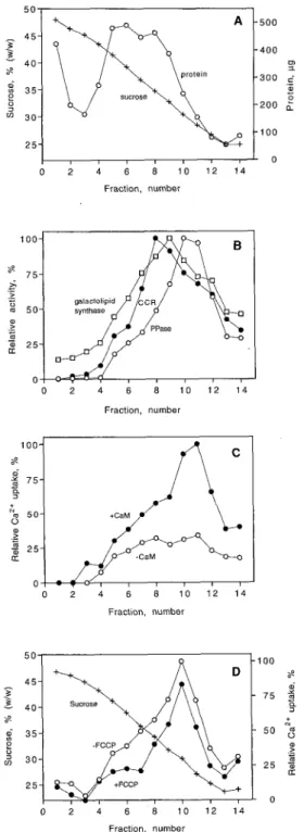
Crude microsomes were fractionated by continuous Suc gradient centrifugation and the distribution of ATP- dependent Ca2+ uptake was compared with various enzy- mic membrane markers (Fig. 1). Membrane protein was mainly found in a broad peak in the middle of the gradient. Since membranes were loaded from the bottom of the tube, soluble protein was found in fraction 1 (Fig. 1A). Antimy- cin A-insensitive NADH-Cyt c reductase activity (ER mark- er), galactolipid synthase (plastid inner envelope; Douce and Joyard, 1980), PPi-dependent H + pumping (vacuolar
O 2 4 6 8 1 0 1 2 1 4 Fraction, number Fraction, number O 2 4 6 8 1 0 1 2 1 4 Fraction. number 1 O0 8 o) a 2 7 5 5 N 5 0 2
2
2 5 O O 2 4 6 8 1 0 1 2 1 4 Fraction, numberFigure 1. Distribution of ATP-dependent, CaM-stimulated Ca’+ uptake and different marker enzyme activities after continuous Suc gradient centrifugation of a microsomal membrane fraction from cauliflower inflorescences. Membranes were loaded from the bottom (fraction 1 ). A,
+,
In percentage (w/w) of SUC; O, in micrograms of protein. B, O, Antimycin A-insensitive NADH-Cyt c reductase activity (CCR; 100% = 68 nmol min-’); O, PPi-dependent Ht pumping (PPase; 100% = a change in A495 min-’ of 0.86); O, galactolipid synthase (100% = 12 nmol h-’). C, ATP-dependent Caz+ uptake (100% = 1 .O nmol min-‘) in the presence (O) or absence (O) of 1 p~ CaM. D, ATP-dependent Ca’+ uptake (100% = 1.2 nmol min-’) measured in the presence of 1p~ CaM in the presence (O) or absence (O) of 5 p~ FCCP. The data shown in A to C were obtained from the same Suc gradient separation, and the data shown in D are from a separate experiment.
1002 Askerlund Plant Physiol. Vol. 114, 1997
membrane marker; Chanson, 1990), and ATP-dependent Ca2+ uptake were all located mainly in the lighter part of
the gradient (Fig. 1, B and C). The protonophore FCCP had only a small effect on Ca2+ uptake, indicating that the
major part of the Ca2+ uptake was catalyzed by a Ca2+
-ATPase rather than by a Ca2+ / n H+ antiporter (Fig. ID; see
also "Discussion"). The distribution of Ca2+ uptake (clear
optimum visible only when measured in the presence of CaM) correlated very closely with PPi-dependent H+
pumping but was different from that of NADH-Cyt c re-ductase (Fig. 1, B and C). This is in contrast with the earlier report in which the major part of the CaM-stimulated Ca2+
uptake was suggested to be located in the ER (Askerlund and Evans, 1992). In the earlier experiments, PPi hydrolysis rather than PPi-dependent H+ pumping was measured.
The current view is that PPi hydrolysis is a much less specific marker for the vacuolar membrane than PPi-dependent H+ pumping, possibly due to binding of soluble
PPiases to membranes. In contrast, PPi-dependent H+
pumping may be considered a relatively specific marker for the vacuolar membrane, although this activity has been reported to be present in the plasma membrane (Chanson, 1990; Robinson, 1996).
Use of Antibodies and 125l-CaM to Study the Distribution of Ca2+-ATPases and Membrane Markers after Continuous
Sue Gradient Centrifugation
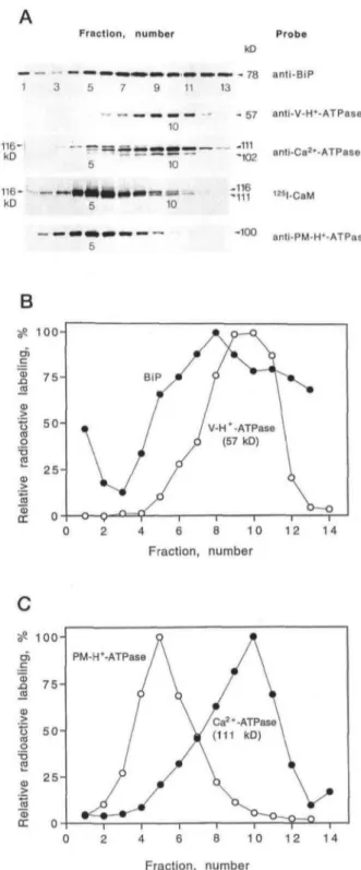
Antibodies were also used as probes for Ca2+-ATPases
and different membranes on the Sue gradient (Fig. 2). Antibodies against the BiP detected a polypeptide of 78 kD, in agreement with the molecular mass of BiP (Denecke et al., 1991; Fig. 2, A and B). The BiP that was found at the bottom of the gradient (fraction 1) may have been released from the ER lumen during homogenization (the mem-branes applied to the Sue gradient were not extensively washed, since pelleting increases the risk of membrane aggregation). Like the amount of antimycin A-insensitive NADH-Cyt c reductase, the amount of BiP was maximal in fraction 8 (34.76% [w/w] Sue); however, BiP showed a wider distribution on the gradient (compare Figs. IB and 2B). Antibodies against the 57-kD subunit of the vacuolar H+-ATPase detected a 57-kD polypeptide, which peaked in
fractions 9 and 10 (Fig. 2, A and B). The amount of plasma membrane H+-ATPase (100 kD) was maximal in fraction 5
(Fig. 2, A and C). Finally, antibodies against the 111-kD CaM-stimulated Ca2+-ATPase from cauliflower
(Asker-lund, 1996) detected a polypeptide of 111 kD, which peaked in fraction 10 (Fig. 2, A and C).
In agreement with an earlier report (Askerlund, 1996), the same antiserum also recognized a minor polypeptide band at 102 kD (Fig. 2A). The 102-kD band was suggested to be a truncated form of the intact 111-kD Ca2+-ATPase,
since it accumulated during the trypsin treatment of low-density membranes (Askerlund, 1996). This hypothesis is supported by the fact that the 111- and 102-kD poly pep-tides showed a perfect correlation on the Sue gradient (Fig. 2A), indicating that they are present on the same mem-brane. In addition to the 111- and 102-kD bands, the anti-serum against the cauliflower Ca2+-ATPase recognized a
Fraction, number 1 3 5 7 9 11 13 10 116-kD kD Probe kD > - 78 anti-BiP - 57 anti-V-H+-ATPase -111 10 10 -102 anti-Ca2*-ATPase -116 -111 125l-CaM -100 anti-PM-H»-ATPase
B
100-0) .Q a o at o T3 ra CD DC 75-Fraction, number 100- 75- 502 5 -J5 CD DC PM-H*-ATPase 10 12 14 Fraction, numberFigure 2. Western analysis of fractions after Sue gradient
centrifuga-tion of a microsomal fraccentrifuga-tion from cauliflower inflorescences. A,
Autoradiographs of a 125l-CaM blot and immunoblots with antisera
against BiP (anti-BiP), the 57-kD subunit of the vacuolar H+-ATPase
(anti-V-H+-ATPase), the 111-kD CaM-stimulated Ca2+-ATPase in
low-density membranes from cauliflower (anti-Ca2 + -ATPase), and
the plasma membrane H + -ATPase (anti-PM-H+-ATPase). 125
I-labeled secondary antibodies were used. A sample volume corre-sponding to an 11-/xL Sue gradient fraction was loaded in each lane. B and C, Phosphor imager quantification of immunoblots in A. Data were obtained from the same Sue gradient separation that is shown in Figure 1, A to C.
Calmodulin-Stimulated Ca2+-ATPases in Cauliflower 1.5- 1 - e
o,
.- o) I a 0,5- 1003 C 116-kD polypeptide, which peaked in fraction 5 (Fig. 2A).The co-localization of this polypeptide with the plasma membrane H'-ATPase (Fig. 2A) agrees with the presence of a 116-kD Ca2+-ATPase in plasma membranes from cau- liflower (Askerlund and Evans, 1993). The same antiserum also detected a minor polypeptide band at about 107 kD (clearly visible in fractions 5 and 6 only). This band peaked
in fraction 5 and may therefore be a truncated form of the
116-kD Ca2+-ATPase (see also the data for phase- partitioned plasma membranes below).
CaM-binding polypeptides of 116 (very strong) and 111 kD were identified in lz5I-CaM overlays (Fig. 2A). The 116-kD CaM-binding band peaked in fraction 5, whereas the much weaker 111-kD CaM-binding band peaked in fraction 10. These results agree with the suggestion (Asker- lund, 1996) that the 116-kD CaM-binding polypeptide rep- resents a plasma membrane Ca2+-ATPase with very high affinity for CaM and that the 111-kD CaM-binding polypeptide represents the Ca2+-ATPase in low-density membranes.
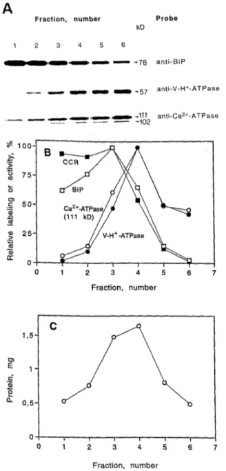
CCD of Low-Density lntracellular Membranes
Low-density membranes from cauliflower, prepared by discontinuous Suc gradient centrifugation (Askerlund, 1996), were further fractionated by CCD in an aqueous two-phase system (Fig. 3). The ER membranes (as detected by anti-BiP and antimycin A-insensitive NADH-Cyt c re- ductase) had a high affinity for the bottom phase and ended up mainly in CCD fractions 1 to 4 (Fig. 3, A and B). In contrast, the vacuolar membranes (as detected by anti- bodies against the 57-kD subunit of the vacuolar H+- ATPase) had a higher affinity for the upper phase and ended up mainly in CCD fractions 3 to 6 (Fig. 3, A and B).
Thus, CCD resulted in a relatively good separation of the ER and vacuolar membranes. The 111-kD Ca2+-ATPase showed an almost perfect correlation with the 57-kD sub- unit of the vacuolar H+-ATPase (Fig. 3, A and B), in agree- ment with a vacuolar membrane location of this Ca2+- ATPase. The co-migration of the 111- and 102-kD polypeptides after CCD further support the suggestion (Askerlund, 1996) that the 102-kD polypeptide is a trun- cated form of the intact 111-kD Ca2+-ATPase.
Comparison of the Ca2+-ATPases in Low-Density
lntracellular Membranes and Phase-Partitioned Plasma Mem branes
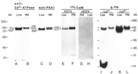
The Ca2+-ATPases in cauliflower plasma membranes (prepared by two-phase partitioning) and low-density membranes (prepared as described by Askerlund, 1996) are compared in Figure 4. As shown earlier (Askerlund, 1996), the antiserum against the 111-kD CaM-stimulated Ca2+- ATPase detected the intact Ca2+-ATPase and the 102-kD degradation product when reacted with low-density mem- branes (Fig. 4, lane A). with plasma membranes, the same antiserum detected a band at 116 kD and weaker bands at
111 and about 107 kD (Fig. 4, lane B). A comparison with Figure 2A suggests that the 116-kD band observed with
Fraction, number Probe
A
kD i 2 3 4 5 6---
-,-
-78 anti-BiP_-
-
-
-
-
-57 anti-V-H+-ATPase , ,111 anti-Ca**'-ATPase -102___
-
-
-
O 1 2 3 4 5 6 7 Fraction, number Fraction, numberFigure 3. Analysis of fractions after CCD of low-density membranes from cauliflower inflorescences. A, Autoradiographs of immunoblots with antisera against BiP (anti-BiP), the 57-kD subunit of the vacuolar H+-ATPase (anti-V-H+-ATPase), and the 1 1 1 -kD CaM-stimulated Ca*+-ATPase i n low-density membranes from cauliflower (anti- CaZt-ATPase). '251-labeled secondary antibodies were used. All
lanes received 6 pg of protein. B, Phosphor imager quantification of immunoblots in A and antimycin A-insensitive NADH-Cyt c reduc- tase activity (CCR; 100% = 0.65 p m o l min-') expressed as relative amount of labeling or activity in each CCD fraction: O, BiP; O, V-H+-ATPase; O, Ca2+-ATPase; and H, CCR. C, Distribution of protein between CCD fractions.
plasma membranes represents the plasma membrane Ca2+-ATPase and that the 111-kD band is due to contam- ination by the vacuolar membranes. The presence of a 107-kD band (probably a degradation product of the plasma membrane Ca2+-ATPase) is also in agreement with
1004 Askerlund Plant Physiol. Vol. 114, 1997
a n t i
-Ca2»-ATPase antl-PEA1 E-«P
kD Low FM Low PM -EGTA Low PM PM Low 200-116- 111-t 97-102"' 66- 45- 31-[ -116- '-111--La3* +La3' Low PM Low PM kD -200 •116 -97 -66 -45 -31 C D E F G H -21 I J K L
Figure 4. Characterization of Ca2 + -ATPases in low-density intracellular membranes (Low) and phase-partitioned plasma
membranes (PM). Lanes A and B, Immunoblot with antibodies against the 111-kD Ca2 + -ATPase in low-density
mem-branes (anti-Ca2 + -ATPase; Askerlund, 1996); lanes C and D, immunoblot with antibodies against the putative Ca2+
-ATPase from A. thaliana chloroplast envelope (anti-PEA1; Huang et al., 1993); lanes E and F, autoradiograph of I25l-CaM
overlay; lanes G and H, same as E and F, but blot was incubated with 125l-CaM in the presence of 1 mM EGTA; lanes I
and J, autoradiograph showing phosphorylated intermediates of Ca2 + -ATPases (E-32P); and lanes K and L, same as I and
J, but phosphorylation was carried out in the presence of LaCI3. Lanes A to H received 6 ^ig of protein, and lanes I to L
received 80 jj,g of protein.
the data from the Sue gradient centrifugation (compare Figs. 2A and 4, lane B).
Antibodies against a region of PEA1 (ACA1), the puta-tive Ca2+-ATPase in the A. thaliana chloroplast envelope
(Huang et al., 1993), detected the 111- and 102-kD bands when reacted with low-density membranes (Fig. 4, lane C). This is in agreement with the relatively high homology between the 111-kD intracellular Ca2+-ATPase in
cauli-flower (BCA 1; Malmstrom et al., 1997) and the A. thaliana Ca2+-ATPase. With plasma membranes these antibodies
gave essentially the same result as the antibodies against the 111-kD Ca2+-ATPase in cauliflower, except that the
116- and 107-kD bands were more strongly stained and the 111-kD band was more weakly stained than when reacted with the antiserum against the cauliflower Ca2+-ATPase
(Fig. 4, lane D). 125I-CaM overlays of low-density
intracel-lular membranes and phase-partitioned plasma mem-branes confirmed the presence of CaM-binding Ca2+
-ATPases with apparent masses of 111 and 116 kD in low-density membranes and plasma membranes, respectively (Fig. 4). As was indicated by the data in Figure 2A, the plasma membrane Ca2+-ATPase was more strongly
la-beled by 125I-CaM. As was suggested earlier (Askerlund,
1996), the 116-kD CaM-binding band visible in the low-density membrane fraction therefore probably represents contamination by a very small amount of plasma mem-branes. The stronger labeling of the plasma membrane Ca2+-ATPase may reflect a higher affinity for CaM.
Anal-ysis of phosphorylated intermediates also indicated that the plasma membrane Ca2+-ATPase is slightly larger than
the intracellular Ca2+-ATPase (Fig. 4, lanes I-L). [32P]ATP
labeling of phosphoproteins was intensified by La3+ in
both fractions, in agreement with earlier reports (Asker-lund and Evans, 1993; Asker(Asker-lund, 1996). Furthermore, la-beling of both bands were Ca2+ dependent and sensitive to
hydroxylamine (data not shown).
Experiments with Isolated Vacuoles
To further establish that the 111-kD CaM-stimulated Ca2+-ATPase is situated in the vacuolar membrane of
cau-liflower inflorescences, intact vacuoles were isolated from cauliflower by slicing and flotation. This resulted in a preparation that looked very pure by light microscopy (not shown). Antimycin A-insensitive NADH-Cyt c reductase activity in vacuole preparations was only 20 nmol min"1
mg"1 protein or less (data not shown), indicating that the
levels of contaminating ER were very low. The vacuoles were small, only 4 to 15 /u,m in diameter, compared with vacuoles isolated from red beet roots using the same tech-nique (15-30 /n,m). The cauliflower vacuoles were able to accumulate 45Ca2+ in the presence of ATP (Table I).
Possi-bly because of their small size, at least part of the vacuoles remained intact during the filtration step used in the
45Ca2+ uptake procedure. Ca2+ uptake by the vacuoles was
CaM stimulated and only partly sensitive to the protono-phore FCCP (Table I), indicating that a CaM-stimulated
Table I. The effect of CaM (1 ^M) and FCCP (5 on 45Ca2+
up-take in intact cauliflower vacuoles in the presence (+) and ab-sence (-) of ATP
The assay included 50 juL of vacuoles corresponding to about 5 fj.g
of protein. Data are means ± so of three Ca2+ uptake measurements
with a single vacuole preparation. A typical experiment is shown. Addition
ATP CaM FCCP Ca2+ Uptake
pmol min~' 0.306 ± 0.036 0.407 ± 0.009 0.186 ± 0.001 0.235 ± 0.021 0.026 ± 0.002
Calmodulin-Stimulated Ca2+-ATPases in Cauliflower 1005
anti-Ca2+-ATPase
anti-V-H+-ATPase
kO Vac Low Vac Low 200-116-.. 97-" 66- 45- 31- -111- -102-.-57-. B
Figure 5. Immunodetection of Ca2+-ATPase and vacuolar
H*-ATPase in isolated vacuoles (Vac) and low-density membranes (Low) from cauliflower. Lanes A and B, Blots incubated with an antiserum
against the 111-kD Ca2 + -ATPase from cauliflower; lanes C and D,
blots incubated with an antiserum against the 57-kD subunit of the
vacuolar H+-ATPase. Alkaline-phosphatase-conjugated secondary
antibodies were used. Lanes A and C received 200 /xL of cauliflower vacuoles (about 20 /j.g), and lanes B and D received 6 /ng of protein.
Ca2+-ATPase was responsible for at least part of the Ca2+
accumulation (see also "Discussion"). The highest degree of CaM stimulation seen in any preparation (n = 5) was 112%, but usually it was much lower (Table I).
The vacuole preparation was probed with the antiserum against the 111-kD Ca2+-ATPase and with the antiserum
against the vacuolar H+-ATPase. Because of the very small
yield of vacuoles, the whole-vacuole preparation, rather than the vacuolar membrane, was analyzed. In all of the preparations that were tested, the 111-kD Ca2+-ATPase
and the 102-kD degradation product were detected (Fig. 5 lane A). In the vacuoles the relative amount of the 111-kD band in comparison with the 102-kD band was usually smaller than in the low-density membranes (Fig. 5, lanes A and B). This is in agreement with the lower degree of CaM stimulation of Ca2+ uptake that was observed with the
intact vacuoles (Table I) than with the low-density mem-branes (Askerlund, 1996) and the fact that the CaM-binding region is lost when the size of the Ca2+-ATPase is reduced
from 111 to 102 kD (Askerlund, 1996). The Ca2+-ATPase in
isolated vacuoles is probably very susceptible to attack by proteases released from broken vacuoles. Indeed, the larg-est relative amount of the 111-kD band, in comparison with the 102-kD band, was observed with the vacuole prepara-tion that showed the highest degree of CaM stimulaprepara-tion. The antiserum against the 57-kD subunit of the vacuolar H+-ATPase detected a 57-kD band in the isolated vacuoles,
as well as in the low-density membranes (Fig. 5, lanes C and D). In the vacuole preparation the labeling of Ca2+
-ATPase (the sum of the 111- and 102-kD bands) was some-what weaker than the labeling of the 57-kD vacuolar H+
-ATPase subunit, whereas in the low-density membranes this relation was the opposite (Fig. 5). The reason for this discrepancy is probably that the Ca2+-ATPase in the
vac-uole preparation is proteolyzed to a greater extent than the vacuolar H+-ATPase. The fragments resulting from
prote-olysis may rapidly be degraded to even smaller fragments and thus be undetected in the immunoblot.
DISCUSSION
In this paper several lines of evidence for a vacuolar membrane location of the 111-kD CaM-stimulated Ca2+
-ATPase in cauliflower are presented. First, CaM-stimulated Ca2+ uptake and the 111-kD Ca2+-ATPase showed the best
correlation with the vacuolar membrane markers PPi-dependent H+ pumping and vacuolar H+-ATPase after
continuous Sue gradient centrifugation of microsomal membranes (Figs. 1 and 2). The vacuolar H+-ATPase and
PPiase have both been reported to be present in mem-branes other than the vacuolar membrane (Herman et al., 1994; Robinson, 1996), but the main location of both of these proteins is the vacuolar membrane. The fact that both of these markers were used must have reduced the risk of incorrectly identifying the vacuolar membrane to a mini-mum. Second, the 111-kD Ca2+-ATPase followed the
vac-uolar H+-ATPase after CCD of low-density membranes in
an aqueous two-phase system but showed a completely different distribution than the ER markers BiP and NADH-Cyt c reductase (Fig. 3). This is a new application of aque-ous two-phase partitioning and shows that the composition of the two-phase system can be adjusted to give separation of vacuolar and ER membranes. Third, ATP-dependent Ca2+ uptake by intact vacuoles was found to be
CaM-stimulated and partly protonophore-insensitive (Table I). Fourth, immunoblotting showed that the 111-kD Ca2+
-ATPase (and the 102-kD degradation product) was present in the isolated vacuoles (Fig. 5).
PEA1 (ACA1), the putative Ca2+-ATPase in A. thaliana,
was suggested to be located in the chloroplast inner enve-lope (Huang et al., 1993). Antibodies against PEA1 reacted with the intracellular (and plasma membrane) Ca2+
-ATPase in cauliflower (Fig. 4, C and D), in agreement with the relatively high homology between PEA1 and BCA1 (62% identity at the amino acid level; Malmstrom et al., 1997). Thus, the possibility existed that the 111-kD Ca2+
-ATPase in cauliflower was located in the plastid envelope. However, galactolipid synthase, a marker for the plastid inner envelope, did not correlate with the 111-kD Ca2+
-ATPase after Sue gradient centrifugation (Fig. 1). Further-more, attempts to identify the Ca2+-ATPase in a crude
preparation of plastids from cauliflower (prepared as de-scribed by Journet, 1987) by immunoblotting were negative (data not shown).
After Sue gradient fractionation a 116-kD band was iden-tified that bound 125I-CaM and showed a cross-reaction
with the antiserum against the 111-kD CaM-stimulated Ca2+-ATPase (Fig. 2). The 116-kD band correlated with the
plasma membrane H+-ATPase, indicating that it
repre-sented a CaM-binding Ca2+-ATPase in plasma membranes
(Fig. 2). This was confirmed by experiments with high-purity plasma membranes obtained by two-phase parti-tioning (Fig. 4) and is in agreement with earlier observa-tions (Askerlund and Evans 1993; Askerlund, 1996). The
1006 Askerl u nd Plant Physiol. Vol. 1 14, 1997 much stronger labeling of the 116-kD Ca2+-ATPase than of
the 111-kD Ca2+-ATPase by lZ5I-CaM indicates that the plasma membrane Ca2+-ATPase h a s a higher affinity for CaM (at least CaM from bovine brain) than the vacuolar Ca2+-ATPase (Figs. 2A and 4, lanes E and F). This is consistent with the difficulties in observing the CaM stim- ulation of Ca2+ uptake in plasma membrane vesicles (for reviews, see Briskin [1990]; Evans et al. [1991]; Askerlund and Sommarin [1996]); the plasma membrane Ca2+- ATPase in many preparations is probably saturated with endogenous CaM. In some cases, CaM may even be per- manently bound to the Ca2+ pump, as was suggested for the hepatocyte plasma membrane Ca2+-ATPase, which shows a much higher CaM affinity than most other animal plasma membrane Ca2+-ATPase isoforms (Carafoli, 1994).
It has been reported that a CaM-stimulated Ca2+-ATPase is present in the ER from barley aleurone (Gilroy and Jones, 1993) and carrot (Hsieh et al., 1991). In contrast, the major part of the CaM-stimulated Ca2’-ATPase in wheat aleu- rone was localized to the vacuolar membrane (Bush and Wang, 1995). A recent study (Hwang et a]., 1997) suggested that both the ER and vacuolar membrane in carrot may harbor CaM-stimulated Ca2*-ATPases. The data shown in Figure 3 indicates that the 111-kD CaM-stimulated Ca2”- ATPase in cauliflower is enriched in the vacuolar mem- brane, although the presence of a small amount of the 111-kD ATPase in the ER cannot be excluded. However, if a more abundant Ca2+-ATPase is present in the ER, it must
show little homology with the 111-kD Ca2+-ATPase, since it was not detected by the antiserum (Fig. 3). Ca2+-ATPases insensitive to CaM have been identified in the ER (Buck- hout, 1983; Thomson et al., 1993; Hwang et al., 1997). The Ca*+-ATPase activity in the absence of CaM was Iow and much more difficult to localize than the activity in the presence of CaM (Fig. 1C). Therefore, Ca2+-ATPases that are not stimulated by CaM may be present in severa1 different membranes in cauliflower, including the ER. Also, if such pumps were immunologically distinct from the 111-kD Ca2+-ATPase, they would not have been de- tected in the western blots (Figs. 2A and 3A).
The presence of a CaM-stimulated Ca2+-ATPase in the vacuolar membrane agrees with the demonstration by Fu- kumoto and Venis (1986) that accumulation of Ca2+ into tonoplast vesicles from apple fruit was CaM-stimulated and directly coupled to ATP hydrolysis. Additional reports supporting the presence of a Ca2+-ATPase in the vacuolar membrane have appeared (Malatialy et al., 1988; DuPont et al., 1990; Gavin et al., 1993; Pfeiffer and Hager, 1993). Recently, it was reported that antibodies against a fusion protein encoding a portion of LCA1, the Ca2+-ATPase previously cloned in tomato (Wimmers et al., 1992), reacted specifically with two polypeptides of 116 and 120 k D that were localized in the vacuolar and plasma membrane, re- spectively (Ferrol and Bennett, 1996). This is very similar to the situation in cauliflower (Fig. 2). This, however, is sur- prising, since LCAl is related to the sarco/ER-type Ca2+- ATPases (Wimmers et al., 1992), whereas the 111-kD CaM- stimulated Ca2”-ATPase (BCA1) in cauliflower is more closely related to the plasma membrane-type Ca2+-
ATPases (Malmstrom et al., 1997). It was suggested by Ferrol and Bennett (1996) that both Ca2+-ATPase polypep- tides detected in tomato are encoded by the same gene. At present it is not possible to say if the 111- and 116-kD CaM-binding Ca2+-ATPases in cauliflower are encoded by the same or different genes.
The partia1 inhibition of ATP-dependent Ca2+ uptake by FCCP (Fig. 1D and Table I) indicates that secondary as well as primary Ca2+ pumps are present in cauliflower vacuolar membranes. However, since it has been suggested that Ca2+-ATPases show an obligatory transport of protons in the opposite direction (Rasi-Caldogno et al., 1987; Da Costa and Madeira, 1994), the vacuolar Ca2+-ATPase may itself be inhibited by protonophores (Rooney and Gross, 1992; Rooney et al., 1994; Bush and Wang, 1995); the observed inhibition by FCCP may therefore not be conclusive evi- dence for the presence of a secondary Ca2+/nHt anti- porter. This may explain why the CaM-stimulated part of the CaZt uptake by membrane vesicles (Fig. 1D) and intact vacuoles (Table I) was partly inhibited by FCCP. Most likely, however, both a C a 2 + / n H + antiporter (Hirschi et al., 1996) and a Ca2+-ATPase are present in cauliflower vacuolar membranes, in analogy to the situation in Saccha-
romyces cerevisiae (Cunningham and Fink, 1994). These
transporters may be active under different conditions, and the much higher Ca2+ affinity of the Ca2’-ATPase than of the secondary Ca2+ /n H + antiporter may be necessary to deplete the cytosol of Ca2+.
A C K N O W L E D G M E N T S
I wish to thank Drs. Jürgen Denecke, Neil E. Hoffman, Ronald J. Poole, and R a m h Serrano for thejr generous gifts of antisera, and Dr. Christer Larsson for helpful discussions.
Received January 15, 1997; accepted April 10, 1997. Copyright Clearance Center: 0032-0889/97/ 114/0999/09.
LITERATURE ClTED
Askerlund P (1996) Modulation of an intracellular calmodulin- stimulated Ca2+-pumping ATPase in cauliflower by trypsin. The use of Calcium Green5N to measure Ca2+ transport in mem- brane vesicles. Plant Physiol 110 913-922
Askerlund P, Evans DE (1992) Reconstitution and characterization of a calmodulin-stimulated Cazt-pumping ATPase purified from Brassica oleracea L. Plant Physiol 100: 1670-1681
Askerlund P, Evans DE (1993) Detection of distinct phosphory- lated intermediates of CaZt-ATPase and H+-ATPase in plasma membranes from Brassica oleracea. Plant Physiol Biochem 31: AskerIund P, Laurent P, Nakagawa H, Kader J-C (1991) NADH- ferricyanide reductase of leaf plasma membranes. Partia1 puri- fication and immunological relation to potato tuber microsomal NADH-ferricyanide reductase and NADH-nitrate reductase. Plant Physiol 95: 6-13
Askerlund P, Sommarin M (1996) Calcium efflux transporters in higher plants. In M Smallwood, JP Knox, DJ Bowles, eds, Mem- branes: Specialised Functions in Plants. Bios Scientific, Oxford, Bennett AB, O’Neill SD, Spanswick RM (1983) H+-ATPase ac- tivity from storage tissue of Beta vulgaris. I. Identification and characterization of an anion-sensitive H+-ATPase. Plant Physiol
7 4 538-544 787-791
Calmodulin-Stimulated Ca2+-ATPases in Cauliflower 1007
Bérczi A, Larsson C, Widell S, Meller I M (1989) Separation of wheat root microsomal membranes by countercurrent distribu- tion. An evaluation of plasma membrane markers. In BC Lough- man, O Gasparikova, J Kolek, eds, Structural and Functional Aspects of Transport in Roots, Kluwer Academic, Dordrecht, The Netherlands, p 69-72
Briskin DP (1990) Ca’+-translocating ATPase of the plant plasma membrane. Plant Physiol 94: 397-400
Buckhout TJ (1983) ATP-dependent Ca’+ transport in endoplas-
mic reticulum isolated from roots of Lepidium sativum L. Planta
159: 84-90
Bush DS (1995) Calcium regulation in plant cells and its role in signaling. Annu Rev Plant Physiol Plant Mo1 Biol 46: 95-122
Bush DS, Wang T (1995) Diversity of calcium-efflux transporters in wheat aleurone cells. Planta 197: 19-30
Canut H, Carrasco A, Rossignol M, Ranjeva R (1993) 1s the vacuole the richest store of Ir,-mobilizable calcium in plant cells? Plant Sci 90: 135-143
Carafoli E (1994) Plasma membrane calcium ATPase: 15 years of work on the purified enzyme. FASEB J 8: 993-1002
Chanson A (1990) Use of the pyrophosphatase activity as a reliable tonoplast marker in maize roots. Plant Sci 71: 199-207
Cunningham KW, Fink GR (1994) Calcineurin-dependent growth
control in Saccharomyces cerevisiae mutants lacking PMC1, a ho- molog of plasma membrane Ca2+ ATPases. J Cell Biol 1 2 4
Da Costa AG, Madeira VMC (1994) Proton ejection as a major feature of the Ca’+-pump. Biochim Biophys Acta 1189: 181-188
Denecke J, Goldman MHS, Demolder J, Seurinck J, Botterman J
(1991) The tobacco luminal binding protein is encoded by a multigene family. Plant Cell 3: 1025-1035
Douce R, Joyard J (1980) Chloroplast envelope lipids: detection
and biosynthesis. Methods Enzymol 69: 290-301
DuPont FM, Bush DS, Windle JJ, Jones RL (1990) Calcium and proton transport in membrane vesicles from barley roots. Plant Physiol 94: 179-188
Evans DE, Briars S-A, Williams LE (1991) Active calcium trans-
port by plant cell membranes. J Exp Bot 236: 285-303
Ferro1 N, Bennett AB (1996) A single gene may encode differen-
tially localized Ca2+-ATPases in tomato. Plant Cell 8: 1159-1169
Fukumoto M, Venis MA (1986) ATP-dependent Ca’+ transport in
tonoplast vesicles from apple fruit. Plant Cell Physiol27 491497
Gavin O, Pilet P-E, Chanson A (1993) Tonoplast localization of a calmodulin-stimulated Ca2+-pump from maize roots. Plant Sci
Gilroy S, Bethke PC, Jones RL (1993) Calcium homeostasis in
plants. J Cell Sci 106: 453-462
Gilroy S, Jones RL (1993) Calmodulin stimulation of unidirec-
tional calcium uptake by the endoplasmic reticulum of barley aleurone. Planta 190: 289-296
Herman EM, Li X, Su RT, Larsen P, Hsu H-T, Sze H (1994) Vacuolar-type H+-ATPases are associated with the endoplasmic reticulum and provacuoles of root tip cells. Plant Physiol 106
Hirschi KD, Zhen R-G, Cunningham KW, Rea PA, Fink G (1996)
CAX1, an H+/Ca2+ antiporter from Arabidopsis. Proc Natl Acad Sci USA 93: 8782-8786
Hsieh WL, Pierce WS, Sze H (1991) Calcium-pumping ATPases in
vesicles from carrot cells. Stimulation by calmodulin or phos- phatidylserine, and formation of a 120 kilodalton phosphoen- zyme. Plant Physiol 97: 1535-1544
351-363
92: 143-150
1313-1324
Huang L, Berkelman T, Franklin AE, Hoffman NE (1993) Char- acterization of a gene encoding a Ca’+-ATPase-like protein in the plastid envelope. Proc Natl Acad Sci USA 90: 10066-10070 and Correction (1994) Proc Natl Acad Sci USA 91: 9664
Hwang I, Ratterman DM, Sze H (1997) Distinction between en-
doplasmic reticulum-type and plasma membrane-type Ca’+ pumps. Plant Physiol 113: 535-548
Journet E-P (1987) Isolation of plastids from buds of cauliflower (Brassicu oleracea L.). Methods Enzymol 148: 234-240
Larsson C (1983) Partition in aqueous polymer two-phase systems:
a rapid method for separation of membrane particles according to their surface properties. In JL Hall, AL Moore, eds, Isolation of Membranes and Organelles from Plant Cells. Academic Press, San Diego, CA, pp 277-309
Logan DC, Venis MA (1995) Characterization and immunological
identification of a calmodulin-stimulated Ca2+-ATPase from maize shoots. J Plant Physiol 145: 702-710
Malatialy L, Greppin H, Pene1 C (1988) Calcium uptake by tono-
plast and plasma membrane vesicles from spinach leaves. FEBS Lett 233: 196-200
Malmstrom S, Askerlund P, Palmgren MG (1997) A calmodulin-
stimulated Ca2+-ATPase from plant vacuolar membranes with a putative regulatory domain at its amino terminus. FEBS Lett
400: 324-328
Manolson MF, Percy JM, Apps DK, Xie X-S, Stone DK, Harrison M, Clarke DJ, Poole RJ (1989) Evolution of vacuolar Hf-AT-
Pases: immunological relationships of the nucleotide binding subunits. Biochem Cell Biol 67: 306-310
Pfeiffer W, Hager A (1993) A Ca’+-ATPase and a Mg2+/H+-
antiporter are present on tonoplast membranes from roots of Zea
mays L. Planta 191: 377-385
Pohl T (1990) Concentration of proteins and remova1 of solutes.
Methods Enzymol 182: 68-83
Rasi-Caldogno F, Carnelli A, De Michelis MI (1992) Plasma
membrane Ca2+-ATPase of radish seedlings. 11. Regulation by calmodulin. Plant Physiol 103: 385-390
Rasi-Caldogno F, Pugliarello MC, D e Michelis MI (1987) The Ca2+ transport ATPase of plant plasma membrane catalyzes a nH+ / Ca’+ exchange. Plant Physiol 83: 994-1000
Robinson C, Larsson C, Buckhout TJ (1988) Identification of a calmodulin-stimulated (Ca’+
+
Mg2+)-ATPase in a plasma membrane fraction isolated from maize (Zea mays) leaves. Physiol Plant 72: 177-184Robinson DG (1996) Pyrophosphatase is not (only) a vacuolar marker. Trends Plant Sci 1: 330
Rooney EK, Gross JD (1992) ATP-driven Ca’+/H+ antiport in
acid vesicles from Dictyostelium. Proc Natl Acad Sci USA 89:
Rooney EK, Gross JD, Satre M (1994) Characterisation of an intra- cellular Ca’+ pump in Dictyostelium. Cell Calcium 16: 509-522
Stoscheck CM (1990) Quantitation of protein. Methods Enzymol
182: 50-68
Thomson LJ, Xing T, Hall JL, Williams LE (1993) Investigation of the calcium-transporting ATPases at the endoplasmic reticulum and plasma membranes of red beet (Beta vulgavis). Plant Physiol
Wimmers LE, Ewing NN, Bennett AB (1992) Higher plant Ca2+-
ATPase: primary structure and regulation of mRNA abundance by salt. Proc Natl Acad Sci USA 89: 9205-9209
8025-8029