Three-dimensional analysis of effects of rapid maxillary expansion on facial
sutures and bones
A systematic review
Farhan Bazargani
a; Ingalill Feldmann
b; Lars Bondemark
cABSTRACT
Objective: To evaluate the evidence on three-dimensional immediate effects of rapid maxillary expansion (RME) treatment on growing patients as assessed by computed tomography/cone beam computed tomography (CT/CBCT) imaging.
Materials and Methods: The published literature was searched through the PubMed, Embase, and Cochrane Library electronic databases from January 1966 to December 2012. The inclusion criteria consisted of randomized controlled trials, prospective controlled studies, and prospective case-series. Two reviewers extracted the data independently and assessed the quality of the studies. Results: The search strategy resulted in 73 abstracts or full-text articles, of which 10 met the inclusion criteria. When treating posterior crossbites with a RME device, the existing evidence points out that the midpalatal suture opening is around 20%–50% of the total screw expansion. There seems to be no consistent evidence on whether the midpalatal sutural opening is parallel or triangular. The effect on the nasal cavity dimensions after RME seems to be apparent and indicates an enlargement between 17% and 33% of the total screw expansion. Circummaxillary sutures, particularly the zygomaticomaxillary and frontomaxillary sutures and also spheno-occipital synchondrosis, appear to be affected by the maxillary expansion. Overall, however, the changes were small and the evidence not conclusive. Conclusions: CT imaging proved to be a useful tool for assessment of treatment effects in all three dimensions. The majority of the articles were judged to be of low quality, and therefore, no evidence-based conclusions could to be drawn from these studies. (Angle Orthod. 2013;83:1074–1082.)
KEY WORDS: Rapid maxillary expansion; Skeletal changes; Rapid palatal expansion; 3D imaging; Systematic review
INTRODUCTION
When a skeletal constricted maxillary arch is diag-nosed in adolescents, orthopedic skeletal expansion involving separation of the midpalatal suture is the
treatment of choice.1Various appliances and treatment
protocols have been developed and the most common is rapid maxillary expansion (RME). The first use of RME was described by Angell2in 1860. Because RME
treatment exerts forces of 15–50 N on the maxilla and paramaxillary structures, changes in other skeletal structures beside the maxilla are possible.1
Thus, widening of the nasal cavity and reformation of the maxillary sinus3–8 and changes in circumaxillary
su-tures9,10and even the sphenoid bone of the cranial base
have been reported after RME treatment.11
Previous investigations of the effects of the RME treatment have been carried out through two-dimen-sional radiographic examination, which has its limita-tions and does not allow an accurate assessment of the structures involved in all three dimensions without structure overlap. Computed tomography (CT) and cone beam computed tomography (CBCT) provide a scanning technique of much greater resolution and
aSenior Consultant, Department of Orthodontics,
Postgradu-ate Dental Education Center, O¨ rebro, Sweden.
bSenior Consultant, Orthodontic Clinic, Public Dental Service,
Ga¨vleborg County Council and Centre for Research and Development, Uppsala University/Ga¨vleborg County Council, Ga¨vle, Sweden.
cProfessor and Chair, Department of Orthodontics, Faculty of
Odontology, Malmo¨ University, Malmo¨, Sweden.
Corresponding author: Dr Farhan Bazargani, Senior Consul-tant, Department of Orthodontics, Postgraduate Dental Educa-tion Center, PO Box 1126, SE 701 11 O¨ rebro, Sweden (e-mail: farhan.bazargani@orebroll.se)
Accepted: April 2013. Submitted: February 2013. Published Online: June 7, 2013
G2013 by The EH Angle Education and Research Foundation, Inc.
allow the investigator to obtain three-dimensional (3D) measurement of treatment-related bony structural changes with minimal image distortion.12,13 The use
of 3D imaging is recommended in orthodontics for several purposes, such as assessing the positions of impacted teeth and evaluating bone grafts in cleft regions and RME effects on nasomaxillary struc-tures.14 Although there are several studies
investigat-ing 3D effects of RME, there is no consensus in the literature regarding these effects and a lack of systematic reviews of the published data is apparent.
The basis for evidence-based health care is system-atic reviews. These are compilations from all available scientific evidence for a certain question/problem concerning the benefits or risks of different methods of diagnosis, prevention, or treatment. As it is almost impossible for the clinician to profit by all information available, systematic reviews are excellent tools to provide comprehensive summaries on evidence of a certain scientific field. Therefore, the purpose of this systematic review is to evaluate the evidence on immediate 3D effects of the RME treatment assessed by CT or CBCT imaging in growing patients.
MATERIALS AND METHODS Search Strategy
A literature survey was conducted to identify all studies that examined the effect of RME on dentoal-veolar and/or skeletal structures measured on com-puted tomography. PubMed, Embase, and Cochrane Library databases from January 1966 to December 2012 were applied, and the MeSH terms used in this literature search were ‘‘palatal expansion technique’’ and ‘‘tomography, x-ray computed.’’ The computerized search was accomplished with the assistance of a senior health sciences librarian.
Selection Criteria
Randomized controlled trials, prospective controlled studies, and prospective case-series were considered eligible to be included in this review. The studies had to be written in English and had to concern human subjects up to 18 years of age, with quantitative data on the immediate effect of RME assessed by CBCT or CT. Studies considering surgical treatment and/or surgery in combination with RME and papers regarding syndromic or medically compromised patients were excluded.
Eligibility of potential studies was determined by reading the title and abstract of each article identified by the search, and then full text articles from the selected abstracts/titles were retrieved. A study was ordered in full text if at least one of the two reviewers considered it to be potentially relevant or if the title and
abstract did not provide sufficient information. The final selection was independently performed by two of the authors. Any discrepancies were solved through discussion until consensus was accomplished. The reference lists of the retrieved articles were also searched manually for additional relevant publications that may have been missed in the search.
Data Analysis
The following data were extracted: author and year of publication, study design, participants/dropouts, intervention, method of outcome measure, imaging parameters, outcome, and conclusions. In addition, to document the methodologic soundness of each article, a quality evaluation was performed. The following variables were evaluated: study design, sample size, selection description, defined diagnosis and endpoints, valid outcome measures, method error analysis, and blinding in measurements.
Each article was graded as high, medium, or low according to predetermined criteria (Table 1). Two authors performed the data extraction and indepen-dently graded the articles without blinding. Based on the evaluated studies, the grading and the final level of evidence was judged according to the protocols of the Centre for Reviewers and Disseminations in York, UK and The Swedish Council on Technology Assessment in Health Care.15,16
RESULTS
The search strategy resulted in 73 articles. All of these articles were analyzed according to the inclusion/exclusion criteria, and 10 articles were
qual-Table 1. Criteria for Quality Grading of Selected Studies High value of evidence (All criteria should be met)
Randomized clinical trial or a prospective study with a well-defined control group
Sufficient sample size
Defined sample selection description Defined diagnosis and endpoints Valid outcome measures Method error analysis performed Blinded measurements performed
Medium value of evidence (All criteria should be met) Prospective study or a retrospective study with a well-defined
control group Sufficient sample size
Defined diagnosis and endpoints Defined outcome measures
Low value of evidence (One or more of the conditions below) Unclear diagnosis and endpoints and outcome measures Unclear outcome measures
Poorly defined patient material Large attrition
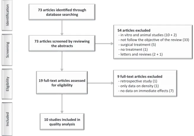
ified for the final analysis. The review details and selection process are given in Figure 1 as described in the PRISMA statement.17The interexaminer agreement
(kappa statistics) for inclusion of studies was 0.90. Summarized data of the 10 studies are presented in Table 2. Two studies were randomized controlled trials and eight were prospective case-series. In the included studies, the age of the participants ranged between 6 and 14.5 years. A number of the retrieved studies had the same patient material but with different aims, so no results have been duplicated in this review. Endpoints
Three studies9,11,18
defined endpoints as when the maxillary lingual cusp of the permanent first molar came into contact with the mandibular buccal cusp of the permanent first molar. Four studies defined endpoints as when the screw expansion reached 7 mm,12,19–21 and
one study14 had the endpoint after 8 mm of screw
expansion. Two studies13,22did not define any endpoints.
Skeletal Changes
In reference to the skeletal changes, the majority of the measurements conducted in the reviewed studies were linear and carried out either on coronal or axial 2D slices.
Midpalatal Suture
Measurements of changes in midpalatal suture width during RME were presented in six stud-ies,9,12,14,20–22 and five of them presented data for both
the anterior and posterior region. The mean expansion changes in the midpalatal suture in the posterior region ranged from 1.6 to 4.33 mm, which corresponded to 22%–53% of the total screw expansion. Mean expan-sion changes in the anterior midpalatal suture ranged from 1.52 to 4.33 mm, corresponding also to 22%– 53% of total screw expansion. The reviewed articles had different endpoints and slightly different reference points when measuring the midpalatal suture opening, which makes comparisons difficult. Three studies12,14,20
concluded that the suture opened in a triangular pattern with the largest opening at the anterior part. One study22 reported that the midpalatal suture
opening was parallel, while Podesser et al.21reported
that the suture opening was larger anteriorly in some individuals and larger posteriorly in others.
Nasal Cavity
Changes in the width of the nasal cavity were investigated in three studies at the level of the first molars. The nasal width was expanded by 1.2 mm,21
1.4 mm,20
and 2.73 mm,22
which corresponded to 17%, 20%, and 33% of the total screw expansion, respectively.
Circummaxillary Sutures
The changes in the circummaxillary sutures were assessed in one study.9 The following sutures were
reported to be affected: zygomaticofrontal, zygomati-comaxillary, frontomaxillary, zygomaticotemporal, na-somaxillary, frontonasal, and internasal. The changes in these sutures were overall small, ie, between 0.30 and 0.45 mm. The average amount of suture opening was generally higher in the sutures articulating directly to the maxilla (zygomaticomaxillary and frontomaxil-lary), whereas sutures further away from the maxilla showed a lower degree of disarticulation.
Spheno-occipital Synchondrosis
One study demonstrated a mean expansion of 0.6 mm in the spheno-occipital synchondrosis.11
Orbital Structures
A significant increase in orbital volume and aperture width was found in one study.18 However, both the
increase in volume (0.72 mL) and width (1.09 mm) were small and considered clinically insignificant. Dental Structures
The dentoalveolar transverse expansion was larger than the skeletal expansion.13,14,22 Dental tipping was
assessed in four studies.13,14,19,22 Mean buccal tipping
of the first molars was reported to be 7.5u in three studies13,14,22and 1.0u in the study by Podesser et al.21
Alveolar bending was also reported and accounted for about 30% of the total expansion.14
Quality Analysis
A quality analysis of the 10 retrieved studies was accomplished according to the criteria in Table 1. Interexaminer agreement for the quality assessment was 0.95 (kappa statistics).
The judged quality and methodologic soundness for the 10 selected studies are presented in Table 3. Two studies were of medium quality,14,18and the other eight
were of low quality. Obvious shortcomings were study design, sample size, and inadequate selection descrip-tion. Only three studies used blinded measure-ments,14,18
and dropouts were not reported in any study. DISCUSSION
This systematic review is the first review to evaluate the effect of RME treatment on sutures outside the
maxilla in all three dimensions, and the review disclosed some interesting findings.
When treating posterior crossbites with a RME device, the existing evidence points out that the midpalatal suture opening is around 20%–50% of the total screw expansion. There seems to be no consistent evidence on whether the midpalatal sutural opening is parallel or triangular.
The effect on the nasal cavity dimensions after RME seems to be apparent and indicates an enlargement between 17% and 33% of the total screw expansion. If this increase in dimensions facilitates the breathing mode of the patients, an interesting follow-up question would be for how long; this question, however, was not assessed in the included studies.
Circummaxillary sutures, particularly the zygomati-comaxillary and frontomaxillary sutures, appear to be affected by the maxillary expansion, but the changes in these sutures were overall small and the evidence not conclusive. The investigations of the sutures were only done in one plane of space and at the middle point of the sutures, which is a shortcoming because no considerations were taken of the topography of the investigated sutures at different sites. In addition, one study showed a small opening of the spheno-occipital synchondrosis after RME treatment.11
The involvement of both the circummaxillary sutures and the spheno-occipital synchondrosis can have some important clinical implications and may explain the forward and downward displacement of the maxilla after RME treatment, which can be beneficial in Class III correc-tions in young patients.9These findings are, however, a
manifestation of the immediate effects of RME, and a systematic review of the long-term effects and critical analysis of longitudinal studies are essential.
The effect of RME on the dentoalveolar structures was found to be greater than the skeletal expansion. Dental and alveolar tipping toward the buccal aspect was reported to occur in four studies. The clinical implications of these ‘‘side effects’’ could imply overexpansion due to the risk for dental and alveolar relapse.
The impact of age on skeletal vs dental effects of RME is an interesting issue in the clinical situation. The age range in the articles reviewed was 6.0 to 14.5 years. All of the studies except one22 showed,
more or less, the same skeletal expansion rate, but dental tipping varied. In the study by Podesser et al.,21
dental tipping was reported to be about 1u compared to 6.5u as reported in the studies by Christie et al.,22
Lagrave`re et al.,13and Weissheimer et al.14Mean age
for patients in the study by Podesser21was 8.1 years,
which is about 2 years younger than participants in the studies conducted by Christie et al.22
and Weissheimer et al.14
Table 2. Summarized Data of the 10 Included Studies
Author Year Country Study Design Participants Size, Gender, Age Intervention Type, Activation Protocol, Endpoints
Weissheimer et al.14 2011 Brazil Randomized controlled trial (RCT) Group I: 18 subjects Group II: 15 subjects 22 girls, 11 boys 10.7 y, (7.2–14.5)
Type:
Group I: Haas expander Group II: Hyrax expander
Activation: Initial activation of four quarter turns (0.8 mm) followed by two quarter turns per d (0.4 mm)
Endpoint: 8-mm screw activation Sicurezza et al.18 2011 Italy Prospective case-series 18 girls, 12 boys 9.8 y (SD 1.8)
Type: Hyrax expander
Activation: three quarter turns per d (0.75 mm) Mean treatment time: 18 d
Endpoint: palatal cusp of the first maxillary molar in contact with the facial cusp of the mandibular first molar Leonardi et al.9 2011 Italy1 Prospective case-series 6 girls, 2 boys 9.8 y (SD 1.8)
Type: Hyrax expander
Activation: three quarter turns per d (0.75 mm) Mean treatment time: 18 d
Endpoint: palatal cusp of the first maxillary molar in contact with the facial cusp of the mandibular first molar Ballanti et al.20 2010 Italy2 Prospective case-series 10 girls, 7 boys 11.2 y (8–14)
Type: Haas expander
Activation: two quarter turns (0.5 mm) per d for 14 d (7 mm)
Endpoint: 7-mm screw activation
Christie et al.22 2010 USA Prospective case-series 10 girls, 14 boys 9.9 y (7.8–12.8)
Type: bonded Haas expander
Activation: two quarter turns per day (0.4 mm) Endpoint: no endpoint defined
Mean screw activation: 8.19 mm
Lagrave´re et al.13
2010 Canada RCT Group I:
13 girls and 8 boys 14. 2 y (SD 1.32) Group II:
15 girls and 5 boys 14.1 y (SD 1.35)
Type:
Group I: bone-anchored maxillary expander Group II: tooth-anchored maxillary expander Activation:
Group I: 1 healing week followed by 1 turn every other d Group II: two quarter turns (0.5 mm) per d
Endpoint: overcorrection of the crossbite Leonardi et al.11 2010 Italy1 Prospective case-series 6 girls, 2 boys 9.8 y (SD 1.8)
Type: Hyrax expander
Activation: three quarter turns (0.75 mm) per d; mean treatment time 18 d
Endpoint: palatal cusp of the first maxillary molar in contact with the facial cusp of the mandibular first molar Ballanti et al.19 2009 Italy2 Prospective case-series 10 girls, 7 boys 11.2 y (8–14)
Type: Haas expander
Activation: two quarter turns (0.5 mm) per d for 14 d Endpoint: 7-mm screw activation
Lione et al.12 2008 Italy2 Prospective
case-series
10 girls, 7 boys 11.2 y (8–14)
Type: Haas expander
Activation: two quarter turns (0.5 mm) per d for 14 d Endpoint: 7-mm screw activation
Podesser et al.21
2007 Austria Prospective
case-series
6 girls and 3 boys 8.1 y (6.1–9.8)
Type: cemented RME splint device Activation: two quarter turns (0.5 mm) per d Endpoint: 7-mm screw activation
1Patient material in Leonardi et al.9and Leonardi et al.11are the same.
Outcome Measure Imaging Parameters Outcome and authors conclusions Comparison of linear and angular measurement
changes in the transverse plane: - Intermolar width
- Molar angulation
- Anterior and posterior midpalatal suture opening
- Anterior and posterior apical base width
Type: cone beam computed tomography (CBCT) Measurements on coronal slices
perpendicular to the nasal plane Resolution:
0.3 mm voxel size
Rapid maxillary expansion (RME) produced signifi-cant increase in all transverse dimensions. The expansion pattern was triangular with smaller effects
at the skeletal level compared to dental level. The opening of the midpalatal suture was larger in
the anterior part compared to the posterior part. The Hyrax produced significantly larger skeletal
effects and less tipping than the Haas. Linear measurement of aperture width
Orbital volume was calculated by defining contours on a series of slices
Type:
Low-dose computed tomography (CT) (80 kV) Measurements on axial slices
Resolution:
Slice-thickness 0.5 mm
RME produced a small but significant increase in orbital volume and aperture width.
Linear measurement of circummaxillary suture width changes:
- Nasomaxillary - Frontomaxillary - Zygomaticomaxillary - Internasal - Zygomaticotemporal - Frontonasal Type: Low-dose CT (80 kV) measurements on axial and sagittal slices Resolution:
Slice-thickness 0.5 mm
RME produced significant bony displacement by circummaxillary suture opening. Sutures that articulate directly with the maxilla had larger displacement than those located further away.
Linear measurement changes in the transverse plane:
- Interincisor width - Midpalatal suture width - Nasal cavity width
Type:
Low-dose CT (80 kV)
Measurements on coronal slices perpendicular to the occlusal plane Resolution:
Slice-thickness 1.25 mm, interval 0.6 mm
RME produced sign increase in all transverse dimensions.
Midpalatal suture and nasal width increased in a parallel manner on coronal scans but were larger anteriorly compared to posteriorly. Linear and angular measurement
changes in the transverse plane: - Intermolar angle
- Midpalatal suture width - Basal bone width - Nasal cavity width
Type: CBCT
Measurements on coronal and sagittal slices Resolution:
No data
RME produced significant increase in transverse dimensions of the nasal cavity, maxillary basal bone, and midpalatal suture.
The midpalatal suture opened in a parallel fashion but had the largest increase in the suture level followed by basal bone and nasal cavity level.
Comparison of linear and angular measurement changes in the transverse, vertical,
and sagittal plane: - Intermolar angle - Midpalatal suture width - Maxillary basal bone width - Nasal cavity width
Type: CBCT
Measurements on sagittal, axial, and coronal slices
Resolution: 0.25 mm voxel size
Transverse changes were significant and with no differences between groups.
Only minor changes in vertical and sagittal dimensions. Dental expansion was greater than skeletal expansion.
Linear measurement of spheno-occipital synchondrosis width changes
Type:
Low-dose CT (80 kV) Measurements on axial slices Resolution:
Slice-thickness 0.5mm
RME led to a small immediate widening of the spheno-occipital synchondrosis.
Linear measurement changes in the transverse plane:
- Intermolar width (crown and apex) - Lingual bone plate thickness Buccal bone thickness
Type:
Low-dose CT (80 kV)
Measurements on coronal scans perpendicular to the hard palate Resolution:
Slice-thickness 1.25 mm, interval 0.6 mm
RME induced a significant increase in the trans-verse dimension of the maxillary arch without causing permanent injury to the periodontal bony support of anchoring teeth.
Linear measurement changes in the transverse plane:
- Midpalatal suture width - Pterygoid width
Type:
Low-dose CT (80 kV)
Measurements on coronal scans perpendicular to the hard palate Resolution:
Slice-thickness 1.25 mm, interval 0.6 mm
RME produced significant increase in the trans-verse dimensions of the midpalatal suture, more anteriorly than posteriorly.
Pterygoid width was also significantly increased.
Linear and angular measurement changes in the transverse plane:
- Intermolar width - Intermolar angle - Maxillary alveolar width - Midpalatal suture width - Maxillary basal bone width - Nasal cavity width
Type: Low-dose CT
Measurements on coronal scans Resolution:
Slice-thickness 1.5 mm, interval 1.5 mm
The relative contribution of dentoalveolar and skeletal changes varied from subject to subject. Almost 50% of the changes were a result of
dentoalveolar changes
1Patient material in Leonardi et al. 2011 and Leonardi et al. 2010 are the same.
2Patient material in Ballanti et al. 2010, Ballanti et al. 2009, and Lione et al. 2008 are the same.
conducted by Lagrave`re et al.13 This age difference
could be one of the factors explaining the lesser dental tipping in younger children.
In contrast to the 2D imaging, CT/CBCT and its impact on more accurate diagnostics in all three dimensions is very encouraging due to greater resolution and without structure. CT/CBCT enables assessment of root resorption, determine positions of impacted teeth, evaluation of bone grafts in cleft regions and treatment effects of RME. If the structural overlap is the main issue in the diagnostics, the use of the low-dose CT/CBCT imaging can be justified for gathering the most adequate information for the treatment. However, the radiation dosage and its bearing on growing patients must be kept in mind. Even though CT-imaging may be available, regular use of it for orthodontic or dental care may not be justified.
The restrictions concerning the number of databas-es and languagdatabas-es when searching the literature might imply that some studies were not identified. Studies that are difficult to find are, however, often of lower quality. The strength of the evidence in a systematic review is probably more dependent on assessing the quality of the included studies than on the degree of comprehensiveness.23
The kappa scores measuring levels of agreement between the two reviewers in assessing data extraction and quality scores of the included articles were very good, and thus, indicated that the results were reliable. Limitations
No meta-analysis was carried out because of the diversity in the methodologic aspects in the selected studies such as different landmarks, reference points, and endpoints.
A notable finding was that none of the selected studies were of high value of evidence, and only two randomized controlled trials were identified. From an evidence-based point of view, the scientific value of a case-series study is limited. Some authors, however,
have argued that well-designed prospective or even retrospective studies should not be ignored when assessing scientific literature.24
Nevertheless, it should be emphasized that the randomized controlled trial with adequate sample size and power is the most powerful tool to evaluate treatment, and the quality of the trial significantly affects the validity of the conclusions.
Many of the studies had serious defects, and according to the criteria used, the majority of the articles were judged to be of low quality. The most serious shortcomings were the study design in combination with small sample size and inadequate selection description. Other examples of shortcomings were lack of method error analysis and the absence of blinding in measure-ments. Furthermore, several studies used the same material (patient data). In addition, all studies used measurements on 2D slices, which imply accuracy problems when identifying the same slice and land-marks between the baseline and posttreatment slices. 3D modeling and superimposition on stable structures is one way to solve this problem and makes it easier to identify the same landmark, and thus, gives rise to more accurate and reliable measurements.
In the reviewed articles, different endpoints and different CT/CBCT protocols with various slice thick-nesses and somewhat different reference points were used as well as different resolutions. This can compromise the quality of the images, and thereby have an impact on the reliability of the results. Therefore, it might be sound to interpret the results from these studies with caution.
This review of the literature has disclosed that additional randomized controlled trials with sufficient power are required to add further insight into the 3D effects of rapid maxillary expansion on sutures and bones of the face, and thus, has exposed a need for future studies in this area. CONCLUSIONS
The available evidence indicates that:
N Midpalatal suture opening during orthodontic treat-ment with RME amounted to 20%–50% of the total
Table 3. Quality Evaluation of the 10 Selected Studies According to Predetermined Criteria (Table 1)
Author Year Country Study Design
Adequate Sample Size
Weissheimer et al.14 2011 Brazil Randomized controlled trial (RCT) Partly adequate
Sicurezza et al.18 2011 Italy Prospective case-series Yes
Leonardi et al.9 2011 Italy Prospective case-series Inadequate
Ballanti et al.20 2010 Italy Prospective case-series Partly adequate
Christie et al.22 2010 USA Prospective case-series Yes
Lagrave´re et al.13 2010 Canada RCT Yes
Leonardi et al.11 2011 Italy Prospective case-series Inadequate
Ballanti et al.19 2009 Italy Prospective case-series Partly adequate
Lione et al.12 2008 Italy Prospective case-series Partly adequate
screw expansion, but there was no consistent evidence on whether the midpalatal sutural opening was parallel or triangular.
N RME produced immediate significant changes in transverse dimensions of the nasal cavity, circum-maxillary sutures, spheno-occipital synchondrosis, and aperture width. Structures that articulated directly with the maxilla had larger displacement than those located further away.
N The majority of the articles were judged to be of low quality therefore, no evidence-based conclusions could be drawn from these studies. Additional randomized controlled trials with sufficient power are required to add further insight into the 3D effects of RME on sutures and bones of the face.
REFERENCES
1. Lagravere MO, Major PW, Flores-Mir C. Long-term skeletal changes with rapid maxillary expansion: a systematic review.Angle Orthod. 2005;75:1046–1052.
2. Angell E. Treatment of irregularity of the permanent or adult teeth.Dental Cosmos. 1860:540–544.
3. Chung C, Font B. Skeletal and dental changes in the sagittal, vertical, and transverse dimensions after rapid palatal expansion. Am J Orthod Dentofacial Orthop. 2004; 126:569–575.
4. Haralambidis A, Ari-Demirkaya A, Acar A, Kucukkeles N, Ates M, Ozkaya S. Morphologic changes of the nasal cavity induced by rapid maxillary expansion: a study on 3-dimensional computed tomography models. Am J Orthod Dentofacial Orthop. 2009;136:815–821.
5. Cross D, JP M. Effect of rapid maxillary expansion on skeletal, dental, and nasal structures: a postero-anterior cephalometric study.Eur J Orthod. 2000;22:519–528. 6. Basciftci F, Mutlu N, Karaman A, Malkoc S, Kucukkolbasi H.
Does the timing and method of rapid maxillary expansion have an effect on the changes in nasal dimensions?Angle Orthod. 2002;72:118–123.
7. Pangrazio-Kulbersh V, Wine P, Haughey M, Pajtas B, Kaczynski R. Cone beam computed tomography evaluation of changes in the naso-maxillary complex associated with two types of maxillary expanders. Angle Orthod. 2012;82: 448–457.
8. Haas A. Rapid expansion of the maxillary dental arch and nasal cavity by opening the mid palatal suture. Angle Orthod. 1961;31:73–89.
9. Leonardi R, Sicurezza E, Cutrera A, Barbato E. Early post-treatment changes of circumaxillary sutures in young patients treated with rapid maxillary expansion. Angle Orthod. 2011;81:36–41.
10. Ghoneima A, Abdel-Fattah E, Hartsfield J, El-Bedwehi A, Kamel A, Kula K. Effects of rapid maxillary expansion on the cranial and circummaxillary sutures.Am J Orthod Dentofa-cial Orthop. 2011;140:510–519.
11. Leonardi R, Cutrera A, Barbato E. Rapid maxillary expan-sion affects the spheno-occipital synchondrosis in young-sters. A study with low-dose computed tomography.Angle Orthod. 2010;80:106–110.
12. Lione R, Ballanti F, Franchi L, Baccetti T, Cozza P. Treatment and posttreatment skeletal effects of rapid maxillary expansion studied with low-dose computed to-mography in growing subjects. Am J Orthod Dentofacial Orthop. 2008;134:389–392.
13. Lagrave`re MO, Carey J, Heo G, Toogood RW, Major PW. Transverse, vertical, and anteroposterior changes from bone-anchored maxillary expansion vs traditional rapid maxillary expansion: a randomized clinical trial.Am J Orthod Dentofacial Orthop. 2010;137:304.e1–12.
14. Weissheimer A, de Menezes LM, Mezomo M, Dias DM, de Lima EM, Rizzatto SM. Immediate effects of rapid maxillary expansion with Haas-type and hyrax-type expanders: a randomized clinical trial.Am J Orthod Dentofacial Orthop. 2011;140:366–376.
15. National Health Service (NHS) Centre for Reviews and Dissemination. Undertaking Systematic Reviews of Re-search on Effectiveness. University of York, UK: York Publishing Services; 2001.
16. Bondemark L, Holm AK, Hansen K, et al. Long-term stability of orthodontic treatment and patient satisfaction. A system-atic review.Angle Orthod. 2007;77:181–191.
17. Moher D, Liberati A, Tetzlaff J, Altman DG. Preferred reporting items for systematic reviews and meta-analyses: the PRISMA statement.PLoS Med. 2009;6:e1000097. 18. Sicurezza E, Palazzo G, Leonardi R. Three-dimensional
computerized tomographic orbital volume and aperture width evaluation: a study in patients treated with rapid maxillary expansion.Oral Surg Oral Med Oral Pathol Oral Radiol Endod. 2011;111:503–507.
19. Ballanti F, Lione R, Fanucci E, Franchi L, Baccetti T, Cozza P. Immediate and post-retention effects of rapid maxillary
Adequate Selection Description Defined Diagnosis and Endpoints Valid Outcome Measures Method Error Analysis Blinding in Measurements Judged Quality Grading
Inadequate Yes Yes Yes Yes Medium
Inadequate Yes Yes No Yes Medium
Inadequate Unclear Yes No Yes Low
Inadequate Yes Partly Yes No Low
Inadequate No Yes Yes No Low
Partly adequate No Yes Yes No Low
Inadequate Unclear Yes No No Low
Inadequate Yes Partly Yes No Low
Inadequate Yes Partly Yes No Low
Inadequate No Partly No No Low
expansion investigated by computed tomography in growing patients.Angle Orthod. 2009;79:24–29.
20. Ballanti F, Lione R, Baccetti T, Franchi L, Cozza P. Treatment and posttreatment skeletal effects of rapid maxillary expansion investigated with low-dose computed tomography in growing subjects.Am J Orthod Dentofacial Orthop. 2010;138:311–317.
21. Podesser B, Williams S, Crismani AG, Bantleon HP. Evaluation of the effects of rapid maxillary expansion in growing children using computer tomography scanning: a pilot study.Eur J Orthod. 2007;29:37–44.
22. Christie KF, Boucher N, Chung CH. Effects of bonded rapid palatal expansion on the transverse dimensions of the maxilla: a cone-beam computed tomography study. Am J Orthod Dentofacial Orthop. 2010;137:S79–85. 23. Egger M, Juni P, Bartlett C, Holenstein F, Sterne J. How
important are comprehensive literature searches and the assessment of trial quality in systematic reviews? Empirical study.Health Technol Assess. 2003;7:1–76.
24. Ioannidis JP, Haidich AB, Pappa M, et al. Comparison of evidence of treatment effects in randomized and nonran-domized studies.JAMA. 2001;286:821–830.