ACTA UNIVERSITATIS
UPSALIENSIS
Digital Comprehensive Summaries of Uppsala Dissertations
from the Faculty of Science and Technology
1308
Palaeobiology and diversification
of Proterozoic-Cambrian
photosynthetic eukaryotes
ISSN 1651-6214 ISBN 978-91-554-9389-9
Dissertation presented at Uppsala University to be publicly examined in Hambergsalen, Geocentrum, Villavägen 16, 752 36, Uppsala, Friday, 11 December 2015 at 10:15 for the degree of Doctor of Philosophy. The examination will be conducted in English. Faculty examiner: Professor Shuhai Xiao (Geosciences, Virginia Polytechnic Institute and State University).
Abstract
Agić, H. 2015. Palaeobiology and diversification of Proterozoic-Cambrian photosynthetic eukaryotes. Digital Comprehensive Summaries of Uppsala Dissertations from the Faculty of Science and Technology 1308. 47 pp. Uppsala: Acta Universitatis Upsaliensis. ISBN 978-91-554-9389-9.
One of the most important events in the history of life is the evolution of the complex, eukaryotic cell. The eukaryotes are complex organisms with membrane-bound intracellular structures, and they include a variety of both single-celled and multicellular organisms: plants, animals, fungi and various protists. The evolutionary origin of this group may be studied by direct evidence of past life: fossils. The oldest traces of eukaryotes have appeared by 2.4 billion years ago (Ga), and have additionally diversified in the period around 1.8 Ga. The Mesoproterozoic Era (1.6-1 Ga) is characterised by the first evidence of the appearance complex unicellular microfossils, as well as innovative morphologies, and the evolution of sexual reproduction and multicellularity. For a better understanding of the early eukaryotic evolution and diversification patterns, a part of this thesis has focused on the microfossil records from various time periods and geographic locations. Examination of microfossil morphology, cell wall microstructure and biochemical properties, reflect their intracellular complexity and function, and allow reconstructions of their life cycle, as well as observing the evolutionary pattern of change from Mesoproterozoic, to Cambrian-Ordovician transition. Several case studies included assemblages deriving from Mesoproterozoic, Neoproterozoic and early Paleozoic time intervals that show disparate morphotypes and innovative features indicative of algal clades. The Mesoproterozoic Ruyang Group in northern China has yielded a diverse microfossil assemblage that provides important clues about the diversification of different eukaryotic groups. Furthermore these microfossils contributed an additional evidence for the emergence of the crown group Eukarya by 1.7-1.4 Ga. In another part of this thesis, examination of wall microstructure and chemical properties via Raman spectroscopy has been used to assess the biological affinities of various Neoproterozoic problematic carbonaceous compression fossils. Studies on the early Phanerozoic (c. 545-485 Ma) assemblages from Estonia reconstructed patterns of the early radiations of phytoplankton and its evolutionary innovations. A continuing theme in this thesis has been using a combination of evidence of microfossils’ fine-scale morphology, ecology and chemical properties to determine their function in life, in addition to their systematic position.
Keywords: microfossils, eukaryotes, Mesoproterozoic, Neoproterozoic, Cambrian, Ordovician, palaeobiology, biostratigraphy, phylogeny, China, Estonia, Siberian Platform, prasinophytes, microstructure, Raman spectroscopy
Heda Agić, Department of Earth Sciences, Palaeobiology, Villav. 16, Uppsala University, SE-75236 Uppsala, Sweden.
© Heda Agić 2015 ISSN 1651-6214 ISBN 978-91-554-9389-9
List of Papers
This thesis is based on the following papers, which are referred to in the text by their Roman numerals.
I Agić, H., Moczydłowska, M., Yin, L. (2015) Affinity, life
cy-cle, and intracellular complexity of organic-walled microfossils from the Mesoproterozoic of Shanxi, China. Journal of
Paleon-tology, 89(1):28–50. doi:10.1017/jpa.2014.4.
II Agić, H., Moczydłowska, M., Yin, L. Diversity of
organic-walled microfossils from the early Mesoproterozoic Ruyang Group, North China Craton – a window into the early eukaryote evolution. Manuscript submitted, Precambrian Research. III Agić, H., Moczydłowska, M. Raman spectroscopy and
micro-structural comparison of carbonaceous compression and body fossils from the Neoproterozoic of Siberian and Eastern Euro-pean platforms. Manuscript for submission to Geological
Mag-azine.
IV Moczydłowska, M., Budd, G. E., Agić, H. (2015) Ecdysozoan-like sclerites among Ediacaran microfossils. Geological
Maga-zine, 152(6):1145–1148. doi: 10.1017/S001675681500045X.
V Agić, H., (2015) A new species of small acritarchs with porous
wall structure from the early Cambrian of Estonia, and implica-tions for the fossil record of eukaryotic picoplankton.
Paly-nolgy, 40, in production. doi: 10.1080/01916122.2015.1068879.
VI Agić, H., Moczydłowska, M., Canfield, D. E. Reproductive
cyst and operculum formation in the Cambrian-Ordovician ga-leate-plexus microfossils. Manuscript accepted for publication
in GFF. doi: 10.1080/11035897.2015.1116603.
Reprints of published papers were made with kind permission from the re-spective publishers.
Paper I © Cambridge Journals and The Paleontological Society Paper II © by the authors
Paper III © by the authors
Paper IV © Cambridge University Press Paper V © Taylor & Francis
Paper VI © by the authors Statement of authorship
Paper I: H.A. analysed the material, interpreted the results and shared writ-ing.
Paper II: H.A. analysed the material, collected data, interpreted the results and shared writing.
Paper III: H.A. analysed the material, collected and interpreted the data and shared writing.
Paper IV: H.A. assisted in data collection.
Paper V: H.A. analysed the material, interpreted the results and wrote the manuscript.
Paper VI: H.A. analysed the material, collected the data, interpreted the re-sults and shared writing.
Disclaimer: The papers presented herein are for the purpose of public exam-ination as a doctoral thesis only. They are not considered valid publications for taxonomic and nomenclatural purposes according to ICZN article 8.2. Accordingly, all new taxonomic names and emendations in papers I-VI are void. Authority for taxonomic work is retained by the original publications. Front cover: Scanning electron micrograph of the pores in the organic wall of Tasmanites volkovae (Paper V) from the early Cambrian Lükati For-mation, northern Estonia.
Contents
1. Introduction ... 9
Introduction to Proterozoic micropalaeontology and the aims of this thesis ... 9
The Precambrian biosphere ... 10
Organic-walled microfossils and their phylogenetic implications ... 12
2. Mesoproterozoic eukaryotic microfossils from northern China ... 15
Ruyang Group: a hotspot of early eukaryotic biodiversity ... 15
Microstructure and the biological affinities of the Ruyang taxa ... 17
3. Neoproterozoic carbonaceous fossils ... 20
Raman spectroscopy as a tool in investigating phylogenetic affinities .... 20
Animals in the Proterozoic ... 22
4. Early Phanerozoic radiations of phytoplankton ... 24
Possible Cambrian picoplankton ... 24
Cyst formation in the galeate plexus ... 25
5. Functional morphology and palaeobiology ... 29
Eukaryotic cysts ... 29
Vesicle morphology as a result of intracellular complexity ... 30
6. Conclusions and future directions ... 33
7. Svensk sammanfattning ... 35
Introduktion ... 35
Livet i proterozoikum ... 35
Vad är organiska mikrofossil? ... 36
Fossil av proterozoiska eukaryoter ... 37
Diversifiering av acritarcher vid början av fanerozoikum ... 37
8. Acknowledgements ... 39
Abbreviations
Ma – million years (milium annum) Ga – billion years (giga-annum) OWM – organic-walled microfossil VSM – vase shaped microfossil SSF – small shelly/skeletal fossil SCF – small carbonaceous fossil LM – light microscopy
SEM – scanning electron microscopy TEM – transmission electron microscopy
ECAP – Ediacaran complex acanthomorph Palynoflora GOBE – Great Ordovician Biodiversification Event GOE – Great Oxygenation Event
1. Introduction
Introduction to Proterozoic micropalaeontology and the
aims of this thesis
The Proterozoic Eon, lasting from c. 2500 to 542 million years ago (Ma) (Fig. 1), is one of the most important time intervals for providing infor-mation about the evolution of life on Earth, especially the origin and diversi-fication of one of the three domains of life: Eukaryota. The eukaryotes are complex organisms with membrane-bound intracellular structures such as nucleus and mitochondria (and chloroplasts, for some groups). They include a variety of both single-celled and multicellular life: animals, plants, fungi, algae, amoebae and various other protists. Based on the molecular data from present-day organisms, the origin of eukaryotes has been estimated back to 2.5-2.0 Ga. Although the origin of animals and true plants apparently oc-curred much later on in Earth’s history, close to the Ediacaran-Cambrian transition (c. 555 Ma), most of the Proterozoic fossil record of the eukary-otes comprises microscopic protists.
Some of the oldest eukaryotic fossils are morphologically simple, small cells with organic, carbonaceous cell-walls, dating back to c. 2000-1800 Ma (Paleoproterozoic). While initially simple, these fossils have undergone sev-eral diversification events through the rest of the Proterozoic Eon and the early Phanerozoic (the time of visible life, c. 542 Ma).
In this thesis, I aim to address some questions concerning the nature of early fossils with eukaryotic and phytoplanktonic morphology, and docu-ment the appearance of certain innovative characters in the microfossil rec-ord, for a better understanding of the eukaryotic evolution. Additionally, I intend show the diversification patterns of these organisms as recorded by various assemblages. For that purpose, I have investigated the material from separate geographic localities and time periods. These topics include: recon-structing the biological affinities of the early eukaryotes and their diversity patterns in Mesoproterozoic oceans; comparing the enigmatic carbonaceous compressions and body fossils from the Neoproterozoic Era and their chemi-cal properties; examining the evolutionary trends of the phytoplanktonic microfossils at the beginning of the Phanerozoic; and understanding how the morphology of these microfossils reflects their intracellular complexity.
10
The Precambrian biosphere
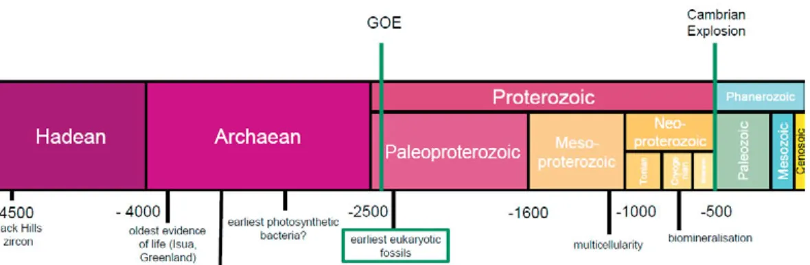
Figure 1. Geological time scale of the Earth, showing four Eons (Hadean, Archaean, Proterozoic, Phanerozoic) that span c. 4.567 billion years (colour coded according to the standards of the International Commission on Stratigraphy). Time is given in millions of years before present. Major events in the history of Earth’s biosphere are listed at the bottom. Two significant events that have drastically shaped life and planet (Great Oxygenation Event and the Cambrian Explosion) are marked with green lines. Material studied in this thesis derives from various ages in the time interval between the Mesoproterozoic and the early Paleozoic eras.
Throughout most of the Earth’s history (c. 85%), life was inconspicuous, microscopic, and did not produce biomineralized elements that are easily preserved in the fossil record. This absence of fossils has puzzled Victorian geologists, especially Charles Darwin (1859) who wondered why there was seemingly no life before the Cambrian system, when skeletal fossils sudden-ly occur in abundance. Due to a relative scarcity of Precambrian rocks and the lack of macroscopic organisms, it took a whole century until the first discovery of Precambrian fossils, originally reported by Barghoorn and Ty-ler (1965) from the 1.8 Ga Gunflint chert exposed on the shore of Lake Su-perior. Since then, the field of the Precambrian palaeontology has flourished and a lot of progress has been done investigating the antiquity and the early evolution of life. The oldest trace of life, carbon isotopic fractionation signa-ture from the Isua peninsula, Greenland (Mojzsis et al., 1996, Fig. 1), dates back to c. 3.8 Ga (or even as far back as 4.1 Ga, as possible biogenic carbon within a zircon; Bell et al., 2015). More robust evidence of the earliest life comes from the fossilized cells from the c. 3.4 Ga old rocks in East Pilbara Terrane, Western Australia (Schopf et al., 2002; Wacey et al., 2011), and
Archaean stromatolites - accretionary structures formed by trapping and binding of sediment particles by microbial mats (Allwood et al., 2006). Such chemotrophic or phototrophic bacterial mat-building communities seem to have been predominant organisms on the early Earth.
Even though still mostly microscopic, life became more complex and di-verse in the Proterozoic Eon (Porter, 2004; Cohen & Macdonald, 2015). This time in Earth’s history was characterised by a stable δ13C record, which
in-spired the eponym “the Boring Billion” (Buick et al., 1995; Brasier & Lind-say, 1998). However, despite this catchphrase, a lot of innovations were developing on a cellular level, and complex, eukaryotic cells originated dur-ing this time. The earliest bona fide eukaryotic fossils generally lack diag-nostic characters apart from size, like Grypania (Han & Runnegar, 1992), and date back to 2.4 Ga. This group might be even more ancient if the large carbonaceous cells reported from South Africa by Sugitani et al. (2015) are accepted as true eukaryotes. More convincingly eukaryotic fossils are the 1.8 Ga leiosphaerids (round, organically preserved unicells) from the North Chi-na Craton (Lamb et al., 2009). Subsequent divergence of eukaryotic lineages from the last eukaryotic common ancestor (LECA) probably took place dur-ing the Mesoproterozoic Era, as estimated by molecular clock analyses (Yoon et al., 2004; Parfrey et al., 2011). Fossils from this time illustrate the appearance of novel cell morphologies, evolution of sexual reproduction, and the origin of multicellularity (Hermann, 1990; Knoll, 2011; Moczydłow-ska et al., 2011; Butterfield, 2014). The oldest eukaryote exhibiting cellular differentiation is a red alga Bangiomorpha pubecens from the 1.2 Ga strata of arctic Canada (Butterfield, 2000).
Additional steps in the eukaryotic evolution took place later on, in the Neoproterozoic Era, along with environmental changes (rise in atmospheric oxygen concentrations; Lenton et al., 2014). New morphologies and metabo-lisms represented in the fossil record from this interval include macroscopic algae (Ye et al., 2015), the early testate amoebae preserved as c. 742 Ma old vase-shaped microfossils (Martí Mus & Moczydłowska, 2000; Porter & Knoll, 2000), and possible ciliates 715-635 Ma (Bosak et al., 2011). Neopro-terozoic Era also marks the onset of biomineralization, as evidenced by 812-717 Ma old protistan phosphatic scales (Cohen et al., 2011). Finally, in the terminal Proterozoic (Ediacaran), large, macroscopic and in some cases, motile organisms appear in the fossil record, leading to the evolution of ani-mals (e.g. Narbonne, 2005).
Despite their relative rarity, sedimentary rocks of Proterozoic age are vital for an understanding of the most critical time in the history of the biosphere (e.g. eukaryogenesis – Sagan, 1967; Brasier, 2012), and how microscopic life coped during the extensive Neoproterozoic glaciations (Corsetti et al., 2003; Moczydłowska, 2008), and through the onset of animals which have significantly changed the environment (Bottjer et al., 2000). A great part of the Precambrian biosphere is composed of small, organically preserved cells
12
or filaments, called the “organic-walled microfossils”, which are a focus of this thesis.
Organic-walled microfossils and their phylogenetic
implications
Defining organic-walled microfossils (OWM) is not an easy task. This is a broad grouping including mostly single-celled (but also some multicellular and filamentous) microfossils from a few microns to a millimetre in size, which possess a non-mineralized, carbonaceous vesicle wall (Fig. 2). Even though OWMs have been studied from different continents and time periods for over a century, their exact phylogenetic affinities have not been fully understood. As a result, Evitt (1963) introduced and named the polyphyletic group Acritarcha, from ancient Greek words achritos and arche, meaning “uncertain” and “origin”, to serve as an umbrella term for early Paleozoic, marine palynomorphs. This name is now becoming somewhat obsolete, since more and more of these taxa have been assigned to known groups of microorganisms (Loeblich &Wicander, 1976; Tappan, 1980; Colbath & Grenfell, 1995; Arouri et al., 2000; Butterfield, 2000, 2004; Knoll et al., 2006; Moczydłowska & Willman, 2009; Moczydłowska et al., 2011; Chen et
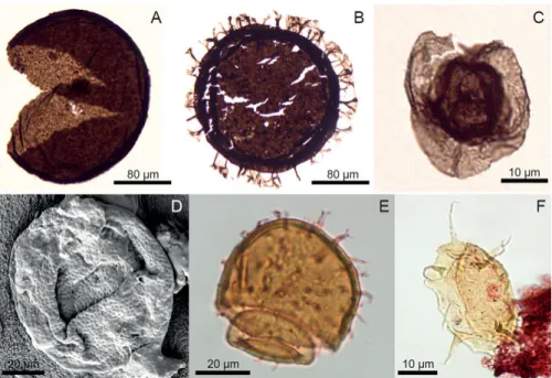
Figure 2. Some representative morphologies of Proterozoic and early Phanerozoic organic-walled microfossils (OWM). A-C, Mesoproterozoic (1.7-1.4 billion years old) fossils from the Ruyang Group, China. A, Dictyosphaera macroreticulata. B, Shuiyousphaeridium macroreticulatum. C, Pterospermopsimorpha insolita. D, Tas-manites volkovae from the lower Cambrian Lükati Formation in Estonia. E-F, Early Ordovician OWMs from the Varangu Formation, Estonia. E, Priscogalea distincta. F, Actinotodissus achrasii.
The fossil record is strongly biased for shallow-marine organisms with skeletal elements (Allison & Briggs, 1993), which have long played a major role in palaeontological research. However, organically-preserved body fos-sils of either prokaryotes or eukaryotes, or their biosedimentary signatures (e.g. stromatolites), are dominant throughout the Precambrian times (Men-delson & Schopf, 1992; Brasier, 2012; Ward & Kirschvink, 2015). There-fore, they are crucial to our understanding of the early evolution of complex life, and more specifically, the radiation of the major eukaryotic kingdoms.
Organic-walled microfossils are most commonly preserved in cherts, shales and siltstones, but can occur in other lithologies as well. They are prepared for microscopic observation via palynological acid-extraction. The rock matrix is dissolved in hydrofluoric and hydrochloric acids, and the re-maining organic residue, including the fossils, is mounted onto glass slides (e.g. Grey, 1999). Rocks samples containing the specimens (usually in case of cherts) may also be cut and polished into thin sections and the fossils may then be observed in cross-section.
Recalcitrant, sporopollenin-like biopolymers comprising the organic body of OWMs allow its preservation in the fossil record and resistance to acetol-ysis (Horodyski et al. 1992; Graham et al., 2013). Modern analogues that
14
utilise such compounds in cytogenesis are the encysting eukaryotes, specifi-cally green algae that produce protective cysts during reproduction (Arouri
et al., 2000; Versteegh & Blokker, 2004, Damiani et al., 2006;
Moczydłow-ska, 2010). While bacteria may be preserved in secreted sheaths, or permin-eralised in cherts (Bartley, 1996; Schopf et al., 2015), and their traces fossil-ised as microbialites (Noffke & Awramik, 2013), the only extractable indi-vidual organic fossils are eukaryotes.
There is a lot of variation in size and shape among these microfossils, but three main features of their body plan are shared: 1) resilient carbonaceous vesicle wall enclosing a central cavity, able to withstand acid maceration; 2) presence/absence of processes (spiny protrusions); 3) round or split openings in the wall, called the excystment structure or a pylome. Processes are thought to have been formed during the organism's reproductive stage, when it begun to develop a protective cyst, and the openings likely enabled the release of daughter-cells, as in many living protists (Tappan, 1980).
Being abundant, planktonic, usually globally distributed and with rapidly evolving form-species (Vidal & Moczydłowska-Vidal, 1997), OWMs are excellent index fossils that can be used in stratigraphic correlation of Prote-rozoic and Paleozoic rocks (e.g. Grey, 2005; Willman & Moczydłowska, 2008; Munnecke et al., 2011). If a species occurs worldwide and over a short time period, it may be used to identify different stratigraphic units, or bi-ozones. This is helpful in estimating the age of the rocks, especially where other dating methods are not available.
2. Mesoproterozoic eukaryotic microfossils
from northern China
Ruyang Group: a hotspot of early eukaryotic
biodiversity
The first part of this thesis focuses on the organic-walled microfossils from the Mesoproterozoic Ruyang Group, exposed in Shanxi Province, North China Craton. They have been recognized for a few decades (Yin, 1987; Xiao et al., 1997) and have played a crucial role in investigations on the palaeobiology of early eukaryotes.
We have assessed the taxonomic diversity in the Ruyang Group, dis-cussed the implications for the potential use of specific OWMs in Mesopro-terozoic biostratigraphy, and finally, investigated the biological affinities of several taxa by the use of LM, SEM, and comparison with living protistan clades. The age of the Ruyang Group has recently been constrained to c. 1700-1400 Ma (Hu et al., 2014). This antiquity is important because it vides a temporal constraint on the capability of complex organisms to pro-duce certain intricate morphologies. Consequently, it records one of the old-est divergence nodes in the eukaryote phylogeny.
Paper II documents a relatively high OWM diversity by Mesoproterozoic
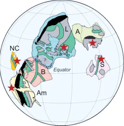
standards, and the earliest appearance of distinct morphologies, such as the ornamented and envelope-bearing forms, in addition to fossils with sculp-tured vesicles. The studied assemblage contains 26 species belonging to 16 genera, of which a few are newly recorded and named. This is a considerably higher diversity than previously recorded in the Mesoproterozoic succes-sions (Cohen & Macdonald, 2015), so we recognize the Ruyang Group as a critical window into the early eukaryotic radiation. A few taxa are endemic to northern China, however most also occur in Proterozoic successions worldwide (Australia, Canada, Greenland, India, Russia (Urals and the Sibe-rian Platform), Svalbard, and western USA (Fig. 3), and have substantially longer stratigraphic ranges than previously known, of up to a billion years (Schopf & Klein, 1992).
16
Figure 3: Paleomagnetic reconstruction of continental position at 1.53 Ga, based on data from Amazonia (Am), Australia (A), Baltica (B), North China (NC), Laurentia (L), Siberia (S). Green colour indicates orogenic belts, and grey is for Archaean belts. Stars mark the location of major Mesoproterozoic microfossil assemblages (not to scale), showing the global distribution of the early eukaryotes during the Mesoproterozoic. Modified from Mertanen & Pesonen (2012) and used kind permis-sion from the authors.
Owing to this longevity in the rock record, most of the early Proterozoic OWMs are poor candidates for biostratigraphic purposes. However, a few palaeogeographically widespread species with well-constrained ages may prove to be useful in relative dating and subdividing the Mesoproterozoic on local and global scales, and we have proposed Tappania plana as a potential index fossil. Additional studies from coeval strata on other continents will be helpful in evaluating the application of microfossils in the early Proterozoic biostratigraphy.
Most significantly, some of these Mesoproterozoic OWMs recorded in northern China display a sufficient level of morphological complexity to be identified as crown group eukaryotes, and even positioned in major eukary-otic clades like the prasinophyte algae and the crown group Chloroplastida. Following this, the main eukaryotic clades have started to branch out already
prior to 1.7-1.4 Ga. These results enhance the understanding of the timing of the early eukaryotic evolutionary history.
Microstructure and the biological affinities of the
Ruyang taxa
In Paper I, the focus of study is on the reproductive function and life cycle reconstruction as well as the biological affinity of the Mesoproterozoic uni-cellular microfossils Dictyosphaera and Shuiyousphaeridium.
Standard palynological light microscopy is useful for identification of ma-jor morphological features of OWMs, but many taxa possess a suite of fine-scale characters such as cell wall ultrastructure and the patterns of intricate vesicle surface sculpture that require additional methods in order to be de-tected. Scanning electron microscope (SEM) observations on the wall micro-structure of these microfossils have expanded on previous studies (Kaufman & Xiao, 2003; Javaux et al., 2004; Schiffbauer & Xiao, 2009), and have given new insights into the microfossils’ palaeobiology. Dictyosphaera and
Shuiyousphaeridium share a unique, complex cell wall composed of an
ace-tolysis-resistant, reticulated outer layer displaying a pattern of honeycomb-like pits. We have documented that these depressions are infilled by polygo-nal organic platelets on the interpolygo-nal side of the vesicle, constituting a second-ary wall which presumably served for additional strengthening of the vesicle, akin to the processes occurring in modern cyst-forming protists.
Javaux et al. (2004) and Knoll et al. (2006) have linked the complexity of the wall structure, in addition to the presence of ornamentation, to eukaryotic identity. Indeed, taken together, large size, presence of processes and surface sculpture, as well as a composite vesicle wall, indicate that Dictyosphaera and Shuiyousphaeridium are early eukaryotes. Whereas some prokaryotes like the vacuolate sulphur bacteria may grow to considerable sizes (Bailey et
al., 2007), no bacteria are known to produce a combination of composite and
resilient walls, and processes.
Expanding on this, we have further compared Ruyang OWMs to various unicellular protists (Table 1, Paper I) to narrow down their phylogenetic affinity. Several Ruyang microfossils were previously assigned to phyto-plankton (Yin et al., 2005), undefined green algae (Kaufman & Xiao, 2003), dinoflagellates (Meng et al., 2005), chlorophytes (Moczydłowska et al., 2011), or broadly identified as protists without distinguishing between auto- or heterotrophic mode of life (Javaux et al., 2004; Knoll et al, 2006). The results of our comparative morphology investigations have identified
Dicty-osphaera-Shuiyousphaeridium as a primitive green alga with shared
charac-ters of the crown group Chloroplastida (Viridiplantae) and Chromalveolata (see Baldauf, 2008 for a model of eukaryote phylogeny). Considering the
18
sequences of endosymbiotic events, and a much later evolution of chromal-veolates (Yoon et al., 2004), Dictyosphaera-Shuiyousphaeridium plexus was likely an autotrophic protist in the early lineage of Chloroplastida.
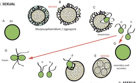
Figure 3: Life cycle model for Dictyosphaera-Shuiyousphaeridium. A, Zygote, start of the sexual reproduction. B, Shuiyousphaeridium morphotype, cyst shrinkage produces processes. Outer membrane is discarded, meiosis occurs. C, Haploid off-spring is released through the excystment structure. It may fuse into another zygote (G) or form a resting cyst (D). E, Start of asexual reproduction and resting cyst formation, mitosis occurs in Dictyosphaera morphotype. F, Offspring is released through the excystment opening. G, Flagellated haploid offspring fuses into a zy-gote.
Unique vesicle wall structure and the presence of characters analogous to modern photosynthetic eukaryotes, have also led to a proposal of a hypothet-ical life-cycle model for the Dictyosphaera-Shuiyousphaeridium organism. Both microfossils are differentiated only by the absence or presence of pro-cesses respectively, which has led workers to suggest that they were a part of the same biological species (Xiao et al., 1997), probably as distinct cyst stages (asexual and zygotic respectively) in the life cycle as discussed in
Paper I (Fig. 3). Sexual heteromorphism and reproduction via the
alterna-tion of generaalterna-tions is common throughout Eukaryota and often reflects envi-ronmental conditions. The inferred life cycle, reconstructed by comparison to living protists (Golenkinia radiata, Micrasterias thomasiana and
Stau-rastrum furgicerum), consists of two stages (asexual and sexual), represented
In Paper II, we have documented the presence of several, abundant OWM species consisting of a central body surrounded by a thick envelope. This morphology appears throughout the geological record from the Protero-zoic to Recent, and it is identical to a reproductive structure formed by the extant prasinophyte algae – a phycoma. Although the prasinophytes in gen-eral are paraphyletic (Leliaert et al. 2012), their ancient fossil forms (like
Tasmanites/Pachysphaera and Pterosperma-like taxa) cluster within the
same prasinophyte division Pyramimonadales (Turmel et al., 2009). Recent species within this group also exhibit phycoma-like (cyst) vesicle morpholo-gy, so it is likely that this is a synapomorphy between the modern taxa and the Ruyang fossils, rather than a result of convergent evolution. As a result, the presence of phycoma-producing forms in the Ruyang strata, suggests the evolution of the order Pyramimonadales [and by extension, the crown-group Chloroplastida (Adl et al., 2012; Leliaert et al., 2012)] as early as 1.7-1.4 Ga.
20
3. Neoproterozoic carbonaceous fossils
Raman spectroscopy as a tool in investigating
phylogenetic affinities
Apart from OWMs, important components of the mid-late Neoproterozoic fossil record are small yet macroscopic carbonaceous compression and or-ganic body fossils preserved on bedding planes (Xiao & Dong, 2006; Ye et
al., 2015). Most of these macrofossils have been accepted as algae, however
narrowing down their exact biological affinities is quite challenging, due to the lack of diagnostic morphological characters.
Additional methods aside from morphological observations are therefore required for palaeobiological investigation of these fossils. One of such in-novative tools is Raman spectroscopy (e.g. Schopf et al., 2002). This tech-nique enables observations on vibrational modes of different molecules in a system, and it may be used to detect a fingerprint-like signature of specific compounds. It has been valuable in palaeobiological studies either to ascer-tain the chemical properties of the organic matter between specific groups of organisms that are known to produce diagnostic biopolymers like algaenan, sporopollenin, and chitin groups. It may also be used to determine the level of the maturity of the organic matter within the rock (Marshall et al., 2005; Moczydłowska et al., 2014).
In Paper III, we have studied the microstructure and the chemical proper-ties of carbonaceous compressions of the assumed algae Chuaria, Tawuia and Beltanelliformis, and the organic body-fossil of an early annelid worm
Sabellidites to test the recognition of chemical compounds in the fossil algae
and the early metazoans. The material was collected from the well-preserved and diagenetically unaltered shales in the boreholes from the Tonian-Cryogenian interval in northeastern Siberia, and the end-Ediacaran in the western Russia (East European Platform). Despite the different age and geo-graphic position, both successions record abundant and pristinely preserved carbonaceous fossils.
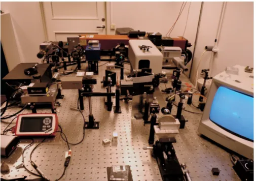
Figure 4: Research Raman System setup at Uppsala University.
Individual fossils were investigated with SEM to identify new microstructur-al characters, and probed using the laser Raman spectroscopy. We have used the Research Raman System (Fig. 4) at the Department of Earth Sciences, Uppsala University, which consists mainly of a green laser (λ=532 nm) and several different types of optical accessories. Its light source is the argon ion laser with 514.5 nm wavelength. Spectral resolution in the case of the UU facilities is 2.15 cm-1. This allows separation of peaks when the distance
between them is >2.15 cm-1. We have used low laser-beam power of 1.5-2.0
mW to prevent heat damage and scorching of the delicate carbonaceous ma-terial.
Our study recorded differences in the Raman shift signature between var-ious taxa. Vibrational absorption bands similar to those in α-chitin signature were observed in the organic wall of Sabellidites. Chitin, a biopolymer com-prising the cells walls of fungi and integuments of various animal groups, was already indirectly detected in this fossil (Moczydłowska et al., 2014). Following the ultrastructural and chemical analysis, Sabellidites was previ-ously positioned in the annelidan family Siboglinidae. The signature similar to α-chitin (Ehrlich et al., 2007) described in Paper III may corroborate its metazoan affinity. More importantly, the Raman results have revealed dis-tinct organic matter spectra of macroalgae Chuaria-Tawuia plexus and
Bel-tanelliformis, and the possible early annelid Sabellidites, which suggests that
Raman spectroscopy could be a useful method in identifying different branches (kingdoms, phyla) of the early eukaryotes.
22
Additionally, Paper III also discusses caveats and limitations of the Ra-man spectroscopy in palaeobiology. In conclusion, a combination of mor-phological, palaeoenvironmental and chemical studies will provide the most conclusive evidence when exploring the biological affinities of enigmatic Proterozoic fossils.
Animals in the Proterozoic
In addition to the presence of a probable annelid worm in the terminal Edia-caran successions, there is other evidence to corroborate the presence of specific metazoan phyla in the Precambrian. Paper IV records the occur-rence of organic-walled sclerites similar to those produced by modern Ecdy-sozoa, in the upper Ediacaran strata from the Włodawa Formation (c. 551 to 541 Ma) in the Łopiennik IG-1 borehole in southern Poland, geologically on the East European Platform (Moczydłowska, 1991).
Organically preserved sclerites from the Włodawa Formation co-occur with OWM taxa and macroalgal filaments (vendotaenids), and have long been recognized as the Ediacaran-Ordovician enigmatic fossil Ceratophyton
vernicosum Kirjanov. Initially, it had been grouped with palynomorphs of
uncertain affinities (Volkova et al. 1979), but Fatka & Konzalová (1995) placed Ceratophyton into the phylum Crustacea, based on its great similarity to the copepod exopodite segments.
Intensive research in the Cambrian palaeobiology and biostratigraphy was lately dedicated to so-called small carbonaceous fossils (SCF) – microscopic organic carbonaceous fragments that may be extracted from the rock matrix in a similar way to OWMs (Butterfield & Harvey, 2012). This group essen-tially includes palynomorphs with organically-preserved disarticulated scle-rites and cuticular fragments belonging to various animal clades. SCFs rep-resent an important component of the Cambrian fossil record, and have con-tributed greatly to the overall understanding of the Cambrian explosion (Harvey et al., 2012).
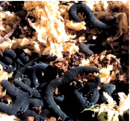
Recently, Smith & Ortega-Hernández (2014) have demonstrated morpho-logical similarities between the sclerites of the Middle Cambrian lobopodian fossil Hallucigenia and the claws and the jaw apparatus of living onychoph-oran worms (Fig. 5),which suggests a close relationship between the phyla Euarthropoda and Onychophora. SCF material from the Ediacaran Włodawa Formation strongly resembles those elements. Although the onychophoran origin in the Ediacaran is debatable, the morphology of the SCFs studied in
Paper IV conforms to the estimates for the presence of Bilateria (Erwin et
al., 2011) and their trace fossils (Jensen, 2003) in the terminal Proterozoic.
Ultrastructural studies and investigation of their chemical properties may shed further light on the nature of the Włodawa sclerites.
Figure 5. Euperipatoides kanangrensis (Onychophora) collected in 2012 in the Blue Mountains, New South Wales, Australia, for Evo-Devo studies.
24
4. Early Phanerozoic radiations of
phytoplankton
Possible Cambrian picoplankton
Studies on the early Cambrian palynomorph assemblage from the Lükati Formation in northern Estonia (Paper V) have documented a diverse assem-blage of organic-walled microfossils that have previously been allocated to phytoplankton (Volkova et al., 1979; Moczydłowska, 2011). The Estonian specimens reflect the trend of the morphological change and size-reduction in OWMs at the Proterozoic-Phanerozoic transition (Butterfield, 1997; Vidal & Moczydłowska, 1995; Vidal & Moczydłowska-Vidal; 1997), in contrast to the earlier, larger, Meso-Neoproterozoic (e.g. Paper II) and Ediacaran paly-nofloras (ECAP) (Grey, 2005; Yuan et al., 2011).
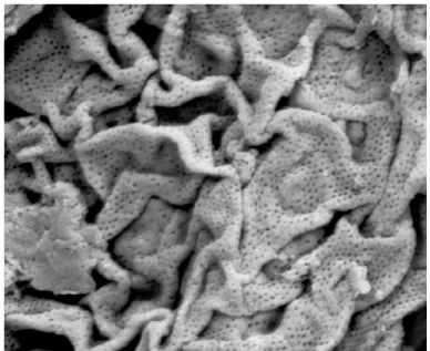
The Lükati material has also yielded a new species of a minute spheroidal microfossil with eukaryotic morphology that was described – Reticella
cor-rugata. SEM examination of the fossil’s wall microstructure has revealed a
unique surface sculpture consisting of regular, nanometre-scale pores in the flexible and corrugated organic wall (Fig. 6). Furthermore, R. corrugata is peculiar in its particularly small size (4-10 μm).
Comparison of the main characters revealed by the microstructural analy-sis to other fossil and extant species, indicates that R. corrugta shares its diagnostic characters with the prasinophyte algae (Pyramimonadales) of the family Tasmanaceae (Guy-Ohlson, 1996). A porate vesicle wall is assumed to be a rare and conserved feature. Due to its tiny dimensions, R. corrugata also falls into the broad microfossil size-class of the so-called “small acri-tarchs”. This grouping was originally erected by Habib & Knapp (1982) for a Cretaceous assemblage of tiny palynomorphs, and subsequently recognized in numerous successions from the lower Paleozoic to present (Loeblich & Wicander, 1976; Le Hérissé, 2002; Schrank, 2003; Moczydłowska, 2011). Those abundant assemblages of minute phytoplanktonic fossils seem to oc-cupy the same ecological niche as present-day eukaryotic picoplankton. Ex-tant picoeukaryotes (ranging from 0.8-7 μm in size) also include the smallest living eukaryote, the prasinophyte Ostreococcus tauri (Courties et al., 1994). Small prasinophyte algae are the dominant component of the modern eukar-yotic picoplankton. Although this group has only recently been defined, it is acknowledged to constitute a major portion of the biomass in the modern
oceans (Lewin et al., 2000). A combination of notably small size and prasi-nophyte-characteristic microstructure documented in Paper V, suggests that
R. corrugata was a likely member of the Cambrian eukaryotic picoplankton.
Figure 6: SEM micrograph of the unique wall structure of the Cambrian microfossil Reticella corrugata.
This case study also stressed the importance of the use of SEM in micropal-aeontological research. Diagnostic, nano-scale characters of the new species are impossible to recognize with the conventional transmitted light micros-copy, and it is difficult to distinguish it from other early Cambrian OWMs without microstructural examination. Therefore, the palaeobiodiversity of such minute microfossils is probably even higher than estimated, for two reasons: significant amount may be overlooked in the studies utilising only LM (discussed by Schrank, 2003), and the fossil record is generally biased against the smaller organisms (Cooper et al., 2006).
Cyst formation in the galeate plexus
The final part of this thesis (Paper VI) continues the study of the phyto-plankton evolution in the early Phanerozoic, by looking at the microfossil assemblages from northern Estonia; the transitional Cambrian-Ordovician
26
Kallavere Formation and the lowermost Ordovician Varangu Formation (c. 490-485 Ma).
The trend of small size and relatively simple processes among OWMs discussed in Paper V, changes around the Cambrian-Ordovician boundary, during the Great Ordovician Biodiversification Event (GOBE) when phyto-plankton disparity and species-richness increase substantially (Webby et al., 2004). This time marks the first appearance of new OWM body plans, such as vesicles with polar symmetry (Actinodissus), composite processes
(Vul-canisphaera), polygonal flanges (Cymatiogalea), and remarkable
excyst-ment structure–opening with a rim covered by opercula in the galeate plexus microfossils. The Ordovician radiation represents the second largest diversi-fication of photosynthesising microbiota in Phanerozoic, after the Cambrian one (almost reaching the present-day levels) (Servais et al., 2008).
Although the modern phytoplankon (dinoflagellates, diatoms and cocco-lithophores) did not diversify until the Mesozoic, quite late in the evolution-ary history of algae and parallel to the Modern evolutionevolution-ary fauna (Sepkoski, 1981), the Ordovician radiation was important for the increasing diversifica-tion of the Paleozoic biota. The early Paleozoic phytoplankton is represented by fossils with innovative complex ornamentation, excystment structure with an operculum, and polar symmetry of the vesicle. Some of the morphologi-cally most striking members of the Cambro-Ordovician microbiota are the microfossils of the galeate (“helmet-like”) plexus (Servais et al., 2004), which includes form-genera Caldariola, Priscogalea, Stelliferidium and
Cymatiogalea. Galeates are defined by a rare element among organic-walled
microbiota–a reproductive opening (excystment) with a lid structure (oper-culum).
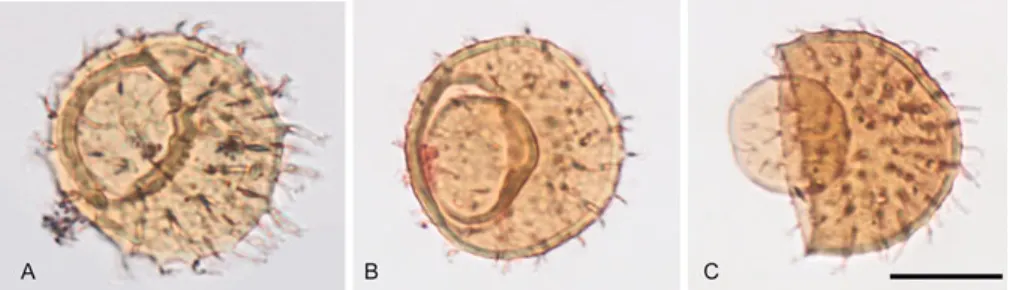
Figure 7: Different stages of operculum formation in galeate plexus acritarchs. Scale bar represents 20 μm. A, Priscogalea distincta with formed operculum at-tached to the cyst. B, Priscogalea simplex with partially open operculum. C, Pris-cogalea barbara vesicle with excystment opening and dislocated operculum partial-ly collapsed into the empty cyst.
In the microfossil assemblage studied in Paper VI, different stages of the operculum formation have been observed in all galeate morphotypes: from an encysted vesicle with a faint fault line developing on the wall, to a vesicle with a fully formed operculum still attached to the excystment, and eventual-ly to an opened vesicle with the operculum completeeventual-ly detached or collapsed inside the empty vesicle, presumably upon gametes/offspring release by the matured cyst (Fig. 7). We have compared this anatomy and its inferred func-tionality with present-day analogues among photosynthesizing protists. Similar morphology is observed in the extant marine green alga
Acetabular-ia mediterranea (Dasycladales, Chlorophyta). Although AcetabularAcetabular-ia is a
derived unicellular alga with complex morphology and a very large cell (up to 1 mm in length), its reproductive cysts are very small, smooth, thick-walled and possess a lid-structure like the one seen in galeate microfossils (Fig. 8). A series of measurements on the Estonian galeates (Table 2; Paper
VI) have shown that all form-species have the same operculum to vesicle
diameter ratio. This value is almost identical to the lid / vesicle diameter ratio in Acetablularia, despite the overall size difference between the
Ace-tabularia cysts and the galeate plexus microfossils.
Figure 8. Reproductive cyst of the extant green alga Acetabularia acetabulum (Dasycladales). Gametes are released through the excystment opening after the lid became detached. Image used with kind permission from Sigrid Berger (Heidelberg University).
28
Morphologically, Acetabularia cysts are identical to the process-lacking morphotype of Caldariola. Culture studies on Acetabularia have shown that the development of the operculum during the cyst formation is regulated by the so-called lid-forming apparatus (LFA) organelle.
In addition to the phenotypic similarity, the Estonian galeates have inhab-ited a similar environment as both extant (Berger et al., 1987), Paleozoic and Mesozoic Dasycladales (e.g. Young et al. 2007). Palaeocontinent Baltica, which included the present-day Estonia, was positioned in the mid latitudes during the early Ordovician, and it had a subtropical climate (Munnecke et
al., 2011) similar to the present-day Mediterranean where Acetabularia lives
in abundance (Berger et al., 1987). Considering the similarities in morpholo-gy, cyst formation and ecology between the galeate plexus and the living dasycladalean Acetabularia, as well as the known Ordovician record of Dasycladales, one of the conclusions from Paper VI is that the galeate mi-crofossils likely belong to this order of green algae. Moreover, extant and rare operculate microorganisms utilise a complex organelle to form the oper-culum structure, so it is likely that LFA or its precursors were needed to produce that kind of vesicle morphology in the galeates.
5. Functional morphology and palaeobiology
Eukaryotic cysts
In addition to the broad classification among Eukaryota, Proterozoic organ-ic-walled fossils studied as a part of this thesis exhibit diagnostic features that were used to narrow down their phylogenetic position further, and com-pare them to extant clades (Moczydłowska et al., 2011). Throughout most of the papers presented here, these features are regarded as diagnostic charac-ters that can provide information on the functional palaeobiology of OWMs. These are: excystment openings, processes, wall structure, and the acetoly-sis-resistant organic vesicle (Tappan, 1980; Colbath & Grenfell, 1995; Wicander, 2002; Moczydłowska, 2010). In combination, the above charac-ters define the function of some unicellular organic-walled microfossils as reproductive cysts.
Excystment opening structures are formed in the vesicle wall during the excystment process, presumably when gametes/daughter-cells are being released from the matured reproductive cyst (Vavrdová, 1992), as seen in extant algae (e.g. Neuhaus-Url & Schweiger, 1984). It is the most direct evidence of the fossil’s reproductive function (Moczydłowska, 2010). Varie-ty of opening structures is found throughout Proterozoic and Phanerozoic organic-walled microfossils, in rare cases bearing additional features like a neck or an operculum. Partial or median-split openings and round openings (pylomes) occur in acritarchs of all ages (Papers I, II, VI), but the later are more common in the Phanerozoic. Recent biomechanical studies by Pang et
al. (2015) have shown that the complex striation pattern in a Ruyang OWM Valeria lophostriata served as a control mechanism for the excystment
struc-ture. This find further stresses the importance of the excystment character in the early eukaryotes.
Processes in unicellular organic-walled microfossils show comparable complexity and morphological patterns as those of extant algal cysts. They are formed during the contraction of the cyst from the vegetative cell wall (e.g. Dale, 2001), and are used in buoyancy control and sensory activity. Upon discarding the remnants of the vegetative wall / outer membrane, the mature, process-bearing (acanthomorphic) cyst will settle to the substrate and eventually release the offspring through the excystment opening. Orna-mented microfossils in particular bear strong similarities with encysted
stag-30
es (zygotes) of extant unicellular algae in orders Chlorococcales, Volvocales and Zygnematales (Moczydłowska, 2010).
OWMs are extracted using a harsh method of maceration in hydrofluoric acid (HF) that dissolves the surrounding siliciclastics rock within a few days. Fossils themselves are unaffected by HF. This property of acetolysis-resistance is a result of complex, sporopollenin-like biopolymers building the vesicle wall (Horodyski et al. 1992; Damiani et al., 2006; Javaux & Mar-shall, 2006), synthesised primarily by autotrophic eukaryotes (Hagen et al., 2002; Knoll et al., 2006; Moczydłowska et al., 2011). Presence of such stur-dy walls during reproductive stages (Damiani et al., 2006) requires signifi-cant metabolic investment by the microorganism, which suggests wall’s function in protection of daughter-cells, that is, in propagation of genetic material. Tough walls are also considered to be ecological indicators due to their presence in the benthic sphaeromorph assemblages (Mendelson & Schopf, 1992), which probably represent sunken resting cysts.
Separated by a span of almost billion years, assemblages from the Ruyang Group, China, and the Estonian Lükati, and Kallavere-Varangu successions, exhibit these diagnostic, eukaryotic reproductive features and likely repre-sent cysts of the extinct microbiota.
Vesicle morphology as a result of intracellular
complexity
Mechanism of the fossil cyst formation is a major theme in this thesis, espe-cially explored in Papers I, II, V, VI. In several case studies, suites of dif-ferent morphological characters of OWMs from various time intervals have been compared to extant protists. Proterozoic and Paleozoic microfossils share diagnostic processes, encystment structures, composite walls, and ad-ditional cyst-like elements, such as necks, opercula or plugs (Tappan, 1980). These features are present in a variety of living groups, predominantly algae, and their formation has been studied in cultures. However, many of these elements are considered to be derived and require a certain degree of devel-oped intracellular machinery for their formation.
As shown in Paper I, Dictyosphaera-Shuiyousphaeridium organism’s cyst is composed of two layers: the primary wall reinforced by a secondary layer of polygonal platelets. Similar wall construction during the reproduc-tive phase is known in Prasinophyceae, Chlorophyceae and Chrysophyceae (Melkonian et al., 1991), where those additional wall elements are produced and secreted through the Golgi apparatus (GA) and the endoplasmatic reticu-lum (ER) organelles. By comparison, the organic platelets building the sec-ondary wall in Dictyosphaera-Shuiyousphaeridium may be interpreted as the controlled secretion in cyst formation, governed by GA and ER.
Sophisticat-ed vesicle-wall patterning on the fossil sphaeromorphs Valeria lophostriata,
Kildinosphaera verrucata and the younger Cerebrosphaera buickii would
have also required a certain degree of complexity for their formation. This indirect evidence for GA and ER may provide the time constraint on their origin in the eukaryotic cell, by at least 1700-1400 Ma, in case of the Ruyang biota. Accepting that the organism was photosynthetic (Kaufman & Xiao, 2003), a plastid may also be added on the list of present intracellular components.
The Golgi apparatus is considered to be one of the least understood orga-nelles and research into it has been marked by controversy (Hua & Graham, 2009). However its three main functions are clear: 1) serving as the cell’s secretory pathway, protein sorting and trafficking station; 2) processing the waste material from the cytoplasm; and 3) cell wall construction. The last may be seen as a secondary function, that is, a means of dealing with and putting the excreted material to use. The timing of the GA invention in eu-karyotic evolution is also unclear.
Genomic data for the GA throughout Eukaryota, suggest that the stacked GA had already been present in the last eukaryotic common ancestor (LECA) and was subsequently lost or underwent changes eight times during cellular evolution (Mowbrey & Dacks, 2009). Dyer & Obar (1994) also ar-gue for its presence in a “proto-eukaryote” before the endosymbiotic events that gave rise to a eukaryotic cell (e.g. Margulis (Sagan), 1967), while others propose its evolution along with the ER after the endosymbiosis of the ar-chaeon and the bacterium (summarized by O’Malley, 2010). Nevertheless, considering this antiquity of the GA and its function of wall secretion in eukaryotic cells, this organelle may be expected as a cellular control for ves-icle construction and ornamentation in the Proterozoic microbiota.
Another example of complex morphology depending upon certain cellular components, is found in the Cambrian-Ordovician galeate plexus microfos-sils, specifically Caldariola, Priscogalea and Stelliferidium. These taxa pos-sess a very rare character among palynomorhs–a lid/operculum. The lid structure attached to the excystement structure is rare among the early Paleo-zoic fossils, and so far unknown from the Precambrian time interval (Tap-pan, 1980). However, such morphology is present in the extant chlorophyte genus Acetabularia, as discussed in Paper VI (Fig. 8). Operculum for-mation in Acetabularia is governed by the “lid-forming apparatus”. LFA is an array of microtubules adjacent to the cell membrane. Microtubules regu-late the formation of a circular band on the cyst wall, which deepens into a fault line on the cyst surface. This grove-like band will eventually separate the operculum from the cyst and allow a more controlled reproductive open-ing and gamete release through the protective cyst wall (Neuhaus-Url & Schweiger, 1984).
As in the case of the Proterozoic fossils (Paper I), the LFA organelle or its precursors necessary to construct the recorded morphologies, are inferred
32
to have been active as early as the Cambrian-Ordovician transition (c. 490-480 Ma). Cellular control on cyst production and regulated excystment has probably evolved much earlier, as evidenced by the Mesoproterozoic taxa with large, regular openings (Paper II), or the elaborate mechanisms guid-ing the biologically programmed excystment in Valeria lophostriata (Pang et
al., 2015).
Whereas intracellular activity is impossible to observe like in modern
Ac-etabularia culture studies, and intracellular structures are only preserved in
exceptional circumstances, external features of the cyst may also indirectly reveal significant information about past processes inside the cell, and pre-sent organelles.
6. Conclusions and future directions
This doctoral thesis consists of four papers that investigate the nature and evolutionary patterns of organic-walled microfossils, and two papers that explore organically preserved macrofossils of algae and early animals. Case studies presented herein have provided insight into the biological affinities of OWMs, and into evolutionary patterns of the early eukaryotes and the early phytoplankton.
The Ruyang Group encompasses well-preserved OWMs, which are very diverse by Mesoproterozoic standards. Recently, the age of this succession has been constrained to 1.7-1.4 Ga. This has significant implications for the early eukaryotic evolution, because it means that complex microfossil morphologies have already evolved by this time. Diagnostic characters of these microfossils, shared by some living protistan groups, also suggest that Eukaryo-ta has sEukaryo-tarted to diverge into different clades at this time.
The use of Raman spectroscopy in addition to SEM was used to differentiate between morphologically similar Neoproterozoic macrofossils of photosynthetic taxa and the early animals.
Organically preserved sclerites of the earliest metazoans co-occurring with palynomorphs in the terminal Ediacaran assem-blage provided insight into the parallel evolution of the metazoans that facilitated the biosynthesis of resilient polymers in their body walls, as photosynthesizing microorganisms have done much ear-lier in their cysts though of different chemistry.
The Cambrian and Ordovician successions from Estonia also doc-ument a variety of OWMs, record a trend in their size reduction and morphology innovations, and indirectly reveal information about the intracellular complexity in the early Phanerozoic.
The results of my studies are presented in a chronological/stratigraphical order, from the oldest, to the youngest fossils. Although this allows a good insight into patterns of early evolution of OWMs, there are temporal gaps in this study. Additional work on certain characters (e.g. openings, microscale vesicle sculpture) in Ediacaran taxa could expand the understanding of evo-lutionary innovations in OWMs.
34
An ongoing theme of this thesis has been using the comparative morphol-ogy to answer the questions about the microfossils biological affinity. A caveat that needs to be considered with this approach is convergent evolu-tion, i.e. the same functional morphology arising independently in unrelated clades. In order to address this, increased phylogenomic studies on extant protistan clades are needed to understand the distribution of certain diagnos-tic characters throughout the Eukaryota, and especially the photosynthesiz-ing lineages. Moreover, understandphotosynthesiz-ing the gene regulatory pathways produc-ing specific morphologies in livproduc-ing organisms (e.g. genes regulatproduc-ing the LFA organelle in Acetabularia) can shed further light on their evolutionary histo-ry.
A few papers in this thesis have also re-evaluated older material. Novel techniques and ideas have yielded new information, indicating the im-portance of re-examination in palaeontology. Revisiting old material from exceptionally preserved assemblages from critical time periods, such as Lakhanda Formation, with novel techniques, will likely provide more infor-mation about the patterns of the early eukaryotic radiation.
Raman spectroscopy has proven to be useful, but not conclusive to ascer-taining phylogenetic affinities of organically preserved fossils, stressing that morphological approach is still very important for both taxonomy and pal-aeobiology, since it provides direct information about the main fossil fea-tures. Furthermore, the use of SEM for microstructural analysis was espe-cially important in several papers, as it revealed relevant structures (and a new specie) that would not have been recognized via transmitted light mi-croscopy.
Palynological material from the late Ediacaran Period also encompasses fossils of the early Metazoa. Future studies on the chemical properties of the Włodawa sclerites and the tubular body-fossil Sabellidites may be helpful for interpreting their affinities with a greater degree of confidence.
Use of transmitted electron microscopy (TEM) and focussed ion beam (FIB) will shed further light on the ultrastructural properties of the presented microfossils.
Lastly, combined micropalaeontological and geochemical investigations of the successions studied in this thesis will provide a better insight into how the environmental conditions play the role in the early eukaryotic evolution, as well as how the emergence of complex cells in turn affected the early Earth.
7. Svensk sammanfattning
Introduktion
Proterozoikum varade från cirka 2500 till 542 miljoner år (Ma) sedan och är en viktig tidsperiod då den ger grundläggande information om den tidiga utvecklingen av livet på jorden, särskilt inom domänen Eukaryota. Eukaryo-terna är organismer med komplexa intracellulära strukturer såsom till exem-pel en cellkärna, mitokondrier och kloroplaster avgränsade av ett cellmem-bran. De omfattar en mängd både encelliga och flercelliga organismer: djur, växter, svampar, alger, amöbor och diverse andra protister. Baserat på mole-kylära data har man uppskattat eukaryoternas uppkomst till 2,5-2,0 miljarder år (Ga) sedan och de flesta fossila fynd av eukaryoter från proterozoikum representerar mikroskopiskt små protister. Djurens och växternas uppkomst sker förmodligen långt senare i jordens historia, strax innan edicaraperioden övergår i kambrium (för cirka 555 Ma sedan).
De äldsta eukaryota fossilen är små och morfologiskt enkla celler med or-ganiska karbonatiska cellväggar och hittas i bergarter daterade kring 2000-1800 Ma sedan (paleoproterozoikum). Dessa ursprungligen enkla organ-ismer har sedan dess genomgått flera perioder av utveckling genom resten av proterozoikum till tidiga fanerozoikum (perioden av “synligt liv” ca 545 Ma sedan).
I min avhandling har jag undersökt material från olika lokaler och från olika tidsperioder för att besvara frågor som rör några av de tidigaste fossilen med eukaryo-liknande morfologi. Några av dessa frågor handlar om hur dessa eukaryoter är besläktade och hur de har utvecklats under mesoprotero-zoikum: Jag har också studerat avtryck av eukaryoter från neoproterozoi-kum; undersökt de evolutionära trenderna hos fossil av fytoplankton under början av fanerozoikum, samt försökt förstå hur morfologin hos dessa mikro-fossil återspeglar deras intracellulära komplexitet.
Livet i proterozoikum
Proterozoikum omfattar en väsentlig del av jordens historia (44 %) och är en tid av omfattande förändringar i jordens miljö och följaktligen också biosfä-ren. Under proterozoikum så ökar syrehalten i både atmosfären och haven, och jättekontinenten Rodinia splittras och bildar ett antal mindre plattor.
36
Dessutom pågår flertalet stora nedisningar (så kallade snöbollsjordar) samti-digt som ett antal grundläggande evolutionära innovationer, såsom sexuell reproduktion och flercellighet uppkommer, utöver en generell diversifiering av flera olika eukaryota grupper.
Mikrofossil från denna tid ger en direkt inblick i den tidiga biosfären, och är särskilt intressanta för att bättre förstå dessa föränderliga miljöer.
Vad är organiska mikrofossil?
I den här avhandlingen har jag studerat mikrofossil med cellväggar upp-byggda av organiskt material (jmf engelskans organic walled microfossils, OWM) en grupp som traditionellt kallats acritarcher. Proterozoiska och ti-digpaleozoiska acritarcher har studerats i över ett sekel, men deras exakta fylogenetiska släktskap är inte klarlagt därav namnet acritarcher som kan härledas till grekiskan och betydelsen “okänt ursprung”. Namnet börjar, enligt vissa, bli mer och mer förlegat eftersom fler och fler fossil nu kan härledas till kända grupper av mikroorganismer. Därför använder jag här förkortningen OWM som ett samlingsnamn.
OWM prepareras fram genom syraextraktion från sedimentära bergarter (som skiffer, lera, silt, eller kalksten) med hjälp av fluorväte-, och saltsyra. De utlösta fossilen filtreras för att ta bort kvarvarande mineralkorn och andra oönskade partiklar och monteras sedan på ett objektglas. Därefter kan fossi-len undersökas med olika mikroskoperingstekniker, vanligast är ljus-, och svepelektronmikroskopi (SEM). Stuffer av andra bergarter (t.ex. hornsten) kan skäras och poleras till tunnslip där fossilen studeras i tvärsnitt.
Det finns en hel del variation i storlek och form hos dessa mikrofossil men det finns tre gemensamma huvuddrag 1) en syramoståndig, kolhaltig cellvägg; 2) processer (taggiga utskott); 3) runda eller delade öppningar i cellväggen. Processerna bildades förmodligen under organismens reproduk-tiva stadie, när det börjat utveckla ett skyddande hölje. Öppningarna tjänade sannolikt som utsläppspunkter av dotterceller, som hos många nu levande protister.
Flertalet OWM är talrika, planktoniska, och oftast globalt spridda. Dessu-tom är många lätta att identifiera och i kombination med att de existerade under en specifik period gör att de är utmärkta som så kallade indexfossil. Dessa indexfossil kan användas i stratigrafisk korrelation av proterozoiska och paleozoiska bergarter och är därför väldigt viktiga för att uppskatta en bergarts ålder, speciellt då andra dateringsmetoder inte går att använda.
Fossil av proterozoiska eukaryoter
Artikel I och II handlar om mikrofossil från bergarter från den mesoprotero-zoiska Ruyang-gruppen, i norra Kina, bergarter som avsattes i en grund havsmiljö. De två vanligaste mikrofossilen som hittas i Ruyang-gruppen är
Dictyosphaera och Shuiyousphaeridium och artikel I fokuserar på deras
pa-leobiologi. Med hjälp av SEM-studier har jag kunnat avslöja att båda fossi-len har en identisk och komplex cellvägg, en ultrastruktur som även återfinns hos en del moderna eukaryoter. Baserat på ytterligare jämförelser med nu levande alger, har den fossila organismens livscykel rekonstruerats. I nu levande mikroorganismer konstrueras motsvarande morfologi av komplexa organeller inuti cellen, såsom golgiapparaten och det endoplasmatiska nät-verket. En liknande morfologi hos fossila celler skulle förmodligen ha krävt en liknande grad av intracellulär komplexitet och därför föreslås det i artikel I att golgiapparaten var närvarande redan i mesoproterozoikum.
I artikel II tittar jag på den bredare förekomsten av fossil i Ruyang-gruppen i syfte att uppskatta den biologiska mångfalden på platsen. 26 arter har registrerats, vissa förekommer endast i norra Kina, medan andra hittas i bergarter av motsvarande ålder över hela världen. I artikeln diskuteras också möjligheterna att använda dessa fossil för biostratigrafi samtidigt som stu-dien ökar förståelsen för den globala biologiska mångfalden av mikroorgan-ismer.
Förutom mikrofossil så har jag även studerat makroskopiska proterozo-iska fossil, bevarade som kolhaltiga avtryck. Släktskapen mellan dessa fossil är till stor del osäkra, men många anses ändå vara besläktade med alger. Flera neoproterozoiska taxa har undersökts med hjälp av en kombination av SEM och ramanspektroskopi. Raman är en spektroskopisk teknik som an-vänds för att observera vibrationslägen inom och mellan molekyler och när det används på fossil så ger det information om de kemiska egenskaperna hos cellväggarna. I artikel III, diskuterar vi hur ramanspektra och mikro-strukturer kan användas för att särskilja de kolhaltiga fossilen från varandra.
Diversifiering av acritarcher vid början av fanerozoikum
Planktoniska, fotosyntetiserande eukaryoter diversifierade kraftigt i edia-caraperioden, de genomgick en omfattande förändring i antalet arter när ediacaraperioden övergick i kambrium, och blev mycket mindre i storlek i början av den kambriska perioden (cirka 545 Ma). I artikel V beskrivs ett nytt ovanligt litet organiskt mikrofossil kallat Reticella corrugata, som här identifieras som en grönalg tillhörande Prasinophyceae. Med referens till hur den gruppen lever i dag, som en viktig grupp av marina, eukaryota picoplankton, indikerar det också att picoplankton utvecklades så långt till-baka som kambrium.
38
Artikel VI behandlar den efterföljande utvecklingen av fotosyntetiserande organiska mikrofossil när kambrium övergår i ordovicium (för ungefär 490-485 Ma sedan). En speciell grupp av fossila acritarcher (kallade galeatea acritarcher) med en komplicerad morfologi studerades och jämfördes med den nu levande algen Acetabularia i syfte att rekonstruera olika morfolo-giska anpassningar för reproduktion.
8. Acknowledgements
First and foremost, I wholeheartedly thank my supervisor Małgorzata Moczydłowska-Vidal for her guidance and support during my time at Upp-sala University. I have learned a lot from you, and I’ve especially enjoyed our chats about the history of science. Sebastian Willman, my second super-visor, has always been there when I needed help, with good advice at hand, and I appreciate that greatly. My additional advisor Frances Westall (Centre de biophysique moléculaire, CNRS Orléans, ESA) is thanked for warmly welcoming me during my visit to CNRS, and for inspirational discussions at various conferences, both about geology and being a geologist.
Yin Leiming at the Nanjing Institute of Geology and Palaeontology is sincerely thanked for providing the Ruyang material, which turned out to be a huge part of this thesis and an absolute pleasure to study. He was of great help via emails when I needed urgent translations of systematics. I would also like to thank Kath Grey (Geological Survey of Western Australia) for helpful discussions on microfossils and labwork during her stay at UU. Nick Butterfield at University of Cambridge is gratefully acknowledged for agree-ing to be my Licentiate thesis opponent back in 2014. Our discussions have led me to critically examine some ideas, for which I am thankful.
The Palaeobiology Programme at UU has been a good environment of pal-aeontological schooling. I am especially grateful to Graham Budd for excel-lent advice and discussions, as well as for jollies around the Cambrian or the world, like Skåne and Australia. John Peel is thanked for easing my transi-tion from the world of small shellies to acritarchs when I arrived at Uppsala. I am also thankful to Lars Holmer for introducing me to Estonian geology, and for generally emitting knowledge of all things brachiopod. Ralf Janssen is thanked for being persistent about organising the Wednesday Seminars, which were a great help for conference preps and becoming a better present-er.
I would also like to thank the rest of my colleagues (present and past) in Team Palaeo, roughly in the order in which we had crossed paths: Oskar J., Oskar B., Aodhán, Jenny, Linda, Michi, Jorijntje, Ralf, Haizhou, Emma, Tim, Thessa, Luka, Baptiste, Steve, Åsa, Miloš, Manuela, Mattias, Lewis, Giannis, Alba, Lanyun, Zhilliang, as well as colleagues from MPT (ex-Berg) and Geophysics. Thanks for all the support and various TGIFs over the years. To my officemate Luka, thanks for putting up with the shear chaos in