Cancer and reconstructive
surgery in inflammatory
bowel disease
Linköping University Medical Dissertation No. 1670Maie Abdalla
M
aie
A
bd
al
la
Ca
nc
er a
nd r
ec
on
str
uc
tiv
e s
urg
ery i
n i
nfla
m
m
ato
ry b
ow
el d
ise
as
e
20
19
FACULTY OF MEDICINE AND HEALTH SCIENCES
Linköping University Medical Dissertation No. 1670, 2019 Department of clinical and experimental medicine (IKE) Linköping UniversitySE-581 83 Linköping, Sweden
www.liu.se
3
Linköping University, Medical Dissertation, No. 1670
Cancer and reconstructive surgery in
Inflammatory bowel disease
Maie Abdalla
Linköping 2019
Division of Surgery,
Department of clinical and experimental medicine,
Faculty of Medicine and Health Sciences,
4
© Maie Abdalla 2019
The published articles have been reprinted with permission of the copyright
holder
Printed in Sweden by LiU-Tryck, Linköping, Sweden
ISBN: 978-91-7685-109-8
ISSN: 0345-0082
5
ميحرلا نمحرلا الله مسب
{
ُهاَض ْرَت اًحِّلاَص َلَمْعَأ ْنَأ َو َّيَدِّلا َو ٰىَلَع َو َّيَلَع َتْمَعْنَأ يِّتَّلا َكَتَمْعِّن َرُكْشَأ ْنَأ يِّنْع ِّز ْوَأ ِّ ب َر
يِّنْل ِّخْدَأ َو
ني ِّحِّلاَّصلا َكِّداَبِّع يِّف َكِّتَمْح َرِّب
}
ميظعلا الله قدص
ةيأ لمنلا ةروس
١٩
In the name of God, the merciful
( My Lord enable me to be grateful for your favour which you have bestowed
upon me and upon my parents and to do righteousness of which you approve,
and admit me by your mercy into the ranks of your righteous servants)
The Holly Quran
Surat Al-naml: Aya 19
6
Principle Supervisor
Pär Myrelid, Associate professor
Division of surgery, Department of clinical and experimental medicine,
Faculty of Medicine and Health sciences,
Linköping University, Linköping, Sweden
Assistant supervisors
Johan D Söderholm, Professor
Division of surgery, Department of clinical and experimental medicine,
Faculty of Medicine and Health sciences,
Linköping University, Linköping, Sweden
Roland E Andersson, Professor
Division of surgery, Department of clinical and experimental medicine,
Faculty of Medicine and Health sciences,
Linköping University, Linköping, Sweden
Department of surgery, Ryhov County Hospital, Jönköping, Sweden
Kalle Landerholm, Ph.D.
Division of surgery, Department of clinical and experimental medicine,
Faculty of Medicine and Health sciences,
Linköping University, Linköping, Sweden
7
Opponent
Yves Panis, Professor
Division of surgery,
Beaujon hospital, University hospitals of Paris the North valley of the Seine
Clichy, France
Committee board
Urban Karlbom, Associate professor
Division of surgery,
Akademiska Sjukhuset,
Uppsala, Sweden
Christopher Sjöwall, Assistant professor,
Division of Neuro and inflammation/ Rheumatology,
Department of clinical and experimental Medicine,
Faculty of Medicine and Health sciences,
Linköping University, Linköping, Sweden
Substitute
Stergios Keshagias, Professor
Division of Cardiovascular Medicine (KVM),
Department of Medical and Health Sciences (IMH),
Faculty of Medicine and Health sciences,
8
List of Contents
Abstract ... 9 List of papers ... 11 List of figures ... 12 List of tables ... 14 Abbreviations ... 15 Introduction ... 17Background to the study ... 20
Aims of the study ... 38
Methods ... 39
Results ... 46
Discussion ... 66
Conclusions ... 80
Future studies ... 81
Clinical applications of the thesis ... 82
Populärvetenskaplig sammanfattning ... 83
Acknowledgments ... 87
References ... 88
Appendix ... 109
9
Abstract
Ulcerative colitis (UC) is a chronic inflammatory disease that affects the colon. According to the literature, some thirty percent of UC patients may require a subtotal colectomy and ileostomy due to failure of medical treatment, acute toxic colitis or dysplasia/cancer diagnosis. Some patients choose to get continence restored with either an ileorectal anastomosis (IRA) or an ileal pouch-anal anastomosis (IPAA). Worldwide most surgeons prefer an IPAA to an IRA, despite reports of pouchitis, impaired fertility and fecundity. Fear of recurring proctitis and fear of rectal cancer in the remaining rectum is contributing to the choice of an IPAA. Little is known regarding the outcomes of IRA compared with IPAA in UC patients. We aimed to investigate the anorectal function, quality of life (QoL), risk of failure and rectal cancer in patients with UC restored with IRA and IPAA respectively.
Methods: Data about all Inflammatory bowel disease (IBD) patients was obtained from the
Swedish National Patient Register (NPR) between 1964-2014 and in one study from the Linköping University Hospital medical records 2006-2012. Patients who developed cancer were identified from the Swedish National Cancer Register. We investigated the risk of cancer and inflammation, functional outcome and failure as well as the quality of life for IRA and IPAA patients. Investigation of risk for cancer in IRA and IPAA compared with the background population was performed using survival analytic techniques: uni-and multivariate regression, Kaplan Meier curves and standardized incidence ratio.
Results: Twelve percent (7,889 /63,795) of UC patients required colectomy according to the
NPR. The relative risk for rectal cancer among patients with an IRA was increased (SIR 8.7). However, the absolute risk was 1.8% after a mean follow up of 8.6 years and the cumulative risk 10- and 20-years after IRA was 1.6% and 5.6%, respectively. Risk factors for rectal cancer were primary sclerosing cholangitis in patients with an IRA (hazard ratio 6.12), and
10
severe dysplasia or cancer of the colon prior to subtotal colectomy in patients with a diverted rectum in place (hazard ratio 3.67). Regarding IPAA, the relative risk to develop rectal cancer was (SIR 0.4) compared with the background population and the absolute risk was only 0.06% after a mean of 12.2 years of follow up.
Among patients operated at the Linköping University Hospital: IRA patients reported better overall continence according to the Öresland score with in median3 (IQR 2–5) for IRA (n=38) and 10 (IQR 5–15) for IPAA (n=39, p<0.001). There were no major differences regarding the QoL.
According to the NPR, after a median follow up of 12.4 years failure occurred in 265(32%) out of 1112 patients, of which 76 were secondarily reconstructed with an IPAA. Failure of the IPAA occurred in 103 (6%) patients with primary and in 6 (8%) patients after secondary IPAA (log-rank p=0.38).
Conclusion: IRA is a safe restorative procedure for selected UC patients. Patients should be
aware of the annual postoperative endoscopic evaluation with biopsies as well as the need to the use of local anti-inflammatory preparations.
However, IRA should not be offered for UC patients with an associated primary sclerosing cholangitis diagnosis due to the increased risk to develop rectal cancer in their rectal mucosa. In such case, IPAA is probably the treatment of choice.
11
List of papers
Paper I: The risk of rectal cancer after colectomy for patients with ulcerative colitis: A national cohort study.
Paper II: Survival of ileal pouch anal anastomosis constructed after colectomy or secondary after an ileorectal anastomosis in ulcerative colitis patients: a population-based cohort.
Paper III: Quality of life in ulcerative colitis patients restored with pelvic pouch or ileo-rectal anastomosis after colectomy.
Paper IV: Impact of inflammatory bowel disease and primary sclerosing cholangitis on risk and location of colorectal cancer. A national cohort study.
12
List of figures
Figure 1: Grades of inflammation detected in Ulcerative colitis via endoscopy. ... 21
Figure 2: Microscopic picture of ulcerative colitis ... 22
Figure 3: Endoscopic and histologic picture of Crohn’s colitis ... 23
Figure 4: Endoscopic and microscopic pictures of a colonic adenocarcinomas. ... 25
Figure 5: MRCP of the biliary tree showing dilated biliary ducts typical for PSC ... 27
Figure 6: Ilea-rectal anastomosis (IRA). ... 30
Figure 7: A simplified drawing of the Kock pouch ... 32
Figure 8: Ileal-pouch anal anastomosis (IPAA). ... 33
Figure 9 :The studied populations in the thesis. ... 46
Figure 10: Kaplan Meier curve investigating the effect of possible risk factors on the rectal cancer free survival ... 48
Figure 11: Kaplan Meier curve analysing the effect of possible risk factors on the rectal cancer free survival of ulcerative colitis patients with an intact and diverted rectum. ... 50
Figure 12: The risk of IPAA failure was similar after primary and secondary reconstruction ... 52
Figure 13: Flow chart of the included study participants in Paper Ⅲ. ... 53
Figure 14: Boxplots of the medians and CI 95% of Öresland score in UC patients operated with IRA and IPAA. ... 54
Figure 15: Boxplots of the medians and 95% confidence interval of SF-36 in ulcerative colitis patients operated with IRA or IPAA. ... 56
Figure 16: Flow chart of the studied population in paper Ⅳ. ... 59
Figure 17: Cumulative risk of developing CRC in patients with inflammatory bowel disease. with and without primary sclerosing cholangitis as well as matched controls according to age of IBD diagnosis and length of follow up. ... 62
13
Figure 18: Hazard ratio and (95% CI) of cancer distribution in inflammatory bowel disease patients with and without primary sclerosing cholangitis compared to controls from the Swedish population. 63
14
List of tables
Table 1 Demographics of the studied population (paper Ⅰ) of 5,886 patients with ulcerative colitis that
were operated with subtotal colectomy during 1964-2010 ... 47
Table 2: The demographics of the studied population paper Ⅱ ... 51
Table 3: Results of pathology reports: histological grades of inflammation, types and site of dysplasia and number of polyps. ... 57
Table 4: Incidence rate ratio of age groups to develop colo-rectal cancer for inflammatory bowel disease patients with and without a concomitant diagnosis of primary sclerosing cholangitis, all compared with controls without an IBD diagnosis. ... 61
Table 5: Type of resection in IBD patients diagnosed with metachronous cancers classified by sub-diagnoses. ... 65
Supplementary table 1: ICD codes for the studied populations in the thesis. ... 109
Supplementary table 2: the Nordic Medico-Statistical Committee (NOMESCO) codes for operations included in the thesis. ... 110
Supplementary table 3: Öresland score ... 111
Supplementary table 4: Short form-36 (SF-36) ... 112
Supplementary table 5: Short health scale (SHS) ... 115
15
Abbreviations
UC Ulcerative Colitis
IBD Inflammatory Bowel Disease
IRA Ileo-rectal Anastomosis
IPAA Ileal Pouch Anal Anastomosis
DR Diverted Rectum
PSC Primary Sclerosing Cholangitis
RC Rectal Cancer
SC Subtotal Colectomy
SIR Standardized Incidence Ratio
SCR Swedish Cancer Register
ICD International Classification of Diseases
NOMESCO Nordic Medico-Statistical Committee
IQR Inter-quartile range
QOL Quality of Life
SF-36 Short Form 36 questions
SHS Short Health Scale
16
VAS Visual analogue scale
17
Introduction
Ulcerative colitis (UC) and Crohn’s disease (CD) are usually referred to as inflammatory
bowel disease (IBD). UC is a colonic disease spreading in a distal to proximal location while CD may involve all parts of the gastrointestinal tract. Ileorectal anastomosis (IRA) was long considered an inappropriate option for reconstruction of patients with UC after colectomy(1)
while it often has been considered appropriate among patients with colitis due to Crohn’s disease (CD).(2) Despite the advantages of IRA in form of short operative time, less
intraoperative bleeding and less postoperative fertility problems, many surgeons hesitate to offer IRA for their patients as an alternative reconstruction after colectomy.(3, 4) The reasons
behind their hesitation can be summarized into three factors:
a. The remaining rectal mucosa in IRA is exposed to the same underlying pathology that caused UC in the first place which leads to proctitis. In some instances, symptoms of proctitis can be controlled by anti-inflammatory treatment.(5) If treatment fails, patients
will suffer from poor reservoir function, mainly in form of urgency and
incontinence.(6, 7) Subsequently, UC patients operated with an IRA will report a poor
QoL.
b. Moreover, there are reports of an 18% increased risk to develop colorectal cancer (CRC) within 30 years from the UC diagnosis itself.(8) Therefore, one of each five
patients who chose an IRA as reconstruction would develop a rectal cancer.
c. IRA would fail in any of the above situations. Then patients are offered a completion proctectomy and either a permanent ileostomy or a secondary reconstruction in form of an ileal pouch anal anastomosis (IPAA) or a Kock pouch. However, success of the secondary reconstruction is not guaranteed.
18
All above mentioned factors reasonably contribute not to choose an IRA reconstruction for UC patients.
Additionally, there is also a subpopulation of IBD patients with special characteristics: the primary sclerosing cholangitis (PSC) patients. (9) These patients are known to, in some ways,
have a more aggressive disease outcome than the IBD patients without an associated PSC diagnosis.(10, 11) It is also known that they develop colorectal cancer (CRC) and
cholangiocarcinoma more frequently than IBD patients without a PSC.(12, 13) However, little is
known about the most common anatomic location of the primary CRC in this subpopulation compared to the total IBD population. The type of colectomy will be customized according to the tumor location and stage, even though restorative proctocolectomy and IPAA is preferred in UC.(14)
It is difficult to find a proper surgical option after colectomy for PSC-IBD patients. PSC patients may develop portal hypertension which causes open porto-systemic shunts, also called bowel wall varicosities. (15) Therefore, many surgeons avoid bowel diversion in form of
ileostomy or a Kock pouch. Accordingly, patients are left with one of two reconstruction options, IRA or IPAA. (16), (17) The choice between both types of reconstruction are not
properly studied in the case of PSC in UC patients. Previous reports detected an increased risk of pouchitis in UC patients with a concomitant PSC (UC/PSC+) diagnosis when compared to those without PSC (UC/PSC-). However, UC/PSC+ patients did not manifest worse surgical complications after reconstruction with an IPAA than UC/PSC- patients. Subsequently, UC/PSC+ patients did not risk more or earlier failure of their pouches.(18, 19) In case of IRA, failure was more frequently reported in UC/PSC+ compared to UC/PSC- patients.(20) Further,
we could not find any study commenting on the risk of rectal cancer after colectomy in PSC patients with IRA or IPAA.
19
We designed this thesis to critically appraise the claimed disadvantages of IRA as a reconstruction compared to IPAA for UC patients. Moreover, we wanted to evaluate the magnitude of surgical problems that a concomitant PSC diagnosis carries for IBD patients in general and UC patients particularly. Our hypothesis is that IRA may be a good surgical alternative in properly selected UC patients. In such instances, an extra step will be safely added to the stairway of surgical treatment in UC patients and thus helping patients maintain continence for more years after colectomy.
20
Background to the study
Inflammatory bowel disease
Inflammatory bowel disease (IBD) includes a group of chronic inflammatory disorders that affects mainly the gastro-intestinal tract leading to mucosal inflammation, injury and damage. There are two major IBD categories, ulcerative colitis (UC) and Crohn’s disease (CD).
UC is one of the major subtypes of IBD. A recent study on the prevalence of UC reported the highest prevalence in the world to be in Europe with 505/100,000 in Norway compared to 286/100,000 and 319/100,000 in USA and Canada respectively.(21) Several factors are involved in the pathogenesis of UC, including genetic, immunological, familial and environmental factors.(22, 23)
UC affects the colonic mucosa, usually the inflammation starts in the rectal mucosa and then extends to affect the mucosa in proximal segments of the colon. In 10-20% of UC patients, the inflammation spreads beyond the ileocecal valve due to valve incompetence causing backwash ileitis, in which up to 30 cm of the terminal ileum may be inflamed. (24, 25)
UC patients complain of insidious onset abdominal pain and cramps, diarrhea, rectal bleeding, tenesmus, mucous and in severe cases purulent discharge from the rectum. UC disease course is variable with few or frequent flares interrupted with variable periods of clinical
remission.(26)
Endoscopic examination of UC patients shows inflamed, hyperemic mucosa that bleeds easily, mucosal ulcerations and in some instances, mucus or purulent discharge. The inflammation has a continuous appearance. Histological examination shows an inflammation confined to the mucosa, mainly in the intestinal crypts. Crypt abscess stimulates the goblet
21
cells to increase their mucus discharge. Superficial ulceration occurs with consequent healing with fibrosis causing inflammatory polyps. Serosal inflammation is rare and detected only in patients presented with toxic megacolon. Intestinal dysplasia is an important feature that may present on pathology reports.
UC patients may present with some extra-intestinal manifestations such as uveitis, arthritis, arthralgia, primary sclerosing cholangitis, erythema nodosum and pyoderma gangrenosum.(27) (28)
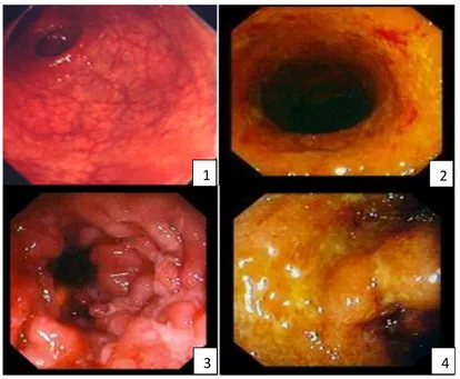
Figure 1: Grades of inflammation detected in Ulcerative colitis via endoscopy.
Photo taken by Pär Myrelid. 1 mild inflammation of mucosa (hyperaemia). 2 moderate inflammation 3 severe inflammation with a visible ulcer. 4 severe inflammation with exudate.
1 2 3 3 3 4 3
22
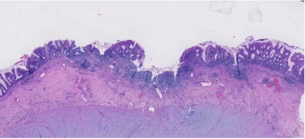
Figure 2: Microscopic picture of ulcerative colitis. Photo taken by Marina Perdiki Grigoriadi
CD is the other major IBD subtype affecting any part of the gastrointestinal tract from the mouth to the anus. CD is characterized by transmural inflammation of the bowel wall, and often discontinuous areas with inflammation” skip lesions” separated by segments of normal bowel and commonly associated with extra-intestinal manifestations as well.
CD patients commonly complain of intermittent abdominal pain and diarrhea, fatigue. In addition, some patients complain of rectal bleeding, low-grade fever and weight loss. The disease causes malnutrition and thus lead to bone loss and vitamin deficiencies. (29, 30) (31) CD patients also have a variable disease course with flares and periods of remission. (32)The
transmural inflammation can complicate the disease, causing obstruction due to intestinal fibrosis and/or fistulation.
Endoscopic examination of CD patients reveals areas of inflamed mucosa with superficial ulcerations over the granulomas, which spread in serpiginous pattern causing the
23
normal appearing mucosa causing the characteristic finding of skip lesions. Both granulomas and skip lesions are characteristic of CD.
Histological examination may show inflammatory infiltration in and around the crypts causing crypt abscesses and superficial mucosal ulcerations. Acute and chronic inflammatory infiltrates form non-caseating granulomas that extends in the submucosa, up to the mesentery and lymph nodes, which may cause inflammatory strictures. Serositis is also common, leading to formation of fibrotic strictures as well as bowel adhesions. Thus, it is common for CD patients to present with an intestinal obstruction. Crypt destruction commonly occur leading to the pipe-like appearance of the bowel. (33)
Figure 3: Endoscopic and histologic picture of Crohn’s colitis. Photo taken by Pär Myrelid (right) and Marina Perdiki Grigoriadi (left)
There is a third sub-diagnosis among IBD patients: indeterminate colitis (IC). According to the Montreal classification, IC diagnosis is reserved only for patients having had a colectomy and where pathologists still are unable to make a definitive diagnosis of either UC or CD. Moreover, they have suggested a new term “inflammatory bowel disease, type unclassified” (IBD-U). This term entitles to IBD patients, who have colon still in place, with a clinical and
24
endoscopic chronic inflammation affecting the colon, without small bowel involvement, and without a definitive histological/ clinical evidence in favor of UC or CD. (34) Little is known about the pathogenesis and clinical course of IBD-U, but it may not be correct to force this group of patients under either a UC or a CD diagnosis. Approximately 10-11% of adult and 12-18% of paediatric IBD patients will remain as IBD-U. (35) Another study reported that
paediatric IBD-U patients have molecular and serological characteristics more similar to UC than CD. However, 60 % of paediatric IBD-U patients will keep the same diagnosis of an IBD-U even after two years of follow up. (36) It is possible that IBD-U isa true separate IBD
sub-diagnosis or that some UC and CD patients are misdiagnosed. Unfortunately, most of previous cohort studies have excluded IBD-U patients from their analyses. Further research is required to better understand the behaviour of an IBD-U.
It is also known that IBD patients are more susceptible to develop colorectal cancer (CRC) compared with the general population. Previous studies have also established that long duration, severe and/or wide-spread colonic inflammation in IBD patients increase the risk to develop CRC. (37) (38)Several genetic and environmental factors contribute to the development
of CRC such as genetic instability, epigenetic alteration, oxidative stress that attacks cell and nuclear proteins, immune response caused by mucosal inflammatory mediators, as well as alteration of the intestinal microbiota species, concentration and distribution in the colon.
(39-42)(43)
The introduction of novel biological therapy and cancer screening programs with colonoscopy and biopsies offered to IBD patients have raised hope of decreased risk to develop CRC.(44) However, recent reports about the incidence of CRC in IBD patients are conflicting. One recent Danish population-based study concluded that the CRC risk is decreasing in patients diagnosed with UC.(45) On the contrary, another study from the US states that the incidence of
25
Additionally, they stated that this increased risk was stable over the 20 years duration of the study.(46) We believe that more large-scale cohorts from publicly funded health care systems, that offers biological interventions as well as surveillance to IBD patients, are required to determine the actual risk of CRC.
Figure 4: Endoscopic and microscopic pictures of a colonic adenocarcinomas. Photo taken by Pär Myrelid (right) and Marina Perdiki Grigoriadi (left).
IBD patients are more vulnerable to develop synchronous and/or metachronous CRC. (47) This
could be due to genetic predisposition(48), persistence of the inflammatory process(49) or
missed diagnosis of cancer in case of synchronous cancer. Cancer growth in IBD patients presents as a locally raised mucosa that could be missed in the presence of ulcerative disease, inflamed mucosa, inflammatory polyps and bleeding. Thorough endoscopic surveillance is thus crucial for young IBD patients, because they have a high risk to develop a post colonoscopy colorectal cancer.(50) Studies are required to evaluate the actual risk of
synchronous and metachronous cancers in IBD patients to be able to tailor the management plans accordingly.
26
Primary sclerosing cholangitis
PSC is a chronic liver disease characterized by a progressive course of cholestasis with inflammation and fibrosis of the intrahepatic and extrahepatic bile ducts.(51) The progressive fibrosis of biliary ducts leads to liver cirrhosis and portal hypertension and is one of the most
common causes of liver transplantation.(52) Between 80-90% of PSC patients have a
diagnosis of IBD as well(53) but usually have an insidious onset of their bowel inflammation
and may be asymptomatic or present with mild symptoms, which may delay their diagnosis of IBD.(9) (54-59) The problem is that IBD patients with a PSC diagnosis not uncommonly develop
cholangiocarcinoma and/or CRC. (9, 60, 61) One proposed contributing factor that increases the
risk of cancer is that cholestasis causes an accumulation of secondary bile acids, both in the liver and the colon, which is proven to have a carcinogenic effect in animal models. (53, 62)
Another proposed contributing factor is that IBD/PSC+ patients have a prolonged course of unmanifested subclinical inflammation of the colon that causes no or very mild symptoms. Consequently, it takes time for patients to get diagnosed and properly treated and previous studies detected that a long duration of colonic inflammation increases the risk to develop CRC in UC. (37) (38)
27
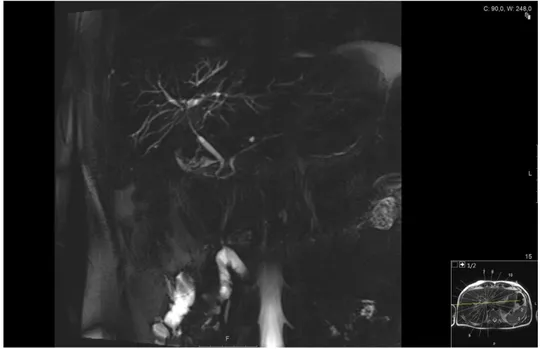
Figure 5: MRCP of the biliary tree showing dilated biliary ducts typical for PSC. Photo taken by Jenny Öman
It is also essential to identify the most common location for CRC in IBD/PSC+ patients to determine the required extent of resection. A recent study reported that CRC are commonly located in the right side of the colon in IBD patients with a concomitant PSC.(63) Another
study reported that UC/PSC+ patients who underwent a subtotal colectomy with an ileostomy and a disconnected rectal remnant in situ did not risk to develop neoplasia in their remaining rectal stump .(64) Thus it seems that the existence of the enterohepatic circulation and/or
preserved faecal stream to the colon or rectum are crucial for the cancer process to occur in IBD/PSC patients. In order to confirm such theory, we need to assess the CRC location for a large sample of IBD patients with PSC.
Theoretically, if PSC increases the risk for primary CRC, it should also increase the risk of synchronous and metachronous cancers. Unfortunately, most studies for CRC in IBD patients
28
excluded synchronous and metachronous cancer patients. Studies with focus on the risk for synchronous cancers and metachronous cancer after management of the primary cancer for IBD-PSC patients are required. First to evaluate the effectiveness of the current surgical resections for PSC patients diagnosed with primary CRC in preventing future cancers. Secondly to adjust the overall management plans for PSC-IBD patients before they get their CRC diagnosis.
Surgical reconstruction for ulcerative colitis
Approximately one-third of patients diagnosed with ulcerative colitis involving the colon proximal to the splenic flexure will require surgery within 15 years of diagnosis in the form of subtotal colectomy and ileostomy(65), either due to a severe flare, medically refractory chronic
disease or due to development of dysplasia or invasive cancer. The choice of definitive surgery is made after patients’ counseling with surgeon, which depends on patients’ age, general condition, persistence of rectal inflammation as well as the results of the pathology report.
Severely ill and malnourished patients are preferred to keep their ileostomy functioning with a diverted rectum (DR) left in place or with a later completion proctectomy. (10) If high grade
dysplasia or cancer of the resected colon were detected on the pathology report, completion proctectomy should be performed as soon as the patient is deemed fit. In the meantime, tight endoscopic surveillance should be offered for early detection of rectal dysplasia/cancer. Apart from dysplasia and cancer some patients with DR suffer from symptomatic proctitis that may require a completion proctectomy.
Surgically fit patients, without dysplasia or cancer in their pathology report, who underwent a subtotal colectomy usually request for restoration of their bowel continuity, with either an ileorectal anastomosis (IRA) or an ileal pouch anal anastomosis (IPAA).(66) DR patients who
29
future risk of IRA failure. Instead, patients should undergo a completion proctectomy and choose another form of reconstruction.(7, 67)
IRA was first introduced by Stanley Aylett more than 50 years ago(68). It was the standard
surgery for treatment of UC for years. Unlike Brooke’s ileostomy, patients got to keep their bowel continuity and continence. IRA surgery can be performed as one step surgery at the same session of subtotal colectomy or as two step surgery. Especially in severely ill patients a stepwise approach is recommended, with a subtotal colectomy and ileostomy as a first step followed by an ileorectal anastomosis later.(6)IRA surgery, compared with IPAA, is not as
complex as a procedure, with shorter operating time and requires minimal pelvic dissection and causes minimal blood loss. However, concerns are raised about a possible future risk of cancer in the remaining rectal tissue.(69)
30
Figure 6: Ilea-rectal anastomosis (IRA).
A)The colon parts marked in red will be resected (subtotal colectomy) leaving the rectum behind. B)The ileum anastomosed to the remaining rectum.
Reprinted with permission from Springer international Publishing AG, from the book: The Ileoanal Pouch 2019; Chapter 14, page 174, figure 14.1 a and b.
Proctitis is another not uncommon problem that concerns UC patients reconstructed with an IRA. Patients who suffer from proctitis complain from abdominal pain, urgency, diarrhea and bleeding. A previous study reported that 22% of UC patients with a grossly normal rectum at time of subtotal colectomy developed proctitis, of which 11% underwent a completion proctectomy for IRA failure.(70) A previous study reported that 27% of UC patients will
manifest IRA failure and will require a secondary proctectomy, of which two thirds were due to proctitis. (71)
The search for a novel surgery that preserves continence and avoids the risk of rectal cancer was on. The year 1969, Prof Nils Kock fashioned a reservoir constructed of the small bowel. It had the advantage of preserved continence while having an external opening on the abdominal
31
wall. To reconstruct the reservoir, an intestinal loop was opened longitudinally and sutured together to form U shaped limbs. Then the reservoir’s corner was sutured to the abdominal wall, and an efferent limb was added. Initially, continence was obtained from the rectus abdominis muscle. Later, continence was maintained through a nipple valve formed by retrograde intussusception of the efferent intestinal limb into the pouch.(72) Despite continence, several
surgeons reported the valve slippage and detachment from the abdominal wall. Further modifications were developed in order to staple the nipple valve by Kock, Stein and Fazio. (73-75) Addition of synthetic material was discouraged due to risk of fistulas.(76) Barnett introduced
another type of valve, the isoperistaltic valve from the afferent limb, to overcome valve slippage. Furthermore, a conduit of ileum that forms a living intestinal collar was added around the reservoir outlet. When the reservoir is full the pressure increases inside the collar, subsequently maintain continence of the pouch. This modification is called the Barnett continent ileostomy reservoir (BCIR). Further modification was added by stapling the valve into the reservoir and moreover placing the stapling at the same line of pouch sutures.(75, 77)
Trials to avoid valve slippage lead to the development of the new T shaped reservoir with a serosal lined anti reflux mechanism is reported by Kaiser.(78) However, this T shaped reservoir
32
Figure 7: A simplified drawing of the Kock pouch
Reprinted with permission from Springer international Publishing AG, from the book: The Kock pouch 2019; Chapter 4, page 36, figure 4.1.
There are several problems with continent ileostomy other than valve slippage. First, continent ileostomy requires proper training on catheter intubation for evacuation and a cooperative patient without any mental, physical or psychological obstacles. Second, continent ileostomy is contraindicated for patients with past/ family history of desmoid tumors due to risk of tumor recurrence. Third, it requires approximately 50 cm of small bowel to fashion a continent ileostomy. Therefore, patients with short bowel or those who underwent a pouchectomy of their pelvic pouch are not recommended to perform a continent ileostomy.(79, 80)
33
maintain continence and discard most of the rectal tissue. Thus, he could minimize future risk of rectal cancer as well as restore the bowel stream via an ileal-pouch anal anastomosis (IPAA).
(6, 81, 82) Details of the procedure were first published in the British Medical Journal in 1978. (83)
Since then, IPAA has been the gold standard in most parts of the world. (84, 85)
Figure 8: Ileal-pouch anal anastomosis (IPAA).
A) Colon and rectum (marked in red) will be surgically resected. B) formation of a J shaped pouch of ileum then anastomosis to the anal canal is performed.
Reprinted with permission from Springer international Publishing AG, from the book: The Ileoanal Pouch 2019; Chapter 14, page 174, figure 14.2 a and b.
Reconstruction with an IPAA may today be performed through an open or laparoscopic technique. Studies reported that there is no clear evidence that laparoscopic IPAA is superior to open IPAA regarding operative time, blood loss or postoperative hospital stay.(86, 87) They
did not suffer from more postoperative comorbidities compared to open IPAA but they were able to close their ileostomy earlier.(86, 88)Additionally, another study reported that there was no
34
IPPA.(89) One more study concluded that the rate of pregnancy in females with UC was higher
in patients reconstructed with laparoscopic restorative proctocolectomy compared to those with a conventional restorative proctocolectomy. This is probably due to less bleeding with subsequent reduction of pelvic adhesions.(90) Prospective large scale studies are required to determine the potential superiority of the laparoscopic approach.
Moreover, IPAA can be constructed in one step at the same session of the proctocolectomy, which may be safe for selected UC patients without severe disease activity, e.g. elective surgery in patients who are not steroids. (91) IPAA can also be performed as a two- or three-staged
procedure. The most common method in Sweden is a three-staged procedure with subtotal colectomy and end-ileostomy first. When the patient has recovered after approximately six months, a proctectomy is performed with creation of the IPAA and a diverting loop-ileostomy. The diversion is closed after approximately 3 months. It can also be performed as a two-stage procedure when proctocolectomy and IPAA is performed directly with a diverting loop-ileostomy, followed by closure of the ileostomy as in the three-stage procedure. Recently a modified two-stage procedure has been winning ground. In these cases, a subtotal colectomy and end-ileostomy is done first while the second step includes proctectomy with IPAA but without the use of a diverting ileostomy. Thus patients have shorter hospital stays and avoid the burden of a third surgery.(92) A recent study concluded that patients with the modified
two-stage IPAA reported significantly lower anastomotic leaks, despite suffering from more severe UC activity and higher preoperative use of steroids.(93) The advantage of the three-stage and
the modified two-stage IPAA is that it can be performed in emergency settings as it allows time to improve the patients’ nutritional status, treat anemia, and withdraw from immunomodulating medications before going through a more complex pelvic dissection with a high risk anastomosis. (92, 94) Other studies concluded that the three-stage IPAA is associated with more
35
scale study reported that there were no differences in morbidity or mortality of patients with a three-stage versus two-stage IPAA.(96)
Surgeons have developed several shapes of the reservoir the S-, the J- and the W-reservoir. These reservoirs can be performed via hand-sewn (with or without mucosectomy) or via trans-anal stapling. Studies that compared the stapled and hand-sewn anastomosis reported no significant differences in early postoperative complications or bowel function.(97) However,
some studies reported more night seepage and incontinence of liquid stools in the handsewn group(98)and that a stapled anastomosis is superior regarding continence, need of using pads,
restrictions in the diet as well as restrictions in work and social life.(87, 99) Additionally, there
were no significant differences between the three shapes of pouches regarding the early/late postoperative complications, e.g. anastomotic leak or strictures, infection of the wound or pelvic sepsis, pouchitis or pouch failure.(100, 101) S-shaped and W-shaped pouches manifested lower
frequency of defecation and thus less need for anti-diarrheal treatment.(102) Surgeons who prefer the S-pouch, mostly handsewn, motivate that because the efferent limb fits well in the anal canal and the body lies above the levator muscles of the pelvic floor. (87) Surgeons prefer J- pouch because it can be formed by staplers, thus minimize operative time, and requires less intubation to evacuate. However, the blunt end of the J-pouch can be distorted because it gets forced in the muscular anal canal. On the another hand, K-reservoir is time-consuming, complex procedure that requires experienced hands for optimal results.(103) However, it has
somewhat better long term functional outcomes than J- reservoir.(104)
It is known that IPAA patient satisfaction is dependent on their ano-rectal function and sexual function.(105-107) It is also known that female UC patients reconstructed with an IPAA suffer
from reduced fecundity compared to other UC patient.(108) Few small-sized studies suggested
that the laparoscopic reconstruction of an IPAA was not different from the conventional IPAA regarding the reduction of intraoperative bleeding, operative time and postoperative
36
morbidities(86, 87) but may have less impairment on female fertility.(89, 90, 109) However, large
scale studies are required to draw firm conclusions.
Recently, IRA has regained popularity in Sweden. (110) Part of the reason why IRA regained popularity is that it lacks some of the disadvantages of the IPAA. The surgical procedure is less complicated, less time consuming and involves minimal bleeding. Additionally, IRA does not impair bowel function or sexual function. IRA patients have even better functional outcomes compared to IPAA patients if strict selection criteria were followed.(111) Evidence has showed that fertility and fecundability of young UC patients with an IRA is not worse than the general population, especially after the introduction of laparoscopy.(112) Accordingly, IRA could
function as an interim procedure for young UC patients in order to postpone pelvic dissection required for an IPAA.(113)
However, IRA as a surgical option for UC patients comes with some concerns. Researchers argue that leaving behind rectal tissue can be problematic. A recent study concluded that UC patients have an increased risk of IRA failure, up to 27% specially if they were reconstructed for chronic refractory disease.(71) On the contrary, another study concluded the risk of IRA
failure to be 15% with minimal functional impairment observed.(114) Additionally, researches
hypothesize that persistent mucosal inflammation may trigger development of dysplasia or cancer in the remaining rectal mucosa. (3, 6, 69) However, this was only investigated from tertiary
care centers and large-scale population-based studies are required to draw firm conclusions.
When IRA fails, it is inevitable to recommend a completion proctectomy. Then rises an important question: could patients keep continence or not, i.e. end ileostomy? In case they request a secondary reconstruction there are two possibilities today, an IPAA or a Kock pouch (a continent ileostomy) The functional outcome of IPAA as a secondary reconstruction has been thoroughly studied in patients with Familial adenomatous polyposis (FAP) and both pouch
37
survival and function were found to be similar to that of primary pouches. (115) By contrast, to
our knowledge no previous study compared the outcome of primary and secondary IPAA in UC patients. More studies are required to investigate the outcome of primary and secondary reconstructed IPAA in IBD patients in general and UC patients in specific.
38
Aims of the study
I. To assess the risk and risk factors for rectal cancer in ulcerative colitis patients after colectomy and reconstruction with an IRA, an IPAA or without reconstruction in form of permanent ileostomy and diverted rectum in place.
II. To compare the colorectal cancer in IBD patients, with and without a PSC diagnosis, to non IBD controls regarding tumor incidence, tumor location as well as risk of synchronous and metachronous cancers after segmental resection.
III. To evaluate the association between the anorectal function and QoL between IRA and IPAA in ulcerative colitis patients and relate their mucosal inflammation, diagnosed via endoscopy and pathology reports, to the anorectal function.
IV. To estimate the survival of primary reconstructed IPAA and secondary reconstructed after a previous IRA in ulcerative colitis patients.
39
Methods
Registers
A unique personal identification number was first introduced in Sweden in 1967 and since then assigned to cover all Swedish residents. This personal number permits the follow-up of patients in national registers, e.g. the Swedish Patient Register (NPR). The NPR was established in 1964–1965 to document individual hospital discharges. The discharge
diagnoses are coded according to the International Classification of Diseases, seventh revision (ICD7) 1964-1968, eighth revision (ICD8) 1969-1986, ninth revision (ICD-9) 1987-1996, and ICD-10 thereafter. Each record corresponds to one in-hospital episode and contains
information about date of hospital admission, main discharge diagnosis and up to seven concurrent diagnoses as well as treating department. The register also includes information about surgical procedures based on the “Klassifikation av operationer” (1964-1996) and the Swedish version of the NOMESCO (Nordic Medico-Statistical Committee) classification of surgical procedures since 1997. As health care service in Sweden is funded by the government and registration in the NPR is mandatory, Swedish hospitals began reporting to the register the year 1964 with complete national coverage since 1987 (116). A previous study estimated the completeness of the NPR to be 85-95%. (117)
The same unique personal number is also used to obtain individual information about the date and site of a diagnosis of dysplasia or cancer from the colon and rectum via linkage to the Swedish Cancer Register (SCR) . The SCR was first introduced in 1958 and it is mandatory according to Swedish law to report to the register. It receives data about cancer diagnoses from clinical, morphological, laboratory as well as autopsy reports.(118, 119) Further,
40
completeness of Cause of Death Register was evaluated in a studies by Brooke et al and Johansson et al. Death due to cancer is well reported in most instances, such as breast cancer, because of the linkage to the National Cancer Register, while some other cancers are underreported e.g., pancreatic cancers.(118, 120, 121)(122)
Patients
Patients with a diagnosis of IBD after 1st of January 1964 were identified from the patient
registers (papers I, II and IV). From the Linköping University Hospital, we identified all UC patients operated with subtotal colectomy between the years 1992-2006 (paper III).
Variables identification
For papers I and II, we identified patients with a diagnosis of Ulcerative colitis (ICD7 572.20, 572.21, 578.03; ICD8 563.10, 569.02; ICD9 556*; ICD10 K51*). For paper IV, we identified UC patients as well as Crohn’s disease patients (ICD 7 572.00, 572.09; ICD 8 563.00; ICD 9 555*; ICD 10 K50*) and indeterminate colitis (ICD 7 572.30; ICD 8 563.98, 563.99; ICD 9 558; ICD 10 K52.3). Even though the ICD-9 era was little problematic because there was no code for IC diagnosis, we selected patients with the code (558) as IC only if it was followed by another IBD diagnosis on a second occasion, otherwise the patient was excluded. For the patient to keep an UC, CD or IC diagnosis he/she should not have any other IBD discharge diagnosis. If the patients had a first diagnosis of an IC and later diagnosed as either having UC or CD, he/she will be accepted as an UC or CD patient respectively. An IBD-Mixed diagnosis (IBD-Mix) was determined as a final diagnosis if the patient had a combination of UC, CD or IC. The last registered diagnosis was used for patients having multiple
registrations with different IBD diagnoses. The first registered date of an IBD diagnosis in the register was accepted as the date of debut of IBD.
41
We investigated PSC as an independent risk factor for cancer in (papers I and IV). This was not easy because there are no specific codes for PSC in the ICD-10, neither was there in the ICD-9, 8 or 7. We identified patients with a cholangitis code associated with an IBD diagnosis as PSC patients, through the codes; ICD-7 (575.05 and 585.29), ICD-8 (575.05), ICD-9 (576B), and ICD-10 (K830A, K830). The date of PSC diagnosis was accepted as the date of hospitalization when PSC diagnosis is made.
We identified surgical procedures (papers I-IV), patients operated with subtotal colectomy without a concurrent anastomosis to the rectum (operation codes 4651, JFH10 or JFH11). We also identified the sub-cohorts of patients that had been operated with colectomy and an ileorectal anastomosis (IRA, operation codes 4650, JFH00, JFH01, JFC40 or JFC41) or an ileal pouch anal anastomosis (IPAA, operation codes 4654, 4823, JFH30, JFH33, JGB50 or JGB60).
Paper specific methodology
Paper I: The follow up started from the date of sub-total colectomy in the group of patients
with a diverted rectum and the date of reconstruction (IRA or IPAA) for reconstructed patients. The follow up ended at the date of a rectal cancer diagnosis, proctectomy, death or emigration or the 31st of December 2010, whichever comes first.
Paper II: The follow up started at the date of reconstruction of ileal-pouch anal anastomosis
(IPAA), both the primary and secondary reconstructed. The follow up ended at the date of pouch failure, death or 31st of December 2010, whichever comes first. In order to understand the follow up, we stated some statistical definitions. A primary IPAA was defined as an IPAA with no previous IRA while a secondary IPAA was defined as an IPAA constructed
42
We defined pouch failure as the removal of the IPAA (i.e. pouchectomy), construction of a diverting ileostomy, including a continent ileostomy, more than 30 days after the
reconstruction of an IPAA. Patients with a redo IPAA after a previous IPAA were excluded from the study after their first failure.
Paper III: UC patients reconstructed at Linköping University Hospital with either an IRA or
an IPAA were invited to undergo an evaluation of their anorectal function and their post-operative QoL. Those who agreed to take part signed the informed consent and answered the following validated questionnaires: Öresland functional score, SF-36, and the Short Health Scale (see appendix).
Three weeks later they were invited to an endoscopic evaluation of their reservoirs or rectums including multiple biopsies taken and sent for histopathological evaluation. The grade of endoscopic inflammation was evaluated according to the Baron-Ginsberg score (BG score). BG score is originally intended for evaluation of rectal mucosa in IRA. However, we chose to standardize the method for both IRA and IPAA (appendix 4).
Multiple biopsies were taken from the ileum, anastomotic site, upper rectum, and ampulla in case of IRA and from the ileum proximal of the pouch, from upper and lower part of the pouch itself, and the rectal cuff in case of IPAA. The pathologist reported the grade of inflammation from all segments, coded as no, mild, moderate or severe inflammation. A global grade of inflammation was estimated for each patient from the highest grade of inflammation found in any part of the reservoir or rectum. Presence of dysplasia or polyps were reported. We investigated for possible associations between the severity of endoscopic and histopathologic inflammation and the anorectal function and quality of life in IRA and IPAA patients.
43
Paper IV: IBD patients with and without a concomitant PSC diagnosis (IBD/PSC+ and
IBD/PSC-) were assigned four matched controls, according to age, gender, time at diagnosis and place of residence at time of diagnosis. We needed to investigate the impact of an IBD diagnosis with a concomitant PSC diagnosis on colorectal cancer location. In order to do this we identified this data for each individual from the Cancer Register using the following SCR coding: right sided colon (ICD-7: 153.4 and 153.0, denoting cancers occurring in the appendix, caecum or ascending colon), transverse colon (ICD-7: 153.1), descending colon (ICD-7: 153.2), sigmoid colon (ICD-7:153.3) and rectum (ICD-7:154.0). Colon cancer with unspecified location was identified by the following ICD code (ICD-7:153.9).
Follow up started at the date of PSC diagnosis for the IBD patients with a concomitant PSC, the date of IBD diagnosis for IBD patients without a concomitant PSC and the matching date for the controls. The follow up ended at the date of first cancer diagnosis, colectomy, death or 31st of December 2014 for the colorectal cancer diagnosis, whichever came first.
Moreover, we needed to identify the risk of synchronous and metachronous cancers in the IBD/PSC- and IBD/PSC+ compared to controls. Since we had data only about tumour location and the date of diagnosis, it was not possible to identify the distance between the cancers to be more than 4 cm in case of synchronous. To overcome this problem, we defined synchronous cancers statistically as two or more CRCs occurring at a time interval equal to or less than 180 days. Meanwhile, metachronous cancer were defined as two or more CRC occurring at a time interval more than 180 days and were in different location of the colon and rectum then the first tumour. The follow up in such cases started at the date of segmental colectomy and ended at the date of synchronous cancer or the date of metachronous cancer, total colectomy, death, emigration or 31st of December 2014, whichever comes first.
44
Statistics
We reported patient’s demographics via mean and SD (papers I, III and IV) and median and
IQR (in paper II, and some analyses in paper III). We used some non-parametric tests: Mann Whitney U test to test the difference between reconstruction groups (paper III) and the Chi squared test as well as the Fisher’s exact to test differences in proportions (papers I, II and
IV).
We used the Kendall-tau correlation and ordered logistic regression (paper III) to analyse the possible association between the answered questionnaires and the grade of inflammation reported on the endoscopic/pathologic examination.
In papers ( I, II and IV ), we performed several types of survival analysis: Cox proportional hazard to identify possible risk factors using both uni- and multivariable analyses. Kaplan Meier survival curves, cumulative hazard and log rank tests were used to identify differences in survival between the studied groups (papers I, II and IV). Standardized incidence ratios (SIR) were estimated using age-, sex- and period-specific incidence rates for the Swedish population obtained from the web-based statistical service of the Swedish National Board of Health and Welfare (paper I). Incidence rate ratio (IRR) of risk factors for CRC in IBD patients were reported after matching to non-IBD controls according to age, sex, period-specific and residence (paper IV).(123) Net survival and excess hazard of death were estimated
for CRC patients with/without PSC diagnosis compared to controls (paper IV).
Handling missing data
Paper III: Some patients did not answer all the questions of the questionnaires of the SF-36. If less than one third of a questionnaire was left unanswered, the missing items scores were calculated through- person specific mean score calculated based on non-missing scores.(124) If
45
from the analysis. Seven patients declined to undergo endoscopy and macroscopic
assessment. Moreover, four endoscopy reports were missing (of which one pathology report was found)while two more pathology reports were missing after complete endoscopy.
All tests were 2-sided, and the results were considered statistically significant if p value was <0.05 or at 95% CI. All calculations were performed using STATA program versions 15 (StataCorp. 2015. Stata Statistical Software: Release 15. College Station, TX: StataCorp LP, USA).
Ethical approval
All studies were approved be Linköping regional ethics review board (Dnr 2011/419-31, papers I, II and IV) and (Dnr:M127-07, paper III).
46
Results
Detailed results are mentioned in the respective papers. Here only the most important findings will be highlighted.
Figure 9 :The studied populations in the thesis.
Data for all studies were nation-wide obtained from the National Patient register, except for paper III were data was obtained from the patient charts at Linköping University hospital.
47
Paper I
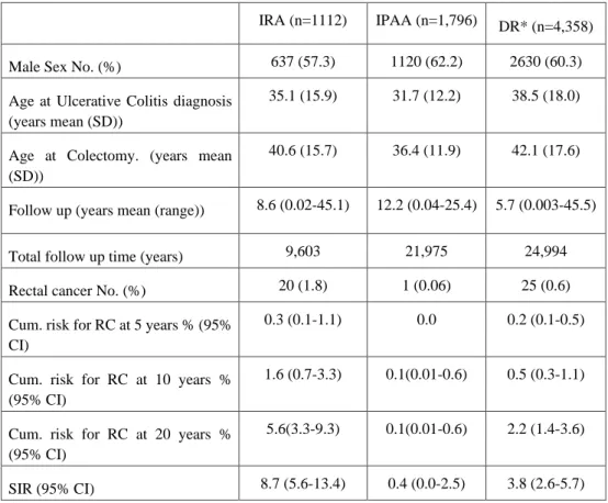
During the study period from 1964 to 2010, a total of 63,795 patients were diagnosed with Ulcerative colitis. Of which 7,889 (12 %) patients underwent subtotal colectomy. Rectal cancer occurred in total of 46 (0.8 %) of the 5,886 patients having had a sub-total colectomy (table 1).
Table 1 Demographics of the studied population (paper Ⅰ) of 5,886 patients with ulcerative colitis that were operated with subtotal colectomy during 1964-2010§
IRA (n=1112) IPAA (n=1,796) DR* (n=4,358)
Male Sex No. (%) 637 (57.3) 1120 (62.2) 2630 (60.3) Age at Ulcerative Colitis diagnosis
(years mean (SD))
35.1 (15.9) 31.7 (12.2) 38.5 (18.0)
Age at Colectomy. (years mean (SD))
40.6 (15.7) 36.4 (11.9) 42.1 (17.6)
Follow up (years mean (range)) 8.6 (0.02-45.1) 12.2 (0.04-25.4) 5.7 (0.003-45.5)
Total follow up time (years) 9,603 21,975 24,994 Rectal cancer No. (%) 20 (1.8) 1 (0.06) 25 (0.6) Cum. risk for RC at 5 years % (95%
CI)
0.3 (0.1-1.1) 0.0 0.2 (0.1-0.5)
Cum. risk for RC at 10 years % (95% CI)
1.6 (0.7-3.3) 0.1(0.01-0.6) 0.5 (0.3-1.1)
Cum. risk for RC at 20 years % (95% CI)
5.6(3.3-9.3) 0.1(0.01-0.6) 2.2 (1.4-3.6)
SIR (95% CI) 8.7 (5.6-13.4) 0.4 (0.0-2.5) 3.8 (2.6-5.7)
*Patients going through ileorectal anastomosis, proctocolectomy, ileal pouch anal anastomosis or completion proctectomy at a later stage are included as patients with de-functioned rectum until proctectomy or
reconstruction took place. §Patients who had proctocolectomy from the start or rectal cancer at time of colectomy
48
Ileorectal anastomosis
Rectal cancer (RC) occurred in 20 out of 1,112 IRA patients. The relative risk for RC was 8.7, while the absolute risk was 1.8%. One third of the patients restored with an IRA who
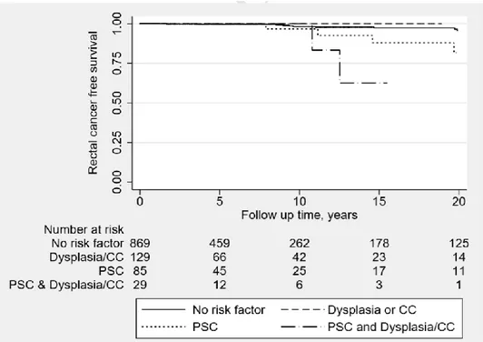
developed rectal cancer had a concomitant primary sclerosing cholangitis (PSC). The hazard ratio of developing rectal cancer in UC patients with a concomitant PSC was 5.95 (95% CI 2.34-15.13, p <0.001) in univariate analysis, and 6.12 (95% CI 2.33-16.03, p <0.001) in multivariate analysis compared to those without a PSC. It is noteworthy that most cancers occurred after 10 years of follow up after an IRA (Table 1, figure 10).
Figure 10: Kaplan Meier curve investigating the effect of possible risk factors on the rectal cancer free survival (p value< 0.001)
49
Ileal-pouch anal anastomosis
One UC patient out of 1,796 reconstructed with an IPAA developed RC. The relative risk to develop cancer was 0.4 (95% CI 0.0-2.5) when compared to the general population while the absolute risk was 0.06%. This RC patients was a young male diagnosed with UC at the age of 14 and was reconstructed at the age of 16. At the age of 25, he developed cancer in the rectal remnant of the pouch without a previous history of severe dysplasia, cancer or PSC. He died one year later due to advanced rectal cancer.
Diverted rectum
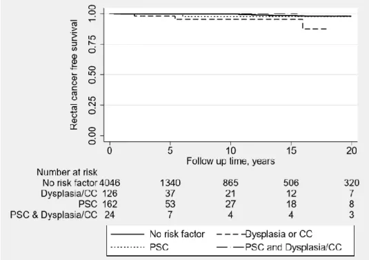
Twenty-five out of 4,358 UC patients with an intact but diverted rectum developed RC. The relative risk for DR patients to develop RC was 3.8 compared with the general population, while the absolute risk was 0.6%. History of severe dysplasia or colon cancer before colectomy was the only risk factor detected in univariate analysis HR was 4.64 (95%CI 1.38-15.58, p=0.0013) and in multivariate analysis HR was 3.67 (95%CI 1.01-13.37, p=0.049). Most cancers occurred after 15 years of follow up after date of subtotal colectomy (Table 1, figure 11).
50
Figure 11: Kaplan Meier curve analysing the effect of possible risk factors on the rectal cancer free survival of ulcerative colitis patients with an intact and diverted rectum.
(p value <0.001)
Paper II
From the same cohort of UC patients, IPAA was the primary reconstruction in 1,720 (95.8%) patients, and the secondary reconstruction after a previous IRA in 76 (4.2%) patients. As expected, time from colectomy to reconstruction was longer in case of secondary IPAA than primary IPAA (p <0.001). Failure occurred in a total of 109 IPAA patients (103 primary and 6 secondary) after a total follow up of 21,202 person-years. There was no difference in survival of primary reconstructed IPAA compared to secondary reconstructed IPAA (Table 2, figure 12)
51
Table 2: The demographics of the studied population paper Ⅱ
Total IPAA Primary IPAA Secondary IPAA p valuea
Number of patients 1796 1720 76 -
Number per year period (%)
1964-1979 0 0 0 - 1980-1989 197(47.0) 187 10 - 1990-1999 897(81.2) 868 29 - 2000-2010 702(54.3) 665 37 - Male sex (%) 62.3 62.6 54.0 0.13 Age at reconstruction median (IQR) 36.9 (28.2-45.8) 36.8 (28.2-45.8) 38.6 (28.8-48.0) 0.31 Duration of UC at reconstruction (years) 3.2 (1.2-8.2) 3.1 (1.2-8.1) 6.1 (2.2-11.7) 0.002
Time from colectomy to reconstruction (years) 0.4 (0.0-1.0) 0.4 (0.0-1.0) 1.9 (0.8-1.8) <0.001
Follow up duration (years) 12.4 (6.5-16.6) 12.6 (6.7-16.6) 10.0 (3.5-15.9) 0.12 Failure n (%) 109 (6.1) 103 (6.0) 6 (8) 0.50 Survival of IPAA, % (95%CI)
5 years 96 (95-97) 96 (95-97) 94 (85-98)
0.38π 10 years 94 (93-95) 94 (93-96) 92 (81-97)
20 years 92 (90-93) 92 (90-93) 88 (73-95)
a Primary versus secondary IPAA. π Log rank test
52
Figure 12: The risk of IPAA failure was similar after primary and secondary reconstruction.
(p value 0.38)
Paper III
In the study, 38 IRA and 39 IPAA patients answered the questionnaires. Most of them were males 29 (52.7%) in IRA and 26 (47.3%) in IPAA. There was no difference in age at diagnosis between IRA patients 25.8 (IQR 6.4-57.2) and IPAA patients 25.2 (IQR 9.1-42.0,
p=0.910) or age at reconstruction with IRA was 33.1 (10.3-75.2) and reconstruction with
IPAA was 35.2 (18.9-58.9, p= 0.424). The median follow-up after the restorative procedure was 12.1 (range 3.5-19.4) years. 68 patients underwent endoscopic evaluation. (Figure 13).
53
Figure 13: Flow chart of the included study participants in Paper Ⅲ.
* Data of the 77 patients were included in demographic table and the questionnaire analyses. However, they were not included for the test of the associations.
168 eligible IRA=87, IPAA=81 6 missing endoscopy and/ or pathology report IRA=2, IPAA=4 IPAA=30 IRA=34 77 signed informed consent and completed
questionnaires 143 invited IRA=72, IPAA=71 25 excluded due to
permanent diversion, excision, rectal cancer or
death 66 declined to participate IRA=34, IPAA=32 7 declined endoscopic assessment IRA=2, IPAA=5 68 underwent endoscopy* IRA=36, IPAA=32
54
The questionnaires
Ano-rectal function (the Öresland Score)
Patients with IRA had better function according to the Öresland score with a median overall score of 3 (IQR 2-5) compared to 10 (IQR 5-15) for IPAA (p<0.001) (Figure 14). Only three (7.9 %) IRA patients had a total Öresland score ≥8 compared to 21 (53.8 %) IPAA patients, (p<0.001).
Figure 14: Boxplots of the medians and CI 95% of Öresland score in UC patients operated with IRA and IPAA.
55
On the other hand, thirteen IRA patients (34.2%) showed a trend towards urgency compared to six (15.4%) IPAA patients (p=0.057) and the number of IRA patients who received medications (occasionally or continuously) was 32 (84.2%) compared to 22 (56.4%) of the IPAA patients (p=0.008). Twenty-eight IRA patients received mesalamine alone or combined with another medication (three combined with prednisolone, two with hydrocortisone foam, and one patient combined with both).
SF-36
The mean SF-36 score for the role limitation due to physical problems was worse for IRA patients compared to IPAA patients, 77.6 and 92.3 (p=0.043). There was also a trend towards worse transition of mental health in IRA patient with a mean of 74.2 in compared to 85.3 in IPAA (p=0.053) (Figure 15).
56
Figure 15: Boxplots of the medians and 95% confidence interval of SF-36 in ulcerative colitis patients operated with IRA or IPAA.
Zero represents the best possible and 100 the worst possible score. * p value <0.05.
Endoscopy & biopsy results
On endoscopy, six (18%) IRA patients had a Baron-Ginsburg (BG) score of two or higher, compared to 11 (37%) IPAA patients (p=0.273). On histopathology, 21 (60%) IRA patients had moderate to severe inflammation in the rectum whereas 24 (77%) of the IPAA patients displayed histological signs of pouch inflammation (p=0.454) (Table 3).
57
Table 3: Results of pathology reports: histological grades of inflammation, types and site of dysplasia and number of polyps.
IRA IPAA p-value
Endoscopy*
Baron Ginsburg score
0 1 2 3 n=34 7 21 5 1 n=30 6 13 11 0 0.173 Pathology reports+ Inflammatory grades None Mild Moderate Severe n=35 4 10 11 10 n=30 0 6 17 7 0.091 Cellular changes
Low grade dysplasia Hyperplasia Squamous metaplasia 1 at anastomosis 1 at ileum 2 at rectal cuff 1 rectal mucosa 0.099
*Missing endoscopy reports from two IRA. Missing both endoscopy and pathology report from two IPAA
patients. So (IRA n=34, IPAA n=30)
+ Found an additional IRA pathology report without endoscopy report. Missing pathology report from two IPAA
patients. So (IRA n=35, IPAA n=30).
Association between mucosal inflammation and function
There was a positive correlation between Öresland score and endoscopic grades of
inflammation (BG score) for IPAA patients only (tau. 0.28, p=0.006) but not for IRA patients. For the IPAA patients the strength of the association was further analysed with ordered logistic regression. This shows that the Öresland score is strongly impaired when BG score increases, with OR 1.3 (CI -0.6-3.2, p=0.188) for grade 1 to OR 3.4 (CI 0.8-6.1, p=0.012) for grade 2.
58
By contrast, there was no correlation between the results of pathology reports and the total Öresland score neither in IRA (p=0.740) or IPAA (p=0.197) patients.
Association between macroscopic and microscopic grades of inflammation
There was a weak correlation between grades of inflammation reported macroscopically using BG endoscopic score and microscopically through pathology reports (tau. 0.19, p=0.01) in all patients. When studying correlation between BG score and pathology reports in IPAA and IRA, respectively, there was a correlation in the previous (tau. 0.26, p=0.021) but not the latter group (tau. 0.16, p=0.125).
59
Paper IV
After 10,645,342 person-years of follow up, a total of 2,854 (2.2%) CRC were diagnosed out of 127,578 IBD patients compared to 8,107(1.3%) CRC diagnosed out of 610,120 matched controls. CRC occurred in 4.5% of IBD/PSC+ compared to 2.2% in IBD/PSC- (figure 16).
Figure 16: Flow chart of the studied population in paper Ⅳ.
€ out of IBD/PSC+ with primary cancer. * out of IBD/PSC- patients with primary cancer
IBD Inflammatory Bowel Disease, PSC primary sclerosing cholangitis, CRC colorectal cancer CRC n=2,677 Total IBD N=127,578 Synchronous cancer n=74 (2.8%) *
IBD patients with PSC diagnosis
n=3,497 IBD patients without
PSC diagnosis n=124,081 Synchronous cancer n=139 (1.3%) Synchronous cancer n=8 (4.5%) € CRC n=177 CRC n=8,107 Controls N=610,120 Metachronous cancer n=215 (2.7%) Metachronous cancer n=86 (3.2%) * Synchronous cancer n=5 (2.8%) €
60
Risk factors for Colo-rectal cancer in IBD patients
IBD patients with PSC+ had a significantly increased risk of CRC compared to those without a PSC diagnosis. The IRR of IBD/PSC+ was 6.6 (95% CI; 5.2-8.4), IBD/PSC- was 1.6 (95% CI; 1.5-1.7; p <0.001).
Additional risk factors
Patients diagnosed before the age of 20 years had the lowest risk in univariate analysis, HR 0.10 (95% CI 0.09-0.12; p<0.001) as well as multivariate analysis, HR 0.08 (95%CI 0.07-0.10; p<0.001). However, compared to controls of the same age the youngest IBD patients had the highest lifetime relative risk (IRR 63.52, 95% CI 39.46-101.39 for PSC+ and IRR 13.87, 95% CI 9.72-20.09 for PSC-).
Patients diagnosed with an IBD diagnosis at the age of 60 or older had the highest risk to develop CRC, HR 3.24 (3.07–3.41, p<0.001), taking middle aged patients (40–59 years) as reference value, whereas they had only a slightly higher risk compared with controls (IRR 1.92, 95% CI 1.27-2.77 for PSC+ and IRR 1.63, 95% CI 1.54-1.74 for PSC-) (table 4).