Faculty of Veterinary Medicine and Animal Science
Reproductive Status
of the Female Baltic Ringed Seal
Reproduktionsstatus
hos vikarehonor i Östersjön
Alexandra Odevall
Uppsala 2019
Reproductive Status of the Female Baltic
Ringed Seal
Reproduktionsstatus hos vikarehonor i Östersjön
Alexandra Odevall
Supervisor: Sara Persson, Department of Clinical Sciences Examiner: Anne-Marie Dalin, Department of Clinical Sciences
Degree Project in Veterinary Medicine
Credits: 30
Level: Second cycle, A2E Course code: EX0869 Place of publication: Uppsala Year of publication: 2019
Online publication: https://stud.epsilon.slu.se
Key words: Baltic ringed seal, occlusion, PCB, DDT, reproduction
Nyckelord: Vikare, säl, Bottniska viken, ocklusioner, PCB, DDT, reproduktion
Sveriges lantbruksuniversitet
Swedish University of Agricultural Sciences
Faculty of Veterinary Medicine and Animal Science Department of Clinical Sciences
SUMMARY
The population of ringed seals in the Baltic Sea was estimated to around 190 000–220 000 individuals in the 1900’s and decreased to approximate 5 000 in the late 1970s. The number of pregnant females also decreased during this time. This decreased pregnancy rate has been explained to be due to hunting and environmental pollutants such as dichlorodiphenyl-trichloroethane (DDT) and polychlorinated biphenyl (PCB) leading to pathological lesions (occlusions) in the uterus blocking the passage from the uterine horn to the ovary. Around 1970, the environmental pollutants were restricted in Sweden and in the 1990s the ringed seal population started to increase. Today the annual growth rate of the ringed seal population is estimated to be 5%. This is still considered to be a slow growth rate compared to other seal populations which can have a growth rate of around 10%.
This master thesis includes a study of the reproductive status of 142 female Baltic ringed seals necropsied by personnel at the Swedish Museum of Natural History from 2002–2018. Reproductive parameters were recorded such as lesions, ovary weight, pregnancy and corpus luteum and corpus albicans size. Specific measurements in ringed seal female reproductive organs, such as length and width, and blastocyst flushing were also performed. Data was grouped based on time of death: spring or fall.
Specific measurements of the size and weight of the female reproductive organs were described which has not been done before. This master thesis found that occlusions were observed in three females (6.8%) in the last 16 years. This could potentially be at least partially explained by bacterial infections causing abortions which in turn lead to occlusions and sterility. However, tissue concentrations of PCB in the affected females were not analyzed. Future studies including the male ringed seals part in the reproductive status of the species has been found to be needed.
SAMMANFATTNING
Vikarepopulationen i Östersjön var under tidigt 1900-tal uppskattad till 190 000–220 000 individer. Denna siffra sjönk till ca 5 000 individer under sent 1970-tal. Även antalet dräktiga honor minskade under denna period vilket förklarats av jakt och miljögifter så som dichlorodiphenyltrichloroethane (DDT) and polychlorinated biphenyl (PCB). PCB och DDT ska ha medfört patologiska lesioner (ocklusioner) i uterus som lett till att passagen mellan uterushorn och äggstock blockerats. Dessa miljögifter förbjöds i Sverige runt 1970 och på 1990-talet sågs en populationsökning. Idag uppskattas den årliga tillväxthastigheten hos vikare i Östersjön att vara ca 5 % vilket anses lågt jämfört med andra sälpopulationer som ligger runt ca 10 %.
Detta examensarb ete har undersökt reproduktionsstatus hos 142 vikarehonor från Östersjön som obducerats av personal på Naturhistoriska Riksmuseet mellan 2002–2018. Parametrar såsom lesioner, ovarievikt, dräktighet, och storlek på corpus luteum och corpus albicans noterades. Specifika mätningar hos vikarehonors reproduktionsorgan, så som längd och bredd, och blastocystsköljning utfördes också. Datan delades sedan in i vår eller höst beroende på när vikaren dog.
Specifika mätningar av storleken och vikten av det honliga reproduktionsorganet har beskrivits, något som inte gjorts tidigare. Detta examensarbete fann att 3 (6,8 %) fall av ocklusioner har observerats under de senaste 16 åren. Detta kan åtminstone delvis förklaras av bakteriein-fektioner som gett upphov till abort som i sin tur gett ocklusioner och sterilitet. PCB koncentrationer hos dessa individer har dock ej undersökts och framtida studier inom området behövs.
TABLE OF CONTENTS
Introduction 1
Literature Review 3
Background 3
Temporal Population Trends of the Ringed Seal 3
Reproductive Biology 4
Female Ringed Seal Reproductive Organ Facts 5
Occlusions in the Uterus 5
Material and Methods 7
Standard Measurements in Ringed Seal Females 7
Specific Measurements in Ringed Seal Female Reproductive Organs 7
Grouping of data 8 Blastocyst Extraction 9 Pregnancy rate 9 Statistical analysis 9 Results 10 Measurements 10 Pregnancy rate 10 Non-Pregnant Females/Pathologies 14 Blastocyst Extraction 16 Discussion 17 Measurements 17 Occlusions 18 Blastocyst Extraction 19
Popular Scientific Summary 21
1
INTRODUCTION
The ringed seal inhabits the northern part of the globe with a circumpolar distribution including the Baltic Sea and they mainly eat fish (ArtDatabanken, 2015; McLaren, 1958; Suuronen & Lehtonen, 2012). In the Baltic sea, the ringed seal is abundant in the Bothnian bay and does not migrate (Härkönen et al., 2008).
In the beginning of the last century, the population of ringed seals declined drastically which was thought to be due to hunting. In the 1970s the population declined even more which has been explained by environmental pollutants such as dichlorodiphenyltrichloroethane (DDT) and polychlorinated biphenyls (PCB). Hunting was stopped around the Baltic Sea in the 1980s and so were the use of DDT and PCB (Bjurlid et al., 2018; Harding et al., 2007; Härkönen et
al., 1998), and the population started to increase but only about 5% (ArtDatabanken, 2015)
which may be considered low compared to other seal species populations which can have a growth rate around 10% (Harding et al., 2007). The decline and the subsequent slow increase of the ringed seal population has been considered to be partly due to environmental pollutants such as DDT and PCB causing connective tissue occlusions in the uterus, consisting of connective tissue so that the passage through the uterine horn closed off (Harding & Härkönen, 1999; Helle et al., 1976 a; b; 1980b). The seals with these pathological changes also had considerably higher levels of DDT and PCB in the blubber (Helle et al., 1976b). The prevalence of occlusions peaked in the 1970s (when the correlation was discovered) and after the environmental pollutants were banned and declined (Bjurlid et al., 2018; Blomkvist et al., 1992; Nyman et al., 2002) so did the number of observed occlusions (Helle et al., 1976b). Declining occurrence of occlusions in uteri correlated with the decline of DDT and PCB found in the blubber. This has also been reported in other species such as grey seals in Sweden (Roos et al., 2012).
The ringed seal does not migrate, but immature seals tend to be in the off-shore waters and older ones keep to the fast ice (McLaren, 1958). Ringed seals follow the expanding fast ice as it expands south in December–January, but no other migration is seen (Härkönen et al., 2008). Therefore, migration as an explanation for the low growth rate of the Baltic ringed seal population can be disregarded (Härkönen et al., 1998; Härkönen et al., 2008) leaving only the explanation of a low rate of reproduction, high mortality rate or a combination of the two (Härkönen et al., 2008).
Since the beginning of the 1970’s, seals found dead in fishing gear or along the coast have been sent to the Swedish Museum of Natural History for investigation of their health status and testing for environmental contaminants. Since 1989 these investigations are included in the national environmental monitoring program. For many years, the number of ringed seals sent to the museum was low, but since the hunting quota increased during recent years, the number of samples collected did the same which in turn has facilitated studies of ringed seals from Swedish waters. The aim of this study was to investigate the general reproductive biology in female ringed seals from the Bothnian bay. The reproductive organs of female seals sampled between 2015–2018 were examined and measured. Also, an attempt to retrieve unimplanted blastocysts (implantation delayed by 3–3.5 months (Kelly et al. 2010; McLaren, 1958; Smith,
2
1970)) was performed. Data from these investigations, together with data collected within the national environmental monitoring program from 2002 and onwards was used to study reproductive parameters such as frequency of pregnancy, occurrence of occlusions and size and weight of potential fetuses. Special attention was given to the pathological findings found during this time period.
3
LITERATURE REVIEW Background
The ringed seal (Phoca hispida, family Phocidae pinniped) inhabits the northern part of the globe with a circumpolar distribution such as Alaska, Canada, Greenland, Russia, Svalbard and Karelian lakes and they live on or near stable ice and breed there (see review by Reeves, 1998). The subspecies are ladogensis, saimensis, ochotensis, botnica. The subspecies botnica, which is the main focus of this master thesis, is located in the Baltic Sea, specifically in the Bothnian Bay (70%), Gulf of Finland (25%) and Gulf of Riga (5%) (ArtDatabanken, 2015; Härkönen et al.et al., 1998).
The ringed seal eats mainly fish (McLaren, 1958; Suuronen & Lehtonen, 2012), such as three-spined stickleback (Gasterosteus aculeatus), Baltic herring, smelt (Osmerus eperlanus) and vendace (Coregonus albula) (Suuronen & Lehtonen, 2012).
In the Arctic ocean, the subnivean period is January–-May and is spent most of the time in the ice. The seals use breathing holes in the ice when in the water and lairs in the ice to rest (Kelly
et al. 2010; Kelly and Quakenbush, 1990; Lydersen, 1991). This is also seen in the Baltic ringed
seal (Almkvist et. al., 1980). The Arctic ringed seals population leave their lairs as the temperature rises in May-June and the snow collapses and lie basking on the ice (Kelly et al. 2010; McLaren, 1958) close to their previous lairs, until the ice coverage melts completely and then in July–December (Kelly et al. 2010).
Temporal Population Trends of the Ringed Seal
The ringed seal world population is estimated to be 6–7 million animals (Härkönen et al. 1998) and about 10 000 individuals inhabited the Bothnian Bay in 2015. The Baltic ringed seal population has an increase rate of ca 5% (ArtDatabanken, 2015). In the beginning of the 20th century, the Baltic ringed seal population was estimated to ca 180 000–200 000 animals (Harding & Härkönen, 1999), but decreased to 25 000 ringed seals in the early 1900’s due to hunting in Sweden and Finland (Harding & Härkönen, 1999).
Between 1940–1965 the population was considered stable, but a further decline was seen in the 1970’s, due to excessive hunting and environmental pollutants that made female ringed seals permanently sterile (Harding & Härkönen, 1999; Helle et al., 1976 a; b; 1980b). The number of pregnant females decreased from 1973 to 1979 with about 4 percentage units per year. (Helle, 1980b). The population was then well below 5 000 animals (Harding & Härkönen, 1999; Helle, 1980a). The population then increased up to about 8 000 individuals in the Bothnian Bay in the mid 1990’s (Härkönen et al., 1998) after hunting was stopped around the Baltic Sea in the 1980s and environmental pollutants concentrations declined (Bjurlid et al., 2018; Harding et
al., 2007; Härkönen et al., 1998). This decline in pollutant concentration was seen after a
Swedish law restricting the use of PCB (Jensen, 1972) and DDT (Olsson & Reutergårdh, 1986) was passed in 1972 and 1969 respectively.
4
Variation in ice conditions has also been suggested as a cause of non-increasing population trends in ringed seals (Sundqvist et al., 2012) where climate change (Sundqvist et al., 2012; Ferguson et al., 2017) and global warmings effect on ice coverage (ArtDatabanken, 2015; Hammar & Mattsson, 2017) are considered to be a potential future threats. It is likely that the population size of ringed seals will be proportional to the availability of the fast ice for making birthing lairs (McLaren, 1958; Sundqvist et al., 2012). Also, by-catch by fisheries, i.e. seals drowning when caught in fishing nets, and ice breaking in breeding areas of the ringed seal have been discussed as pressures for the population (ArtDatabanken, 2015; Hammar & Mattsson, 2017).
Reproductive Biology
The female reproductive organ of the Phocidae consists of a bicornuate uterus. It has a uterine body and two horns with spherical ovaries at the end (See review by Atkinson, 1997). In the caudal end the uterine horns connect to form a short corpus and a hymeneal fold separated the long vagina from the vestibule. They have an os clitoris, but it is small (Berta et al., 2005). The ringed seal sexually matures at the age of 5–7 years (McLaren, 1958; Smith, 1970) but the subspecies Botnica slightly earlier, at 4-5 years (Kauhala et al. 2018). Male maturity is defined by sperm in the testis and epididymis and female maturity by the first ovulation (McLaren, 1958; Smith, 1970) defined as the development of a corpus luteum (McLaren, 1958).
In the Baltic Sea, females have been found pregnant at the age of four and corpus albicans found at the age of three (Kauhala et al. 2018). Pregnancy rate (measured as the frequency of postpartum pregnancy signs such as corpus albicans and placental scars in spring) peaks at age 5–12 years and then declines, particularly after the age of 20. Ovulation rate also declines after the age of 20 but only marginally (Helle, 1980b; Kauhala et al. 2018).
Breeding occurs in May and implantation is delayed by 3 to 3.5 months (Kelly et al. 2010; McLaren, 1958; Smith, 1970). The Baltic ringed seal breeding season is a little earlier, in March-May but implantation delays are the same (Jensen, 2004).
Ringed seals fast from April to June which coincides with the peak of the mating season and the time when most seals are sunning on the ice (Kelly et al. 2010; McLaren, 1958) and molting their fur. During this time, they lose weight which is not gained again until the fall. In the females, the weight gain then is more than double (Härkönen et al., 2008; McLaren, 1958). The gestation period is 240 (McLaren, 1958) to 270 days (Smith, 1970), i.e. 8–9 months, and the birth season is in March–April the following year after mating season (McLaren, 1958). The female hollows out the snow on the fast ice and gives birth in a lair about 3×0.6 m. This requires snow that is deep enough, but sometimes natural pressure ridges are used for whelping (McLaren, 1958; Sundqvist et al., 2012). After this, the lactation period follows which is about 2 months long (McLaren, 1958). In the Baltic sea the whelping season is earlier; February– March, and the lactation period is shorter; about 1–1.5 months long (Jensen, 2004). At birth, the body length of ringed seals average at 65 cm (McLaren, 1958) and the pup weight is 4–5 kg when born in the Baltic sea (Jensen, 2004).
5
Female Ringed Seal Reproductive Organ Facts
The ringed seals are seasonal breeders and signs of ovulation in the ovaries has been found before and after mating season (McLaren, 1958) but ovulation does not necessarily mean conception (Helle1980b; Smith, 1970; McLaren, 1958). The corpus luteum starts to develop after ovulation, and if implantation occurs (August) it increases considerably in size compared to no implantation (McLaren, 1958). The average size of the corpus luteum was 13.6 mm in pregnant females and 9.4 mm in non-pregnant in October-November regardless of the age of female and 11.6 mm in April-May (Helle, 1980b). Before implantation the luteal components are closely packed together but after implantation, with increased vascularization in the corpus luteum (even visible macroscopically), connective tissue is organized in nodules and luteal cells are divided up by fibers of collagen (McLaren, 1958). The same is seen in late pregnancy (McLaren, 1958; Smith, 1970).
The corpus luteum degenerates into a corpus albicans, which is a small hard structure, after birth (Berta et al., 2005). The corpus albicans remains the same size in different reproductive categories (pregnant, non-pregnant without occlusions, occlusions in one horn, occlusions in both horns, pregnancy disturbed in some other way) and size is not dependent on the age of the female. Histologically, the corpus albicans consists of whitish scar tissue forming from the middle and out and measuring on average 4.4 mm in the fall and 6.5 mm in the spring (Helle, 1980b).
Maximum ovary size (average size of 26.1 mm long and 13.3 mm wide) is found in females at the age of seven or eight years (McLaren, 1958). Mature females have on average 13.7 follicles with a size over 3 mm in October-November (Helle, 1980b). After 20 years of age, the follicular activity decreases (Helle 1980b; Kauhala et al. 2018).
An alteration in pregnancy between the uterine horns is also suggested even though this does not always seem to happen (Helle, 1980b). The endometrium of a uterine horn that has been pregnant is less smooth, has more folds and is more vascularized than a horn that has not been pregnant (McLaren, 1958; Smith, 1970). The fetus is in the uterine horn on the same side as the ovary with the corpus luteum, i.e. ipsilateral (Helle, 1980b).
Occlusions in the Uterus
Sampling data from the Baltic Sea ringed seal collected in 1975 showed that about 40% of the females had pathological changes in the uterus, specifically stenoses and occlusions (Helle et
al., 1976b) and the older the female, the more common were the occurrence of occlusions.
Occlusions seemed to be equally distributed between the two sides of the uterus (Helle, 1980b). The occlusions consisted of connective tissue as a thin membrane which formed a closed chamber in the horn of the uterus. This membrane blocked the reproductive tract completely, but partially open occlusions have been seen (stenosis). Cranially to the occlusion, the uterine horn was filled with fluid ranging from light brown, turbid and foul smelling with decomposing particles to reddish brown, clear and odorless (Helle et al., 1976b; Helle, 1980b). The occlusions can sometimes be detected from outside of the uterus. (Helle, 1980b). The occlusions
6
were usually found at about 6 cm from the tip of the horn of the uterus and the width of the occlusion was usually around 3 cm. The uterine wall of the occlusion was thicker than pregnant uterine horn walls but thinner than non-pregnant ones. The uterine walls of the occlusion also varied from very thin, when very filled, to thicker and more grooved when less fluid was present inside (Helle, 1980b). Enlarged blood vessels could be seen in the area of the occlusions (Helle
et al., 1976b). These changes prevented passage from the ovary to the uterine horns and may
explain the low reproductive rate of ringed seals (Helle et al., 1976b). Uterine occlusions were unilateral in 25.5% of the mature females examined and bilateral in 16.5% (Helle, 1980b). In the mid-1970s, 34% of females had occlusions (Helle, 1980b) leading to only 32% of female ringed seals being pregnant (Helle et al., 1976b).
In the late 70s, the frequency of occlusions in Baltic ringed seals had risen to 59% (Helle, 1980b), 48.1% had occlusions before 1997, 22.5% in 1997-2006 and 5.5% 2007-2017 (Kauhala
et al., 2018). This shows a well-defined decline from the 1970s. The youngest age of ringed
seal females with these occlusions increased during this time period. The proportion of females found to have been pregnant also changed with the frequency of the occlusions in the ringed seal population in the Baltic Sea: 29% before 1997, 60% in 1997-2006 and 72.4% in 2007- 2016.
7
MATERIAL AND METHODS
Since the 1970’s, ringed seals that were by-caught or found dead on a shore have been collected at the Swedish Museum of Natural History (SMNH). From 2015 and onwards, it is permitted to hunt ringed seals between first of May and December 31. SMNH received tissues from ringed seals collected by the hunters during the hunt in May–July (reproductive organs, blubber, liver and lower jaw). Body length (nose to tip of tail) and blubber thickness (at the sternum) were measured by the hunters. Seals shot between August and December, with a body length above 100 cm, were transported frozen to the museum, where they were thawed and necropsied. Age was determined by cementum analysis at SMNH (Lockyer et al., 2010).
Standard Measurements in Ringed Seal Females
In this study, data from 142 female ringed seals necropsied by personnel at the SMNH between 2002 and 2018 was used (whole carcasses or samples from the hunt). This is a routine necropsy with a standardized procedure of data collection including recording of reproductive parameters such as lesions, ovary weight, presence and size of corpus luteum and albicans macroscopically and pregnancy. Also, the status of the uterus (juvenile, multiparous etc.) is described and standard data such as age, body weight, body length and blubber thickness is included.
Specific Measurements in Ringed Seal Female Reproductive Organs
The reproductive organs from 20 females were removed from whole bodies (collected during necropsies of females shot in the time period of 2015–2018) were stored frozen until further examination specifically for this study. Another 13 female reproductive organs were sampled by the hunters from seals shot during the hunt from May to the end of July 2018. Because these tissue samples were collected by hunters they were sometimes cut off at the cervix or the vestibulum leading to incomplete data in these cases. This missing data is demonstrated with M/D in the results tables. See figure 1 for uterus anatomy in the ringed seal.
The following specific measurements of the reproductive organs were taken:
Ovary size: longest and widest part was measured. They were inspected for corpus luteum and corpus albicans and cut open.
Corpus luteum (yellow) and corpus albicans (whitish) were identified macroscopically and measured at the widest point. Histology wasn’t performed because transitional stages weren’t seen due to samples being collected in the beginning of December to the end of April.
Uterine horn length: from tip of the horn to bifurcation, and width at the widest point Corpus of uterus, length: from bifurcation to cervix cranial point
Cervix width at the widest point
Vagina length: from cervix caudal part to the urethra opening
Vestibula length: from the urethra opening to the edge of the vulva (were mucous membrane and dermis meet)
8
It was evaluated if the uterus was juvenile (defined as absence of corpus luteum and corpus albicans and an immature uterus), not pregnant, pregnant left horn and pregnant right horn based on the side of the corpus luteum placement and with a fetus present. If there was a placental scar, the size, structure and which horn it was placed in was recorded. The weight, length and width of both ovaries were noted separately and the diameters of corpus albicans and corpus luteum were measured (two perpendicular measurements). Occurrence of uterine occlusions were also noted and described in size, placement from uterine horn tip and eventual content. If there were any pathological findings, these were also noted.
Figure 1. Uterus anatomy in the ringed seal.
Grouping of data
All adult females were divided into two time periods: spring and fall. Spring is the period after gestation and most of the lactation/mating period, up until the end of the implantation period, i.e. April–July. Fall is the period for fetal development, i.e. August–December.
The data set was also grouped according to reproductive stage within these time periods. During spring the adult seals (i.e. seals not having juvenile uteruses) were recorded as “postpartum” (presence of a corpus albicans and/or placenta scar, indicating a recent pregnancy) or “non-postpartum” (no corpus albicans or placenta scar was found). During fall the females were recorded as pregnant (with a visible fetus) or non-pregnant. Juveniles were defined as having no corpus luteum or corpus albicans and an immature uterus, macroscopically evaluated. Subadults were defined as having a CL, but a juvenile uterus.
In some cases, data was incomplete or missing. This is demonstrated in the results with M/D.
Bifurcation Cervix Urethra opening Urinary bladder Clitoris tip Uterine horn tip Bursa ovarica
9
Blastocyst Extraction
Three out of the 13 female reproductive organs sampled by hunters from seals shot during the hunt from May to the end of July 2018 uteruses were flushed with 4% polyvinyl alcohol in phosphate-buffered saline to extract not yet implanted blastocysts. The females were adult, collected in May, had a fresh corpus luteum and therefore could have had a fertilized egg, but implantation had not yet occurred.
This flushing saline solution was used to prevent the blastocyst from sticking to the instruments. Before flushing, the ligaments, ovaries, cervix and vagina were removed. Then the uterus was cut open with scissors dipped flushing saline to prevent the blastocyst from sticking to them. The uterus was held with tweezers without touching the endometrium, then the uterine groves were flushed allowing the flow of the saline solution to drip into a clean container. The flushed solution was then set to sediment. The sediment was thereafter removed by pipette and put into a container to be examined in a microscope.
Pregnancy rate
The pregnancy rate was calculated as the percentage of pregnant females out of all adults during fall with an age of 5–24 years. Postpartum pregnancy rate was calculated from spring as the percentage of females with post-partum signs out of all adult females with an age of 6–25 years (HELCOM, 2019).
Total combined pregnancy rate was calculated using combined data from the two groups.
Statistical analysis
Descriptive statistical calculations were performed using the Means procedure of SAS 9.3 (Milltown, USA). Pairwise t-tests for means were calculated using the GLM procedure. Microsoft Excel was used for calculating pregnancy rates. All P-values below 0.05 were considered to be significant.
10
RESULTS Measurements
The recorded data and measurements are reported in Table 1. It shows that the mean CL diameter was generally small in subadults (all sampled in spring) and adults with no signs of previous pregnancy in spring (non-postpartum), but this was found to be only significantly when compared to the CL of the pregnant (p=0.01 and 0.02, respectively) and non-pregnant females (p=0.03 and 0.02, respectively). No significant difference in CL diameter was found between pregnant and pregnant females during fall, however the CA diameter of non-pregnant females was significantly larger compared to the non-pregnant ones (on average 2 mm larger, p=0.03) but this was only based on two non-pregnant females. There was a significant difference in CL diameter, but not CA diameter, between adults sampled in spring and adults sampled in fall; the mean CL diameter was 10.8 mm and 12.2 mm respectively (p=0.04). The corpus luteum was found to be on average 12.1 mm in October-November and 11.6 mm in April–May and the corpus albicans was found to be an average of 8.0 mm in spring and 7.6 mm in fall.
The blubber layer of pregnant females in the fall was significantly thicker than the blubber layer of non-pregnant females in fall (p=0.003) and adults sampled in spring (both with or without postpartum pregnancy signs, see Table 1, p≤0.0001 for both). Females with no postpartum pregnancy signs in spring did not have a significantly different blubber thickness than females that had been pregnant (showing postpartum pregnancy signs).
Juveniles had significantly shorter length and width of the uterine horns than adults sampled in spring (see Table 1, p<0.0005 for both comparisons). A comparison of sizes between uterine horns that had supposedly been pregnant and those that had not could not be made with the data collected for this study. No significant differences between the different groups were found for corpus length or cervix width. The juveniles also had significantly lower total ovary weight than adults (p<0.0001 for all comparisons). No significant difference in total ovary weight was found between the adult groups (p>0.05).
Pregnancy rate
The pregnancy rate was 77% (20 pregnant females out of all adults (26) during fall with an age span of 5–24 years). The postpartum pregnancy rate was 69% (9 females with post-partum signs out of all adults (13) during spring with an age of 6–25 years). Total combined pregnancy rate was 74% (29/39).
12
Table 1.Mean, standard deviation, SD (within parenthesis) and minimum and maximum for measurements of the reproductive organs of 142 Swedish ringed seal females (collected 2002–2018). Occlusions and leiomyomas not included
Adults, spring Adults, fall
Juveniles N=86 Subadult N=6 Postpartum signs N=15 No postpartum signs N=5 Pregnant N=24 Non-pregnant N=6
Variable N Mean (SD) Min-max N Mean (SD) Min-max N Mean (SD) Min-max N Mean (SD) Min-max N Mean (SD) Min-max N Mean (SD) Min-max Age, years 81 0.6 (0.9) 0-3 6 3.2 (0.8) 2-4 14 9.9 (4.9) 4-20 4 11.0 (1.8) 9-13 24 11.9 (5.6) 3.0-27.0 6 13.7 (5.3) 9.0-23.0 Body weight, kg 77 28.4 (10.6) 5.8-60 5 48.1 (9.6) 37.8-59.3 11 43.7 (5.6) 33.5-54 2 77.5 (3.5) 75-80 22 83.4 (12.0) 51.3-103.0 6 63.8 (27.6) 25.6-99.8 Body length, cm 86 98.7 (12.6) 64-121 6 118.0 (9.5) 105-134 15 128.4 (6.3) 119-140 5 127.6 (19.0) 102-150 24 133.5 (8.6) 121.0-152.0 6 126.0 (8.5) 117.0-136.5 Blubber thickness, mm 83 27 (10) 1-60 5 36 (19) 15-60 15 22 (10) 9-50 3 29 (6) 23-34 24 56 (9) 36-78 6 40 (21) 8-67 CL mean diameter, mm 0 N/Aa N/A 6 9 (1) 7-11 15 11 (2) 9-15 5 10 (4) 5-16 24 12 (2) 10-16 3 13 (5) 10-19 CL mean diameter double, mm
0 N/A N/A 0 N/A N/A 0 M/Db M/D 1 N/A 13 1 N/A 9 0 M/D M/D
CA mean diameter, mm
0 N/A N/A 0 N/A N/A 12 7 (1) 5-9 0 N/A N/A 10 6 (2) 3 2 9 (1) 8-10
Uterus horn length sn, mm
6 71 (17) 46-90 1 N/A 117 2 155 (12) 146-163 1 N/A 181 1 N/A 365 2 202 (17) 190-214
Uterus horn width sn, mm
6 6 (2) 4-9 1 N/A 7 2 17 (3) 15-19 1 N/A 16 1 N/A 154 2 19 (4) 16-22
Uterus horn length dx, mm
6 76 (20) 45-95 1 N/A 110 2 154 (9) 148-160 1 N/A 180 0 M/D M/D 2 173 (17) 161-185
Uterus horn width dx, mm
6 7 (4) 4-14 1 N/A 8 2 18 (4) 15-20 1 N/A 13 0 M/D M/D 2 20 (11) 12-28
Uterus horn length pregnant, mm
0 N/A N/A 0 N/A N/A 0 M/D M/D 0 N/A N/A 14 204 (60) 112-362 0 N/A N/A
Uterus horn width pregnant, mm
0 N/A N/A 0 N/A N/A 0 M/D M/D 0 N/A N/A 14 60 (61) 10-214 0 N/A N/A
Uterus horn length non-pregnant, mm
0 N/A N/A 0 N/A N/A 0 M/D M/D 0 N/A N/A 14 194 (81) 214-324 0 N/A N/A
Uterus horn width non-pregnant, mm
13
a Not applicable
b Missing data Table 1 cont.
Adults, spring Adults, fall
Juveniles N=86 Subadult N=6 Postpartum signs N=15 No postpartum signs N=5 Pregnant N=24 Non-pregnant N=6
Variable N Mean (SD) Min-max N Mean (SD) Min-max N Mean (SD) Min-max N Mean (SD) Min-max N Mean (SD) Min-max N Mean (SD) Min-max
Corpus uteri length, mm
6 18 (9) 9-34 1 N/A 32 2 43 (14) 57-29 1 N/A 20 15 64 (32) 25-136 2 59 (6) 54-63
Cervix width, mm 6 10 (2) 8-13 1 N/A 19 1 N/A 21 1 N/A 26 16 29 (22) 26-103 2 22 (1) 21-23 Vagina length, mm 4 46 (7) 37-54 0 M/D M/D 1 N/A 131 1 N/A 104 16 116 (31) 80-160 2 121 (23) 104-137 Vestibulum length,
mm
1 M/D 48 0 M/D M/D 1 N/A 83 1 N/A 65 13 77 (12) 54-103 2 83 (13) 74-92
Clitoris length, mm 1 M/D 17 0 M/D M/D 1 N/A 67 1 N/A 19 12 54 (6) 40-61 2 53 (17) 41-65 Clitoris width, mm 1 M/D 14 0 M/D M/D 1 N/A 14 1 N/A 10 12 15 (3) 12-21 2 7 (4) 4-10 Uterus weight, g 6 2.0 (0.7) 1.7 1 N/A 13.4 1 N/A 43.0 1 N/A 49.8 14 135.8 (66.1) 50.5-244.0 2 84.5 (6.4) 80.0-89.0 Ovary weight sin, g 81 0.9 (0.4) 0.9-2.7 6 1.9 (0.8) 0.9-2.9 15 3.5 (1.1) 1.8-5.8 5 3.2 (0.9) 1.9-4.1 24 4.3 (1.4) 2.6-7.6 6 4.5 (2.7) 1.3-9.1 Ovary weight dx, g 80 0.9 (0.4) 0.3-2.7 6 1.9 (0.6) 1.2-2.7 15 3.4 (0.8) 2.0-4.9 5 3.1 (0.9) 2.0-3.8 24 3.3 (1.0) 1.7-5.8 6 3.5(1.6) 1.3-6.2 Total ovary weight, g 80 1.8 (0.8) 0.6-5.4 6 3.8 (1.1) 2.1-5.0 15 6.9 (1.6) 3.9-10.2 5 6.3 (1.7) 4.2-7.9 24 7.6 (1.9) 4.2-12.5 6 7.9 (4.3) 2.6-15.4
14
Non-Pregnant Females/Pathologies
Reabsorbed fetus
This individual was found to have a suspected reabsorbed fetus in the right uterine horn. This female was 23 years old and shot 2014 November 3 during hunting. The reproductive organs were removed, frozen and sent in to be examined. The female was measured to be 125 cm long, weighed 99.8kg and a fat layer of 67 mm. The left ovary weighed 4.9g and contained a corpus luteum with a size of 10×9 mm. The right ovary weighed 3.2g. In the right uterine horn signs of fetus reabsorption was found. Blood vessels in the walls were evident and the endometrium was smooth. The left horn had no signs of previous pregnancy and was folded with an immune affected mucus membrane. The vagina contained tough yellowish secretion. Pseudomonas spp. was found in the cervix.
Occluded Females
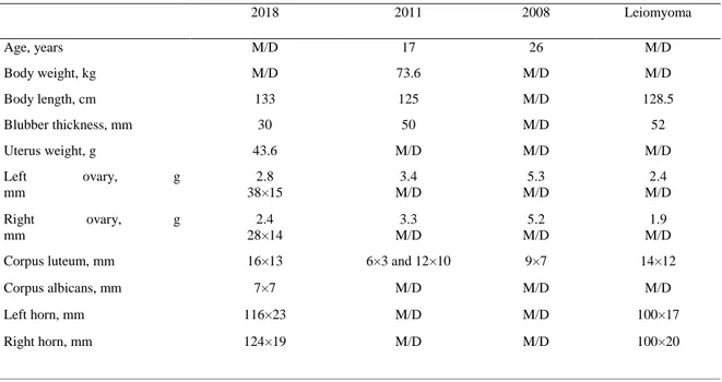
Occlusions were found in three individuals, that had been shot in 2008, 2011 and 2018, corresponding to 6.8% (3/44) of adult females (based on reproductive status (as seen in Table 1), not actual age), and one female was found to have leiomyoma in the uterus. None of these females were pregnant.
It can be noted, even though this is not statistically proven due to lack of sample quantity, that the occluded uterus' horns appear to be shorter and thicker than non-occluded adult uterine horns. Also, the ovaries seem to weigh less, corpus uteri are longer and cervix less wide. There appears to be no difference in blubber thickness or weight of uterus. See Table 2 for standard and specific measurements.
Table 2. Measurements of seals with uterine occlusions and leiomyoma
2018 2011 2008 Leiomyoma Age, years M/D 17 26 M/D Body weight, kg M/D 73.6 M/D M/D Body length, cm 133 125 M/D 128.5 Blubber thickness, mm 30 50 M/D 52 Uterus weight, g 43.6 M/D M/D M/D Left ovary, g mm 2.8 38×15 3.4 M/D 5.3 M/D 2.4 M/D Right ovary, g mm 2.4 28×14 3.3 M/D 5.2 M/D 1.9 M/D Corpus luteum, mm 16×13 6×3 and 12×10 9×7 14×12
Corpus albicans, mm 7×7 M/D M/D M/D
Left horn, mm 116×23 M/D M/D 100×17
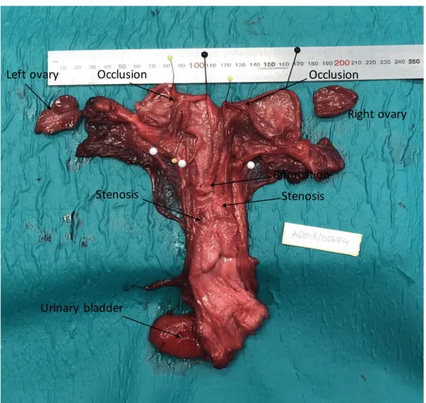
15 Occluded Uterus in a Female Shot in 2018:
This 28-year old individual shot on May 23, 2018 was found to have bilateral uterine horn occlusions and stenosis in the corpus and left horn. The left ovary contained a corpus luteum, the right ovary contained a corpus albicans and no follicles over the size of 1 mm were found. In the left uterine horn, the occlusion was 10 mm long and placed 30 mm from the tip of the horn, see figure 2. The occluded area was dilated with a thin and smooth mucous membrane and filled with 2ml of turbid yellowish-brown fluid. In the right uterine horn, the occlusion was about 4 mm long and placed 40 mm from the tip of the horn. The occluded area was dilated with a thin and smooth mucous membrane and filled with 3ml of turbid yellowish-brown fluid. No fluid was found distally of the occlusions. In the corpus there was a stenosis of 20 mm long placed close to the cervix. Next to this in the left uterine horn was another stenosis about 4 mm long.
Bacterial culture from the uterus was negative for Brucella spp. and positive for Streptococcus
marimammalium.
Figure 2. Occluded uterus in a female shot in 2018.
Occlusion
Bifurcation
Right ovary Left ovary Occlusion
Stenosis Stenosis
16 Occluded Uterus in a Female Shot in 2011:
This 17-year-old individual was found to have a unilateral uterine horn occlusion and the female was shot 2011 December 2.
In the right horn the occlusion was membranous and 20 mm from the bifurcation containing a turbid fluid. The endometrium is smooth with dark dots. The uterine wall in the right horn is very thin and looked to be juvenile even though two corpus luteum were present (6×3 and 12×10 mm). Left horn uterine wall was thicker in the middle area and looked as if it had been pregnant at least once.
Occluded Uterus in a Female Shot in 2008:
This 26-year-old individual was found to have bilateral uterine horn occlusions and was found drowned in 2008.
The left horn had a thin membranous occlusion ca 30 mm from the bifurcation containing a yellowish-green transparent fluid. The uterus wall was thinner and smoother in the area of the occlusion. The right horn occlusion with a length of 23 mm was found ca 10 mm from the bifurcation containing a yellowish-green transparent fluid. The cervix opening was only 1 mm and the corpus had some transverse structures without being occluded.
Leiomyoma
This individual was found to have myoma in the corpus and vagina. She was shot 2016 August 29.
In the corpus close to the cervix a myoma with a size of 4×25 mm with a dark mélange surface was found. There was also a smaller myoma with a light cut surface ca 10×7 mm in diameter. In the vagina close to cervix there was myoma a measuring 12×10 mm in diameter with a light cut surface and another two smaller myomas. A few 1mm myoma were palpable in the wall of the vagina.
Blastocyst Extraction
17
DISCUSSION Measurements
In this study the uterine horns of postpartum and non-pregnant females were found to be 146– 214 mm long and 12–28 mm wide which is much longer than the corpus (20–63 mm) which was found to be quite short in comparison. The vagina was 104–137 mm long and vestibulum 65–92 mm. These specific measurements of the female reproductive organ except the ovaries, corpus luteum and albicans of the ringed seal has not been described in this detail before, but rather in more general terms for the Phocidae (See review by Atkinson, 1997; Berta et al., 2005) and further studies are needed.
The corpus luteum of the Baltic ringed seal which was found to be on average 12.1 mm in October–November and 11.6 mm in April–May was much larger compared to 11.6 and 3.6 mm respectively in the arctic ringed seal (McLaren, 1958). Also, the corpus albicans was found to be larger in this study measuring an average of 8.0 mm in spring and 7.6 mm in fall compared to the arctic ringed seal (4.4 mm and 6.5 mm respectively) (McLaren, 1958). This can be explained by a normal variation in the Baltic populations, but it cannot be confirmed without further studies. It would be interesting to study the rest of the female Baltic ringed seal reproductive organ parameters to see if other differences occur between populations and what impact this could have for the reproductive status and population growth.
This study found that the youngest female with a corpus luteum was only two years old. This is younger than earlier literature has reported (McLaren, 1958; Smith, 1970) but matches the Kauhala et al. (2018) study which focuses on the Baltic ringed seal. Kauhala et al. (2018) also found corpus luteum and corpus albicans in female seals at the age of three. The female Baltic ringed seal seems to mature earlier than other ringed seal populations even in this study. The pregnancy rate was 77% and the postpartum pregnancy rate was 69%. Total combined pregnancy rate was 74%. Using corpus albicans and placental scars as a pregnancy rate measurement does not say anything if the female has been aborting the fetus, it only shows that she has been pregnant at one point the last year but not whether she has been full term or not, with the exception of early abortions not necessarily leaving placenta scars. Also, after ovulation, female ringed seals develop a corpus luteum (McLaren, 1958) that may persist and lead to a corpus albicans whether or not they were pregnant, making corpus albicans even more unsure as a measurement of pregnancy rate unless the placental scars are evaluated at the same time. This has been found in grey seals in North Sea were corpus albicans may remain in the ovaries for more than one year and ovulations from more than one follicle may occur leading to multiple corpus luteum (Boyd, 1982). Therefore, the most reliable way of calculating may be the total combined pregnancy rate were both postpartum signs and fetuses are taken into account.
Interestingly, this study showed no significant difference in CL diameter between pregnant and non-pregnant females during fall, but the CA diameter of non-pregnant females was significantly larger compared to the pregnant ones, but this was only based on two non-pregnant
18
females and further study is needed to draw conclusions. Also, further studies within female blubber thickness comparing females with no postpartum signs to females with postpartum signs would be interesting because this study showed no significant difference and comparisons haven’t been reported in other studies.
Occlusions
This study only showed three occluded uteruses (PCB and DDT levels yet to be determined) between the years of 2002 and 2018 corresponding to 6.8% of non-juvenile females compared to 59% in the 1970s (Helle, 1980b). The environmental pollutants PCB and DDT most likely played an important role in the occurrence of these and this is supported by the correlation of the legislation restricting the use around 1970 in Sweden (Jensen, 1972; Olsson & Reutergårdh, 1986) and the decrease in occluded ringed seal females after this ban (Harding et al., 2007; Härkönen et al., 1998). The restriction of environmental pollutants is a contributing factor, but it may not completely explain the low population increase of 5% reported by ArtDatabanken, 2015 compared to other seals, 10%, (Harding et al., 2007) since they only represent 6.8% of fertile females but must be studied further because population development was outside the scope of this study.
The seals with these occlusions were also reported to have significantly higher levels of DDT and PCB than non-occluded uteruses when blubber was tested (Helle et al., 1976b). This is a rather weak argument for PCB being the reason for the occlusions, as organochlorines cross over to the fetus via lactation (Wolkers et al., 2004). Females with bilateral occlusions are much less likely to lactate so increased concentrations are to be expected. A stronger argument for PCB being the underlying cause is that it has been shown to cause abortions. In mink, the number of implantation sites were not considerably different in PCB-fed minks compared to the control group, but the number of whelping females and number of whelps born differed significantly in favor of the control group (Jensen & Olsson, 1977). It has also been shown in mink that PCB fed females had embryos with various degrees of toxicity (Aulerich & Ringer,1977) which could lead to abortion. It is possible that this happens in ringed seals and that the subsequent endometrial healing leads to occlusions, making it impossible for the female to get pregnant again in the affected horn.
If abortions can cause occlusions in general, which has been seen in humans were miscarriages lead to uterine adherences (Jaslow & Kutteh, 2013), this might be why we are still seeing occlusions even if they are rare. This is supported by the fact that occlusions have not been described in juvenile females suggesting that occlusions are preceded by pregnancy. Also, the occlusions are in the same place as the placenta scars which also supports that they are preceded by pregnancy. The occlusions observed in this study were found consistent in placement, size, structure and content with previous studies (Helle et al., 1976b; Helle, 1980b) except one occlusion containing turbid yellowish-green fluid which has not been described before. This female also had transverse structures in the corpus without occluding it which also hasn’t been described in connection to occlusions before. This difference may be a sign of something else causing abortions. On the other hand, the fact that organochlorines are passed on through
19
lactation might be why we are still seeing occlusions even though there has been a decline in PCB and DDT after the mid-1970s (Bjurlid et al., 2018; Harding et al., 2007; Härkönen et al., 1998). It is possible that the occluded females in this study had these pathological lesions due to higher levels of organochlorines due to a combination of “inherited” organochlorines through lactation combined with their long lives giving them time to accumulate these environmental pollutants over time. The occluded females were determined to be the age of 28 (2018), 26 (2008) and 17 (2011) years old making it possible that they inherited PCBs and DDTs from their mothers and therefore were more sensitive to the environmental pollutants still in the Baltic Sea. The generally longer lactation period of ringed seals compared to grey seals (see review by Lydersen and Kovacs, 1999) could be the reason for the ringed seals being “more sensitive” than grey seals. The last case of occlusion in grey seals was found in 1993 (Roos et
al., 2012).
The occluded female from 2018 and the female with the reabsorbed fetus (2014) were both positive for infections in their reproductive organs. In the occluded uterus from 2018
Streptococcus marimammalium was found. This bacterium has been described in 2005 in the
lungs of seals as a novel species (Lawson et al., 2005) and has been shown to give bronchopneumonia in South American fur seal pups (Seguel et al., 2018). Maybe this can also lead to abortions and/or occlusions in Baltic ringed seals when pregnant females are infected. This female was also negative for Brucella spp. which is interesting because even though brucellosis is generally known to cause abortions in mammals (see review by Hull and Schumaker, 2018) this was not the case in this particular female. The 2014 female with the reabsorbed fetus in the right uterine horn was positive for Pseudomonas spp. in the cervix.
Pseudomonas pseudomallei has been shown to cause abortions in other mammals such as goats
(Thomas et al., 1988). This could have been the reason for her reabsorbed fetus.
Also, one female was found to have leiomyomas in her reproductive organs. She was not pregnant, and it can be discussed whether this has an impact on reproduction in ringed seals. Myomas have been reported in humans to have a negative impact on reproductive outcome such as abortions (Vlahos et al., 2017). This is true in grey seals were females with uterine leiomyomas did not have corpus luteum or corpus albicans in 63% of the cases (Bäcklin et al., 2003).
It would be interesting for future research to look at the male ringed seals reproductive status to see if this might have an impact on population growth as a whole.
Blastocyst Extraction
No blastocysts were found by flushing the uterus with 4% polyvinyl alcohol in phosphate-buffered saline. This can be due to there not being any blastocysts present, but also that it was quite difficult holding the uterus in a way that opens the lumen up to flushing without touching the endometrium, but the most likely explanation would be decay of the sample.
20
Another technique could be to turn the uterus inside out after cutting it open and dipping it in a 4% polyvinyl alcohol in phosphate-buffered saline bath before flushing it with a pipette. This way, if the blastocyst is not in a uterine groove, it is not accidentally transferred to an instrument. Even flushing the uterus without cutting it open would minimize transfer risks. In cattle, flushing embryo techniques are practiced in live animals where the embryo is recovered by using Foley catheters inserted through the cervix so that the cuff can be inflated in the cervix. Then an inflow and outflow channels are used to flush the uterus several times to extract the embryo (Seidel, 1981). This is a technique well worth trying in ringed seal uteruses before cutting them open. The risk of transferring the blastocyst while cutting the uterus open is eliminated and so is the accidental transfer through handling an open uterus.
21
POPULAR SCIENTIFIC SUMMARY
The ringed seal inhabits the northern part of the globe with four different subspecies. This study focuses on the subspecies botnica which lives in the northern part of the Baltic sea. They eat mainly fish and live on the ice but spent a lot of time in the water, for example when they hunt for food. The ringed seal is not a migrating animal but follow the ice when it melts and can also rest on moving ice.
The Baltic ringed seal sexually matures at the age of 4–5 years which is defined by sperm in the testicles in the male and first ovulation in the female. They have been found to be pregnant at the age of four but most commonly pregnant at the age of 5–12 years of age. Breeding occurs during March–May but the fertilized egg (blastocyst) does not implant until about 3.5 months later, this is called delayed implantation. During mating season, the seals fast and bask in the sun molting their fur. The ringed seal female is pregnant 240–270 days (8–9 months) and give birth in snow lairs in the ice in February–March the following year. The pup is about 65 cm when born and weighs about 4–5kg. The female lactates for about 1–1.5 months and during this time mating may occur.
The female ringed seal has been noted to ovulate before and after mating season, but ovulation does not always mean conception and pregnancy. After ovulation a corpus luteum (yellowish in color) develops in the ovary. If implantation of a blastocyst in the uterus occurs, then the corpus luteum stays throughout the whole pregnancy. When the pregnancy is over the corpus luteum regresses into a small whitish nodule called the corpus albicans consisting of scar tissue. In the early 1900’s the Baltic ringed seal population was estimated to be about 180 000–200 000 animals but quickly decreased to 25 000 individuals due to hunting in Sweden and Finland. During the mid-1900’s the population was stable, but a further decline was seen in the 1970’s due to excessive hunting and environmental pollutants such as the organochlorines dichloro-diphenyltrichloroethane (DDT) and polychlorinated biphenyls (PCB) which have been suggested to make many female ringed seals sterile through occluded uteruses. After hunting stopped in the 1980’s the population started to increase slightly.
In the 1970’s it was shown that 40% of the investigated Baltic ringed seal females had occluded uteruses making it impossible for sperm to reach the egg and therefore making them sterile leading to only 32% of the females getting pregnant. The occlusions consisted of connective tissue and were millimeter thin up to 3 cm wide. They can block the passage to one or both ovaries and were often filled with a discolored fluid. A law was passed in Sweden in 1969 restricting the use of DDT and 1972 PCB lowering the levels and the occlusions declined to only 5.5% during 2007–2017 and pregnancy was at 72.4%.
In this study, 142 female ringed seal reproductive status were looked at. They were collected by hunters or whole bodies were sent in by hunters during the years 2002 and 2018. They were then frozen before necropsied by personnel at the Swedish Museum of Natural History. Parameters such as lesions, ovary weight, presence and size of corpus luteum and albicans and
22
pregnancy was recorded. Out of these, 33 female reproductive organs were examined for specific measurements such as ovary size, corpus luteum and corpus albicans size, length and width of different parts of the uterus and clitoris size. It was also evaluated if the uterus was juvenile, not pregnant, pregnant (including fetus size) or postpartum and the individuals were divided into spring or fall depending on when their time of death. Statistical analysis comparing these different variables was performed.
It was shown that the ovary, corpus luteum and albicans size in the Baltic ringed seal is larger than in other ringed seal populations but the different parts of the uterus could not be compared since this type of data haven’t been collected before. Juveniles reproductive organs were generally smaller than adult females´.
The total combined pregnancy rate was 74% and only three occluded uteri were found 2002– 2018 which is equivalent to only 6.8% but since they still occur it was discussed that if abortions can cause occlusions then occlusions can occur with or without the presence of PCB and DDT. Instead, bacterial infections could be the explanation for the occlusions although it was only supported by two occluded uteruses found in this study actually having bacterial infections. Also, the environmental pollutants can be passed on through lactation to the pup which could have given the occluded females in this study a bad start by already having a high level of organochlorines in their bodies and therefore not needing it to accumulate much more for the environmental pollutants to lead to occlusions.
23
REFERENCES
Atkinson, S. (1997). Reproductive biology of seals. Reviews of Reproduction, 2(3), pp. 175–194. ArtDatabanken (2015). Pusa hispida, Vikare. Tillgänglig:
http://artfakta.artdatabanken.se/taxon/100104 [2018-12-04]
Almkvist, L., Olsson, M. and Söderberg, S. (1980). Sälar i Sverige 1. uppl. Stockholm: Svenska naturskyddsföreningen, pp 80.
Aulerich, R. J. and Ringer, R. K. (1977). Current status of PCB toxicity to mink, and effect on their reproduction. Archives of Environmental Contamination and Toxicology, 6(2–3), pp. 279–92. Berta, A., Sumich, J. L., Kovacs, K. M., Folkens, P. A and Adam, P. J. (2005). Marine Mammals. 2nd
Edition. USA: Elsevier. Pp 367–371.
Bjurlid, F., Roos, A., Ericson Jogsten, I. and Hagberg, J. (2018). Temporal trends of PBDD/Fs, PCDD/Fs, PBDEs and PCBs in ringed seals from the Baltic Sea (Pusa hispida botnica) between 1974 and 2015. Science of The Total Environment, 616–617, pp. 1374–1383.
Blomkvist, G., Roos, A., Jensen, S., Bignert, A. and Olsson, M. (1992). Concentrations of sDDT and PCB in seals from Swedish and Scottish waters. Ambio, 21(8), pp. 539–545.
Boyd, I. L. (1982). The use of corpora albicantia for determining pregnancy rates in seals with special reference to grey seals (Halichoerus grypus). International Council for Exploration of the Sea, 14. Bäcklin, B.-M., Eriksson, L. and Olovsson, M. (2003). Histology of uterine leiomyoma and
occurrence in relation to reproductive activity in the baltic gray seal (Halichoerus grypus). Veterinary Pathology, 40(2), pp. 175–180.
Ferguson, S. H., Young, B. G., Yurkowski, D. J., Anderson, R., Willing, C. and Nielsen, O. (2017). Demographic, ecological, and physiological responses of ringed seals to an abrupt decline in sea ice availability. PeerJ Preprints, 5, p. e2957.
Hammar, J. and Mattsson, M. (2017). Underlag för klimatrefugier i havsplaneringen 2017.Havs- och vattenmyndigheten (Havs-och vattenmyndighetens rapport 2017:37).
Harding, K. C. and Härkönen, T. J. (1999). Development in the Baltic grey seal (Halichoerus grypus) and ringed seal (Phoca hispida) populations during the 20th century. Ambio, 28(7), pp. 619–627. Harding, K. C., Härkönen, T., Helander, B. and Karlsson, O. (2007). Status of Baltic grey seals:
Population assessment and extinction risk. NAMMCO Scientific Publications, 6(0), pp. 33–56. HELCOM (2019). Assessment Protocol. Tillgänglig:
http://www.helcom.fi/baltic-sea-trends/indicators/reproductive-status-of-seals/assessment-protocol/ [2019-01-03]
Härkönen, T., Stenman, O., Jüssi, M., Jüssi, I., Sagitov, R. and Verevkin, M. (1998). Population size and distribution of the Baltic ringed seal (Phoca hispida botnica). NAMMCO Scientific
Publications, 1(0), pp. 167–180.
Hörkönen, T., Jüssi, M., Jüssi, I., Verevkin, M., Dmitrieva, L., Helle, E., Sagitov, R. and Harding, K. C. (2008). Seasonal activity budget of adult baltic ringed seals. PLOS ONE, 3(4), p. e2006. Helle, E. (1980a). Aerial census of ringed seals Pusa hispida basking on the ice of the Bothnian Bay,
24
Helle, E. (1980b). Lowered reproductive capacity in female ringed seals (Pusa hispida) in the
Bothnian Bay, northern Baltic Sea, with special reference to uterine occlusions. Annales Zoologici Fennici, 17(3), pp. 147–158.
Helle, E., Olsson, M. and Jensen, S. (1976a). DDT and PCB levels and reproduction in ringed seal from the Bothnian Bay. Ambio, 5(4), pp. 188–189.
Helle, E., Olsson, M. and Jensen, S. (1976b). PCB levels correlated with pathological changes in seal uteri. Ambio, 5(5/6), pp. 261–262.
Hull, N. C. and Schumaker, B. A. (2018). Comparisons of brucellosis between human and veterinary medicine. Infection Ecology & Epidemiology, 8(1), pp. 1–12
Jaslow, C. R. and Kutteh, W. H. (2013). Effect of prior birth and miscarriage frequency on the prevalence of acquired and congenital uterine anomalies in women with recurrent miscarriage: a cross-sectional study. Fertility and Sterility, 99(7), pp. 1916–1922.e1.
Jensen, B. (2004). Nordens däggdjur. 2. uppl. Stockholm: Prisma, pp. 324. Jensen, S. (1972). The PCB Story. Ambio, 1(4), pp. 123–131.
Jensen, S. and Olsson, M. (1977). Effects of PCB and DDT on mink (Mustela vision) during the reproductive season. Ambio, 6(4), pp. 239–239.
Kauhala, K., Bergenius, M., Isomursu, M. and Raitaniemi, J. (2018). Reproductive rate and nutritional status of Baltic ringed seals. Mammal Research, pp. 109–120.
Kelly, B. P., Badajos, OH., Kunnasranta, M., Moran, JR., Martinez-Bakker, M., Wartzok, D. and Boveng, P.( 2010). Seasonal home ranges and fidelity to breeding sites among ringed seals. Polar Biology, 33(8), pp. 1095–1109.
Kelly, B. P. and Quakenbush, L. T. (1990). Spatiotemporal use of lairs by ringed seals (Phoca hispida). Canadian Journal of Zoology, 68(12), pp. 2503–2512.
Lawson, P. A., Foster, G., Falsen, E. and Collins, M. D. (2005). Streptococcus marimammalium sp. nov., isolated from seals. International Journal of Systematic and Evolutionary Microbiology, 55(1), pp. 271–274.
Lockyer, C. Mackey, B., Read, F., Härkönen, T. and Hasselmeier, I. (2010). Age determination methods in harbour seals (Phoca vitulina) with a review of methods applicable to carnivores. NAMMCO Scientific Publications, 8(0), pp. 245–263.
Lydersen, C. (1991). Monitoring ringed seal (Phoca hispida) activity by means of acoustic telemetry. Canadian Journal of Zoology, 69(5), pp. 1178–1182.
Lydersen, C. and Kovacs, K. M. (1999). Behaviour and energetics of ice-breeding, North Atlantic phocid seals during the lactation period. Marine Ecology Progress Series, 187, pp. 265–281. McLaren, I.A. (1958). The biology of the ringed seal (Phoca hispida Schreber) in the eastern Canadian
Arctic. Fisheries Research Board of Canada, Arctic Unit, Montreal, P.Q. Bulletin No. 118. Nyman, M., Koistinen, J., Fant, M. L., Vartiainen, T. and Helle, E. (2002). Current levels of DDT,
PCB and trace elements in the Baltic ringed seals (Phoca hispida baltica) and grey seals (Halichoerus grypus). Environmental Pollution, 119(3), pp. 399–412.
25
Olsson, M. and Reutergårdh, L. (1986). DDT and PCB pollution trends in the Swedish aquatic environment. Ambio, 15(2), pp. 103–109.
Reeves, R. R. (1998). Distribution, abundance and biology of ringed seals (Phoca hispida): an overview. NAMMCO Scientific Publications, 1(0), pp. 9–45.
Roos, A. M., Bäcklin, B-M. V. M., Helander, B. O., Rigét, F. F. and Eriksson, U. C. (2012). Improved reproductive success in otters (Lutra lutra), grey seals (Halichoerus grypus) and sea eagles
(Haliaeetus albicilla) from Sweden in relation to concentrations of organochlorine contaminants. Environmental Pollution (Barking, Essex: 1987), 170, pp. 268–275.
Seguel, M., Gutiérrez, J., Hernández, C., Montalva, F. and Verdugo, C. (2018). Respiratory mites (Orthohalarachne diminuata) and β-hemolytic streptococci-associated bronchopneumonia outbreak in South American fur seal pups (Arctocephalus australis). Journal of Wildlife Diseases, 54(2), pp. 380–385.
Seidel, G. E. (1981). Superovulation and embryo transfer in cattle. Science, 211(4480), pp. 351–358. Shaw, S. D., Brenner, D., Bourakovsky, A., Mahaffey, C. A. and Perkins, C. R. (2005).
Polychlorinated biphenyls and chlorinated pesticides in harbor seals (Phoca vitulina concolor) from the northwestern Atlantic coast. Marine Pollution Bulletin, 50(10), pp. 1069–1084. Smith, T. G. (1970). Population dynamics of the ringed seal in the Canadian Eastern Arctic. Diss.
Montreal: McGill University.
Sundqvist, L., Härkönen, T., Svensson, C. J. and Harding, K. C. (2012). Linking climate trends to population dynamics in the Baltic ringed seal: Impacts of historical and future winter temperatures. AMBIO, 41(8), pp. 865–872.
Suuronen, P. and Lehtonen, E. (2012). The role of salmonids in the diet of grey and ringed seals in the Bothnian Bay, northern Baltic Sea. Fisheries Research, 125–126(C), pp. 283–288.
Thomas, A. D., Forbes‐Faulkner, J. C., Norton, J. H. and Trueman, K. F. (1988). Clinical and pathological observations on goats experimentally infected with Pseudomonas pseudomallei. Australian Veterinary Journal, 65(2), pp. 43–46.
Vlahos, N. F., Theodoridis, T. D. and Partsinevelos, G. A. (2017). Myomas and adenomyosis: Impact on reproductive outcome, BioMed Research International, p. 14.
Wolkers, H., Lydersen, C. and Kovacs, K. M. (2004). Accumulation and lactational transfer of PCBs and pesticides in harbor seals (Phoca vitulina) from Svalbard, Norway. The Science of the Total Environment, 319(1–3), pp. 137–146.