UMEÅ UNIVERSITY MEDICAL DISSERTATIONS New Series No 829 – ISSN 0346-6612 – ISBN 91-7305-398-8
From the Department of Public Health and Clinical Medicine, Rheumatology, Umeå University, Umeå, Sweden
A Clinical and Genetic Study
of
Psoriatic Arthritis
Gerd-Marie Alenius
CONTENTS
CONTENTS ………. 1 ABSTRACT ………. 3 ABBREVIATIONS ……….. 4 ORIGINAL PAPERS ……… 5 INTRODUCTION ……… 7Classification and epidemiology ………. 7
Clinical aspects ……… 9 Pathogenesis ……… 13 Genetic factors ……… 13 Immunological factors ..………. 15 Environmental factors ..………. 16 Vascular factors ..……….. 17 AIMS .……….. 18
PATIENTS AND METHODS .……… 19
Patients and controls .……….. 19
Clinical assessments ..………. 21 Diagnostic criteria ...……… 22 Laboratory tests ……… 23 Statistical analyses ..……… 24 STUDY DESIGN..………. 26 Paper I ...………... 26 Paper II ……… 28 Paper III ..……… 28 Paper IV ..……… 29
RESULTS AND COMMENTS ……… 30
A Prevalence study and an evaluation of a questionnaire (Paper I) 30 HLA antigens and inflammatory joint manifestations (Paper II) 38
Analysis of five disease susceptibility loci for PsA (Paper III) 44
Renal abnormalities in PsA patients (Paper IV) .……… 46
Additional observations ………...………. 48 CONCLUDING REMARKS ……… 52 CONCLUSIONS .………..……… 55 SAMMANFATTNING PÅ SVENSKA ...……… 56 ACKNOWLEDGEMENT ……… 58 REFERENCES ………. 59 PAPERS I – IV
Abstract
A Clinical and Genetic Study of Psoriatic Arthritis
Psoriatic arthritis (PsA) is an inflammatory joint disease associated with psoriasis. PsA has a heterogeneous pattern, expressed by different manifestations such as mild mono-oligoarthritis or very severe, erosive and destructive polyarthritis. Measurable inflammatory activity is not always prominent. The aetiology is unknown but genetic factors are believed to be of importance. The pattern of inheritance is proposed to be polygenic. The aim of this study was to estimate the prevalence of joint and axial manifestations, characterise the disease in relation to inflammatory and genetic markers, and to identify disease susceptibility gene(s) for PsA in patients from northern Sweden.
All patients from the city of Umeå (n=276), selected from a community and hospital based psoriasis register (n=1737) at the Dept of Dermatology, were invited to a prevalence study. Two hundred-two patients were examined and 97 (48%) had inflammatory manifestations such as peripheral arthritis, axial disease, undifferentiated spondylarthropathy (uSpA) and enthesopathies. Of the 67 patients (33 %) with peripheral arthritis and/or axial disease, 30 were not previously diagnosed.
The association of clinical manifestations and potential markers of aggressive joint disease with HLA associations were analysed in 88 patients with PsA. We were not able to confirm findings of other groups reporting strong association with several HLA-antigens. The prevalence of HLA-B17, B37 and B62 was increased compared with controls, but the strongest predictive factors among our patients for an aggressive disease, in a multiple logistic analysis, were polyarthritic disease and distal interphalangeal engagement.
In order to investigate for disease susceptibility genes, five genetic loci were analysed with microsatellites and single nucleotide polymorphisms in an association study of 120 patients with PsA. There was a significant association with the TNFB locus on chromosome 6p but not with any other loci examined; 1q21 (PSORS4), 3q21 (PSORS5), 8q24 and CTLA4. When stratifying for the TNFB alleles the association was confined to allele 123. In a subgroup of patients who were HLA-typed (n=83), we were not able to verify linkage disequilibrium with the TNFB allele 123 and the HLA antigens; B17, B27, B37, B62 or Cw*0602.
The presence of renal abnormalities was evaluated as a manifestation of systemic inflammation in 73 patients with PsA. Renal abnormalities defined as decreased creatinine-clearance (≤ mean - 2SD) and/or urinary albumin >25 mg/24 h was found in 23% of the patients. The predictive factors for renal abnormalities was inflammatory activity (ESR > 25 mm/h and/or CRP >15 mg/L) indicating a systemic effect in some of the patients.
In conclusion, we found high prevalence of inflammatory manifestations in patients with psoriasis. There was no strong association between PsA and HLA antigens and predictive factors for aggressive disease were polyarthritic disease and DIP joint engagement. The TNFB locus was associated with PsA and there were no linkage disequilibrium with the HLA antigens B17, B27, B62 or Cw*0602. There were evidence for systemic effects as renal abnormalities in patients with PsA and measurable inflammatory activity.
Key words: Psoriatic arthritis, prevalence, inflammatory manifestations, genetic loci, HLA,
Abbreviations
________________________________________________________________________
ABBREVIATIONS
PsA psoriatic arthritis
AS ankylosing spondylitis SpA spondylarthropathy
uSpA undifferentiated spondylarthropathy RA rheumatoid arthritis
SI joints sacroiliac joints
DIP joints distal interphalangeal joints PIP joints proximal interphalangeal joints MCP joints metacarpophalangeal joints MTP joints metatarsophalangeal joints
NSAID non-steroidal anti-inflammatory drug DMARD disease modifying anti-rheumatic drug PAQ psoriatic and arthritic questionnaire HLA human leucocyte antigen
MHC major histocompatibility complex
PCR-SSP polymerase chain reaction - sequence specific primers SNP single nucleotide polymorphism
PSORS psoriasis susceptibility RF rheumatoid factor
ESR erythrocyte sedimentation rate CRP c-reactive protein
CTLA4 cytotoxic T-lymphocyte antigen 4 TNF tumor necrosis factor
IL-1ra interleukin-1 receptor antagonist IL-2sRalpha soluble interleukin-2 receptor alpha IL-6 interleukin-6
Original papers
________________________________________________________________________
ORIGINAL PAPERS
I. Alenius G-M, Stenberg B, Stenlund H, Lundblad M and Rantapää
Dahlqvist S. Inflammatory joint manifestations are prevalent in psoriasis: prevalence study of joint and axial involvement in psoriatic patients, and evaluation of a psoriatic and arthritic questionnaire. J Rheumatol
2002;29:2577-82.
II. Alenius G-M, Jidell E, Nordmark L and Rantapää Dahlqvist S. Disease Manifestations and HLA antigens in psoriatic arthritis in Northern Sweden. Clin Rheumatol 2002;21:357-62.
III. Alenius G-M, Friberg C, Nilsson S, Wahlström J, Rantapä Dahlqvist S and Samuelsson L. Analysis of five susceptibility loci in psoriatic arthritis. In manuscript
IV. Alenius G-M, Stegmayr BG, and Rantapää Dahlqvist S. Renal
abnormalities in a population of patients with psoriatic arthritis. Scand J Rheumatol 2001;30:271-4.
Introduction
________________________________________________________________________
INTRODUCTION
Psoriatic arthritis (PsA) is an inflammatory arthritic disease associated with psoriasis. Throughout history there are descriptions of skin diseases, e.g., in the Old Testament and by Hippocrates (460-377 BC), but no distinctions were made between leprosy and psoriasis at that time. Psoriatic arthritis was probably first described in 1818 by Alibert [1] but there may be a description of a monk in Peru with PsA from 1674. He was described as having fish-like skin covered with scars and his fingers were crooked. He was thought to have leprosy but it is tempting to think of his disease as PsA.
Classification and Epidemiology
Since the first descriptions of PsA, whether this disease is a distinct clinical entity has been a topic of debate. Attempts to define criteria for PsA have been beset by difficulties because the disease has a heterogeneous pattern. Furthermore, it has been difficult to classify sub-groups because of the variable disease expression, individual changes over time and overlaps between the suggested classification sub-groups. The most common classification still in use is that of Moll and Wright from 1973 [1] with five sub-groups (Table 1). However, this classification does not include other manifestations such as undifferentiated spondylarthopathy (uSpA), and enthesitis [2], which often are present in PsA. Patients with PsA are usually seronegative for rheumatoid factor and the disease is consequently classified among the seronegative spondylarthropathies including; ankylosing spondylitis (AS), reactive arthritis, arthritis and spondylitis associated with inflammatory bowel disease, and uSpA [3]. In clinical practice in Sweden, the most common criteria used, is a modification of Wright and Moll and presented in Table 2.
Introduction
________________________________________________________________________
Table 1. The classification according to Moll and
Wright [1] with five sub-groups.
Distal interphalangeal (DIP) joint involvement, predominantly Spondylitis
Asymmetric mono-oligoarthritis Symmetric/asymmetric polyarthritis Arthritis mutilans
Table 2. Modified criteria1 for Psoriatic arthritis according to the Swedish Society for Rheumatology (1-3 are required for diagnosis).
1. Arthritis in ≥ 3 joints, or sacroiliitis with/without spondylitis 2. Skin- or nail-lesions similar to psoriasis
3. Absence of rheumatoid factor (RF) and rheumatic nodules
1Wright & Moll: In: Seronegative polyarthritis. Amsterdam: North Holland Publ Comp,
1976:Ch 4:169, modified by Helliwell et al, Br J Rheumatol 1991;30:339 [4].
The prevalence of psoriasis, with or without PsA, varies between populations. The prevalence seems to be highest in the northern parts of Russia and Norway (5-10%), moderate in Germany, United States and the Netherlands (2-3%) and low in North and Latin American Indians and West Africans (0-0.3%) [5]. The prevalence of PsA in the world population is not known, different studies report prevalence in psoriatic patients between 7 and 40% [1, 6-10]. The variability can be explained by selection bias, differences in the definition of PsA, and differences in the expression and prevalence within any given population, or a combination of these points.
Introduction
________________________________________________________________________
Clinical aspects
Disease manifestationsThe onset of psoriasis of the skin seems to have three peaks, one about puberty and the others about the ages of 30 and 50 years [11], whilst the onset of PsA seems to occur between the ages of 30-55 years [12]. In 75-80% of patients with PsA, skin manifestations precede the joint disease and most previous studies report no relationship between type or engagement of the skin disease and the joint manifestations. Most patients with PsA have a mild or moderate skin disease [2, 13]. Psoriasis in patients with onset before the age of 40 years is referred to as psoriasis type I compared with late onset (≥ 40 years) which is referred to as psoriasis type II. Inheritance is reported more prevalent in psoriasis type I [14].
Psoriatic arthritis has a heterogeneous pattern and patients can present with different symptoms such as mild mono-oligoarthritis or very severe, erosive and destructive polyarthritis [1]. Those joints frequently involved are, according to Moll and Wright [1], distal interphalangeal (DIP) and proximal interphalangeal (PIP) joints, the wrists, the metatarsophalangeal (MTP) joints, the joints of the lower extremities, the sacroiliac (SI) joints and the spinal column. Measurable inflammatory activity is not always evident despite ongoing arthritis [1, 15].
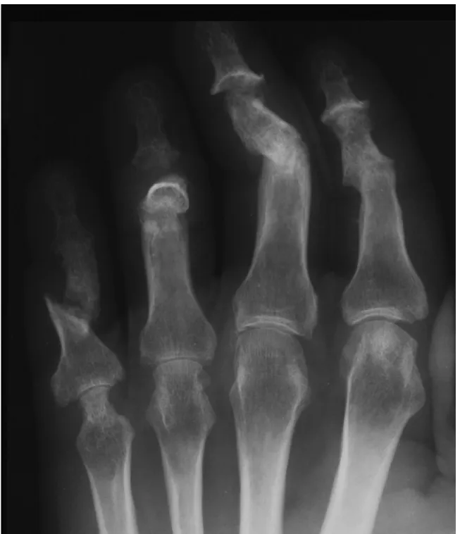
Extra-articular manifestations such as dactylitis (painful swelling, inflammation) of fingers or toes and enthesitis, especially at the insertion site of the achilles tendon, or plantarfascia are common in patients with PsA [2]. These extra-articular symptoms differ from patients with rheumatoid arthritis (RA) but occur among patients with other diseases within the seronegative spondylarthropathy group. Other manifestations in patients with PsA include uveitis [15, 16], distal extremity swelling with pitting oedema [17], possibly because of lymphatic obstruction [18], and discoloration of the skin over affected joints [19]. To date, neither psoriasis nor PsA has been described as a disease with systemic manifestations. However, there are reports of renal abnormalities, as well as increased mortality of patients with psoriasis and PsA indicating systemic effects, at least in a proportion of patients [20-24]. The radiological changes in PsA includes; erosion of terminal phalangeal tufts, whittling of bone ends, cupping of proximal ends of bones, severe
Introduction
________________________________________________________________________
destruction of isolated small joints, predilection of DIP and PIP joins with relative sparing of MP joints (illustrated in Figure 1), sacroiliitis or ankylosing spondylitis [1]. Recently, there have been findings giving rise to the hypothesis that the primary sites for inflammation in PsA, and other diseases within the spondylarthropathy group, are the enthesites (sites where ligament, tendon, joint capsule and fascia are inserted into bone) and that synovitis is secondary. Evidence for this hypothesis are the findings from radiological and MRI examinations of the spinal column, peripheral joints and peripheral enthesites which reveal signs of inflammation, new bone formation and erosions at these sites both early and late in the disease process (illustrated in Figure 2) [25, 26].
Treatment
The non-steroidal anti-inflammatory drugs (NSAIDs) are the basic treatment in PsA as in other arthritic diseases for pain, stiffness and, in mild cases, the inflammation. The most widely used disease modifying anti-rheumatic drugs (DMARDs) in PsA are sulfasalazine, methotrexate and cyclosporin, used either as a single treatment or in combinations [27]. New biological agents, such as anti-tumour necrosis factor-α (anti-TNFα), seem to improve signs and symptoms of PsA and psoriasis [28] and will be of interest in the future. It is difficult to evaluate treatment in PsA since there is neither uniform diagnostic criteria for the disease nor criteria for response of treatment.
Prognosis
During recent years it has become apparent that PsA is not the harmless, mild disease as previously thought [15]. The disease can be very destructive and almost 20% of patients with PsA develop severe, deforming arthritis [29]. Measurable inflammatory parameters and the number of swollen joints, especially when presenting at a rheumatology clinic, seem to be predictive for a poorer prognosis [30, 31]. In one recent study the occurrence of remission lasting for at least 12 months was studied [32]. Of the 391 patients included in the study, 69 (17.6%) entered remission with or without treatment and only 6 patients were determined to have “true remission”, i.e., they showed no evidence of damage and were on no medication. More than half (52 %) of the patients with remission, had a flare after an average of 2.5 years of remission.
Introduction
________________________________________________________________________
Figure 1. Radiographic examination of the hand showing; ankylosis of the
PIP-joints II, III and DIP-joint V, arthritis mutilans in DIP-joint IV, whittling and cupping in DIP-joint III, severe erosions in MCP-joints IV and V, and pseudostoarthritis dig V.
Introduction
________________________________________________________________________
Introduction
________________________________________________________________________
Pathogenesis
Several factors such as genetic, immunological, environmental and vascular factors have been proposed to be of importance for the aetiology, the expression and prognosis of PsA.
Genetic factors
During the past decades, statistical methods and molecular methodology have improved and the genetic research field is growing. Linkage and association studies are used in the analysis of the genetic aetiological contributions to a given disease. In linkage analysis, the inheritance pattern of a genetic marker is compared with the inheritance pattern of a familial disease, in order to estimate the frequency by which the marker and the disease segregate together. In an association study, the association of a genetic marker, or known locus, with a disease is analysed by comparing individuals having the disease with healthy controls.
Psoriasis and PsA probably have, in concordance with other autoimmune diseases, a polygenic background. The contribution of genetic factors for susceptibility for psoriasis has been analysed in linkage and association studies [5, 33, 34] whilst genetic studies in PsA have concentrated on association studies concerning the HLA region. However, a recent linkage study reported identification of a susceptibility gene on chromosome 16q [35]. Previous familial studies suggest first-degree relatives to be at risk of developing PsA [1]. The sibling recurrence risk (λs) is estimated to be approximately four for psoriasis [36], but Moll and Wright’s family studies suggest that the λs for PsA may be higher [1].
The Human Leucocyte Antigens (HLA)
Several association studies of the major histocompatibility complex (MHC) region on chromosome 6p have reported associations with various HLA-antigens in psoriasis and/or PsA. HLA HLA-antigens Cw6 and Cw*06 are more prevalent among patients with psoriasis, as well as those with PsA, compared with healthy controls [37-40].
Introduction
________________________________________________________________________
This is particularly so for patients with an early onset of skin involvement [14, 41, 42]. Some studies, however, suggest that the primary association of this gene is with psoriasis rather than PsA [38, 39, 43] Various other associations between PsA and other HLA-antigens, e.g., HLA B13, B17, B27, B38, B39, DR4, DR7 and DQ3, have also been reported [39, 40, 43-46]. HLA B27 has consistently been associated with axial disease [39, 47, 48], independent of psoriasis [38, 39], whilst HLA B39 has been associated with peripheral disease [39]. HLA DQ3 is suggested to be a marker of disease progression [49] and an increased frequency of DR4 has been associated with severity of the disease [50] development of polyarticular symmetrical arthritis [49] and with presence of joint erosions [48]. The presence of erosions has also been associated with expression of HLA DR3 [38].
The reported identification of a relatively large number of HLA-antigens may be due to the widely varying ethnic backgrounds and disease expression in the study populations, and therefore varied expression of HLA-antigens. Of particular interest is HLA B27, which has a higher prevalence in blood donors [51, 52].
Non-HLA genes on chromosome 6
Evidence for the possibility that a non-HLA gene, mapping close to the MHC region of chromosome 6p (PSORS1) [53], could be a susceptibility gene in PsA has been reported. MHC chain-related gene A (MICA) [54, 55] has been considered to be a candidate gene because it is in linkage disequilibrium with HLA-B alleles and is reported to have an increased frequency in psoriatic and PsA patients [55].
Other possible susceptibility genes within the MHC complex are the tumour necrosis factor (TNF) region, and the S-gene. The tumour necrosis factor alpha (TNFA) gene maps close to the HLA B locus and associations have been found between haplotypes of microsatellite markers mapping close to the TNFA gene and PsA, independent of psoriasis and HLA class I associations [56]. Other loci within the TNF region are described in psoriasis [57], RA [58] and AS [59]. The S-gene, which encodes for corneodesmosin, lies close to the HLA C and a disease association has been observed in psoriatic patients [60, 61].
Introduction
________________________________________________________________________
Other disease susceptibility genes
Studies of PsA and disease susceptibility genes are scarce. More information is available for psoriasis, with or without PsA, where disease susceptibility genes with a high significance have been described as being on chromosome 1q (PSORS4) [33], 3q (PSORS5) [34] 4q (PSORS3) [62], 17q (PSORS2) [63] and 19p (PSORS6) [64]. Weaker association have been identified for chromosome 2p, 8q [53], 16q and 20p [57].
Usually the investigators have not diagnosed the patients for arthritis and will not, therefore, be able to evaluate the possible differences between psoriatic patients with and without arthritic disease. There have, however, been reports of associations of chromosome 7q25 as well as chromosome 3q21 [65] with psoriatic patients with self-reported joint complaints and in a recent study linkage between PsA and chromosome 16q [35], especially with paternal inheritance, has been reported. This chromosome is of interest because of a reported association with ankylosing spondylitis [66].
Immunological factors
T-lymphocytes are thought to play a major role in the pathogenesis of both psoriatic skin disease and PsA [67], which is in accordance with the immunopathogenetic concept of the spondylarthropathies [68]. Activation of T-lymphocytes requires two signals, the first involving the binding of the antigen presenting cell (APC) to the T-lymphocyte receptor mediated by the MHC molecules situated on the APC. The antigen non-specific co-stimulatory pathway, in which the CD20 or CTLA4 molecules, present on the T-lymphocyte surface are important for binding to the B7-ligands, mediates the second signal [69]. Genetic studies have shown associations of CTLA4 polymorphism with susceptibility for RA [70], and with other autoimmune diseases [71-73], indicating an altered activation of T-lymphocytes in autoimmune diseases.
In psoriatic skin lesions, increased levels of CD4+ T-lymphocytes have been found in the dermis [67]. Cytokines released by these CD4+ T-lymphocytes may activate CD8+ T-lymphocytes. Previous studies have described the presence of T-lymphocytes in the synovium of patients with PsA [74]. However, recent studies have shown that the CD8+ T- lymphocyte popu-lation is significantly increased in synovial fluid from patients with PsA [67].
Introduction
________________________________________________________________________
Synovial fluid-derived CD8+ T -lymphocytes from patients with PsA were mature (CD45RO+), activated (HLA-DR+) and expressed low levels of CD25 (IL-2R), indicating a non-classical activation pattern [75]. The predominance of CD8+ T-lymphocytes in inflamed psoriatic joints may indicate a role for a MHC class I-mediated cellular immune response with cytotoxic T-lymphocytes as effector lymphocytes. The suggestion that specific CD8+ T-lymphocytes may play a more significant role than CD4+ T -lymphocytes in the pathogenesis in both skin and joint psoriatic disease is also proposed by Fearon et al. [76].
Other authors have studied the cytokines released by activated T-lymphocytes. Ritchlin et al. [77] have, in their study, shown that the cytokine profile in PsA is characterised by the presence of Th1 cytokines such as interleukin (IL)-1β, IL-2, interferon (IFN)-γ and TNFα but also very elevated levels of IL-10. Interferon-γ plays a role in the activation and induction of class I and class II MHC antigen expression on monocytes, synovial fibroblastoid cells and keratinocytes [78, 79] and are supposed to be important in PsA [77].
Another cell population important in the pathogenesis of PsA is activated macrophages [77]. Increased levels of metalloproteases (MMPs), which derive from macrophages and leucocytes, have been shown to be present in both synovial fluid and the synovium [80, 81].
Environmental factors
Environmental factors affecting the aetiology of PsA are difficult to separate from the immunological factors [68]. It is well known that psoriasis can be trigged by infection with group A β-haemolytic streptococci and a superantigen model of T –lymphocyte activation has been proposed [67]. Bacterial infections and exacerbations of PsA have been discussed due to findings of high titres of antibodies against bacterial cell wall peptidoglycan [82]. However, the Th1 cytokine pattern suggests that synovial inflammation is not driven by an aberrant immune response to a persistent bacterial antigen [77].
Introduction
________________________________________________________________________
Another environmental factor discussed, independent of the immunological factors, is physical trauma. The Köbner phenomena is known to be important for developing psoriatic lesions at the site of skin injury. Studies have also shown that physical trauma can trigger PsA [83]. Punzi and co-workers have shown that physical injury more often triggers PsA than other arthritic diseases such as RA and AS. The underlying pathogenesis is not clear but the release of neuropeptides, capable of stimulating the synovial membrane and the following hypervascularization of the synovium is discussed [76, 83].
Vascular factors
The PsA synovium is characterised by increased vascularity and fibrosis than in normal subjects [74] and when compared with patients with RA [84, 85]. The synovial lining layer, consisting of the synoviocytes, is not especially thickened which contrasts with the synovium of patients with RA [74, 84, 85]. The most dominant findings in the synovium are: the increased number of blood vessels and the vascular changes with prominent endothelial cell swelling, inflammatory cell infiltration (lymphocytes); marked thickening of the vessel wall; fibrosis which not is restricted to patients with longstanding disease; and dilated and tortuous papillary blood vessels [74, 84-86].
Aims
________________________________________________________________________
THE AIMS OF THIS THESIS
Psoriatic arthritis (PsA) is a heterogeneous disease with similarities with other inflammatory arthritic diseases; e.g., rheumatoid arthritis (RA) and ankylosing spondylitis (AS). In this study I have analysed clinical and genetic aspects in patients with PsA from northern Sweden with special emphasis on:
the prevalence of inflammatory manifestations, such as peripheral arthritis, axial disease, undifferentiated spondylarthropathy (uSpA) and enthesopathies in patients with psoriasis
the clinical manifestations of PsA, associations with human leucocyte antigens (HLA-antigens) and identifying markers for aggressive joint disease
identifying potential PsA susceptibility genes by association analyses the presence of systemic manifestations
Patients and Methods
________________________________________________________________________
PATIENTS AND METHODS
Patients and controls
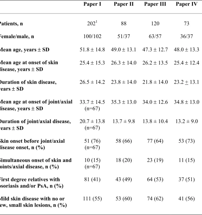
The patients in this study were all assessed using a standardised protocol, at one time point at the Department of Rheumatology, University Hospital, Umeå. Their previous disease history from disease onset was collected from the patients and from their hospital records at the Departments of Rheumatology and Dermatology. Laboratory and radiological examinations were performed concurrently with the actual clinical examination. The patients had psoriasis of the skin diagnosed either by a dermatologist or a general practitioner. The distribution of the patients included in the Papers is presented in Figure 3, and the characteristics of the patients are presented in Table 3.
The controls were healthy blood donors attending the Department of Blood and Transfusion Medicine (Paper II), and randomly selected individuals from the population register (Paper III). All the controls originated from the same geographic area of northern Sweden as the patients.
Paper IV
73 patients Paper III
120 patients Paper II 88 patients Paper I 202 patients
Figure 3. Number of patients included in paper I-IV and the
Patients and Methods
________________________________________________________________________
Table 3. Characteristics of the patients in Papers I-IV.
Paper I Paper II Paper III Paper IV
Patients, n 2021 88 120 73
Female/male, n 100/102 51/37 63/57 36/37
Mean age, years ± SD 51.8 ± 14.8 49.0 ± 13.1 47.3 ± 12.7 48.0 ± 13.3
Mean age at onset of skin
disease, years ± SD 25.4 ± 15.3 26.3 ± 14.0 26.2 ± 13.5 25.4 ± 12.4
Duration of skin disease,
years ± SD 26.5 ± 14.2 23.8 ± 14.0 21.8 ± 14.0 23.2 + 13.1
Mean age at onset of joint/axial
disease, years ± SD 33.7 ± 14.5 (n=67) 35.3 ± 13.0 34.0 ± 12.6 34.8 ± 13.0
Duration of joint/axial disease,
years ± SD 20.7 ± 13.8 (n=67)
13.7 ± 9.8 13.8 ± 10.4 13.2 ± 9.0
Skin onset before joint/axial disease onset, n (%)
51 (76) (n=67)
58 (66) 77 (64) 53 (73)
Simultaneous onset of skin and joints/axial disease, n (%)
10 (15) (n=67)
18 (20) 23 (19) 11 (15)
First degree relatives with
psoriasis and/or PsA, n (%) 81 (41) 43 (49) 64 (53) 37 (51)
Mild skin disease with no or
few, small skin lesions, n (%) 111 (55) 53 (60) 74 (62) 41 (56)
Patients and Methods
________________________________________________________________________
Clinical assessments
Clinical examinationsClinical examinations and medical history of peripheral arthritis, deformities and functional disability, including distal interphalangeal joints as well as spinal pain, buttock pain, chest wall pain, inflammatory spinal pain (presented in Table 4), spinal mobility (flexion, extension and rotation), chest expansion (abnormal < 2.5 cm), dactylitis and enthesitis were evaluated. Measurements of anterior lumbar flexion were made according to Schober [87].
Table 4. Symptoms of inflammatory spinal pain. History or present
symptoms of spinal pain in back, dorsal, or cervical region. At least four of the symptoms are required for diagnosis [3].
1. Onset before the age of 45 2. Insidious onset
3. Improved by exercise
4. Associated with morning stiffness 5. At least 3 months duration
The affected skin area was graded using a five point scale: 0 = no actual lesion, 1 = a few small lesions, 2 = many small or few large lesions, 3 = many large lesions and 4 = extensive involvement. The activity of the skin involvement (erythema, induration, and scaling) was graded as: 0 = none, 1 = mild, 2 = moderate and 3 = severe.
Radiographic examination
Radiographs of the peripheral joints were evaluated for erosions (≥ grade 2) according to Larsen´s grading system [88]. The spinal column and/or sacroiliac joints were examined radiologically in patients with actual or
Patients and Methods
________________________________________________________________________
previous history of back pain and/or decreased movement of the spine diagnosed in at least two directions as described in ESSG criteria [3] and/or according to Schober [87]. The diagnosis of axial disease was based on radiological findings in the sacroiliac joints according to the New York criteria (≥ 2) [89] and/or syndesmophytes, ligamentous ossification, vertebral squaring and shining corners of the spine [90]. The radiographs were evaluated by two separate radiologists, one for each of the four studies.
Diagnostic criteria
Peripheral arthritisPeripheral arthritis was diagnosed when a swollen and tender joint, with duration of more than 6 weeks, located outside the spine and/or sacroiliac joints was present. Patients with peripheral arthritis were further classified as having oligoarthritis when less than 5 joints were affected and polyarthritis when 5 or more joints were affected [4]. Symmetrical and asymmetrical polyarthritis were defined according to description by Helliwell and co-workers [4]. Aggressive joint manifestations were defined as radiological erosions and/or irreversible deformities (e.g., ankylosis, subluxation and/or loss of function or reduced mobility) of peripheral joints. Enthesitis was diagnosed when the patients had signs of tenderness, swelling, redness, warmth, loss of function and/or radiographic destruction at the insertion site of the Achilles tendon, plantarfascia, lateral or medial epicondyle [2]. Dactylitis was defined as painful swelling and inflammation of a finger or a toe [2].
Axial disease
The diagnosis of sacroiliitis and axial disease was based on radiological findings while uSpA was diagnosed when the patients, in addition to psoriasis, had inflammatory back pain [3] and decreased mobility of the spine in at least two directions without fulfilling the criteria for sacroiliitis or axial disease.
Patients and Methods
________________________________________________________________________
Disease pattern
The classification of the disease pattern, at onset and during disease course, was based on actual and/or previous findings of peripheral/axial engagement diagnosed by a rheumatologist and as reported in the hospital records. Patients reporting pain but not fulfilling any of the cited criteria were grouped as having other joint complaints (including osteoarthritis, fibromyalgia, chronic pain syndrome).
Renal abnormalities
Renal abnormality was defined as endogenous creatinine clearance decreased to less than the mean and 2 SD of the published normal distribution, corrected for age, according to reference values by Graneus and Aurell [91] and/or increased urinary albumin >25 mg/24 h.
Laboratory tests
In all of the papers the erythrocyte sedimentation rate (ESR, mm/h), C-reactive protein (CRP, mg/L) orosmucoid (g/L), haptoglobin (g/L) and rheumatoid factor (RF, according to Waaler-Rose) were determined using routine methods. In Paper IV analysis of serum levels of ß2-microglobulin
(mg/L), lipoprotein (a) (mg/L), cholesterol (mmol/L), HDL-cholesterol (mmol/L), LDL-cholesterol (mmol/L), triglycerides (mmol/L), immuno-globulin (Ig)G (g/L), IgA (g/L) and IgM (g/L) whilst aldosterone (pmol/L) concentration was measured in plasma using routine methods. Twenty-four hour urine collection was used for calculation of endogenous creatinine clearance (mL/min/1.73 m2) and measurement of albumin (mg/24h).
HLA typing (Paper II)
HLA typing of both patients and controls (i.e., healthy blood donors from the same area as the patients) was performed for HLA A, HLA B, HLA Cw*, DR B1* and DQ B1* antigens. The serological technique was used for the class I antigens A (23 subtypes) and B (41 subtypes) [92].
Patients and Methods
________________________________________________________________________
The Class I allele Cw* (14 subtypes) and the class II alleles DR B1* (15 subtypes) and DQ B1* (7 subtypes) were identified with a plymerase chain reaction-sequence specific primers (PCR-SSP) technique using low resolution primers from Dynal (Oslo, Norway). DNA from the samples was extracted with DTAB/CTAB (decyltrimethylammoniumbromide/cyltrimetyl- ammoniumbromid) [93]. Amplification of the DNA was performed in a thermal cycler (Model 9600; Perkin-Elmer, Applied Biosystems Inc., Foster City, CA) according to the protocol of the manufacturer after which the reaction produckt were visualized following electrophoresis using a 2% agarose gel.
Genotyping of single nucleotide polymorphisms (SNPs) and microsatellites (Paper III)
Genomic DNA was extracted from the peripheral blood lymphocytes, anti coagulated with EDTA, using standard phenol-chloroform procedures [94] and a modified salt-out method [95].
A total of 34 markers were analyzed, 32 microsatellites and 2 single nucleotide polymorphisms (SNP). All single nucleotide polymorphisms were genotyped by TaqmanR fluorogenic 5’ nuclease assays (Applied Biosystems Inc.). TaqmanR primers and probes were designed using Primer Express software (Applied Biosystems Inc.). TaqmanR assays were performed according to the manufacturers guidelines. The microsatellite markers were amplified by PCR with optimized annealing temperature. PCR products were separated by electrophoresis on an ABI PRISM 3100 Genetic Analyzer (Applied Biosystems Inc.) and on an ABI 377 (Applied Biosystems Inc.) according to the manufacturers protocols. Genotyping was performed using GENESCAN ANALYSIS 3.7 / 2.1 and GENOTYPER 3.7 / 2.0.
Statistical analysis
Student’s t-test was usually used to test for differences for continuous data. Anyhow, for some variables there were small samples and then was the Mann-Whitney U test used. Chi-square with Yates’ correction was used for testing categorical data between groups and Odds Ratio (OR), as an estimate of the relative risk, was calculated with 95% confidence interval (CI).
Patients and Methods
________________________________________________________________________
Spearman´s correlation was used to test for correlation between variables in small samples. Multiple logistic regression model was used to identify the predictive values of different variables.
To assess the utility of the PAQ in detecting arthritis in Paper I, sensitivity, specificity and predictive values were calculated. The receiver operating characteristic (ROC) curve was used to display the relationship between sensitivity and specificity.
In Paper III the Chi-square test was used for comparing allele frequencies between cases and controls. In each of the regions the achieved nominal P-values were Bonferroni corrected by the number of markers tested in that region. For the AT repeat at CTLA4 the alleles were first dichotomised into repeat lengths ≤82bp and >82bp in accordance with the hypothesis of the existence of a functional cutoff value [96].
Study Design
________________________________________________________________________
STUDY DESIGN
Paper I
During 1995 and 1996, the Department of Dermatology, University Hospital, Umeå, collected and administered a register including information about patients (n=1737) diagnosed as having psoriasis and living in the County of Västerbotten [97]. The register which was community-, as well as hospital-based, included diagnostic information about psoriatic patients of all ages from the hospital records at the Department of Dermatology, University Hospital, Umeå, the Departments of Internal Medicine of the local hospitals in Lycksele and Skellefteå, 10 out of 36 primary health care centres in the county of Västerbotten, and from the register of the members of the Swedish Psoriasis Association (Svenska Psoriasisförbundet) living in Västerbotten (n=602). All patients had psoriasis of the skin diagnosed by a dermatologist or a general practitioner.
Of the 291 patients, older than 16 years, registered as living in the city of Umeå, two were deceased and 13 patients had moved from the area at the time for our study. The remaining 276 patients were asked to answer a psoriatic and arthritic questionnaire (PAQ) and to participate in a prevalence study of joint and/or axial inflammatory involvement, and 202 patients (73.2%) accepted to fulfil the study including clinical, laboratory and radiological assessments. The inflammatory conditions examined for included peripheral arthritis, axial disease, uSpA and enthesopathies.
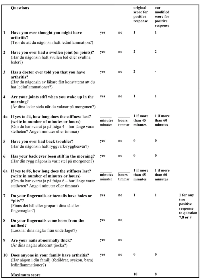
An evaluation of the PAQ was undertaken to identify patients with arthritis in this psoriatic population. The questionnaire was constructed by Peloso et al. at the University of Saskatchewan, Saskatoon, Canada [98] and is reproduced as Table 5. As we wanted to evaluate the PAQ in a population not knowing that they had an arthritic disease we excluded patients with known arthritic disease as well as the third question, “Has a doctor ever told you that you have arthritis?”, from our analysis.
Table 5. The Psoriatic and Arthritis Questionnaire (PAQ). Questions original score for positive response our modified score for positive response
1 Have you ever thought you might have arthritis?
(Tror du att du någonsin haft ledinflammation?)
yes no 1 1
2 Have you ever had a swollen joint (or joints)?
(Har du någonsin haft svullen led eller svullna leder?)
yes no 2 2
3 Has a doctor ever told you that you have arthritis?
(Har du någonsin av läkare fått konstaterat att du har ledinflammationer?)
yes no 2 -
4 Are your joints stiff when you wake up in the morning?
(Är dina leder stela när du vaknar på morgonen?)
yes no 1 1
4a If yes to #4, how long does the stiffness last? (write in number of minutes or hours)
(Om du har svarat ja på fråga 4 – hur länge varar stelheten? Ange i minuter eller timmar)
______ minutes minuter ______ hours timmar 1 if more than 45 minutes 1 if more than 60 minutes
5 Have you ever had back troubles?
(Har du någonsin haft ryggvärk/ryggbesvär?)
yes no 0 0
6 Has your back ever been stiff in the morning?
(Har din rygg någonsin varit stel på morgonen?)
yes no 0 0
6a If yes to #6, how long does the stiffness last? (write in number of minutes or hours)
(Om du har svarat ja på fråga 6 – hur länge varar stelheten? Ange i minuter eller timmar)
______ minutes minuter ______ hours timmar 1 if more than 45 minutes 1 if more than 60 minutes
7 Do your fingernails or toenails have holes or “pits”?
(Finns det hål eller gropar i dina tå eller fingernaglar?)
yes no 1 1
8 Do your fingernails come loose from the nailbed?
(Lossnar dina naglar från underlaget?)
yes no
9 Are your nails abnormally thick?
(Är dina naglar abnormt tjocka?)
yes no 1 for any two positive response to question 7,8 or 9
10 Does anyone in your family have arthritis?
(Har någon i din familj (föräldrar, syskon, barn) ledinflammationer?)
yes no 0 0
Maximum score 10 8
The third question was not included in the statistical analyses and the duration of morning stiffness was expanded from the original described 45 minutes to at least 60 minutes.
Study Design
________________________________________________________________________
The duration of morning stiffness was expanded to at least 60 minutes in peripheral joints in accordance with the ARA criteria for rheumatoid arthritis [99]; the increased time-scale was also applied to the morning stiffness of the spine. Consequently, the original total PAQ score of 10 was reduced to 8 in our analysis (Table 5).
In order to evaluate the PAQ in detecting arthritis the sum of PAQ scores were compared with clinical diagnoses. Furthermore, the utility of the individual PAQ questions was assessed. The total PAQ, with all questions, was also analysed in the whole study population (n=202).
Paper II
In this cross sectional study, 88 patients (51 females and 37 males) were consecutively recruited at the Department of Rheumatology, University Hospital, Umeå. The disease manifestations at onset and during disease course were evaluated and the patients were HLA typed. The patients were also classified according to disease severity (deformities and/or radiological erosions) and statistical analyses were performed in order to find clinical and/or HLA antigens as predictive factors for aggressive disease as defined in diagnostic criteria.
Paper III
Based on the results of previous genetic studies on psoriasis, RA, AS and other autoimmune diseases [33, 53, 58, 59, 62, 70-73], the association between several interesting loci and PsA was analysed in 120 psoriatic patients with a defined joint disease, recruited from the Department of Rheumatology. The study was performed with particular focus on the TNF locus, 1q21 (PSORS4), 3q21 (PSORS5), 8q24 and the CTLA4 gene with microsatellites and SNPs.
Study Design
________________________________________________________________________
Paper IV
In this study the systemic influence of inflammation in patients with psoriatic arthritis with the kidneys as a target organ was estimated. Seventy-three patients (36 females/37 males) were included. Twenty-four of the patients had received DMARD treatment for at least 6 months (methotrexate, sulphasalazine, cyclosporin, and intramuscular gold in combination with methotrexate) at the start of the study. Twenty-nine patients were receiving daily NSAID treatment during this study period and nine had oral corticosteroids daily. Eight of the 73 patients had hypertension, five diabetes mellitus, one known hyperlipidaemia and one had been treated with lithium for many years because of a unipolar affective disorder. Laboratory measurements were made to estimate the presence of renal abnormalities (see diagnostic criteria) and to evaluate laboratory-measured inflammatory activity defined as ESR >25 mm/h and/or CRP > 15 mg/L. Measurement of serum, or urine, immunoglobulins (IgA, IgG, IgM), serum lipids, serum and urine β2-microglobulin, and plasma aldosterone were also made.
Results and Comments
________________________________________________________________________
RESULTS AND COMMENTS
A prevalence study and an evaluation of a psoriatic and
arthritic questionnaire in 202 psoriatic patients (Paper I)
Prevalence of inflammatory joint manifestations Inflammatory manifestations
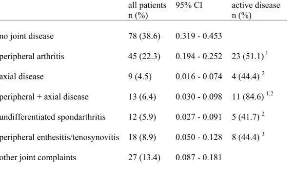
A high prevalence of inflammatory manifestations such as peripheral arthritis, axial disease, uSpA and/or enthesopathies, was found among the psoriatic patient in this study group. The prevalence of these inflammatory manifestations is presented in Table 6, whilst classification of the patient’s disease, according to the Moll and Wright scheme [1] is presented in Table 7. Table 6. Inflammatory joint/axial diagnosis and other joint and pain
involvement in the 202 psoriatic patients at the time of the study.
all patients n (%) 95% CI active disease n (%) no joint disease 78 (38.6) 0.319 - 0.453 peripheral arthritis 45 (22.3) 0.194 - 0.252 23 (51.1) 1 axial disease 9 (4.5) 0.016 - 0.074 4 (44.4) 2 peripheral + axial disease 13 (6.4) 0.030 - 0.098 11 (84.6) 1,2 undifferentiated spondarthritis 12 (5.9) 0.027 - 0.091 5 (41.7) 2 peripheral enthesitis/tenosynovitis 18 (8.9) 0.050 - 0.128 8 (44.4) 3
other joint complaints 27 (13.4) 0.087 - 0.181
1 defined as active, peripheral arthritis with or without increased ESR and/or CRP
2 defined as actual inflammatory back pain and decreased mobility of the spine with or without increased
ESR and/or CRP Multiple logistic regression
Results and Comments
______________________________________________________________________
Table 7. The Moll & Wright classification of PsA for the 202 psoriatic
patients at the time of the study.
classification all patients
n (%) female n=100 male n=102 active diseasen (%)
DIP-joint disease, predominantly 0
axial disease 15 (7.4) 0 15 10 (66.7)
mono/oligoarthritis 30 (14.6) 15 15 15 (50.0)
polyarthritis,
symmetrical/asymmetrical
21 (10.4) 12 9 12 (57.1)
mutilans arthritis, predominantly 1 0 1 1 (100)
Ninety-seven patients (48%) were identified as having inflammatory manifestations with peripheral arthritis and/or axial disease being diagnosed in 67 (33%) patients, of whom 9 had axial disease exclusively. More than half of the 58 patients with peripheral arthritis had ongoing arthritis at the time for the study and 29/58 (50%) had destructive changes of the joints defined as radiographic evidence of erosions and/or deformities of joint(s). No patients had predominantly DIP involvement but 28% had involvement in these joints during disease history which is less than previously described [15]. Only about 40% of the psoriatic patients could be classified as healthy based on the absence of joint/back pain overall.
Patients with inflammatory manifestations not previously diagnosed
Thirty patients (45%) with peripheral arthritis and/or axial disease who had not previously been diagnosed were identified. Of these 22 had peripheral arthritis, 6 had axial disease and 2 had both peripheral and axial disease. Of the patients with peripheral arthritis (n=24), there were 17 (71%) with ongoing arthritis and 10 had radiological changes and/or deformities of the joints.
Results and Comments
________________________________________________________________________
The patients with axial disease had, by definition, radiological changes. Almost half of the patients not previously been diagnosed, had destructive changes in the joints indicating that they may not have been receiving sufficient treatment for their joint disease. These observations suggest that patients are not diagnosed at an appropriate time.
Inflammatory back pain
Back pain is common among patients in general and is difficult to evaluate. In this study, 20 (62%) out of 32 patients with inflammatory back pain and decreased mobility of the spine had radiological changes in the sacroiliac joint or the spine. Sacroiliitis, with no clinical symptoms has been described in patients with psoriatic arthritis [15] To evaluate the presence of sacroiliitis in patients without any clinical signs, 20/47 patients without decreased mobility or any back pain were randomly chosen for radiographic examination of the sacroiliac joints. None of these patients had any radiological changes indicating that symptoms of inflammatory back pain, in combination with decreased mobility of the spine, is an important clinical diagnostic sign for axial disease as suggested by Viitanen et al [100].
Measurable inflammatory activity
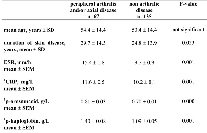
Measurable inflammatory activity is not always prominent in patients with psoriatic arthritis. The patients with peripheral arthritis and/or axial disease (n=67) in this study, had, as a group, significantly higher ESR, CRP, p-orosmucoid and p-haptoglobin (Table 8) suggesting that joint manifestations have higher systemic effect compared with patients with skin disease only. However, the lower ranges of the inflammatory variables were within the normal values in some of the patients.
Results and Comments
________________________________________________________________________
Table 8. Demographic and laboratory data for the 202 psoriatic patients at
the time of the study.
peripheral arthritis
and/or axial disease n=67
non arthritic disease
n=135
P-value
mean age, years ± SD 54.4 ± 14.4 50.4 ± 14.4 not significant
duration of skin disease,
years, mean ± SD 29.7 ± 14.3 24.8 ± 13.9 0.023 ESR, mm/h mean ± SEM 15.4 ± 1.8 9.7 ± 0.9 0.001 1CRP, mg/L mean ± SEM 11.6 ± 0.5 10.2 ± 0.1 0.001 1p-orosmucoid, g/L mean ± SEM 0.81 ± 0.03 0.70 ± 0.01 0.000 1p-haptoglobin, g/L mean ± SEM 1.40 ± 0.08 1.09 ± 0.05 0.001
1reference values: CRP <10 mg/L, p-orosmucoid; (0.54 – 1.17) g/L, p-haptoglobin; 18-50
yrs ; (0.35 – 1.85) g/L, >50 yrs ; (0.47 – 2.05) g/L
Drop out analysis
Of the 28 patients who completed the questionnaire but were unwilling to participate in the full study, 7 (25%) answered that they had had a swollen joint, 4 (14 %) that a clinician had diagnosed arthritis, and another 4 patients (14 %) had back pain in combination with morning axial stiffness for at least 60 minutes. Of the remaining 46 patients not answering the questionnaire, one patient was regularly seen at the Department of Rheumatology, University Hospital, Umeå, for his psoriatic arthritis and another patient have been diagnosed according to given criteria with peripheral arthritis and ankylosing of the sacroiliac joints. The mean age of the patients in the drop-out group did not differ from the study group although they were more representative of the younger and the older patients.
Results and Comments
________________________________________________________________________
If it is assumed that the lower prevalence of arthritic disease was valid for the whole drop-out group, these patients seemed to be healthier than in the study group, and this may introduce an unintentional selection bias into the study. A hypothetical prevalence analysis within the whole group of psoriatic patients (n=276), including the 8 out of 28 patients answering the PAQ and 2 out of 46 patients not answering the PAQ but describing inflammatory symptoms, showed a prevalence of peripheral arthritis and/or axial disease of 28%. When uSpA and enthesitis were included in the analysis the prevalence increased to 39%. This is still a high prevalence of inflammatory manifestations in the psoriatic population.
Analysis of the Psoriatic & Arthritic Questionnaire (PAQ) The questionnaire
The aim was to evaluate whether a questionnaire, and the separate questions contained in it, could discriminate for arthritis in patients with psoriasis who were unaware of having an arthritic disease. A total score of 4 or higher as the optimal cut-off score for predicting peripheral arthritis and/or axial disease in this psoriatic group (area under the curve (AUC)=0.640, OR=2.343, CI=1.224-4.482, P=0.010) when estimated by the ROC curve (Figure 4). The same cut-off score was attained for any inflammatory manifestation(s) such as peripheral arthritis, axial disease, uSpA and/or enthesitis with a sensitivity of 55% and a specificity of 65.7% (AUC=0.647, OR=2.471, CI=1.100-5.548, P=0.028; data not shown). When analysing the complete PAQ in the whole study population (n=202) a higher sensitivity and lower specificity compared with the modified analysis, was attained. The ROC curve indicated a total score ≥ 6 for predicting peripheral arthritis and/or axial disease. The sensitivity was then 68.7%, specificity 77.8% and the positive predictive value 60.5% (Table 9).
Results and Comments
________________________________________________________________________
Figure 4. The ROC curve indicating optimal cut-off value of ≥ 4.
Table 9. The PAQ-score excluding patients with known arthritic disease and
the third question (pop A) and the total PAQ-score including all patients and all questions (pop B).
PAQ-score sensitivity % pop A pop B specificity % pop A pop B positive predictive value % pop A pop B negative predictive value % pop A pop B score ≥ 3 73.3 83.6 44.4 52.6 22.7 46.7 88.2 86.6 score ≥ 4 60.01 82.1 62.2 57.0 26.1 48.7 87.5 86.5 score ≥ 5 46.7 77.6 72.6 65.9 27.5 53.1 86 85.6 score ≥ 6 30.0 68.72 83.0 77.8 28.1 60.5 84.2 83.3 score ≥ 7 16.7 53.7 91.1 86.7 29.4 66.7 83.1 79.1 score ≥ 8 10.0 41.8 97.0 94.1 42.9 77.8 82.9 94.1
1TheROC curve indicated a total score ≥ 4 for predicting peripheral arthritis and/or axial disease for pop A 2The ROC curve indicated a total score ≥ 6 for predicting peripheral arthritis and/or axial disease for pop B
Results and Comments
________________________________________________________________________
Analyses of separate questions
A positive response to the first PAQ question “Have you ever thought you might have arthritis?” (OR=2.36, CI=1.026-5.407, P=0.043) and to the question about morning stiffness in peripheral joints lasting for at least 60 minutes (OR=3.43, CI=1.329-8.844, P=0.011) significantly predicted peripheral arthritis and/or axial disease. When analysing the two questions together the sensitivity was 30%, the specificity 91.1%, the positive predictive value 42.9% and the negative predictive value was 85.5% (OR=4.39, CI=1.648-11.709, P=0.003).
Modified scoring of the questions
As the results obtained in this study were different from those achieved with the original questionnaire used in Canadian patients, when analysing separate questions, the scoring of the questions was weighted and those questions most strongly predicting arthritis assigned a double score compared with the other questions. Thus, the maximum score in this analysis was 9. Using this weighted modification, the ROC curve indicated a score of 5 or higher as the optimal cut-off score for predicting peripheral arthritis and/or axial disease (sensitivity=50%, specificity=73.3%, positive predictive value=29.4%, negative predicted value=86.8%). The same optimal cut-off score was calculated when uSpA and/or enthesitis was included in the analysis (sensitivity=45%, specificity=77.1%, positive predicted value=52.9%, negative predicted value=71.1%; data not shown).
Summary of the univariate analyses
From analysis of the PAQ, with and without the third question, in all patients and those not knowing they had an arthritic disease, was concluded that for detecting arthritis in a psoriatic population the whole PAQ is relatively appropriate. However, for identification of new cases within a psoriatic population the sensitivity and positive predictive value were fairly low, although the specificity was slightly higher. The weighting of the scoring for each question, based on the different result in analysing the separate questions among patients from northern Sweden compared with the Canadian patients, did not improve the predictive value. The first question in the PAQ, “Have you ever thought you might have arthritis?” and the information about morning stiffness for at least 60 minutes were found to be more valuable than the total PAQ score in detecting arthritis, and this observation was supported by the multiple logistic regression analysis.
Results and Comments
________________________________________________________________________
A multiple regression analysis including clinical data and the PAQ
The duration of skin involvement and a positive response to the question ”Have you ever thought you might have arthritis?” in combination with morning stiffness for at least 60 minutes, predicted peripheral arthritis and/or axial disease in a multiple logistic regression model (Table 10).
Table 10. The logistic regression analysis for identifying patients with
peripheral arthritis and/or axial disease, adjusted for sex.
variables univariate analysis multivariate analysis OR 95% CI P-value OR 95% CI P-value
total score ≥ 4 2.717 1.189 – 6.210 0.018 1.744 0.656-4.634 0.265
positive response for the first PAQ question and morning stiffness for at least 60 minutes
4.540 1.681 – 12.265 0.003 3.949 1.185-13.158 0.025
duration of skin
involvement, years 1.033 1.005 – 1.062 0.022 1.038 1.007-1.071 0.018 ESR, mm/h 1.035 1.007 – 1.064 0.015 1.025 0.994-1.057 0.115
In the analysis of the modified PAQ scores with the same dependent
variable and the same covariates, only the duration of skin involvement remained significant (data not shown). An interesting finding from these analyses was that the duration of skin involvement was the variable that most strongly predicted peripheral arthritis and/or axial disease. This finding indicates that time is important for the development of joint disease in psoriatic patients.
In order to evaluate a possible interaction between the PAQ-score ≥ 4 and the first PAQ-question in combination with morning stiffness, these variables were excluded one by one in the multiple regression analyses (Table 11).
Results and Comments
________________________________________________________________________
When excluding a PAQ-score ≥ 4, the significance for duration of skin disease and the first PAQ question in combination with morning stiffness became even stronger. When excluding the first PAQ-question in combi-nation with morning stiffness, the skin duration remained significant and the PAQ-score ≥ 4 became significant. This analysis did not change the conclusion of the study with the combination of the two questions remaining significant and duration of skin involvement as a strong predictive factor.
Table 11. OR and P-values within parentheses when excluding variables for
valuating a possible influence between a total score
≥
4 and positive response for the first PAQ question in combination with morning stiffnessfor at least 60 minutes.
OR (P-value) (P-value) OR (P-value) OR
total score ≥ 4 (0.265) 1.744 - (0.023) 2.716
positive response for the first PAQ question and morning stiffness for at least 60 minutes
3.949
(0.025) (0.002) 5.465 -
duration of skin involvement, years 1.038 (0.018) 1.039 (0.015) 1.031 (0.044) ESR, mm/h 1.025 (0.115) (0.111) 1.025 (0.052) 1.030
HLA antigens and inflammatory joint manifestations (Paper II)
Joint disease manifestationsIn this paper, 88 patients were included and 83 of the patients had peripheral arthritis of whom 16 (8 females/8 males) had simultaneously axial disease. Five males, but no female, had axial disease without peripheral
Results and Comments
________________________________________________________________________
manifestations. Fifty-four patients (65%) had ongoing peripheral arthritis at examination with a mean number (±SEM) of 3.2 (± 0.5) arthritis.
The number of ongoing arthritis correlated with ESR (rs=0.278, P=0.012) and
CRP (rs=0.440, P<0.0001). The disease manifestations at onset, and
accumulated during the disease course are presented in Table 12.
Table 12. Disease manifestations at onset and accumulated manifestations
during disease course in 88 patients with psoriatic arthritis.
onset
n (%) during disease course n (%)
monoarthritis1 16 (18) 1 (1)
oligoarthritis1 25 (28) 22 (25)
polyarthritis1 27 (31) 44 (50)
DIP joint involvement 3 (3) 32 (36)
inflammatory back pain2 11 (13) 5 (6)
axial disease3 nd 5 (6)
axial disease3 + polyarthritis nd 16 (18)
dactylitis4 4 (5) 19 (22)
enthesitis4 2 (2) 31 (35)
1 includes 3, 9 and 12 patients, respectively, with simultaneously inflammatory
back pain
2 inflammatory back pain as defined in ref. 3
3axial diseaseincludes radiological sacroiliitis (≥ grade 2)[89]
and/or spinal involvement as defined in ref. 90
4dactylitis and enthesitis defined as in ref. 2
nd = radiological examination not done
Small joints were the first sites of joint manifestations in 27 patients (31%), mostly in fingers or toes, whilst in another 18 patients the knee was the first joint affected. When the peripheral joint disease started as polyarthritis, small joints were engaged in 86% of the patients, while onset as mono/oligo-
Results and Comments
________________________________________________________________________
arthritis more often affected large joints, (70% of the patients, χ2 =22.17,
p<0.0001, data not shown). In almost 2/3 of the patients with onset in large joints, the disease progressed to polyarticular disease.
HLA antigens and inflammatory joint manifestations Joint disease expression
The frequency of the HLA antigens in patients with PsA is presented in Table 13. HLA B17, B37 and B62 remained significantly increased in the patients after correction for the number of comparisons. Two of the 5 patients with axial disease without peripheral arthritis were HLA B27 positive. The associations between HLA antigens and disease onset and disease course are presented in Table 14. The associations remaining significant, after correction for the number of tests performed, were onset of skin disease and PsA with HLA B37 and onset in large joints with HLA B62.
The association with HLA antigens and disease expression varies and there are inconsistencies between studies [39, 45, 46, 101]. Association with HLA B17 and Cw*0602 in patients with PsA is well documented [39] as it is for HLA B27. The latter is, as in this study, almost exclusively increased in patients with axial disease [39, 45-48, 101] whilst HLA Cw6 or Cw*0602, have repeatedly been shown to determine early onset of psoriasis [14, 36, 41]. HLA B37 was present at a low frequency in this patient group (9.9%) but, because it is rare in blood donors (2.7%), the increase was significant. However, all of those patients positive for HLA B37 were also Cw*0602 positive implying a linkage disequilibrium between them which has been shown by others [102]. An association of B62, a split of B15, has not, as far as is known, been previously reported in PsA.
The presence of nail lesions were in this study associated with B17 (Pc=0.004) but not with Cw*0602. Patients with HLA DR B1*03 did have polyarthritis more often than oligoarthritis at onset of joint disease (Pc=0.0000, OR=133.33, CI=14.52-5633.00; data not shown). It was not possible to confirm previous findings of an association with HLA DR4 and symmetrical polyarticular disease [49], erosive disease [48] or more severe disease [50].
Results and Comments
________________________________________________________________________
Table 13. HLA-antigens in patients with psoriatic arthritis and
controls from northern Sweden.
Antigen Patients
% Controls % P-value Pc OR 95%CI
B17 12.2 4.0 0.001 0.010 3.36 1.38-6.64
B 27 25.7 15.2 0.017 0.171 1.93 1.12-3.34
B 37 9.5 2.7 0.001 0.011 3.80 1.69-9.44
B 62 18.9 7.6 0.001 0.006 2.85 1.53-5.32
DQ B1*06 44.0 71.2 0.0004 0.004 0.32 0.17-0.62
Table 14. HLA-antigens and disease expression with significant corrected
P-values in patients with PsA (n and per cent). The frequencies are compared to controls and presented with P-value, corrected P-value (Pc) and Odds Ratio (OR) with 95% confidence intervals (CI).
Disease expression HLA
antigen
n (%) P-value Pc OR 95% CI
skin onset <40 years B37 7 (11.1) 0.0002 0.0044 4.55 1.91-10.83 PsA onset <40 years B37 5 (11.1) 0.0012 0.0264 4.55 1.67-12.37 onset in large joints B62 7 (24.1) 0.0012 0.0264 3.89 1.61-9.38 nail lesions B17 5 (19.2) 0.0002 0.0044 5.77 2.08-16.03
Results and Comments
________________________________________________________________________
Aggressive joint manifestations
Aggressive joint manifestations, defined as radiologically detectable erosions and/or irreversible deformities, were present in 50 (60%) of the patients with peripheral disease (n=83). These patients had significantly longer skin disease duration, (mean ± SD of 26.7±13.7 v 19.8±14.1 years, P=0.029), longer joint disease duration (15.6±10.0 v 10.0±9.0 years, P=0.013), more often had involvement of DIP joints (χ2 =20.07, P=0.0001), usually a polyarticular
disease course (χ2 =15.5, P=0.0001) and more frequently symmetrical
engagement (χ2 =5 04, P=0.025) compared with patients with non-aggressive
manifestations. The HLA antigens B37 and B62 remained significantly associated with joint destruction and/or deformity after correction for the number of tests performed (Table 15).
HLA DR B1*03 was significantly increased (χ2 = 4.87, P<0.05, OR=3.34,
CI=1.10-10.13) with respect to deformity only while HLA A3 seems to be a protective antigen for joint erosion(s) and/or deformity. There were no differences between patients with or without aggressive manifestations, in current or previous DMARD therapy, family history of disease, age at onset, manifestations at disease onset or number of active arthritis at examination.
Table 15. Associations between HLA-antigens and aggressive disease defined
as deformities and/or erosions in PsA patients with peripheral disease (n=83) compared to controls.
HLA -antigen n (%) P-value Pc OR 95% CI
A 3 7 (6.3) 0.017 0.068 0.38 0.17-0.86
B 37 5 (11.6) 0.001 0.003 4.79 1.76-13.06
B 62 10 (23.3) 0.0002 0.001 3.71 1.77-7.80