Maternal, fetal and perinatal factors
associated with necrotizing enterocolitis in
Sweden. A national case-control study
Margareta Ahle1*, Peder Drott2, Anders Elfvin3, Roland E. Andersson2,4
1 Department of Radiology and Department of Medical and Health Sciences, Linko¨ping University, Linko¨ping,
Sweden, 2 Division of Surgery, Department of Clinical and Experimental Medicine, Linko¨ping University, Linko¨ping, Sweden, 3 Department of Pediatrics, Institution of Clinical Sciences, Sahlgrenska Academy, University of Gothenburg, Gothenburg, Sweden, 4 Department of Surgery, Ryhov County Hospital, Jo¨nko¨ping, Sweden
*Margareta.Ahle@regionostergotland.se
Abstract
Objective
To analyze associations of maternal, fetal, gestational, and perinatal factors with necrotizing enterocolitis in a matched case-control study based on routinely collected, nationwide regis-ter data.
Study design
All infants born in 1987 through 2009 with a diagnosis of necrotizing enterocolitis in any of the Swedish national health care registers were identified. For each case up to 6 controls, matched for birth year and gestational age, were selected. The resulting study population consisted of 720 cases and 3,567 controls. Information on socioeconomic data about the mother, maternal morbidity, pregnancy related diagnoses, perinatal diagnoses of the infant, and procedures in the perinatal period, was obtained for all cases and controls and analyzed with univariable and multivariable logistic regressions for the whole study population as well as for subgroups according to gestational age.
Results
In the study population as a whole, we found independent positive associations with necro-tizing enterocolitis for isoimmunization, fetal distress, cesarean section, neonatal bacterial infection including sepsis, erythrocyte transfusion, persistent ductus arteriosus, cardiac mal-formation, gastrointestinal malmal-formation, and chromosomal abnormality. Negative associa-tions were found for maternal weight, preeclampsia, maternal urinary infection, premature rupture of the membranes, and birthweight. Different patterns of associations were seen in the subgroups of different gestational age.
Conclusion
With some interesting exceptions, especially in negative associations, the results of this large, population based study, are in keeping with earlier studies. Although restrained by the a1111111111 a1111111111 a1111111111 a1111111111 a1111111111 OPEN ACCESS
Citation: Ahle M, Drott P, Elfvin A, Andersson RE (2018) Maternal, fetal and perinatal factors associated with necrotizing enterocolitis in Sweden. A national case-control study. PLoS ONE 13(3): e0194352.https://doi.org/10.1371/journal. pone.0194352
Editor: Olivier Baud, Hopital Robert Debre, FRANCE Received: August 5, 2017
Accepted: March 1, 2018 Published: March 23, 2018
Copyright:© 2018 Ahle et al. This is an open access article distributed under the terms of the
Creative Commons Attribution License, which permits unrestricted use, distribution, and reproduction in any medium, provided the original author and source are credited.
Data Availability Statement: Data belong to the Swedish National Board of Health and Welfare in Sweden and the Statistics Sweden, and sharing with a third part is not allowed. All requests to obtain the original data must therefore be addressed to these authorities. The Swedish National Board of Health and Welfare may be contacted atregisterservice@socialstyrelsen.se
and Statistics Sweden atmikrodata.individ@scb. se. The authors are willing to assist in the process. Funding: This study was supported by Region O¨ stergo¨tland, Sweden,https://www.fou.nu/is/lio
limitations of register data, the findings mirror conceivable pathophysiological processes and underline that NEC is a multifactorial disease.
Introduction
Necrotizing enterocolitis (NEC) remains a challenge in neonatal care. The incidence rate of NEC in Sweden during 1987 through 2009 was 0.34 in 1,000 live births, with an increasing trend, partly explained by increased survival of the most premature but also seen in higher ges-tational ages (GA)[1].
The etiology is multifactorial and may differ according to the degree of maturity of the patient[2]. Prematurity and low birthweight are the most consistent predisposing factors, whereas other risk factors vary with GA,[3,4] as well as between study populations, and the results of previous studies are sometimes contradictory.
Pathophysiologically, the complex interactions of the innate immunity system and coloniz-ing bacteria, pro-inflammatory factors and modulatory systems, are deranged[5,6]. The imma-turity of intestinal motility and digestion, structural and biochemical barrier functions as well as circulatory regulation contribute to the vulnerability[5,6]. The result is hemorrhagic and necrotizing inflammation, bacterial overgrowth and translocation of bacteria to the intestinal wall and systemic circulation[7].
The presence of bacteria is thus an important prerequisite in the pathogenesis of NEC, but their role as contagions is disputable. Reports of seasonal variation in incident rates, episodic outbreaks and clustering of NEC,[1,8] however, suggest a role for transmissible infectious agents or other environmental factors.
Objective of the study
The aim of the present case control study is to analyze differences in maternal, fetal, gesta-tional, and perinatal factors among NEC cases and matched controls from routinely collected register data from Sweden 1987 through 2009, in order to identify associations of these factors with NEC.
Material and methods
Data were obtained from the National Patient Register (NPR), the Swedish Medical Birth Reg-ister (MBR), and the National Cause of Death RegReg-ister (NCD). All infants born 1987 through 2009 with a registered diagnosis of NEC, according to the World Health Organization (WHO) International Classification of Disease system, ninth or tenth revision (ICD9 or 10), were identified.
A total of 2,399 episodes, i.e. admissions in the NPR and registrations in the MBR and NCD, with a diagnosis of NEC were found. 676 episodes were excluded because of missing personal identification number[9]. 1,723 episodes remained, belonging to 794 individuals with complete identity information, allowing linkage between the registers. Note that several epi-sodes may belong to one individual. 74 of these cases could not be found in the MBR, leaving 720 cases. For each case, we aimed to identify 6 randomly selected controls, matched for birth year and GA. Due to limited number of available controls in some strata, we failed to reach the desired number of controls for some cases, especially in GA < 32 w. The goal of six controls per case was met in 70% of the cases. The mean number of controls per case was 3.9 in infants LiO-107641 (MA); and the Medical Research
Council of Southeast Sweden,https://www.fou.nu/ is/forss/FORSS-77481 (MA). Further support was received through non-specific research funding by Futurum – the Academy of Health Care, Jo¨nko¨ping County Council, Jo¨nko¨ping, Swedenhttp://plus.rjl. se/futurum(RA) and participation in the following grants from Region O¨ stergo¨tlandhttps://www.fou. nu/is/lio: LIO-130291, LIO-204581, LIO-280451, LIO-361481, LIO-449011, and LIO-538881 (MA). The funders had no role in study design, data collection and analysis, decision to publish, or preparation of the manuscript.
Competing interests: The authors have declared that no competing interests exist.
with GA<28 w and 6.0 for the term infants. The resulting study population consisted of 720 cases and 3,656 controls.
Information on morbidity and pregnancy related diagnoses of the mother, perinatal diag-noses of the child, as well as codes of procedures in the perinatal period, was obtained from the MBR, NPR and NCD for all cases and controls. Socioeconomic data about the mothers, such as country of birth, family type (living with the father of the child or not), length of education, work status, and income was obtained from Statistics Sweden. Hospitalizations were consid-ered relevant in infants if the admission occurred within four weeks of birth.
As predisposing factors are known to vary with the degree of prematurity, all analyzes were made in the entire study population and in subgroups of GA defined according to the defini-tions of the World Health Organization (WHO) International Classification of Disease system, tenth revision (ICD 10). Because of the low frequency of NEC among infants with a GA >42 weeks, they were merged with the full-term group.
Descriptive statistics are provided for cases and controls. To identify factors associated with NEC, univariable and multivariable logistic regression was used, reporting results as odd’s ratio (OR) with a 95% confidence interval (CI). The matching variables, i.e. GA and year of birth were included in all regressions. Some children that died early may have died before they developed NEC. This competing risk was treated by including a variable for 7 days survival as a covariate in all regressions.
We used Least Angle Regression (LARS) to identify potential variables to include in the final multivariable models. We made separate LARS analyses in each subgroup of GA and kept all variables that were selected for any of these groups. Variables deemed to be spuriously asso-ciated with NEC on pathophysiological grounds, variables showing signs of multicollinearity, and variables giving unstable models because of few cases in some strata, were sought for and removed. We finally added variables that have previously been reported as risk factors. We included all these variables in all analyses to avoid under specification bias.
Some missing values were found in birthweight (n = 90), length at birth (n = 932), Apgar score at 1 minute (n = 174), Apgar score at 5 minutes (n = 197), Apgar score at 10 minutes (n = 627), maternal weight (n = 1,245), and maternal disposable income (n = 47). Missing val-ues were supplemented through multivariate imputation using chained equations, using pre-dictive mean matching for continuous variables, and ordered logistic regression for ordinal variables[10]. We made sensitivity analyses to analyze the impact of imputed values that were not missing at random, the inclusion of a covariate for >7 days survival, and the effect of the long study period. We used Stata 15 for all statistical analyses (StataCorp. 2017. Stata Statistical Software: Release 15. College Station, TX: StataCorp LLC.
No consent was obtained from the participants, since all data were analyzed anonymously. The study was approved by the Regional Ethical Review Board of Linko¨ping.
Results
The characteristics of the cases and controls are presented inTable 1. The difference in GA is explained by the unequal number of controls in the stratas, with fewer controls in the most premature infants. The difference in 7 days survival, a prerequisite for developing NEC, is evi-dent in GA < 28 w. All the following results are adjusted for these two factors and year of birth. The uni- and multivariable analyses of the associations with NEC for the entire study population, are presented inTable 2, the univariable analyses in subgroups according to GA in S1 Table, and the multivariable analyses in subsets inTable 3.
In GA < 28 w, only lower birth weight, despite matched GA, and gastrointestinal malfor-mation were positively associated with NEC. Negative associations with NEC were noted in
this group for maternal urinary infection during pregnancy, premature rupture of the mem-branes (PROM), and neonatal anemia. In GA 28–31 w, low birth weight had a similar, effect as in < 28 w, but in addition this group followed the more mature groups in positive associations of NEC with isoimmunization, fetal distress, persistent ductus arteriosus (PDA), bacterial infec-tion/sepsis, and erythrocyte transfusion. As for comorbidities typical of prematurity, infant respi-ratory distress syndrome (IRDS), retinopathy of the premature (ROP), intracranial bleeding, predominantly intraventricular bleeding (IVH), and chronic lung disease/bronchopulmonary Table 1. Basic characteristics of cases and controls.
Basic characteristics Cases N = 720 Controls. N = 3656 Numbers
Full term n 138 828
GA 32–36 w n 117 701
GA 28–31 w n 196 1065
GA < 28 w n 269 1062
Mortality at 7 days at 28 days at 7 days at 28 days
Overall n (%) 39 (5%) 107 (15%) 258 (7%) 305 (8%)
Full term n (%) 2 (1%) 5 (4%) 0 0
GA 32–36 w n (%) 6 (5%) 8 (7%) 13 (2%) 17 (2%)
GA 28–31 w n (%) 13 (7%) 33 (17%) 53 (5%) 60 (6%)
GA < 28 w n (%) 18(7%) 61 (23%) 192 (18%) 228 (22%)
Maternal age (years) mean 29.9 29.5
Maternal weight (kg) mean 65.6 67.1
Maternal smoking n (%) 106 (15%) 638 (17%)
Maternal education > 12 years
n (%) 251 (35%) 1137 (31%)
Maternal unemployment n (%) 95 (13%) 587 (16%)
Maternal disposable income (100 E)
mean 143 131
Born in Stockholm n (%) 255 (35%) 804 (22%)
First born n (%) 363 (50%) 1858 (51%)
Male sex n (%) 394 (55%) 2006 (55%)
Gestational age (days) mean 216.5 222.5
Birth weight (g) mean 1618 1887
Full term 3337 3476
GA 32–36 w 2148 2328
GA 28–31 w 1260 1355
GA < 28 w 800 860
Length (cm) mean 40.1 42.4
Ponderal index mean 23.6 24.2
Head circumference (cm) mean 28.3 29.9
Small for gestational age n (%) 176 (24%) 596 (16%)
Spontaneously starting delivery
n (%) 299 (42%) 1767 (48%)
Cesarean section n (%) 409 (57%) 1560 (43%)
Apgar at 1 minute mean 6.3 6.9
Apgar at 5 minutes mean 8.0 8.4
Apgar at 10 minutes mean 8.8 9.0
CI− Confidence Interval; OR–Odds Ratio; GA–Gestational age
dysplasia (BPD) were all more common among NEC cases than controls with a positive relation-ship in the univariable analysis for all except BPD. In the multivariable model only the positive association of NEC with IVH remained statistically significant, whereas a negative association with BPD emerged.
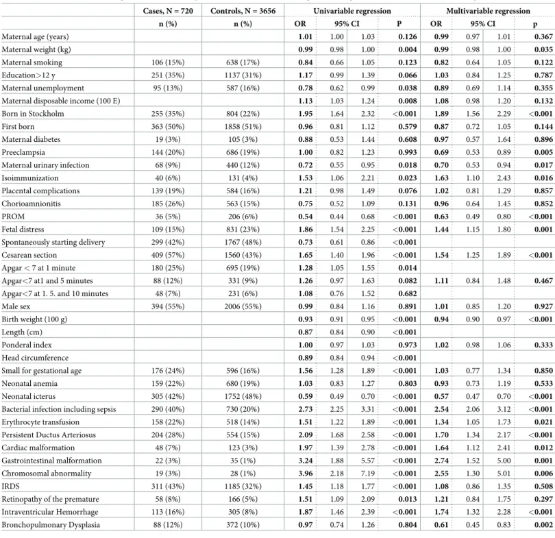
Table 2. Associations of maternal, gestational, fetal, and perinatal factors with NEC in all gestational ages, univariable and multivariable regressions. Cases, N = 720 Controls, N = 3656 Univariable regression Multivariable regression
n (%) n (%) OR 95% CI P OR 95% CI p
Maternal age (years) 1.01 1.00 1.03 0.126 0.99 0.97 1.01 0.367
Maternal weight (kg) 0.99 0.98 1.00 0.004 0.99 0.98 1.00 0.035
Maternal smoking 106 (15%) 638 (17%) 0.84 0.66 1.05 0.123 0.82 0.64 1.05 0.122
Education>12 y 251 (35%) 1137 (31%) 1.17 0.99 1.39 0.066 1.03 0.84 1.25 0.787
Maternal unemployment 95 (13%) 587 (16%) 0.78 0.62 0.99 0.038 0.89 0.69 1.14 0.355
Maternal disposable income (100 E) 1.13 1.03 1.24 0.008 1.08 0.98 1.20 0.132
Born in Stockholm 255 (35%) 804 (22%) 1.95 1.64 2.32 <0.001 1.89 1.56 2.29 <0.001
First born 363 (50%) 1858 (51%) 0.96 0.81 1.12 0.579 0.87 0.72 1.05 0.144
Maternal diabetes 19 (3%) 105 (3%) 0.88 0.53 1.44 0.608 0.97 0.57 1.64 0.896
Preeclampsia 144 (20%) 686 (19%) 1.00 0.82 1.23 0.993 0.69 0.53 0.89 0.005
Maternal urinary infection 68 (9%) 440 (12%) 0.72 0.55 0.95 0.018 0.70 0.53 0.94 0.017
Isoimmunization 40 (6%) 131 (4%) 1.53 1.06 2.21 0.023 1.63 1.10 2.43 0.016
Placental complications 139 (19%) 584 (16%) 1.21 0.98 1.49 0.076 1.02 0.81 1.29 0.857
Chorioamnionitis 185 (26%) 563 (15%) 0.75 0.52 1.09 0.131 0.96 0.64 1.45 0.852
PROM 36 (5%) 206 (6%) 0.54 0.44 0.68 <0.001 0.63 0.49 0.80 <0.001
Fetal distress 109 (15%) 831 (23%) 1.86 1.54 2.25 <0.001 1.44 1.15 1.80 0.001
Spontaneously starting delivery 299 (42%) 1767 (48%) 0.73 0.61 0.86 <0.001
Cesarean section 409 (57%) 1560 (43%) 1.65 1.40 1.96 <0.001 1.54 1.25 1.89 <0.001 Apgar < 7 at 1 minute 180 (25%) 695 (19%) 1.28 1.05 1.55 0.014
Apgar<7 at1 and 5 minutes 88 (12%) 331 (9%) 1.26 0.97 1.63 0.082 1.11 0.84 1.48 0.467 Apgar<7 at 1. 5. and 10 minutes 48 (7%) 231 (6%) 1.08 0.76 1.52 0.682
Male sex 394 (55%) 2006 (55%) 0.99 0.84 1.16 0.891 1.01 0.85 1.20 0.927
Birth weight (100 g) 0.93 0.91 0.95 <0.001 0.94 0.90 0.97 <0.001
Length (cm) 0.87 0.84 0.90 <0.001
Ponderal index 1.00 0.97 1.03 0.973 1.02 0.98 1.06 0.333
Head circumference 0.89 0.84 0.94 <0.001
Small for gestational age 176 (24%) 596 (16%) 1.56 1.28 1.89 <0.001 1.03 0.77 1.34 0.850
Neonatal anemia 159 (22%) 680 (19%) 1.03 0.83 1.27 0.803 0.93 0.73 1.19 0.533
Neonatal icterus 305 (42%) 1752 (48%) 0.59 0.49 0.70 <0.001 0.57 0.47 0.70 <0.001 Bacterial infection including sepsis 290 (40%) 730 (20%) 2.73 2.25 3.31 <0.001 2.54 2.06 3.12 <0.001 Erythrocyte transfusion 158 (22%) 518 (14%) 1.51 1.22 1.89 <0.001 1.34 1.05 1.73 0.021 Persistent Ductus Arteriosus 204 (28%) 554 (15%) 2.09 1.68 2.58 <0.001 1.70 1.34 2.17 <0.001
Cardiac malformation 48 (7%) 123 (3%) 1.97 1.39 2.78 <0.001 1.64 1.12 2.41 0.012
Gastrointestinal malformation 22 (3%) 35 (1%) 3.24 1.88 5.57 <0.001 2.74 1.52 5.00 0.001 Chromosomal abnormality 19 (3%) 28 (1%) 3.96 2.18 7.19 <0.001 2.55 1.30 5.01 0.006
IRDS 311 (43%) 1185 (32%) 1.45 1.18 1.77 <0.001 1.08 0.86 1.35 0.508
Retinopathy of the premature 58 (8%) 166 (5%) 1.51 1.09 2.09 0.013 1.21 0.84 1.75 0.297 Intraventricular Hemorrhage 113 (16%) 305 (8%) 1.87 1.46 2.39 <0.001 1.74 1.32 2.28 <0.001 Bronchopulmonary Dysplasia 88 (12%) 372 (10%) 0.97 0.74 1.26 0.804 0.61 0.45 0.83 0.002 CI–Confidence Interval; IRDS–Infant Respiratory Distress Syndrome; OR–Odds Ratio; PROM–Premature Rupture of the Membranes
In GA > 31 w, positive associations with a diagnosis of cardiac malformations, cesarean section (CS), chromosomal abnormalities, and Apgar < 7 were added. More pronounced dis-tress, i.e. Apgar < 4, did not further influence the risk. A positive association with being small for gestational age (SGA) was seen in term infants only.
There were independent, negative associations of NEC with maternal weight and a mater-nal diagnosis of pre-eclampsia (PE), significant only in the total study population, but with essentially uniform tendencies in the subgroups. Chromosomal abnormalities were positively associated with NEC in the population as a whole but because of low numbers not included in the subgroup models.
Analysis of the birth countries of the mothers did not show any relationship to NEC. Among socioeconomic factors, the only factor that remained significant in the multivariable model was being born in Stockholm. There were no significant associations with parity, smok-ing, maternal diabetes, chorioamnionitis or placental complications. Pregnancy related hyper-tensive disorders, apart from PE, were too few to be included in the models.
A discharge diagnosis of intestinal perforation in the newborn was found in 78 individuals, 64 cases and 14 controls, with a significant increasing trend over time as analyzed with poisson regression: Incidence rate ratio (IRR) 1,11 (95% CI 1,06–1,15) per year in the entire study population, p<0,001; IRR 1,10 (1,05–1,15) per year in cases, p<0,001, and 1,09 (0,996–1,9), p = 0,06, in controls.
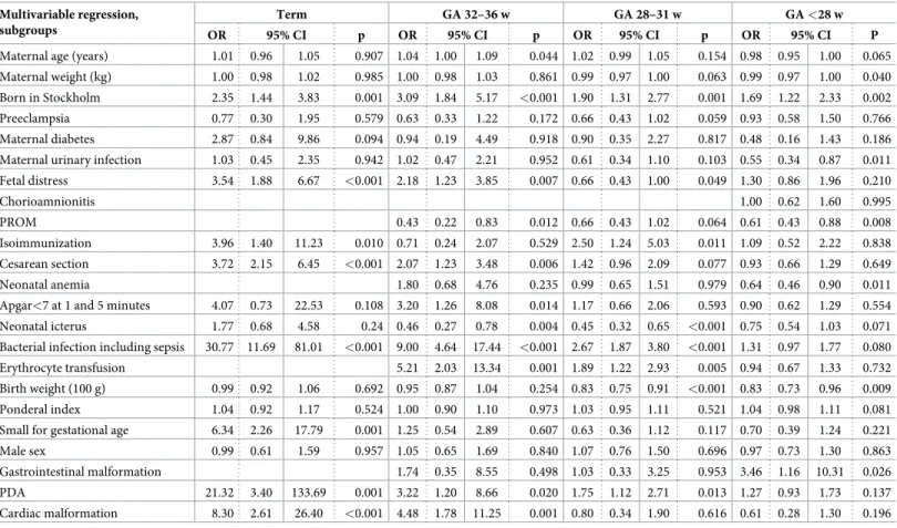
Table 3. Associations of maternal, gestational, fetal, and perinatal factors with NEC in subgroups according to gestational age, multivariable regression. Multivariable regression,
subgroups
Term GA 32–36 w GA 28–31 w GA <28 w
OR 95% CI p OR 95% CI p OR 95% CI p OR 95% CI P
Maternal age (years) 1.01 0.96 1.05 0.907 1.04 1.00 1.09 0.044 1.02 0.99 1.05 0.154 0.98 0.95 1.00 0.065 Maternal weight (kg) 1.00 0.98 1.02 0.985 1.00 0.98 1.03 0.861 0.99 0.97 1.00 0.063 0.99 0.97 1.00 0.040 Born in Stockholm 2.35 1.44 3.83 0.001 3.09 1.84 5.17 <0.001 1.90 1.31 2.77 0.001 1.69 1.22 2.33 0.002 Preeclampsia 0.77 0.30 1.95 0.579 0.63 0.33 1.22 0.172 0.66 0.43 1.02 0.059 0.93 0.58 1.50 0.766 Maternal diabetes 2.87 0.84 9.86 0.094 0.94 0.19 4.49 0.918 0.90 0.35 2.27 0.817 0.48 0.16 1.43 0.186 Maternal urinary infection 1.03 0.45 2.35 0.942 1.02 0.47 2.21 0.952 0.61 0.34 1.10 0.103 0.55 0.34 0.87 0.011 Fetal distress 3.54 1.88 6.67 <0.001 2.18 1.23 3.85 0.007 0.66 0.43 1.00 0.049 1.30 0.86 1.96 0.210 Chorioamnionitis 1.00 0.62 1.60 0.995 PROM 0.43 0.22 0.83 0.012 0.66 0.43 1.02 0.064 0.61 0.43 0.88 0.008 Isoimmunization 3.96 1.40 11.23 0.010 0.71 0.24 2.07 0.529 2.50 1.24 5.03 0.011 1.09 0.52 2.22 0.838 Cesarean section 3.72 2.15 6.45 <0.001 2.07 1.23 3.48 0.006 1.42 0.96 2.09 0.077 0.93 0.66 1.29 0.649 Neonatal anemia 1.80 0.68 4.76 0.235 0.99 0.65 1.51 0.979 0.64 0.46 0.90 0.011
Apgar<7 at 1 and 5 minutes 4.07 0.73 22.53 0.108 3.20 1.26 8.08 0.014 1.17 0.66 2.06 0.593 0.90 0.62 1.29 0.554 Neonatal icterus 1.77 0.68 4.58 0.24 0.46 0.27 0.78 0.004 0.45 0.32 0.65 <0.001 0.75 0.54 1.03 0.071 Bacterial infection including sepsis 30.77 11.69 81.01 <0.001 9.00 4.64 17.44 <0.001 2.67 1.87 3.80 <0.001 1.31 0.97 1.77 0.080 Erythrocyte transfusion 5.21 2.03 13.34 0.001 1.89 1.22 2.93 0.005 0.94 0.67 1.33 0.732 Birth weight (100 g) 0.99 0.92 1.06 0.692 0.95 0.87 1.04 0.254 0.83 0.75 0.91 <0.001 0.83 0.73 0.96 0.009 Ponderal index 1.04 0.92 1.17 0.524 1.00 0.90 1.10 0.973 1.03 0.95 1.11 0.521 1.04 0.98 1.11 0.081 Small for gestational age 6.34 2.26 17.79 0.001 1.25 0.54 2.89 0.607 0.63 0.36 1.12 0.117 0.70 0.39 1.24 0.221 Male sex 0.99 0.61 1.59 0.957 1.05 0.65 1.69 0.840 1.07 0.76 1.50 0.696 0.97 0.73 1.30 0.863 Gastrointestinal malformation 1.74 0.35 8.55 0.498 1.03 0.33 3.25 0.953 3.46 1.16 10.31 0.026 PDA 21.32 3.40 133.69 0.001 3.22 1.20 8.66 0.020 1.75 1.12 2.71 0.013 1.27 0.93 1.73 0.137 Cardiac malformation 8.30 2.61 26.40 <0.001 4.48 1.78 11.25 0.001 0.80 0.34 1.90 0.616 0.61 0.28 1.30 0.196 CI–Confidence Interval; OR–Odds Ratio; PDA–Persistent Ductus Arteriosus; PROM–Premature Rupture of the Membranes; GA–Gestational age
The sensitivity analysis showed only small and nonsignificant differences at the third digit level when imputed variables were included in univariable analyses compared with complete case analyses. Separate multivariable analyses for the study period before and from year 2000 showed a significantly larger association of NEC with gastrointestinal malformations with a very large CI due to the few number of cases in each period. Excluding the covariat for >7 days survival resulted in nonsignificant changes at the second digit level in all variables except persistent ductus arteriosus which became significantly associated with NEC in both uni- and multivariate analyses in GA < 28 w, with an OR 1.49 (p = 0.010) in the multivariable analysis compared with the reported OR 1.27 (p = 0.137).
Discussion
We found a complex pattern of associations between NEC and numerous factors related to the mother, pregnancy, infant and perinatal events. Similar to many previous studies, associations with NEC were more obvious in GA > 31 weeks, than in the more premature, which reflects the overriding effect of prematurity but also supports the notion that different mechanisms are involved at different levels of maturity.
Maternal factors
We have no explanation for the slightly negative association of NEC with increasing maternal weight. Maternal obesity, as well as undernutrition, has been shown to increase the risk of sev-eral complications,[11,12] most of which are controlled for in the multivariable analysis. Maternal obesity, body composition and/or diet have also been shown to influence neonatal body composition [13] as well as maternal milk content of several macronutrients, bioactive substances,[14–16] and microbiota,[17] all of which could possibly influence the complex pathogenetic mechanisms of NEC.
In the multivariable model, no association of NEC with smoking, educational level, income, or unemployment was seen. The association with being born in Stockholm, the capital and largest city of Sweden, was noted in our previous study on incidence of NEC in Sweden[1] and interpreted as a higher tendency to assign the diagnosis.
Pregnancy related factors
Fetal distress mirrors an antenatal asphyxic event, not surprisingly positively associated with NEC in GA > 31 w[3]. Isoimmunization is likely to predispose to a general vulnerability and can be speculated to contribute to imbalance of the developing immune system, which might precipitate NEC. Case reports and retrospective studies have suggested that treatment of hemolytic anemia with intravenous immunoglobulines may precipitate NEC in term and near term infants[18–20]. Whether the association of NEC with isoimmunization is linked to the condition itself or to its treatment, cannot be discerned from our data.
The negative association with maternal urinary infection and PROM, is somewhat surpris-ing, especially as PROM, leading to chorioamnionitis, has been suggested as a risk factor of NEC[21,22]. A meta-analysis from 2013, however, found a tendency towards lower risk for NEC with a shorter course of antibiotics in mothers treated for PROM[23]. We speculate that low grade antenatal exposure to pathogens in maternal urinary infection or PROM, possibly balanced by a short course of antibiotics, may speed up the transfer of immunoglobulines to the fetus and maternal milk, which may in turn contribute to modulation of intestinal inflam-matory responses in the neonate.
Another notable finding is the negative association of NEC with PE, which emerges in the multivariable analysis, significant only in the total study population, but with a uniform
tendency in all sub-groups. The literature is inconclusive. A protective effect of maternal hypertensive disorders has been reported[4] but also positive associations of PE with NEC at least in some subgroups[24,25]. The number of NEC cases in our population was, however, considerably larger than in those studies, and many possible mediating factors were controlled for in the multivariable analysis. There is no obvious explanation for our finding, but negative associations of PE have also been reported with IVH[26] and ROP,[27] maybe above all mir-roring the complexity of interactions in pregnancy and prematurity.
Perinatal factors
Delivery by CS was associated with a greater incidence of NEC except in GA > 31 w. It made no difference if CS was elective or finalized an initially spontaneous delivery. Earlier results have been equivocal, reporting higher risk after CS[28], lower risk [29] or no difference[30]. CS is associated with lower diversity of the gut microbiota of the newborn and differences in dominant species compared to infants born by the vaginal route,[31] which may predispose for NEC[32,33]. In vaginal delivery, colonization of the infant gut is expected to result from the direct exposure to maternal vaginal and intestinal flora, but the composition of the micro-biome in breast milk may also be altered after elective CS compared to normal labor[17].
Apgar scores less than 7 at five minutes were associated with increased NEC incidence in GA 32–36 w. Otherwise there was no association of low Apgar with NEC.
Factors related to the infant
The negative association of birthweight with NEC, even in GA < 28 w, indicates that lower birthweight adds to the risk for NEC among infants of equal degree of prematurity.
Being SGA, according to the routine assessment at birth, was associated with NEC only for full term infants. Earlier results on the impact of being SGA on the risk of NEC in the prema-ture is contradictory with no increased risk for NEC reported for very low birth weight infants in Taiwan,[34] in contrast to increased risk in preterm babies in the USA and Canada[35,36]. There was no association to ponderal index, which means that any growth restriction influenc-ing the risk for NEC would be symmetrical,[37] which in turn is thought to reflect an early cause of growth restriction[38].
Male sex has been suggested as a risk factor of NEC,[4,36] and in our study of the incidence of NEC in Sweden there seemed to be a greater risk of NEC in boys than in girls[1]. In this case-control-study, however, there were 55% boys among NEC cases and controls alike. Com-pared to the national birth cohort, the sex distribution differed significantly in GA 32–36 weeks, where there were 57% boys in the study population and 51% nationally, p<0.001, but not in any other subgroup.
The positive association with cardiac malformation only in GA > 31 w is in keeping with the results of other studies[39–41]. In contrast, for PDA, the association was positive in all sub-groups, although statistically significant in GA > 28 w only. Unlike the findings of Lee et. al.[42], surgical treatment did not further enhance the association, but whether the PDA itself, treatment with indomethacin, or perhaps a combination of both, accounted for the enhanced risk is not possible to discern. Both have been suggested as risk factors of NEC in earlier stud-ies[41,43].
Neonatal complications
Red blood cell transfusion as a risk factor for NEC is under dispute. Evidence is equivocal and mostly from observational studies[44]. In the registers that we have used, the temporal rela-tionship between red cell transfusion and NEC onset cannot be determined, so the positive
association does not imply a causative relationship. It may rather mirror the general vulnerable state of infants susceptible to NEC or, as Hay et.al. suggest, even treatment given for prodromal symptoms of NEC[44] or for sepsis secondary to NEC.
Bacterial infection, including sepsis, was positively associated with NEC. Data on infection with other organisms were too infrequent to be reliably analyzed. Sepsis and bloodstream infections are well-known manifestations of NEC,[45] but have also been suggested as a con-tributing factor in the pathogenesis[46]. Without information on the temporal relationships of the diagnoses, the interpretation of our finding is unclear.
Association with specific complications of prematurity
IRDS, ROP, BPD, and IVH are all morbidities specific for the premature, partially interrelated to one another, with many risk factors in common with NEC. Similar to NEC, IVH is de-scribed as a complex disorder involving changes in cerebral blood flow, coagulation, angiogen-esis, and inflammation, mediated by complicated interactions of various cytokines[47]. The positive relationship between IVH, which normally occurs within the first days of life, and NEC, which rarely occurs before one week of age,[2] is, however, probably explained by patho-genetic mechanisms that predispose for both conditions but are not included in our multivari-able model.
Previous results regarding associations of ROP and BPD with NEC are conflicting[27,48– 50]. Our finding of a negative relationship of BPD with NEC, after correcting for confounders and common risk factors, suggests that there are other factors, not included in the model, that affect the risk of BPD and NEC differently.
SIP contamination of the data set
It is generally accepted that spontaneous intestinal perforation (SIP) should be differentiated from NEC [7]. In this population, a discharge diagnosis of intestinal perforation in the new-born among cases may correspond to SIP that has not been separated from NEC, revised diag-noses after surgery for presumed NEC, misclassification of perforated NEC, or even the development of NEC after surgical recovery from SIP[51]. The awareness of SIP and its dis-tinction from NEC can be assumed to have increased over time, so that the occurrence of SIP is more overt towards the end of the study period, without necessarily having increased. These considerations led to the decision not to exclude individuals with a diagnosis of intestinal per-foration in the newborn.
Strengths and limitations
The comprehensive Swedish national health care registers offer good opportunities for epide-miological research. Despite a small population and, from an international perspective, low incidence rates of NEC in Sweden, the present study represents a large number of subjects over a period of 23 years in which major advances have been made in ante- and perinatal care resulting in a significant growth of the population at greatest risk for NEC.
On evaluation, the registers used have been found to hold high quality [52]. Nevertheless, as in all register studies, the data were routinely collected for administrative reasons rather than specifically for research, which implies that the information collected cannot be influenced to fit the objective of this particular study. The inclusion of a case is based on the discharge diag-nosis without any knowledge about the diagnostic criteria used in each case. Validation of the MBR has shown errors below 5% [53], which is in parity with the findings of Palleri et.al. when manually validating the discharge diagnosis of NEC in Stockholm County 2009–2014 [54]. Hence about 5% of cases can be expected not to have had NEC. The effect of such an error,
however, should be dilution of the dataset, which may weaken actual associations but not introduce false ones. Nevertheless, results must be interpreted with care, baring in mind the risk of spurious associations caused by the great number of variables.
Looking for potential risk factors for NEC, timing is important, but the possibilities to dis-cern timely relationships for events and diagnoses are extremely limited. Information on some potentially important factors such as feeding and medication is lacking all together. There is also a problem with missing values, competing risk of early death in infants that would poten-tially have developed NEC, and eventual difference in associations due to changes in manage-ment during the long study period. We found no clinically important difference in the sensitivity analyses related to these factors.
Missing personal identification numbers in new born infants is another problem with using the registers. The number is given to all Swedish residents at birth or immigration,[9] but dur-ing the study period, there was a delay of a few weeks before newborns received their number, resulting in a substantial proportion of discharge registrations with missing identification number, impeding linkage between individual hospitalizations belonging to one individual as well as between registers. The study population is thus reduced by 22%. In our previous study, diagnoses of prematurity, mortality and sex distribution were compared between the study population and subjects with incomplete identification without demonstrating any significant differences. The missing cases were thus estimated not to represent any systematic error or induce any substantial bias[1]. The risk for such an influence is expected to be even less with the case-control design. We obtained fewer controls in GA < 32 w due to the rarity of eligible matched controls. This potential risk for inducing bias should have been controlled for by the inclusion of GA and year of birth in all analyses.
Conclusion
Our results support the notion that NEC is a common pathophysiological pathway of multifac-torial etiology, rather than a uniform disease entity. Some of the associations identified in this study seem to be primarily related to an unspecific vulnerability, others may have more direct pathophysiologic associations with NEC, and some may affect both. Differences in manage-ment of antenatal and perinatal complications may influence the impact of certain risk factors and contribute to the disparate results of different studies.
Supporting information
S1 Table. Associations of maternal, gestational, fetal, and perinatal factors with NEC in subgroups according to gestational age, univariable regression.
(DOCX)
Author Contributions
Conceptualization: Margareta Ahle, Peder Drott, Roland E. Andersson. Data curation: Margareta Ahle.
Formal analysis: Margareta Ahle, Roland E. Andersson. Funding acquisition: Margareta Ahle, Roland E. Andersson. Methodology: Roland E. Andersson.
Supervision: Roland E. Andersson. Writing – original draft: Margareta Ahle.
Writing – review & editing: Margareta Ahle, Peder Drott, Anders Elfvin, Roland E. Andersson.
References
1. Ahle M, Drott P, Andersson RE. Epidemiology and trends of necrotizing enterocolitis in Sweden: 1987– 2009. Pediatrics. 2013; 132: e443–51.https://doi.org/10.1542/peds.2012-3847PMID:23821702 2. Yee WH, Soraisham AS, Shah VS, Aziz K, Yoon W, Lee SK, et al. Incidence and timing of presentation
of necrotizing enterocolitis in preterm infants. Pediatrics. 2012; 129: e298–304.https://doi.org/10.1542/ peds.2011-2022PMID:22271701
3. Beeby PJ, Jeffery H. Risk factors for necrotising enterocolitis: the influence of gestational age. Arch Dis Child. 1992; 67: 432–435. PMID:1586186
4. Luig M, Lui K, NSW & ACT NICUS Group. Epidemiology of necrotizing enterocolitis—Part II: Risks and susceptibility of premature infants during the surfactant era: a regional study. J Paediatr Child Health. 2005; 41: 174–179.https://doi.org/10.1111/j.1440-1754.2005.00583.xPMID:15813870
5. Denning TW, Bhatia AM, Kane AF, Patel RM, Denning PW. Pathogenesis of NEC: Role of the innate and adaptive immune response. Semin Perinatol. 2017; 41: 15–28.https://doi.org/10.1053/j.semperi. 2016.09.014PMID:27940091
6. Neu J, Pammi M. Pathogenesis of NEC: Impact of an altered intestinal microbiome. Semin Perinatol. 2017; 41: 29–35.https://doi.org/10.1053/j.semperi.2016.09.015PMID:27986328
7. Sharma R, Hudak M. A Clinical Perspective of Necrotizing Enterocolitis. Past, Present, and Future. Clin Perinatol. 201340: 27–51.
8. Magnusson A, Ahle M, Swolin-Eide D, Elfvin A, Andersson RE. Population-based study showed that necrotising enterocolitis occurred in space-time clusters with a decreasing secular trend in Sweden. Acta Paediatr Oslo Nor 1992. 2017;https://doi.org/10.1111/apa.13851PMID:28349558
9. Ludvigsson JF, Otterblad-Olausson P, Pettersson BU, Ekbom A. The Swedish personal identity num-ber: possibilities and pitfalls in healthcare and medical research. Eur J Epidemiol. 2009; 24: 659–667.
https://doi.org/10.1007/s10654-009-9350-yPMID:19504049
10. Bouhlila DS, Sellaouti F. Multiple imputation using chained equations for missing data in TIMSS: a case study. Large-Scale Assess Educ. 2013; 1: 4.https://doi.org/10.1186/2196-0739-1-4
11. Avcı ME,Şanlıkan F, C¸ elik M, Avcı A, Kocaer M, Go¨c¸men A. Effects of maternal obesity on antenatal, perinatal and neonatal outcomes. J Matern-Fetal Neonatal Med Off J Eur Assoc Perinat Med Fed Asia Ocean Perinat Soc Int Soc Perinat Obstet. 2015; 28: 2080–2083.https://doi.org/10.3109/14767058. 2014.978279PMID:25327177
12. Triunfo S, Lanzone A. Impact of maternal under nutrition on obstetric outcomes. J Endocrinol Invest. 2015; 38: 31–38.https://doi.org/10.1007/s40618-014-0168-4PMID:25194427
13. Breij LM, Steegers-Theunissen RPM, Briceno D, Hokken-Koelega ACS. Maternal and Fetal Determi-nants of Neonatal Body Composition. Horm Res Paediatr. 2015; 84: 388–395.https://doi.org/10.1159/ 000441298PMID:26492188
14. Ali MA, Strandvik B, Palme-Kilander C, Yngve A. Lower polyamine levels in breast milk of obese moth-ers compared to mothmoth-ers with normal body weight. J Hum Nutr Diet Off J Br Diet Assoc. 2013; 26 Suppl 1: 164–170.https://doi.org/10.1111/jhn.12097PMID:23627874
15. Ma¨kela¨ J, Linderborg K, Niinikoski H, Yang B, Lagstro¨ m H. Breast milk fatty acid composition differs between overweight and normal weight women: the STEPS Study. Eur J Nutr. 2013; 52: 727–735.
https://doi.org/10.1007/s00394-012-0378-5PMID:22639073
16. Quinn EA, Largado F, Borja JB, Kuzawa CW. Maternal characteristics associated with milk leptin con-tent in a sample of Filipino women and associations with infant weight for age. J Hum Lact Off J Int Lact Consult Assoc. 2015; 31: 273–281.https://doi.org/10.1177/0890334414553247PMID:25348673 17. Cabrera-Rubio R, Collado MC, Laitinen K, Salminen S, Isolauri E, Mira A. The human milk microbiome
changes over lactation and is shaped by maternal weight and mode of delivery. Am J Clin Nutr. 2012; 96: 544–551.https://doi.org/10.3945/ajcn.112.037382PMID:22836031
18. Navarro M, Negre S, Matoses ML, Golombek SG, Vento M. Necrotizing enterocolitis following the use of intravenous immunoglobulin for haemolytic disease of the newborn. Acta Paediatr Oslo Nor 1992. 2009; 98: 1214–1217.https://doi.org/10.1111/j.1651-2227.2009.01279.xPMID:19397554
19. Krishnan L, Pathare A. Necrotizing enterocolitis in a term neonate following intravenous immunoglobulin therapy. Indian J Pediatr. 2011; 78: 743–744.https://doi.org/10.1007/s12098-010-0334-4PMID:
20. Figueras-Aloy J, Rodrı´guez-Migue´lez JM, Iriondo-Sanz M, Salvia-Roiges M-D, Botet-Mussons F, Car-bonell-Estrany X. Intravenous immunoglobulin and necrotizing enterocolitis in newborns with hemolytic disease. Pediatrics. 2010; 125: 139–144.https://doi.org/10.1542/peds.2009-0676PMID:19948572 21. Been JV, Lievense S, Zimmermann LJI, Kramer BW, Wolfs TGAM. Chorioamnionitis as a risk factor for
necrotizing enterocolitis: a systematic review and meta-analysis. J Pediatr. 2013; 162: 236–242.e2.
https://doi.org/10.1016/j.jpeds.2012.07.012PMID:22920508
22. Garcı´a-Muñoz Rodrigo F, Gala´n Henrı´quez G, Figueras Aloy J, Garcı´a-Alix Pe´rez A. Outcomes of very-low-birth-weight infants exposed to maternal clinical chorioamnionitis: a multicentre study. Neonatology. 2014; 106: 229–234.https://doi.org/10.1159/000363127PMID:25011418
23. Kenyon S, Boulvain M, Neilson JP. Antibiotics for preterm rupture of membranes. Cochrane Database Syst Rev. 2013; CD001058.https://doi.org/10.1002/14651858.CD001058.pub3PMID:24297389 24. Perger L, Mukhopadhyay D, Komidar L, Wiggins-Dohlvik K, Uddin MN, Beeram M. Maternal
pre-eclampsia as a risk factor for necrotizing enterocolitis. J Matern-Fetal Neonatal Med Off J Eur Assoc Perinat Med Fed Asia Ocean Perinat Soc Int Soc Perinat Obstet. 2016; 29: 2098–2103.https://doi.org/ 10.3109/14767058.2015.1076386PMID:27480208
25. Cetinkaya M, Ozkan H, Koksal N. Maternal preeclampsia is associated with increased risk of necrotiz-ing enterocolitis in preterm infants. Early Hum Dev. 2012; 88: 893–898.https://doi.org/10.1016/j. earlhumdev.2012.07.004PMID:22831636
26. Ment LR,Åde´n U, Bauer CR, Bada HS, Carlo WA, Kaiser JR, et al. Genes and environment in neonatal intraventricular hemorrhage. Semin Perinatol. 2015; 39: 592–603.https://doi.org/10.1053/j.semperi. 2015.09.006PMID:26516117
27. Seiberth V, Linderkamp O. Risk factors in retinopathy of prematurity. a multivariate statistical analysis. Ophthalmol J Int Ophtalmol Int J Ophthalmol Z Augenheilkd. 2000; 214: 131–135. doi:27482
28. Maayan-Metzger A, Itzchak A, Mazkereth R, Kuint J. Necrotizing enterocolitis in full-term infants: case-control study and review of the literature. J Perinatol Off J Calif Perinat Assoc. 2004; 24: 494–499.
https://doi.org/10.1038/sj.jp.7211135PMID:15229620
29. Guthrie SO, Gordon PV, Thomas V, Thorp JA, Peabody J, Clark RH. Necrotizing enterocolitis among neonates in the United States. J Perinatol Off J Calif Perinat Assoc. 2003; 23: 278–285.https://doi.org/ 10.1038/sj.jp.7210892PMID:12774133
30. Bertino E, Giuliani F, Prandi G, Coscia A, Martano C, Fabris C. Necrotizing enterocolitis: risk factor anal-ysis and role of gastric residuals in very low birth weight infants. J Pediatr Gastroenterol Nutr. 2009; 48: 437–442. PMID:19330932
31. Rutayisire E, Huang K, Liu Y, Tao F. The mode of delivery affects the diversity and colonization pattern of the gut microbiota during the first year of infants’ life: a systematic review. BMC Gastroenterol. 2016; 16: 86.https://doi.org/10.1186/s12876-016-0498-0PMID:27475754
32. Heida FH, van Zoonen AGJF, Hulscher JBF, te Kiefte BJC, Wessels R, Kooi EMW, et al. A Necrotizing Enterocolitis-Associated Gut Microbiota Is Present in the Meconium: Results of a Prospective Study. Clin Infect Dis Off Publ Infect Dis Soc Am. 2016; 62: 863–870.https://doi.org/10.1093/cid/ciw016PMID:
26787171
33. Warner BB, Deych E, Zhou Y, Hall-Moore C, Weinstock GM, Sodergren E, et al. Gut bacteria dysbiosis and necrotising enterocolitis in very low birthweight infants: a prospective case-control study. Lancet Lond Engl. 2016; 387: 1928–1936.https://doi.org/10.1016/S0140-6736(16)00081-7PMID:26969089 34. Tsai L-Y, Chen Y-L, Tsou K-I, Mu S-C, Taiwan Premature Infant Developmental Collaborative Study
Group. The impact of small-for-gestational-age on neonatal outcome among very-low-birth-weight infants. Pediatr Neonatol. 2015; 56: 101–107.https://doi.org/10.1016/j.pedneo.2014.07.007PMID:
25440777
35. Garite TJ, Clark R, Thorp JA. Intrauterine growth restriction increases morbidity and mortality among premature neonates. Am J Obstet Gynecol. 2004; 191: 481–487.https://doi.org/10.1016/j.ajog.2004. 01.036PMID:15343225
36. Bernstein IM, Horbar JD, Badger GJ, Ohlsson A, Golan A. Morbidity and mortality among very-low-birth-weight neonates with intrauterine growth restriction. The Vermont Oxford Network. Am J Obstet Gynecol. 2000; 182: 198–206. PMID:10649179
37. Cooley SM, Donnelly JC, Walsh T, Kirkham C, Gillan J, Geary MP. Ponderal index (PI) vs birth weight centiles in the low-risk primigravid population: which is the better predictor of fetal wellbeing? J Obstet Gynaecol J Inst Obstet Gynaecol. 2012; 32: 439–443.https://doi.org/10.3109/01443615.2012.667172
PMID:22663314
38. Bocca-Tjeertes I, Bos A, Kerstjens J, de Winter A, Reijneveld S. Symmetrical and asymmetrical growth restriction in preterm-born children. Pediatrics. 2014; 133: e650–656.https://doi.org/10.1542/peds. 2013-1739PMID:24488742
39. Ostlie DJ, Spilde TL, St Peter SD, Sexton N, Miller KA, Sharp RJ, et al. Necrotizing enterocolitis in full-term infants. J Pediatr Surg. 2003; 38: 1039–1042. PMID:12861534
40. McElhinney DB, Hedrick HL, Bush DM, Pereira GR, Stafford PW, Gaynor JW, et al. Necrotizing entero-colitis in neonates with congenital heart disease: risk factors and outcomes. Pediatrics. 2000; 106: 1080–1087. PMID:11061778
41. Sankaran K, Puckett B, Lee DS, Seshia M, Boulton J, Qiu Z, et al. Variations in incidence of necrotizing enterocolitis in Canadian neonatal intensive care units. J Pediatr Gastroenterol Nutr. 2004; 39: 366–72. PMID:15448426
42. Lee LCL, Tillett A, Tulloh R, Yates R, Kelsall W. Outcome following patent ductus arteriosus ligation in premature infants: a retrospective cohort analysis. BMC Pediatr. 2006; 6: 15.https://doi.org/10.1186/ 1471-2431-6-15PMID:16689986
43. Ha¨llstro¨m M, Koivisto AM, Janas M, Tammela O. Frequency of and risk factors for necrotizing enteroco-litis in infants born before 33 weeks of gestation. Acta Paediatr Oslo Nor 1992. 2003; 92: 111–113.
44. Hay S, Zupancic JAF, Flannery DD, Kirpalani H, Dukhovny D. Should we believe in transfusion-associ-ated enterocolitis? Applying a GRADE to the literature. Semin Perinatol. 2016;https://doi.org/10.1053/j. semperi.2016.09.021PMID:27866662
45. Heida FH, Hulscher JBF, Schurink M, van Vliet MJ, Kooi EMW, Kasper DC, et al. Bloodstream infec-tions during the onset of necrotizing enterocolitis and their relation with the pro-inflammatory response, gut wall integrity and severity of disease in NEC. J Pediatr Surg. 2015; 50: 1837–1841.https://doi.org/ 10.1016/j.jpedsurg.2015.07.009PMID:26259559
46. Lambert DK, Christensen RD, Henry E, Besner GE, Baer VL, Wiedmeier SE, et al. Necrotizing entero-colitis in term neonates: data from a multihospital health-care system. J Perinatol Off J Calif Perinat Assoc. 2007; 27: 437–443.https://doi.org/10.1038/sj.jp.7211738PMID:17392837
47. Szpecht D, Wiak K, Braszak A, Szymankiewicz M, Gadzinowski J. Role of selected cytokines in the etiopathogenesis of intraventricular hemorrhage in preterm newborns. Childs Nerv Syst ChNS Off J Int Soc Pediatr Neurosurg. 2016; 32: 2097–2103.https://doi.org/10.1007/s00381-016-3217-9PMID:
27541865
48. Mitsiakos G, Papageorgiou A. Incidence and factors predisposing to retinopathy of prematurity in inborn infants less than 32 weeks of gestation. Hippokratia. 2016; 20: 121–126. PMID:28416908
49. Oh W, Poindexter BB, Perritt R, Lemons JA, Bauer CR, Ehrenkranz RA, et al. Association between fluid intake and weight loss during the first ten days of life and risk of bronchopulmonary dysplasia in extremely low birth weight infants. J Pediatr. 2005; 147: 786–790.https://doi.org/10.1016/j.jpeds.2005. 06.039PMID:16356432
50. Kiciński P, Kęsiak M, Nowiczewski M, Gulczyńska E. Bronchopulmonary dysplasia in very and extremely low birth weight infants—analysis of selected risk factors. Pol Merkur Lek Organ Pol Tow Lek. 2017; 42: 71–75.
51. Gordon PV, Swanson JR, MacQueen BC, Christensen RD. A critical question for NEC researchers: Can we create a consensus definition of NEC that facilitates research progress? Semin Perinatol. 2016;
https://doi.org/10.1053/j.semperi.2016.09.013PMID:27866661
52. Ludvigsson JF, Andersson E, Ekbom A, Feychting M, Kim J-L, Reuterwall C, et al. External review and validation of the Swedish national inpatient register. BMC Public Health. 2011; 11: 450.https://doi.org/ 10.1186/1471-2458-11-450PMID:21658213
53. http://www.socialstyrelsen.se/lists/artikelkatalog/attachments/10961/2002-112-4_20021124.pdf
(Swedish only)
54. Palleri E, Aghamn I, Bexelius TS, Bartocci M, Wester T. The effect of gestational age on clinical and radiological presentation of necrotizing enterocolitis. J Pediatr Surg. 2017;https://doi.org/10.1016/j. jpedsurg.2017.09.018PMID:29079313