Twenty-year follow-up of root-filled teeth in a Swedish
population receiving high cost dental care
Kerstin Petersson1, Helena Fransson1, Eva Wolf1, Jan Håkansson2
1Department of Endodontics, Faculty of Odontology, Malmö University, Malmö, 2Department of Periodontology, Institute of Odontology, University of Gothenburg, Gothenburg; Sweden Kerstin Petersson Dept of Endodontics Faculty of Odontology Malmö University SE-205 06 Malmö Sweden Kerstin.Petersson@mah.se
Abstract
Aim To study the 20-year survival rate and periapical status of root-filled teeth in a Swedish population requiring high cost dental care and to identify factors related to survival and normal periapical status at follow-up.
Methodology The study population comprised 104 patients selected from four local health insurance districts with treatment plans including radiographs submitted for approval for reimbursement from the Swedish National Dental Insurance in 1977. In 1998 a clinical and radiographic follow-up examination was conducted, to register the status of 449 teeth identified as root-filled at baseline. Differences in tooth survival and periapical status at follow-up, with reference to periapical status and quality of root-filling at baseline were analyzed by Chi-square tests. Multiple regression analysis was used to describe tooth survival and normal periapical status at follow-up, with the explanatory baseline variables: tooth type, type of restoration, type of post, quality of root-filling, periapical status, marginal bone loss and caries.
Differences were considered significant at a 5% level.
Results Two hundred and ninety (65 %) of the root-filled teeth survived at follow-up Baseline variables associated with low odds for tooth survival were mandibular molar, maxillary premolar, prefabricated posts other than screw-posts, severe marginal bone loss, caries and apical periodontitis (AP).
Normal periapical status at follow-up was registered in 49 % of the root-filled teeth. Baseline variables associated with low odds for normal periapical status (high risk for AP) at follow-up were mandibular molar, maxillary premolar, AP, severe marginal bone loss and inadequate root-filling quality. Of the root-filled teeth with AP
at baseline 42 % had been left untreated during the observation period and at follow-up the AP persisted in 57 % of these teeth.
Conclusions After 20 years 65 % of the root-filled teeth had survived and one third remained in sound periapical conditions, without any further treatment. Almost half of the APs registered at baseline were left without treatment and more than half of them persisted after 20 years.
Key words: Apical periodontitis, endodontics, epidemiology, follow-up studies, treatment outcome
Introduction
The Swedish Council on Health Technology Assessment (SBU, 2010) and The Swedish National Board of Health and Welfare have assessed the scientific basis for treatment of pulpitis and apical periodontitis and concluded that “the scientific
evidence describing the natural course of events and prognosis for root-filled teeth is inadequate and needs further documentation. Important areas deserving
investigation include: long-term survival of root-filled teeth; factors influencing the loss of root-filled teeth and to what extent root canal treatments fail to achieve healthy outcomes and require further treatment” (Bergenholtz & Kvist 2013).The authors also concluded that “It is of utmost importance that clinical research is conducted in
general practice settings, where the majority of endodontic procedures are performed”.
Long-term epidemiological studies of Scandinavian populations have reported the outcome of root-fillings undertaken in general practice (Petersson et al. 1991, Eckerbom et al. 2007, Kirkevang et al. 2012). The 10-year survival was reported to be 88 and 87 % (Petersson et al. 1991, Kirkevang et al. 2012). However, a 20-year follow-up study of a Swedish population disclosed a lower survival (71 %) (Eckerbom
et al. 2007). Thus the survival of root-filled teeth decreased with time.
Of the root-filled teeth diagnosed with apical periodontitis (AP) at the baseline
AP was the most common procedure for root-filled teeth with AP. The 10-year follow-up revealed that the AP persisted in 55 % (Petersson et al. 1991) and 66 %
(Kirkevang et al. 2012) of these teeth.
In the 1970s the Swedish National Dental Insurance Scheme covered all dental treatment, with no limitations. For high-cost treatment, treatment plans including radiographs, had to be submitted to the local health insurance offices for approval. A random sample of the treatment plans and examination protocols (including
radiographs) submitted for approval in 1977-1978 has been studied previously, with special reference to suggested endodontic treatment (Petersson et al. 1989) and a 20-year follow-up of dental health and prosthetic treatment with the observations analysed at the individual level only (Petersson et al. 2006). A more detailed analysis of the single root-filled teeth at the 20-year follow-up would provide further
information relevant to the knowledge gaps defined in the article by Bergenholtz & Kvist (2013). Therefore the aim of the present investigation was to study the survival and periapical status at a 20-year follow-up examination of the teeth identified at baseline as root-filled, in patients selected from four local health insurance districts and receiving high cost dental care. A further aim was to identify factors influencing survival and normal periapical status at follow-up.
Material and methods
The material was selected from all treatment plans made by Swedish dentists and submitted to the local health insurance offices for approval in 1977 and 1978. Every 5th treatment plan from patients born on the 20th day of each month was sampled for baseline studies. In 1998, all such patients with treatment plans sampled for the baseline study (Petersson et al. 1989) in four local health insurance districts: Stockholm, Malmöhus, Örebro and Uppsala (262 patients), were offered a free clinical examination including radiographs. Thirty patients had died and 55 patients could not be contacted by telephone or mail. Telephone interviews were conducted with the remaining 177 patients (68 % of the sample); 104 of them (40% of the sample) agreed to participate in the clinical examination. Among the 73 patients who did not attend the clinical examination, 10 claimed to be edentulous. Other reasons for not attending the clinical examination were illness and weakness, lack of time, or declining the offer of examination. All patients gave informed consent to participation in the study, which was approved by the Central Ethical Review Board, Lund
University, Sweden (LU 593-97).
The baseline examinations were conducted in 1977-1978, and are denoted in the text as “1977”; the follow-up examinations, conducted in 1998-2000, are denoted in the text as “1998”. Examination protocols, radiographs and treatment plans from 1977 were available for all patients. The radiographic examinations in 1977 had been undertaken by the general practitioners who submitted the treatment plans for
remaining teeth) and in 1998 panoramic radiographs, periapical radiographs and bite-wings. Periapical radiographs were taken of teeth in which the periapical
structures could not be clearly interpreted in the panoramic radiographs, leaving only a few root-filled teeth to be examined by panoramic radiography only. All radiographic images were analogue.
Two calibrated observers interpreted the radiographs, each reading one part of the material. At the calibration the observers discussed the criteria and the diagnostic decisions until agreement was reached. Changes in endodontic treatment, periapical status and tooth extractions were analysed in detail at tooth level. At the evaluation, for each individual patient the findings from the clinical and radiographic
examinations carried out in1998 were compared with examination protocols,
radiographs and treatment plans from 1977. The radiographic images from 1977 and 1998 were compared for each tooth and studied side by side. The criteria for
registration are presented in Table 1. In cases of uncertainty about the presence of AP, status was classified as normal. Findings at the individual patient level with respect to remaining teeth, prosthetic reconstruction, restoration, caries, marginal bone loss, root-filled teeth and apical periodontitis have been presented previously, however without analyses using the tooth as the studied unit (Petersson et al. 2006).
Caries and type of restoration were registered both from the clinical examination protocols and the radiographs. The type of endodontic treatment, quality of the root-filling, periapical status, marginal bone loss and type of post retention were registered only from the radiographs. With respect to caries, restoration and post retention, only the status at baseline was registered. The criteria for the registrations are presented
in Table 1. Teeth with completely obturated root canals and no overfillings were considered adequately root-filled.
Statistical methods
Analysis was performed using IBM SPSS Statistics Version 22 (SPSS Inc, Chicago, IL, US). Chi-square tests were applied to detect any significant differences in tooth survival and periapical status at follow-up, with reference to periapical status and quality of root-filling at baseline. Multiple regression analysis was used to describe tooth survival and normal periapical status at follow-up, in terms of the following explanatory baseline variables: tooth type, type of restoration, type of post, quality of root-filling, periapical status, marginal bone loss and caries. . Goodness-of-fit tests of the final models were performed. Differences were considered significant at a 5% level.
Results
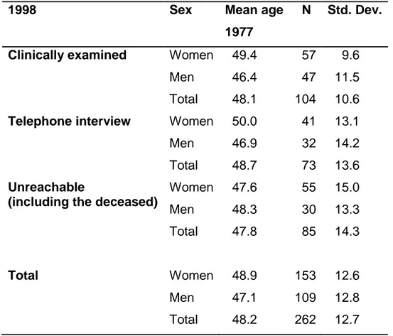
There were no statistically significant sex- or age-related differences among the 104 patients who underwent the follow-up clinical examination, the 73 patients
interviewed by telephone only and the 85 patients who could not be contacted (including the 30 deceased patients) (Table 2).
The ages of the 104 patients who underwent clinical examination (40 % of the sample) ranged from 21-70 years at baseline and from 41-90 years at follow-up. At baseline and follow-up respectively, the mean numbers of remaining teeth were 21.1 and 16.8; endodontically treated teeth 4.5 and 4.6 and teeth with apical periodontitis (root-filled and non-root-filled) 1.7 and 1.6. At baseline 100/104 (96 %) and at follow-up 102/104 (98 %) of the patients had one or more endodontically treated teeth. The 471 endodontically treated teeth present at baseline included 12 teeth that had undergone pulpotomy, 5 teeth that had apical surgery and retrofilling, and 5 teeth with complications (see Table 1). These teeth were excluded leaving a root-filled group of 449 teeth available for study.
At baseline 257 of the root-filled teeth were restored with a laboratory fabricated crown (cast or ceramic), 135 with amalgam, 12 with composite, 18 with a temporary restoration. In 22 teeth the restoration was missing and in 5 teeth the type of
restoration could not be determined.
At baseline, 449 teeth were registered as root-filled and 502 at follow-up. At follow-up significantly more teeth than at baseline were classified as completely obturated (200/502; 40 %) (115/449; 26 %) (p<0.001). The frequency of root-filled teeth with
normal periapical status was also significantly higher at follow-up (369/502; 74 %) than at baseline (273/449; 61 %) (p=0.001).
Survival of root-filled teeth at follow-up
Of the 449 root-filled teeth registered at baseline, 290 (65 %) remained at follow-up: 48 (11 %) had been retreated, 235 (52 %) had no additional endodontic treatment and 159 (35 %) had been extracted (Table 3). At the 20-year follow-up, significantly more teeth with completely obturated root canals at baseline (p<0.001) (Table 3) and significantly more teeth with normal periapical status at baseline (p=0.002) had survived (Table 4). Teeth which could not be assessed were excluded from the statistical calculations.
Multiple regression analysis of baseline factors associated with tooth survival disclosed significantly negative associations for tooth type, caries, marginal bone loss, type of restoration and type of post (these factors were thus significantly associated with tooth extraction). At the second step of the regression analysis the following baseline variables were significantly associated with and had low odds ratio (OR) for tooth survival: mandibular molar (OR 0.06, CI 0.02, 0.21), maxillary premolar (OR 0.20, CI 0.06, 0.62), prefabricated posts other than screw-posts (OR 0.20, CI 0.06, 0.67), marginal bone loss ≥ 1/3 of the root length (OR 0.22, CI 0.13, 0.36), caries (OR 0.26, CI 0.13, 0.55) and AP (OR 0.59, CI 0.35, 0.98). Root-filled teeth restored with amalgam at baseline had three times higher odds for tooth survival than laboratory fabricated crowns (cast or porcelain crowns) (OR 3.34, CI 1.68, 6.63). (Table 5)
Periapical status of root-filled teeth at follow-up
Of the 449 teeth registered at baseline as root-filled, normal periapical status was registered in 221 (49 %) at follow-up. The frequency was significantly greater for root-filled teeth with normal periapical status at baseline 164 / 273 (60%) than for those with AP 47 / 143 (33 %) (p<0.001) (Table 4). Of the 143 root-filled teeth with AP at baseline, 60 (42%) had been left without treatment of the AP during the observation period (19 teeth were retreated, 61 were extracted and 3 could not be assessed).
At follow-up 290 root-filled teeth were remaining. After exclusion of 22 teeth, 268 root-filled teeth were left for further analyses (Table 6). Of the 60 teeth not retreated, the AP had healed in 25 (42%) and persisted at follow-up in 34 teeth (57%). Analysis of the un-retreated and the retreated teeth combined, disclosed that in 32 (41 %) the AP had healed during the follow-up period and persisted in 46 (58 %) (Table 6).
Multiple regression analysis of baseline factors in the root-filled teeth which
correlated with normal periapical status at follow-up disclosed significantly negative associations for tooth type, periapical status, marginal bone loss, quality of root-filling, caries and type of post (these factors were thus significantly associated with AP). Type of restoration at baseline was not significantly related to normal periapical status at follow-up and was therefore excluded from the second step of the regression analysis. At the backward elimination the dynamic of the model was
changed and caries as well as type of post fell out. The second step of the regression analysis showed significant negative associations with normal periapical status at follow-up (and thereby significant associations with AP) for the following baseline variables: mandibular molar (OR 0.14, CI 0.05, 0.39), maxillary premolar (OR 0.25,
CI 0.10, 0.66) AP (OR 0.31, CI 0.19, 0.48), marginal bone loss ≥ 1/3 of the root length (OR 0.35, CI 0.22, 0.55) and inadequate root-filling quality (OR 0.57, CI 0.34, 0.96) (Table 7).
Discussion
The population from which the subjects of the study were sampled for baseline data comprised patients in need of high cost dental care and represents those receiving treatment in general dental practice. At the time of the study the Swedish National Dental Insurance Scheme covered all dental treatments for citizens with 75 % reimbursements for high cost treatments. Although 68 % of the patients sampled at baseline in the selected districts could be reached for follow-up study after 20-23 years, only 40 % attended the clinical and radiographic examination. The low
attendance rate can be explained by the high mean age (48.2 years) of the patients in 1977 in combination with the very long follow-up period of more than 20 years resulting in elderly patients with 11 % of the patients deceased at follow-up and also difficulties to take part in a clinical examination because of disability or sickness. The subjects who presented for clinical follow-up examination, those interviewed by telephone only and those who were deceased or could not be contacted were of similar age and sex at baseline. Thus it is reasonable to assume that although only a limited number was examined at follow-up, these subjects were representative of patients in need of high-cost dental care sampled in 1977.
The baseline radiographic examinations were undertaken by the general dental practitioners who submitted the high-cost treatment plans for approval. At follow-up, the radiographic examinations were carried out at specialist clinics for oral radiology. The difference in image quality between the radiographs used for the baseline
registrations and the follow-up radiographs from the specialist clinics may have influenced the frequency of findings registered. However, in questionable cases, periapical status was classified as normal, hence false negative rather than false
positive findings of periapical status were registered. This should be taken into account in assessment of the results. In order to circumvent individual observer tendencies towards over- or under-registration, two calibrated observers read one part of the material each (Gröndahl 1979).
The aim of the present investigation was to study the survival and periapical status of the root-filled teeth at a 20-year follow-up examination of patients who received high cost dental care. The survival of the root-filled teeth was 65 %. Previous follow-up studies of Swedish and Danish populations have reported 71 % survival after 20 years (Eckerbom et al. 2007) and 88 % and 87 % survival respectively, after 10 years (Petersson et al. 1991, Kirkevang et al. 2012). Higher frequencies were reported in a systematic review of tooth survival following non-surgical root canal treatment: 86-93 % of the pooled proportion of teeth survived over 2-10 years (Ng et al. 2010).
However, several of the 14 studies included were from specialist clinics, and this may contribute to the relatively high survival frequencies reported. It is obvious that
survival of root-filled teeth decrease with time. However the decline in mean number of remaining teeth in the present study from 21.1 at baseline to 16.8 teeth at follow-up is greater than that reported from general Swedish populations during the same decades, with means of 1.4 and 2.9 lost teeth/patient after follow-up periods of 15-18 and 20 years (Norderyd et al. 1999, Jansson et al. 2002). This high general loss of teeth and also of root-filled teeth in the present study can be attributed to the relatively high morbidity of dental diseases in the population examined, which was deemed to be in need of extensive dental care (Petersson et al. 1989) and also by the high age of the patients. However, the reasons for tooth extraction may have
as tooth type, type of post, severe marginal bone loss and caries were negatively associated with tooth survival. It indicates the importance of including oral health variables like marginal bone loss and caries in addition to endodontic and restorative variables in studies of survival of root-filled teeth.
Of the endodontic variables previously shown by logistic regression analyses to be significantly associated with tooth extraction (and thereby negatively associated with tooth survival) such as inadequate root-filling quality and AP at baseline (Cheung & Chan 2003, Ng et al. 2011, Kirkevang et al. 2012), only AP at baseline was found to be significantly negatively associated with tooth survival in the present study.
However when the endodontic variables were studied separately in relation to tooth survival using the chi-square test, teeth diagnosed with completely obturated root canals and those diagnosed with normal periapical status at baseline survived significantly more often than incompletely obturated teeth and root-filled teeth with AP.
It was of interest to note that the odds for survival of teeth restored with amalgam at the baseline examination were three times higher than for those restored with
laboratory fabricated crowns (cast or porcelain crowns). The systematic review by Ng
et al. (2010) concluded that crown restorations increased the proportion of survival of
root-filled teeth. In a prospective study of factors affecting tooth survival of root-filled teeth Ng et al. (2011) found a significantly better 4-year tooth survival for teeth with cast crown restorations. Moreover, Cheung & Chan (2003) found that teeth restored with crowns survived significantly longer in a 10-20 year perspective than those with intracoronal plastic restorations only. However, Skupien et al. (2013), using
regression analysis of the 5-year survival of root-filled teeth, found no difference between composite and crown restorations undertaken in general practice. A possible explanation for the higher OR for survival of root-filled teeth restored with amalgam at baseline in the present study may be that primarily severely damaged teeth were restored with laboratory fabricated crowns, which may have led to the lower OR for long-term survival of these teeth. It is not known to what extent
replacement of the restorations were undertaken during the 20-year follow-up period, making conclusions regarding the association between the restorations present at baseline and the status of the tooth 20 years later, uncertain. The type of post “prefabricated posts other than screw posts” that was significantly associated with low tooth survival was at baseline in 1977 often metal posts with parallel walls. Detailed information on the type of posts were however not available and further conclusions can therefore not be drawn.
The findings in the present study of patients receiving high cost dental care in Sweden may indicate that the survival of root-filled teeth is context dependent, influenced for example by the age of the patients studied, their overall oral health, and social factors such as the availability of dental care (economic and geographic) and cultural acceptance of the level of complexity of dental care usually provided in the community in question. For example, the decision to extract may be influenced by whether or not a patient can afford replacement with a fixed partial denture or an implant.
reported in a Danish study: at the 10-year follow-up, the periapical condition of 50% of the teeth registered at baseline as root-filled remained sound (Kirkevang et al. 2012). Excluding the teeth that were retreated, a lower proportion (37 %) of the teeth registered at baseline as root-filled remained in sound periapical condition after 20 years. Similarly, in the Danish study, when retreated teeth were excluded, normal periapical status was found in 37 % at the 10-year follow-up (Kirkevang et al. 2012). These results indicate that in general Scandinavian populations, half of the teeth registered as root-filled at baseline examinations survived with normal periapical status after 10 and 20 years, but after exclusion of retreated teeth, this decreased to only a third. Thus only a third of the root-filled teeth had a long-term outcome with normal periapical status and no intervention (extraction or retreatment) required during the follow-up period. The health economic consequences of this finding are largely unknown.
In the present study, as in the Danish population (Kirkevang et al. 2012) AP at baseline and inadequate root-fillings were associated with AP of the root-filled teeth at follow-up (negatively associated with sound periapical status). Additionally in the present study mandibular molars, maxillary premolars and severe marginal bone loss at baseline were associated with AP of the root-filled teeth at follow-up. Severe
marginal bone loss has previously been found to be significantly associated with AP at follow-up (Ørstavik et al. 2004, Koch et al. 2014). These findings indicate that the periodontal status of a tooth may be an important determinant of the endodontic treatment outcome.
The frequency of persistent AP in root-filled teeth which were not retreated (57 %) was in accordance with previous observations from Scandinavian general
populations: in root-filled teeth which were not retreated, 55 % and 66 % of the APs persisted after 10 years (Petersson et al. 1991, Kirkevang et al. 2012). As the AP in root-filled teeth was left without treatment (tooth extraction or retreatment) for 20 years, it is unlikely that the patients experienced obvious symptoms. With respect to the relationship between AP and the general health of the patient, some
observations on cardiovascular disease have been presented, but the findings are contradictory (Jansson et al. 2001, Frisk et al. 2003, Caplan et al. 2006, Costa et al. 2014, Petersen et al. 2014, Willershausen et al. 2014): two studies have reported an association between AP and cardiovascular disease (Caplan et al. 2006, Costa et al. 2014), while two earlier studies found no significant associations (Jansson et al. 2001, Frisk et al. 2003). Recently, chronic AP has been reported to have significant associations with atherosclerosis (Petersen et al. 2014) and with acute myocardial infarction (Willershausen et al. 2014). The associations were however found only for endodontically untreated teeth. In one systematic review of the association between AP and systemic levels of inflammatory markers with the potential to lead to
increased systemic inflammation, frequent associations between AP and
inflammatory markers were found. However, when asymptomatic AP was studied separately, such associations were found only occasionally and only for single markers (Gomes et al. 2013). Based on observations in the recent publications it seems possible that the APs persisting for 20 years, probably without symptoms, may not have been harmful to the patients. However, the data collected do not
by Bergenholtz & Kvist (2013), the risk to general health of not intervening in cases of root-filled teeth with apical periodontitis needs further investigation.
It should be noted that the dental care received by the patients in the present study during the follow-up period has provided them with root-fillings of better quality and lower frequency of AP than at baseline. The relatively high frequency of extraction of teeth with inadequate treatment quality and/or AP has obviously contributed to the lower frequency of inadequate root-fillings and of root-filled teeth with AP at follow-up. With respect to periapical disease associated with both the root-filled teeth (the
present study) and with all teeth (Petersson et al. 2006), the patients who received high cost dental care under the Swedish National Dental Insurance Scheme between 1977 and 1998 were healthier at the 20-year follow-up than at baseline. Thus, the dental care provided was effective in reducing disease (AP), albeit mainly by means of tooth extraction and replacement of the lost teeth with fixed partial dentures (Petersson et al. 2006). The patients’ perspectives and the health economic consequences of this approach have yet to be evaluated.
Conclusions
This study has addressed some questions about the long-term fate of root-filled teeth, highlighted by Bergenholtz & Kvist (2013) as a knowledge gap warranting investigation. After 20 years 65 % of the root-filled teeth had survived and one- third of the root-filled teeth remained in sound periapical condition, without any further treatment. Tooth type, AP and severe marginal bone loss were considered to be of importance for both tooth survival and normal periapical status at follow-up. 42 % of
the APs registered at baseline were left without treatment and more than half of them persisted after 20 years.
References
Bergenholtz G, Kvist T (2013) Call for improved research efforts on clinical procedures in endodontics. International Endododontic Journal 46, 697-9.
Caplan DJ, Chasen JB, Krall EA et al. (2006) Lesions of endodontic origin and risk of coronary heart disease. Journal of Dental Research 85, 996-1000.
Cheung GSP, Chan TK (2003) Long-term survival of primary root canal treatment carried out in a dental teaching hospital. International Endodontic Journal 36, 117-28. Costa TH, de Figueiredo Neto JA, de Oliveira AE, Lopes e Maia Mde F, de Almeida AL (2014) Association between chronic apical periodontitis and coronary artery disease. Journal of Endodontics 40, 164-7.
Eckerbom M, Flygare L, Magnusson T (2007) A 20-year follow-up study of endodontic variables and apical status in a Swedish population. International
Endodontic Journal 40, 940-8.
Frisk F, Hakeberg M, Ahlqwist M, Bengtsson C (2003) Endodontic variables and coronary heart disease. Acta Odontologica Scandinavica 61, 257-62.
Gomes MS, Blattner TC, Sant'Ana Filho M et al. (2013) Can apical periodontitis modify systemic levels of inflammatory markers? A systematic review and meta-analysis. Journal of Endodontics 39, 1205-17.
Gröndahl HG (1979) The influence of observer performance in radiographic caries diagnosis. Swedish Dental Journal 3, 101-7.
Jansson L, Lavstedt S, Frithiof L, Theobald H (2001) Relationship between oral health and mortality in cardiovascular diseases. Journal of Clinical Periodontology 28, 762-8.
Jansson L, Lavstedt S, Zimmerman M (2002) Marginal bone loss and tooth loss in a sample from the County of Stockholm – a longitudinal study over 20 years. Swedish
Dental Journal 26, 21-9.
Kirkevang LL, Vaeth M, Wenzel A (2012) Ten-year follow-up observations of periapical and endodontic status in a Danish population. International Endodontic
Journal 45, 829-39.
Koch M, Wolf E, Tegelberg Å, Petersson K (2015) Effect of education intervention on the quality and long-term outcomes of root canal treatment in general practice.
Ng YL, Mann V, Gulabivala K (2010) Tooth survival following non-surgical root canal treatment: a systematic review of the literature. International Endodontic Journal 43,171-89.
Ng YL, Mann V, Gulabivala K (2011) A prospective study of the factors affecting outcomes of non-surgical root canal treatment: part 2: tooth survival. International
Endodontic Journal 44, 610-25.
Norderyd O, Hugoson A, Grusovin G (1999) Risk of severe periodontal disease in a Swedish adult population. A longitudinal Study. Journal of Clinical Periodontology 26, 608-15.
Ørstavik D, Qvist V, Stoltze K (2004) A multivariate analysis of the outcome of endodontic treatment. European Journal of Oral Sciences 112, 224-30.
Petersen J, Glaßl EM, Nasseri P et al. (2014) The association of chronic apical periodontitis and endodontic therapy with atherosclerosis. Clinical Oral Investigation 18, 1813-23.
Petersson K, Håkansson R, Håkansson J, Olsson B, Wennberg A (1991) Follow-up study of endodontic status in an adult Swedish population. Endodontics and Dental
Traumatology 7, 221-5.
Petersson K, Lewin B, Hakansson J, Olsson B, Wennberg A (1989) Endodontic status and suggested treatment in a population requiring substantial dental care.
Endodontics and Dental Traumatology 5,153-8.
Petersson K, Pamenius M, Eliasson A et al. (2006) 20-year follow-up of patients receiving high-cost dental care within the Swedish Dental Insurance System: 1977-1978 to 1998-2000. Swedish Dental Journal 30, 77-86.
Skupien JA, Opdam N, Winnen R et al. (2013) A Practice-based Study on the
Survival of Restored Endodontically Treated Teeth. Journal of Endodontics 39, 1335-40.
Swedish Council on Health Technology Assessment (2010) Rotfyllning–en
systematisk litteraturöversikt (In Swedish), Swedish Council on Health Technology Assessment.Report 203, 2010. English translation, Methods of diagnosis and treatment in endodontics. Accessible at hhtp://www.sbu.se as well as on the ESE website (registered members only - http://www.e-s-e.eu/forum/viewtopic.php?f = 54&t = 87).
Willershausen I, Weyer V, Peter M et al. (2014) Association between chronic
periodontal and apical inflammation and acute myocardial infarction. Odontology 102, 297-302.
Table 1. Criteria for classification of endodontic treatment, periapical status, marginal bone loss, caries, type of restoration and type of post retention. Caries and type of restoration were assessed both from clinical examination protocols and radiographic images.
Endodontic treatment
• Completely obturated root canal: No canal lumen visible, lateral or more than 2 mm apical to the root filling.
• Incompletely obturated root canal: Visible canal lumen lateral or more than 2 mm apical to the root-filling. Teeth with one or more unfilled canals were considered incompletely sealed. • Overfilled root canal: Presence of root-filling material in the periodontal ligament space
or in the surrounding bone.
• Complication: Fractured instrument in the root canal, perforation, longitudinal root fracture. • Retrofilling: Radiographic signs of retrograde filling material.
• Pulpotomy: Radiopaque material only in the pulp chamber.
Periapical status
• Normal periapical conditions: Apical periodontal ligament space (PDL) not more than double the width in other parts of the root.
• Apical periodontitis: Apical PDL more than double the width in other parts of the root or periapical radiolucency observed.
• Marginal periodontitis involving the apex of the tooth: Vertical bone destruction including the apex of the tooth.
Marginal bone loss*
• No marginal bone loss
• Marginal bone loss < 1/3 of the root length • Marginal bone loss ≥ 1/3 of the root length
Caries
• Cavity detectable by probing or in the radiographs • No cavity detectable by probing or in the radiographs
Type of restoration • Cast crown • Ceramic crown • Amalgam restoration • Composite restoration • Temporary restoration • Restoration missing
Type of post retention
• Cast post • Screw post
• Other posts (prefabricated etc.)
* The root length was determined as the distance from the cemento-enamel junction to the apex of the tooth. If the tooth had a full coverage crown restoration the cemento-enamel junction in the adjacent tooth was used for estimation.
Table 2. Distribution according to sex and age of all 262 patients in the sample. There were no significant sex or age differences with respect to the patients examined clinically, those interviewed by telephone only and those who were deceased or could not be contacted.
1998 Sex Mean age
1977
N Std. Dev.
Clinically examined Women Men Total 49.4 46.4 48.1 57 47 104 9.6 11.5 10.6 Telephone interview Women
Men Total 50.0 46.9 48.7 41 32 73 13.1 14.2 13.6 Unreachable
(including the deceased)
Women Men Total 47.6 48.3 47.8 55 30 85 15.0 13.3 14.3 Total Women Men Total 48.9 47.1 48.2 153 109 262 12.6 12.8 12.7
Table 3. Endodontic status at follow-up 1998 in relation to root-filling quality registered at baseline 1977. Unchanged root filling -98 Retreated -98 Extracted -98 Retrofilled -98 Not assessable -98 Total Completely obturated -77 83 (72 %) 4 (3 %) 27 (24 %) - 1 (1 %) 115 Incomplete obturated -77 133 (43 %) 44 (14 %) 124 (40 %) 1 (0.3%) 5 (2 %) 307 Overfilled -77 19 (70 %) - 8 (30 %) - - 27 Total 235 (52 %) 48 (11 %) 159 (35 %) 1 (0.2 %) 6 (1 %) 449
Table 4. Periapical status of root-filled teeth at follow-up in 1998 in relation to baseline in 1977.
Normal -98 AP -98 Perio -98* Extracted -98 Not assess-able -98 Total Normal -77 164 (60 %) 23 (8 %) 4 (2 %) 79 (29 %) 3 (1 %) 273 AP -77 47 (33 %) 33 (23 %) 1 (0.7 %) 61 (43 %) 1 (0.7 %) 143 Perio -77* 1 (7 %) - 1 (7 %) 11 (85 %) - 13 Not assess-able -77 9 (45 %) 3 (15 %) - 8 (40 %) - 20 Total 221 (49 %) 59 (13 %) 6 (1 %) 159 (35 %) 4 (1 %) 449
Table 5. Multiple regression analysis of baseline variables related to 20-year tooth survival of root-filled teeth. Variables not significantly associated to tooth survival in Model 1 were excluded in Model 2. Differences were considered significant at a 5% level.
Model 1 Model 2
Variables Total OR 95% CI (p value) OR 95% CI (p value)
No post Cast post Screw post Other posts (mainly prefabricated) Crown (cast, ceramic) Amalgam Composite Temporary or missing restoration No caries Manifest caries Adequate rf quality Inadequate rf quality Normal periap status AP Marginal bone loss < 1/3 of the root length Marginal bone loss ≥1/3 of the root length Mand incisor Max molar Max premolar Max incisor Mand molar Mand premolar 110 128 153 18 243 124 11 31 359 50 108 301 258 151 279 139 30 55 68 111 48 97 1 0.83 0.58 0.19 1 3.33 2.47 0.82 1 0.27 1 0.63 1 0.62 1 0.22 1 0.51 0.19 0.49 0.06 0.33 Reference 0.38, 1.84 (0.65) 0.31, 1,11 (0.10) 0.06, 0.65 (0.008) Reference 1.67, 6.66 (0.001) 0.27, 22.79 (0.42) 0.31, 2.17 (0.68) Reference 0.13, 0.57 (0.001) Reference 0.35, 1.13 (0.13) Reference 0.37, 1.04 (0.07) Reference 0.13, 0.36 (<0.0001) Reference 0.15, 1.74 (0.28) 0.06, 0.61 (0.005) 0.17, 1.40 (0.18) 0.02, 0.21 (<0.0001) 0.118, 0.98 (0.05) 1 0.88 0.59 0.20 1 3.34 2.92 0.91 1 0.26 1 0.59 1 0.22 1 0.49 0.20 0.53 0.06 0.35 Reference 0.40, 1.94 (0.76) 0.31, 1.12 (0.11) 0.06, 0.67 (0.01) Reference 1.68, 6.63 (0.001) 0.32, 27.03 (0.35) 0.35, 2.40 (0.85) Reference 0.13, 0.55 (<0.0001) Reference 0.35, 0.98 (0.04) Reference 0.13, 0.36 (<0.0001) Reference 0.14, 1.68 (0.26) 0.06, 0.62 (0.005) 0.18, 1.50 (0.23) 0.02, 0.21 (<0.0001) 0.12, 1.04 (0.06) R2=0.321 (goodness-of-fit of the final model)
Table 6. Change in periapical status at the follow-up examination for 268 root-filled teeth (retreated and not retreated) left after exclusion of the following 22 teeth:
• 2 teeth with marginal periodontitis involving the apex of the tooth 1977, • 12 teeth with non-assessable periapical status 1977,
• 4 teeth with non-assessable periapical status in 1998, • 1 tooth retrofilled 1998 and
• 3 teeth impossible to determine whether they were retreated or not.
Normal
periapical status 1977
Apical periodontitis 1977
Remaining normal
New AP Perio* Total Remaining AP Healed AP Perio* Total 1998 Not retreated 139 (86%) 18 (11%) 4 (2%) 161 34 (57%) 25 (42%) 1 (2%) 60 1998 Retreated 23 (82%) 5 (18%) - 28 12 (63%) 7 (37%) - 19 Total 162 (86%)† 23 (12%) 4(2%) 189 46 (58%) 32 (41%)‡ 1 (1%) 79
*Perio = Marginal periodontitis involving the apex of the tooth
† In 2 teeth it was impossible to determine whether they were retreated or not.
Table 7. Multiple regression analysis of baseline variables related to normal periapical status of root-filled teeth at 20-year follow-up. Variables negatively associated with normal
periapical status and variables related at the backward elimination, were not included in Model 2. Differences were considered significant at a 5% level.
Model 1 Model 2
Variables Total OR 95% CI (p value) OR 95% CI (p value)
No post Cast post Screw post Other posts (mainly prefabricated) Crown (cast, ceramic) Amalgam Composite Temporary or missing restoration No caries Manifest caries Adequate root-filling quality Inadequate root-filling quality Normal periap status AP Marginal bone loss < 1/3 of the root length Marginal bone loss ≥1/3 of the root length Mand incisor Max molar Max premolar Max incisor Mand molar Mand premolar 109 128 153 18 243 123 11 31 358 50 108 300 258 150 269 139 30 54 68 111 48 97 1 0.43 0.67 0.33 1 1.47 0.33 0.54 1 0.46 1 0.54 1 0.33 1 0.37 1 0.16 0.13 0.36 0.05 0.22 Reference 0.20, 0.91 (0.03) 0.37, 1.20 (0.17) 0.10, 1.09 (0.07) Reference 0.78, 2.77 (0.23) 0.08, 1.42 (0.14) 0.20, 1.43 (0.21) Reference 0.21, 0.97 (0.04) Reference 0.31, 0.93 (0.03) Reference 0.20, 0.53 (<0.0001) Reference 0.23, 0.60 (<0.0001) Reference 0.05, 0.50 (0.002) 0.05, 0.40 (<0.0001) 0.13, 0.96 (0.04) 0.02, 0.18 (<0.0001) 0.08, 0.62 (0.004) 1 0.57 1 0.31 1 0.35 1 0.38 0.25 0.44 0.14 0.34 Reference 0.34, 0.96 (0.03) Reference 0.19, 0.48 (<0.0001) Reference 0.22, 0.55 (<0.0001) Reference 0.14, 1.01 (0.05) 0.10, 0.66 (0.005) 0.18, 1.07 (0.0.07) 0.05, 0.39 (<0.0001) 0.13, 0.84 (0.02) R2=0.235 (goodness-of-fit of the final model)