https://doi.org/10.1007/s40368-019-00425-9 ORIGINAL SCIENTIFIC ARTICLE
Postoperative pain profile in 10–15-year-olds after bilateral extraction
of maxillary premolars
H. Berlin1 · T. List2 · K. Ridell1 · T. Davidson3 · D. Toft4 · G. Klingberg1
Received: 13 December 2018 / Accepted: 25 February 2019 © The Author(s) 2019
Abstract
Purpose To study pain perception in 10–15-year-olds, during and after uncomplicated extractions of bilateral maxillary premolars. The study investigated pain’s natural course and made comparisons between the first and second extractions.
Methods 31 Swedish children in need of orthodontic treatment were identified and consecutively enrolled. Tooth extractions followed a standardised protocol and the two teeth were extracted with at least 10 days between. The participants rated pain intensity using visual analogue scale (VAS) at 14 different time points from treatment and 7 days forward.
Results The pain intensity profile followed the same pattern for all patients. Pain intensity peaked 2 h after extractions (mean
VASPI 27.3, SD 20.8; median 23.0) when moderate pain intensity (VASPI ≥ 40) was registered for 16 (28%) of 57 cases. After that, there was a rapid decrease in pain intensity notable already at 4 h after extractions. There were no statistically significant differences in any VASPI measurements between the first and second extractions, sexes, or different age groups.
Conclusions The majority of the participants who undergo uncomplicated bilateral extraction of maxillary premolars
experi-ence mild to moderate levels of postoperative pain during a short period of time, with no differexperi-ences between the first and second extractions. Bilateral tooth extractions is a suitable model for further studies on pain management.
Keywords Pain · Child · Adolescent · Visual analogue scale · Self-assessment · Tooth extraction · Postoperative
Introduction
Although extraction is one of the most frequently performed oral surgical therapies (Al-Khateeb and Alnahar 2008; Gha-nei et al. 2018), knowledge about natural course of pain perception during and after tooth extraction in children and adolescents is sparse. Post-extraction pain was investigated in an observational study on 221 children, 2−7 years of age (Acs et al. 1986). Of these, 38% reported pain experience via a questionnaire that the parents filled out (proxy assessment) at home and returned to the dentist. Pain level was recorded
as mild, moderate, or severe. The study did not report the actual time of pain measurement, number of pain recordings, type of tooth extracted, or reasons for extraction. Another study (Ashkenazi et al. 2007), on 2−15-year-old children, described pain after extraction in 43% of the 84 partici-pants. In a telephone interview, usually with the parents, one question to assess pain between 8 and 24 h after extractions was asked. No other details of any pain assessments were reported; the children had received various pharmacologi-cal as well as behaviour management approaches, including sedation with midazolam and inhalation of nitrous-oxide/ oxygen during treatment.
Both of the above studies have several methodological issues. First, the patient material spanned a wide age range, both primary and permanent teeth were included, and diag-noses and reasons for extractions or number of extractions were not described. However, it is reasonable to believe that one common reason for extraction would be teeth with some form of pathology related to it. Second, pain-measuring methods were insufficiently described; the criteria were not defined, and parental reports were the main source of infor-mation. Parental and other proxy reports are problematic: * H. Berlin
henrik.berlin@mau.se
1 Department of Pediatric Dentistry, Faculty of Odontology,
Malmö University, 205 06 Malmö, Sweden
2 Department of Orofacial Pain and Jaw Function, Faculty
of Odontology, Malmö University, Malmö, Sweden
3 Department of Medical and Health Sciences (IMH),
Linköping University, Linköping, Sweden
pain is a subjective experience (IASP 2014) and basing measurements on the patient’s understanding and percep-tion of pain is preferable.
Pain not only harms the patient, but is also an impor-tant concomiimpor-tant factor in the development of dental fear and anxiety (DFA) and behaviour management problems in children and adolescents (Klingberg and Broberg 2007); thus, prevention of pain is important. Today, use of local anaesthetics is well established and regarded as a safe and effective way to minimize pain during treatment (Klingberg et al. 2017). In addition, use of general analgesics, such as oral administration of paracetamol (acetaminophen) to mini-mize the risk of pain in conjunction with dental treatments such as tooth extractions has been proposed. An updated Cochrane-review from 2016 on preoperative administration of analgesics concluded that there was not enough scientific evidence to determine whether analgesics taken before treat-ment were effective for reducing pain after dental treattreat-ment under local anaesthetic in children and adolescents. They also concluded that more well-designed studies are needed (Ashley et al. 2016). No systematic review on postopera-tive administration of analgesics for preventing/reducing postoperative pain has been identified. Despite the lack of scientific support, there are reports of dentists using oral analgesics to reduce pain in conjunction with extractions and filling therapy (Berlin et al. 2018). Thus, there is a need for well-designed clinical studies to evaluate the effect of oral analgesics to prevent pain, and based on this to formulate clinical guidelines. Before doing these studies there are still some methodologic issues to consider. Two questions need answers: (1) what does pain’s natural course, using standard-ised measures made by children, look like after tooth extrac-tions and, (2) if repeated extracextrac-tions—will this affect the perceived pain intensity? The latter question is important as future studies probably would be randomized controlled tri-als using two arms with either two different pharmacologic agents or dosages, or two arms where one is an active drug and the other placebo. It cannot be disregarded that repeated experiences of tooth extraction may affect pain perception: that the child understands and accepts pain differently if the treatment is replicated.
Health economics is important for the development of clinical guidelines. As decisions may be guided by interven-tions’ cost-effectiveness, it is important to know not only the effects but also the societal cost related to all interventions. Because simple or uncomplicated tooth extractions, espe-cially extractions for orthodontic reasons, can be standard-ised, this treatment may serve as a model for studying pain and pain intensity.
The present study aims to investigate pain intensity in 10−15-year-olds during and after uncomplicated tooth extractions—orthodontically indicated and standardised bilateral extractions of maxillary premolars—to understand
the natural course of pain and to make comparisons between the first and second extractions.
The null hypotheses were:
• Children and adolescents experience pain (defined as VASPI ≥ 40) after tooth extraction.
• The first extraction is as painful as the second.
Materials and methods
Eligible participants were identified and consecutively enrolled in the study during their first visit for orthodontic treatment in Malmö, Sweden. Inclusion criteria were good general health, age 10 to 15 years, and requiring extraction of two permanent maxillary premolars (bilateral) before ortho-dontic treatment. Based on the radiographs, the extractions were expected to be uncomplicated. If the patients needed extractions of mandibular premolars, these were done after the study. Extractions in the maxilla were chosen as they are easier to standardise because of both root anatomy and buccal and palatal infiltration for local anaesthetics. In the mandible, infiltration technique is not always sufficient and if inferior alveolar nerve block is required, the additional numbness of the tongue and lower lip may be interpreted as uncomfortable and could affect the VASPI measurement.
Exclusion criteria were the patient needing conscious sedation to manage the extractions, and the patient or legal guardian being unable to understand Swedish.
After verbal and written information, the legal guard-ian signed an informed consent form. All children received age-appropriate information and assented to participate. The Regional Ethics Review Board in Lund, Sweden (#2014/527) approved the study.
Clinical procedures
At the first visit, patients received brief information about the treatment procedure. Which tooth to extract first (on the left or right side) was randomly chosen in a coin toss.
A detailed treatment protocol describing all parts of the treatment including amount of and timing of topical and local anaesthetics was constructed and followed for all extractions. Topical local anaesthetic (lidocaine gel 5%, APL, Sweden) was placed for 2 min both buccal and palatal to the tooth to be extracted (Bhalla et al. 2009). The total amount of topical anaesthetic used was equivalent to the size of a pea (approx. 0.4 g). After that, buccal and palatal injection with 1 cartridge (= 1.8 ml) of room-tempered local anaesthetics (LA), Xylocain Dental Adrenalin (lidocaine hydrochloride 20 mg/ ml, adrenaline 12.5 µg/ ml; Dentsply Pharmaceutical, Weybridge, Surrey, UK) was administered using a 30-gauge (21-mm long) needle. A stopwatch was
used to standardise the length of the injections to 2 min (Maragakis and Musselman 1996). 2 min after injection, the level of anaesthesia was controlled using an explorer, pen-etrating the gingiva around the tooth to be extracted. When anaesthesia was inadequate, additional LA was injected. The clinician extracted the tooth using an elevator and forceps and a gentle, standardised technique. No suggestions or pre-scriptions were given regarding postoperative analgesics. The second visit followed the same treatment procedure as the first visit. All patients, except for two, were treated by the same operator (HB). The other two were treated by one of the other authors (KR). The operators were calibrated by information and discussions about the treatment protocol before performing the tooth extractions.
Pain measurements
At various time points during and after treatment, the patient recorded pain intensity on a 100-mm visual analogue scale (VAS) with the endpoints no pain at all and worst possi-ble pain (Tapossi-ble 1). VAS is a unidimensional scale for esti-mating patients’ perceptions of pain intensity (Huskisson
1974). Patients would place a mark on the VAS at the point representing their perceived pain intensity (VASPI). Later, measurements with a ruler converted the marks to numeri-cal values.
We chose VASPI ≥ 40 to define clinically relevant pain in
the present study. A second threshold was defined as VASPI ≥ 30—to mirror lower levels of moderate pain (Jensen et al.
2003; van Dijk et al. 2002).
Before treatment, patients were given instruction on how to use the VAS. After treatment, they received an envelope with a number of blank VAS and were instructed to fill them out at specific points of time. The time points are pre-sented in Table 1. The evening after treatment, the dentist telephoned the patients to remind them about the VAS. Dental fear and anxiety
Before each visit, participants received a questionnaire with the Children’s Fear Survey Schedule-Dental Subscale (CFSS-DS) (Cuthbert and Melamed 1982) to measure dental fear and anxiety (DFA). This was addressed to ensure that anxiety would not influence the perceived pain intensity. The CFSS-DS is the most frequently used measure of DFA in children and adolescents (Klingberg and Broberg 2007) and comprises 15 items scored on a Likert-type scale rang-ing from 1 (not afraid at all) to 5 (very afraid). The total score ranges between 15 and 75; 38 and above is consid-ered to represent DFA (Klingberg 1994). The children were instructed to fill out the CFSS-DS by themselves and bring it to the dental appointments.
Costs outside the clinic
The patients and their parents or legal guardians were also asked about the time at school or work that had been missed due to the procedure and the related pain, expenditures linked to the dental appointments, and whether the patient’s schoolwork had been affected. Treatment length and num-bers of follow-up visits and contacts with the dental clinic in the 7 days after extractions were also queried. All costs were calculated in Swedish Crowns (SEK) and then converted into Euros (€). Mean exchange rate in 2016 was €1.00 = 9.63 SEK [Sveriges Riksbank (Sweden’s central bank)].
Statistical methods
Data were compiled and analysed using SPSS version 24.0 (IBM Corp. Released 2016. IBM SPSS Statistics for Windows, version 24.0. Armonk, NY: IBM Corp). The independent samples t test analysed differences between groups such as age, sexes, and time of day of the treatment. Paired samples t test was used for comparing treatment time between the first and second extractions. The t tests was used since the data being analysed were judged to be parametric data. The Wilcoxon signed-rank test was used to analyse differences between VASPI at the first and second
extrac-tions, and the Mann–Whitney U test for differences in VASPI between groups at the first and second extractions. A signifi-cance level of p < 0.05 was chosen.
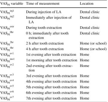
Table 1 Time and location of visual analogue scale pain intensity measurements (VASPI)
LA local anesthetic
VASPI variable Time of measurement Location
VASPIinj1 During injection of LA Dental clinic
VASPIinj2 Immediately after injection of
LA Dental clinic
VASPIextr During tooth extraction Dental clinic
VASPI0hr 0 h; immediately after tooth
extraction Dental clinic
VASPI2hr 2 h after tooth extraction Home (or school)
VASPI4hr 4 h after tooth extraction Home (or school)
VASPIev1 1st evening after tooth extraction Home
VASPImo1 1st morning after tooth extraction Home
VASPIev2 2nd evening after tooth
extrac-tion Home
VASPIev3 3rd evening after tooth extraction Home
VASPIev4 4th evening after tooth extraction Home
VASPIev5 5th evening after tooth extraction Home
VASPIev6 6th evening after tooth extraction Home
Results
Patient characteristics
34 children were initially invited. Three were excluded: one because placement of orthodontic brackets was planned for the day after the first extraction; one, because orthodontic treatment had already begun; and one, who did not understand the VAS. Thus, 31 patients were included and divided into two age groups: 10–13-year-olds, and 14–15-year-olds (Table 2). Thus, 62 extractions were performed, but the number of valid VASPI registrations
made at home or at school, were lower owing to patients failing to return registrations, VAS not correctly filled out, or excluded when patients had self-administered oral analgesics.
Only one individual reported a CFSS-DS score ≥ 38 (40, representing DFA) before the first extraction, and none before the second. There were no statistically sig-nificant differences in mean CFSS-DS scores before the first and second extractions, between sexes or age groups, and there was no relationship between CFSS-DS scores and VASPI. No additional analyses were made regarding
the CFSS-DS scores since DFA did not have any impact on pain intensity.
Clinical procedures
Extraction began on the right side in 20 cases (64.5%); 44 of the 62 extracted teeth were permanent maxillary first premolars; the remaining were permanent maxillary second premolars. The mean time between the first and second extractions was 15 days (range 11–33 days). Mean treatment time, from application of topical anaesthetic to finished extraction of the tooth, was 13.2 min (range 8–22) for the first extraction, and 12.6 min (range 8–19) for the second extraction (p = 0.327, paired-samples t test). 36 of the extractions were done in the morning. 2 h after
treatment, VASPI2hr did not differ statistically significantly
between extractions performed before or after noon. 13 of 29 participants (44.8%; 6 boys and 7 girls) reported previous experience of LA before entering the present study. No statistically significant differences in pain intensity dur-ing injection, extraction, or 2 h after treatment occurred between patients with and without previous experience of LA.
At the first extraction, nine patients reported insufficient anaesthesia after the injection of one capsule LA, and at the second extraction, seven patients. These patients (12 in all) received another 0.5 to 1.5 capsules of LA before extraction. VASPI scores at 2 h after treatment did not differ statistically significantly between patients receiving one capsule of LA and those who received more (Table 3).
Patients reported higher VASPI scores during injection (VASPIinj1) than during extractions (VAS
PIextr) (Fig. 1). There
were no differences in VAS scores between the first and sec-ond appointments (VASPIinj1, VASPIinj2, VASPIextr, VASPI0hr,
VASPI2hr and VAS
PI4hr) (Table 4).
Postoperative pain
Postoperative pain ratings followed the same course for all patients (Figs. 1, 2a–c), with no statistically significant dif-ferences between the first and second extractions, between boys and girls, or between age groups (Tables 3, 4). Pain intensity peaked 2 h after treatment at a mean VASPI for all extractions of 27.3 (SD 20.8; median 23.0) and then decreased to a mean of 18.3 (SD 17.9; median 10.0) at 4 h after treatment. At VASPI2hr, 16 of 57 extractions resulted in
pain (VASPI ≥ 40). This level of pain intensity remained in 6 of 53 cases at VASPI4hr. The corresponding numbers for
VASPI ≥ 30 were 24 of 57 extractions at VASPI2hr and 13 of
53 at VASPI4hr.
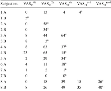
Eight patients took oral analgesics after the first extrac-tion and six of these did so after the second on their own initiative. Table 5 shows the VASPI scores of these patients
made before their intake of oral analgesics. Any scores made after medication were excluded from the statistical analyses. Pain intensity did not differ at any time between patients who took oral analgesics and those who did not.
Costs outside the clinic
Time spent for both extractions showed that parents (and/or legal guardians) spent a mean time of 3.6 h (SD 1.4; median 4.0) for accompanying their children to the two dental treat-ments. The majority of the participants came to the dental clinic by car, but since all treatments within this study, were performed at the university clinic, (a clinic different from their normal dental clinic), the expenditures for their travel was difficult to calculate. Children missed a mean time of Table 2 Characteristics of the 31 participants by sex and age
# Independent samples t test
Boys Girls Total
Signifi-cance p value
No. (%) 15 (48.4) 16 (51.6) 31 (100)
Mean age in years
(SD) 13.5 (1.1) 13.3 (1.1) 13.4 (1.1) 0.591
#
Age group (no.)
10−13 years 6 10 16
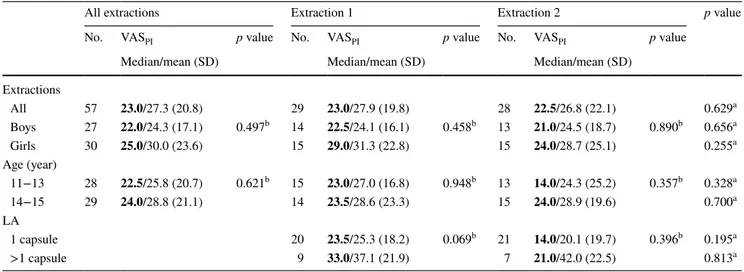
Table 3 Visual analogue scale pain intensity (VASPI) measurements after first and second extraction by sex, age and volume of local anesthetics,
2 h after extraction
no. number of teeth extracted, LA local anesthetics
a Comparisons between the first and second extraction (Wilcoxon signed-ranks test) b Comparisons within groups (sexes, age groups, and amount of LA) (Mann–Whitney U test)
All extractions Extraction 1 Extraction 2 p value
No. VASPI p value No. VASPI p value No. VASPI p value
Median/mean (SD) Median/mean (SD) Median/mean (SD)
Extractions All 57 23.0/27.3 (20.8) 29 23.0/27.9 (19.8) 28 22.5/26.8 (22.1) 0.629a Boys 27 22.0/24.3 (17.1) 0.497b 14 22.5/24.1 (16.1) 0.458b 13 21.0/24.5 (18.7) 0.890b 0.656a Girls 30 25.0/30.0 (23.6) 15 29.0/31.3 (22.8) 15 24.0/28.7 (25.1) 0.255a Age (year) 11−13 28 22.5/25.8 (20.7) 0.621b 15 23.0/27.0 (16.8) 0.948b 13 14.0/24.3 (25.2) 0.357b 0.328a 14−15 29 24.0/28.8 (21.1) 14 23.5/28.6 (23.3) 15 24.0/28.9 (19.6) 0.700a LA 1 capsule 20 23.5/25.3 (18.2) 0.069b 21 14.0/20.1 (19.7) 0.396b 0.195a >1 capsule 9 33.0/37.1 (21.9) 7 21.0/42.0 (22.5) 0.813a
Fig. 1 Box plot of pain intensity measurements (visual analogue scale, VASPI) from injection of local anaesthesia to the evening of
the 7th day post-treatment. Combined measurements from first and
second extractions. Horizontal bars indicate VASPI 40 (threshold for
moderate pain) and 30 (threshold for mild pain). Measurements after intake of analgesics are excluded
6.1 h (SD 4.4; median 4.0) of school. None of the partici-pants sought additional dental treatment for complications after the extractions. Seven children (4 girls and 3 boys) did not return to school after tooth extraction. No detailed data for reason or if they stayed at home after one or both treat-ments is available.
Discussion
In the present study, the majority of the patients who under-went uncomplicated bilateral extractions of maxillary pre-molars experienced mild to moderate levels of postoperative pain. Pain intensity peaked 2 h after tooth extraction, and had declined radically 4 h after extractions. Thus, pain was perceived for a short period of time. There were no differ-ences in pain intensity profiles between the first and second extractions. Nor were there any differences between sexes, or between younger and older patients. Thus, postoperative pain after uncomplicated bilateral extractions of maxillary premolars due to orthodontic indications follows a steady pain intensity profile. As this treatment is relatively com-mon, easy to standardise, and reproducible, we suggest this model as robust and suitable for studies on treatment of in young patients.
Pain studies on children and adolescents can raise ethical issues, why these studies should primarily be done on adult patients. In the present study, however, the natural course of pain during and after a standard procedure that general
dental practitioners perform on a daily basis, was studied. We chose this treatment since it is potentially painful but, according to the literature, not associated with a high fre-quency of any other problems. The patients and their par-ents were informed verbally and in writing that they could withdraw from the study whenever they liked. They also had access to the research group throughout the study, if needed. Parents and guardians signed informed-consent forms before a participant was allowed to enter the study, and the chil-dren received age appropriate information and assented. This study also received ethical approval.
Pain intensity peaked 2 h after treatment in the present study which is concordant with a study from 2013 (Mustafa et al. 2013). It is plausible to speculate that this peak coin-cides with the time when the anaesthesia wears off. Duration of anaesthesia when using Lidocaine 2% (with 1:50,000 to 1:100,000 epinephrine) in the pulp is about 60 min after infiltration injection and in soft tissue, about 170 min (Becker and Reed 2006). The duration of Lidocaine 2% differs from one individual to another; well illustrated in a study from 2016 (Elbay et al. 2016) who reported a vari-ation in durvari-ation of LA after inferior alveolar blocks from 111 to 285 min (mean 149 min). Thus, measurement of pain intensity after both 2 and 4 h appears adequate and crucial, especially when infiltration technique, and not inferior alveo-lar nerve block, is used.
The prevalence of postoperative pain after tooth extrac-tion varies among studies. While 28% (16 of 57) of the extractions in the present study led to moderate postoperative Table 4 Comparisons of first and second extraction, visual analogue scale pain intensity (VASPI) scores (median/mean, [SD]), and numbers of
participants reporting VASPI ≥ 40 and 30, respectively, at the various time points
a Wilcoxon sign-ranks test b McNemar’s test
c Two recordings excluded from analyses due to intake of oral analgesics, affecting the VAS PI
Extraction 1 Extraction 2 p value
VASPI VASPI ≥ 30 VASPI ≥ 40 VASPI VASPI ≥ 30 VASPI ≥ 40
Median/mean (SD) Median/mean (SD) During injection 25.0/29.3 (17.5) 22.5/24.2 (17.2) 0.114a 12 of 29 11 of 30 1.000b 8 of 29 5 of 30 0.508b During extraction 13.0/20.4 (18.6) 11.0/16.0 (13.9) 0.153a 8 of 29 5 of 30 0.375b 4 of 29 2 of 30 0.625b 2 h after extraction 23.0/27.8 (20.3) 22.5/26.8 (22.3) 0.629a 12 of 29 12 of 28 1.000b 11 of 29 5 of 28 0.063b 4 h after extraction 12.0/17.9 (16.1) 10.0/18.7 (19.9) 0.493a 6 of 27c 7 of 26c 1.000b 2 of 27c 4 of 26c 0.625b
Fig. 2 Box plot of pain intensity measurements (visual analogue scale, VASPI) from injection of local anaesthesia to the evening of the 7th day post-treatment. Horizontal bars indicate VASPI 40 (threshold for moderate pain) and 30 (threshold for mild pain). Measurements
after intake of analgesics are excluded. a Measurements are from the first and second extractions. b Measurements for boys and girls. c Measurements for younger (11–13 years) and older (14–15 years) age groups
pain (VASPI ≥ 40), other studies report from around 30%
up to 85% postoperative pain (Acs et al. 1986; Ashkenazi et al. 2007; McGaw et al. 1987; Moore et al. 1985). Fac-tors related to study design may explain the differences; for example, number of participants, age distribution, reasons for extraction, extraction technique, tooth extracted (in max-illa or mandible), and pain measurement method.
An important finding in the present study was that pain intensity did not differ between the first and second extrac-tions, why there is no reason to assume any conditioning effect when repeating the treatment, i.e. the tooth extraction. This has not been studied before. This supports a future ran-domized controlled trial based on split mouth design using bilateral extractions of maxillary premolars as a model to investigate the effect of different pharmacologic agents, dos-ages or placebo in two arms. It is unlikely that there will be any conditioning effect provided a proper sample size calculation precedes the study.
The present study used a standardised protocol with uncomplicated bilateral extractions in the maxilla. This treatment is easy to reproduce, the anatomy of the roots are rather uncomplicated on upper premolars, easily accessible, and the trauma to the bone tissue is limited. Teeth extracted on orthodontic indications are in general healthy and free from inflammatory response in the pulp, which could affect Fig. 2 (continued)
Table 5 Visual analogue scale pain intensity (VASPI) scores of eight patients who self-administered oral analgesics (8 after the first extrac-tion, 6 after the second)
Time points are hours after extraction
A measurements from first extraction, B measurements from second extraction
a Last valid VAS measurement before intake of oral analgesics. All
measurement after intake of oral analgesics were excluded from the analyses
Subject no. VASPI0h VASPI2h VASPI4h VASPIev1 VASPImo1
1 A 0 13 4 4a 1 B 5a 2 A 0 58a 2 B 0 34a 3 A 8 44 64a 3 B 8 3a 4 A 8 63 37a 4 B 23 65 15a 5 A 2 29 34a 6 A 4 11 18a 7 A 1 2 1a 7 B 0 0 0a 8 A 0 18 39 15 26a 8 B 8 26 49 35 40a
the sensation of pain negatively. Administration of local anaesthetics in the maxilla is also less operator sensitive than administration of inferior alveolar nerve block. The present study included a homogeneous group of patients regarding age as well as reason for extractions, and one operator car-ried out the majority of all treatments. The study used a self-reported and patient-centred outcome, which is preferred due to the subjective nature of pain (American Academy of Pediatrics. Committee on Psychosocial Aspects of Child and Family Health, Task Force on Pain in Infants, Children, and Adolescents 2001). Further, pain intensity was measured several times over a 7-day period, enabling an understand-ing of the natural course of pain. These are strengths of the present study.
Previous data on sample size calculation applicable to this study could not be found. In two previous studies (Acs et al. 1986; Ashkenazi et al. 2007), they had 221 and 84 participants, respectively, who underwent dental extractions (tooth type and reason for extraction not explained). On the other hand, a study (Hariharan et al. 2014) included only 27 patients in a split-mouth designed evaluation of pain after use of two different forceps during extraction of upper pre-molars prior to orthodontic treatment. In the light of this, the number of participants in present study is adequate.
Use of the VAS as a pain rating scale for the age group in the present study could be discussed. At some ages, trans-lating pain experiences to a rating tool can be difficult for children, even though they are capable of expressing their pain in words. When children are over the age of 7, various types of scales for self-assessment can be used. Between 7 and 10 years, the literature has advocated face scales, and from the age of 10, numerical rating scales (NRS) and VAS (Norrbrink and Lundeberg 2012; Shields et al. 2003). To ensure comprehension of the VAS in the present study, the participants received thorough information and instruction about the instrument before each extraction.
Cut-off values are always troublesome, especially when dealing with something such as pain, which by definition is subjective (IASP 2014). A fixed cut-off point on the VAS is not relevant due to inter-individual variations in pain expres-sion (van Dijk et al. 2002). A person who scores 45 on the VAS might still not think it is painful, while another might perceive a score of 24 as very painful. Still, there is a need for researchers and clinicians to have some kind of grid or ruler to develop methods for intervening in and prevent-ing pain. To date, no cut-off for postoperative clinical pain requiring pain management in children has been defined. For postoperative pain, some studies have suggested, arbitrarily, VASPI 30 as cut-off for mild to moderate pain (Berde et al.
1991; Breivik et al. 2008; Mustafa et al. 2013; Taddio et al.
2009; van Dijk et al. 2002). VASPI 40 and 44 have been used
as thresholds for moderate pain or need for pain management (Jensen et al. 2003; van Dijk et al. 2002).
In this study, VASPI ≥ 40 succeeded in identifying mod-erate pain 2 h postoperatively in 16 of 57 extractions. A VASPI ≥ 30, however, would identify 26 of 57 scores 2 h
after extraction as mild to moderately painful; this is a higher number, even though lower in intensity. More importantly, the same pattern of a short peak in pain intensity followed by a rapid decrease could be seen. This is important as it puts light on a short window of pain that has to be managed in some way. One strategy, possibly often overlooked, is infor-mation about expected level and the duration of pain and discomfort after the extraction. The finding of a pain profile in this study is thus important as it enables dental health professionals to provide patients with more accurate infor-mation about the expected course of pain after an extraction. Use of pharmacological agents such as oral analgesics is another potential strategy. Still there are some questions. If analgesics were to be used, what would be the best way to administer them? A recently updated systematic review of the effect of preoperative analgesics for additional pain relief in dental treatment of children (Ashley et al. 2016) identified and evaluated five trials. The researchers concluded that it was not possible to determine whether preoperative analge-sics were of benefit in paediatric dentistry for procedures under local anaesthesia, and the authors called for further randomized clinical trials. Based on the pain ratings in this study, and the pain profiles derived from the VASPI ratings
of the participants, would a single dose analgesics after treat-ment be a good alternative? The benefits with this model would be that the onset of the drug would be nearer the pain peak. However, this must be studied further. To our knowl-edge, no systematic review on postoperative administration of analgesics in paediatric dentistry has been published. Thus, there is hardly any evidence for analgesic use, pre- or postoperatively, to reduce pain after uncomplicated dental treatment in children; this remains a knowledge gap. Based on this, and the fact that all administration of pharmacologic agents comes with a risk (Matok et al. 2016; Norman et al.
2014), it can be questioned if there is any reason for clinical guidelines in this area. Analogously, there is no rationale for introducing or routinely using administration of analge-sics in conjunction with routine dental treatments such as filling therapies or uncomplicated extractions (Berlin et al.
2018). When considering use of pharmacological agents with children, there must be a clear-cut reason for doing so. Otherwise, development of general guidelines would be unethical and contraindicated. Instead, the relatively mild pain intensity and short duration of pain indicates that medication should be individually tailored and not a general recommendation.
Another important aspect when making general recom-mendations is health economics. In this study, the cost of the dental treatment could be mirrored by the Swed-ish so-called reference price. For an uncomplicated tooth
extraction, the reference price was €104.3 [Tandvårds- och läkemedelsförmånsverket (The Dental and Pharmaceutical Benefits Agency)] and this is assumed to mirror the total cost in the dental clinic. Other costs are related to travel-ling (which could not be calculated), and the indirect cost for productivity loss related to the parents’ absence from work.
The average wage (year) for individuals aged 40–49, which may be assumed to be parents of children aged 10–15 years, is €40,137 [Statistiska centralbyrån (Statistics Swe-den)] and when the social fees are added (35%) the value is €54,185. Based on number of work hours for 2016 [Arbet-stimmar per månad (work hours per month)], the hourly pro-duction value would be €26.77. Since the parents were away 3.6 h in mean, during the study period, the indirect cost for productivity loss would be €96.4. Adding the cost for the two extractions results in €305.0. This may be considered a low cost for this treatment, especially when comparing the cost for the orthodontic treatment, following (Petrén et al. 2011; Ganzer et al. 2018). In the present study, no side effects, requiring additional treatment were reported. Tooth extraction is a standard procedure and on the right indications, €305.0 has to be considered a reasonable cost and hence, this is a cost-effective procedure.
Conclusion
In conclusion, uncomplicated extractions of premolars caused moderate postoperative pain (VASPI ≥ 40) in 16
(28%) of 57 extractions in young patients. There were no differences in the natural course of pain between first and second extractions, younger and older patients, or between sexes. The pain peaked 2 h after extraction, around the time where LA wears off. After that, there was a rapid decrease in pain intensity notable already at 4 h after extractions. As the pain intensity profiles for both extrac-tions were similar to each other, it is suggested that bilat-eral extractions of maxillary premolars is a suitable model for studies on pain management.
Acknowledgements The study was supported by grants from the Swedish Dental Society, the Swedish Society of Paediatric Dentistry; the Faculty of Odontology, Malmö University; Skåne Regional Coun-cil, Sweden; and the American Dental Society of Sweden.
Author contributions HB and GK conceived the ideas; HB collected the data; HB, GK, TL, TD, KR, and DT analysed the data; HB and GK led the writing; and all authors participated in finalizing the manuscript. Compliance with ethical standards
Conflict of interest The authors declare no conflict of interest.
Open Access This article is distributed under the terms of the Crea-tive Commons Attribution 4.0 International License (http://creat iveco mmons .org/licen ses/by/4.0/), which permits unrestricted use, distribu-tion, and reproduction in any medium, provided you give appropriate credit to the original author(s) and the source, provide a link to the Creative Commons license, and indicate if changes were made.
References
Acs G, Moore PA, Needleman HL, Shusterman S. The incidence of post-extraction pain and analgesic usage in children. Anesth Prog. 1986;33:147–51.
Al-Khateeb TH, Alnahar A. Pain experience after simple tooth extraction. J Oral Maxillofac Surg. 2008;66:911–7.
American Academy of Pediatrics. Committee on Psychosocial Aspects of Child and Family Health, Task Force on Pain in Infants, Children, and Adolescents. The assessment and man-agement of acute pain in infants, children, and adolescents. Pediatrics. 2001;108:793–7.
Arbetstimmar per månad. [Work hours per month]. http://www.arbet stimm arper manad .se/. Accessed September 2018.
Ashkenazi M, Blumer S, Eli I. Post-operative pain and use of anal-gesic agents in children following intrasulcular anaesthesia and various operative procedures. Br Dent J. 2007;202:7.
Ashley PF, Parekh S, Moles DR, et al. Preoperative analgesics for additional pain relief in children and adolescents having dental treatment. Cochrane Database Syst Rev. 2016;(8):CD008392. Becker DE, Reed KL. Essentials of local anesthetic pharmacology.
Anesth Prog. 2006;53:10.
Berde CB, Lehn BM, Yee JD, et al. Patient-controlled analgesia in children and adolescents: a randomized, prospective comparison with intramuscular administration of morphine for postoperative analgesia. J Pediatr. 1991;118:460–6.
Berlin H, List T, Ridell K, et al. Dentists’ attitudes towards acute pharmacological pain management in children and adolescents. Int J Paediatr Dent. 2018;28:152–60.
Bhalla J, Meechan JG, Lawrence HP, et al. Effect of time on clini-cal efficacy of topiclini-cal anesthesia. Anesth Prog. 2009;56:36–41. Breivik H, Borchgrevink PC, Allen SM, et al. Assessment of pain.
Br J Anaesth. 2008;101:17–24.
Cuthbert MI, Melamed BG. A screening device: children at risk for dental fears and management problems. ASDC J Dent Child. 1982;49:432–6.
Elbay US, Elbay M, Kaya E, et al. Effects of two different anesthetic solutions on injection pain, efficacy, and duration of soft-tissue anesthesia with inferior alveolar nerve block for primary molars. J Clin Pediatr Dent. 2016;40:456–63.
Ganzer N, Feldmann I, Petrén S, et al. A cost-effectiveness analysis of anchorage reinforcement with miniscrews and molar blocks in adolescents: a randomized controlled trial. Eur J Orthod. 2018:cjy041.
Ghanei M, Arnrup K, Robertson A. Procedural pain in routine dental care for children: a part of the Swedish BITA study. Eur Arch Paediatr Dent. 2018. https ://doi.org/10.1007/s4036 8-018-0368-2.
Hariharan S, Narayanan V, Soh CL. Split-mouth comparison of phys-ics forceps and extraction forceps in orthodontic extraction of upper premolars. Br J Oral Maxillofac Surg. 2014;52:137. Huskisson EC. Measurement of pain. Lancet. 1974;2:1127–31. IASP. International Association for the Study of Pain. 2014. http://
www.iasp-pain.org/Taxon omy. Accessed September 2016. Jensen MP, Chen C, Brugger AM. Interpretation of visual analog
scale ratings and change scores: a reanalysis of two clinical tri-als of postoperative pain. J Pain. 2003;4:407–14.
Klingberg G. Reliability and validity of the Swedish version of the Dental Subscale of the Children’s Fear Survey Schedule, CFSS-DS. Acta Odontol Scand. 1994;52:255–6.
Klingberg G, Broberg AG. Dental fear/anxiety and dental behaviour management problems in children and adolescents: a review of prevalence and concomitant psychological factors. Int J Paediatr Dent. 2007;17:391–406.
Klingberg G, Ridell K, Brogardh-Roth S, et al. Local analgesia in pae-diatric dentistry: a systematic review of techniques and pharma-cologic agents. Eur Arch Paediatr Dent. 2017;18:323–9. Maragakis GM, Musselman RJ. The time used to administer local
anes-thesia to 5 and 6 year olds. J Clin Pediatr Dent. 1996;20:321–3. Matok I, Elizur A, Perlman A, et al. Association of acetaminophen and
ibuprofen use with wheezing in children with acute febrile illness. Ann Pharmacother. 2016;27.
McGaw T, Raborn W, Grace M. Analgesics in pediatric dental surgery: relative efficacy of aluminum ibuprofen suspension and acetami-nophen elixir. ASDC J Dent Child. 1987;54:106–9.
Moore PA, Acs G, Hargreaves JA. Postextraction pain relief in children: a clinical trial of liquid analgesics. Int J Clin Pharmacol Ther Toxicol. 1985;23:573–7.
Mustafa O, Parekh S, Ashley P, et al. Post-operative pain and anxi-ety related to dental procedures in children. Eur J Paediatr Dent. 2013;14:289–94.
Norman H, Elfineh M, Beijer E, et al. Also ibuprofen, not just par-acetamol, can cause serious liver damage in children. NSAIDs should be used with caution in children, as shown in case with fatal outcome. Läkartidningen. 2014;111:1709–11.
Norrbrink C, Lundeberg T, editors. Om smärta—ett fysiologiskt pers-pektiv. Lund: Studentlitteratur AB; 2012 (In Swedish). Petrén S, Bjerklin K, Marké L, et al. Early correction of posterior
cross-bite—a cost-minimization analysis. Eur J Orthod. 2011;35:14–21. Shields BJ, Cohen DM, Harbeck-Weber C, et al. Pediatric pain meas-urement using a visual analogue scale: a comparison of two teach-ing methods. Clin Pediatr (Phila). 2003;42:227–34.
Statistiska centralbyrån (SCB) [Statistics Sweden]. 2018. http://www. scb.se/. Accessed Sep 2018.
Sveriges Riksbank [Sweden’s central bank]. 2018. https ://www. riksb ank.se/sv/stati stik/sok-ranto r--valut akurs er/?g130-SEKEU RPMI=on&from=2018-08-21&to=2018-09-21&f=Year&c=cAver age&s=Comma . Accessed Sep 2018. Taddio A, O’Brien L, Ipp M, et al. Reliability and validity of observer
ratings of pain using the visual analog scale (VAS) in infants undergoing immunization injections. Pain. 2009;147:141–6. Tandvårds- och läkemedelsförmånsverket [The Dental and
Pharma-ceutical Benefits Agency]. 2018. https ://www.tlv.se/tandv ard/refer enspr islis ta.html. Accessed Sep 2018.
van Dijk M, Koot HM, Saad HH, et al. Observational visual analog scale in pediatric pain assessment: useful tool or good riddance? Clin J Pain. 2002;18:310–6.
Publisher’s Note Springer Nature remains neutral with regard to jurisdictional claims in published maps and institutional affiliations.

![Table 4 Comparisons of first and second extraction, visual analogue scale pain intensity (VAS PI ) scores (median/mean, [SD]), and numbers of participants reporting VAS PI ≥ 40 and 30, respectively, at the various time points](https://thumb-eu.123doks.com/thumbv2/5dokorg/3951907.75484/6.892.79.818.121.430/comparisons-extraction-analogue-intensity-participants-reporting-respectively-various.webp)