R E S E A R C H A R T I C L E
Open Access
Acid tolerance in early colonizers of oral
biofilms
Gabriella Boisen, Julia R. Davies and Jessica Neilands
*Abstract
Background: In caries, low pH drives selection and enrichment of acidogenic and aciduric bacteria in oral biofilms, and development of acid tolerance in early colonizers is thought to play a key role in this shift. Since previous studies have focussed on planktonic cells, the effect of biofilm growth as well as the role of a salivary pellicle on this process is largely unknown. We explored acid tolerance and acid tolerance response (ATR) induction in biofilm cells of both clinical and laboratory strains of three oral streptococcal species (Streptococcus gordonii, Streptococcus oralis and Streptococcus mutans) as well as two oral species of Actinomyces (A. naeslundii and A. odontolyticus) and examined the role of salivary proteins in acid tolerance development.
Methods: Biofilms were formed on surfaces in Ibidi® mini flow cells with or without a coating of salivary proteins and acid tolerance assessed by exposing them to a challenge known to kill non-acid tolerant cells (pH 3.5 for 30 min) followed by staining with LIVE/DEAD BacLight and confocal scanning laser microscopy. The ability to induce an ATR was assessed by exposing the biofilms to an adaptation pH (pH 5.5) for 2 hours prior to the low pH challenge.
Results: Biofilm formation significantly increased acid tolerance in all the clinical streptococcal strains (P < 0.05) whereas the laboratory strains varied in their response. In biofilms, S. oralis was much more acid tolerant than S. gordonii or S. mutans. A. naeslundii showed a significant increase in acid tolerance in biofilms compared to planktonic cells (P < 0.001) which was not seen for A. odontolyticus. All strains except S. oralis induced an ATR after pre-exposure to pH 5.5 (P < 0.05). The presence of a salivary pellicle enhanced both acid tolerance development and ATR induction in S. gordonii biofilms (P < 0.05) but did not affect the other bacteria to the same extent. Conclusions: These findings suggest that factors such as surface contact, the presence of a salivary pellicle and sensing of environmental pH can contribute to the development of high levels of acid tolerance amongst early colonizers in oral biofilms which may be important in the initiation of caries.
Keywords: Streptococci, Actinomyces, Salivary proteins, Pellicle, Acid tolerance response Background
The human oral microbiota comprises more than 600 different bacterial species of which around 100–200 can be found in a single individual [1]. Normally the micro-biota is dominated by species belonging to the genera Streptococcus and Actinomyces as well as Neisseria,
Veillonella and Granulicatella [2]. Bacteria colonizing the oral cavity are mainly found as complex, multispe-cies biofilms attached to the hard and soft-tissue sur-faces. On the teeth, biofilm formation is initiated by interactions between bacteria and the acquired enamel pellicle, a thin film of proteins derived from saliva and gingival crevicular fluid [3]. Early colonizers such as Streptococcus and Actinomyces, can adhere to the tooth surface through non-specific interactions as well as spe-cific binding of bacterial surface adhesins to salivary
© The Author(s). 2021 Open Access This article is licensed under a Creative Commons Attribution 4.0 International License, which permits use, sharing, adaptation, distribution and reproduction in any medium or format, as long as you give appropriate credit to the original author(s) and the source, provide a link to the Creative Commons licence, and indicate if changes were made. The images or other third party material in this article are included in the article's Creative Commons licence, unless indicated otherwise in a credit line to the material. If material is not included in the article's Creative Commons licence and your intended use is not permitted by statutory regulation or exceeds the permitted use, you will need to obtain permission directly from the copyright holder. To view a copy of this licence, visithttp://creativecommons.org/licenses/by/4.0/. The Creative Commons Public Domain Dedication waiver (http://creativecommons.org/publicdomain/zero/1.0/) applies to the data made available in this article, unless otherwise stated in a credit line to the data.
* Correspondence:Jessica.Neilands@mau.se
Section for Oral Biology and Pathology, Faculty of Odontology, and Biofilms -Research Center for Biointerfaces, Malmö University, SE-205 06 Malmö, SWED EN
acid, resulting in a fall in biofilm pH [9]. In health, com-pensatory mechanisms, including buffering by saliva and the generation of alkaline end-products from salivary urea and arginine by bacteria, are able to counteract this perturbation and homeostasis is re-established [10]. This has been termed the ‘dynamic stability stage’ by Nyvad and Takahashi [11] and under these conditions, the composition of the community remains relatively stable. However, when severe ecological stresses overcome the resilience of the biofilm, the balance is perturbed leading to changes in biofilm physiology and composition [12]. According to the‘ecological plaque hypothesis’, frequent episodes of low biofilm pH drive the selection of an acid tolerant microbiota that can continue to metabolize and produce acid even when the pH is low [13]. This corre-sponds to the ‘acidogenic stage’ as described by Nyvad and Takahashi [11]. The increased duration of low pH pushes the de/re-mineralization balance towards net dis-solution of the oral hard tissues, eventually resulting in the development of caries lesions. Dental caries is thought to affect around 2.5 billion people worldwide [14] and estimates suggest that around 5% of the health-care budgets of the Organisation for Economic Co-operation and Development (OECD) countries are con-sumed by the disease [15].
Streptococcus mutans has long been considered as an important agent in caries development largely due to its isolation from carious sites and its ability to withstand low pH [16]. However, since S. mutans can be present at low levels, or even absent, in caries lesions and individ-uals with S. mutans do not always have caries, the pres-ence of this bacteria is more likely to be an indicator of low pH conditions in the biofilm rather than the causa-tive agent [17–19]. Therefore, other prominent members of oral biofilms i.e various species of non-mutans streptococci including Streptococcus sanguinis, Strepto-coccus oralis, StreptoStrepto-coccus gordonii and StreptoStrepto-coccus mitis as well as Actinomyces spp must play a significant role in driving the development of an acid tolerant microbiota that initiates the development of caries [20]. Experiments in planktonic culture have shown that some of these bacteria are able to adapt and survive under low
trast, acid tolerance development in non-mutans streptococci and other oral species has not been as well studied, although changes in fatty acid composition of the membrane, as well as activation of the arginine dei-minase system have been described in S. gordonii and S. sanguinis,respectively in response to low pH [24,25].
Biofilm growth has been shown to have profound ef-fects on the physiological properties of bacteria, and characteristics such as sensitivity to antimicrobial agents and growth rates differ significantly between biofilm bac-teria and their planktonic counterparts [26]. For ex-ample, S. mutans becomes more acid tolerant when grown in a biofilm as compared to planktonic growth [27,28]. In this study we aimed to investigate the role of biofilm formation on development of acid tolerance and ATR induction in clinical and laboratory strains of early oral colonizers (non-mutans streptococci and Actinomy-ces) and to explore the effect of salivary proteins on these processes.
Results
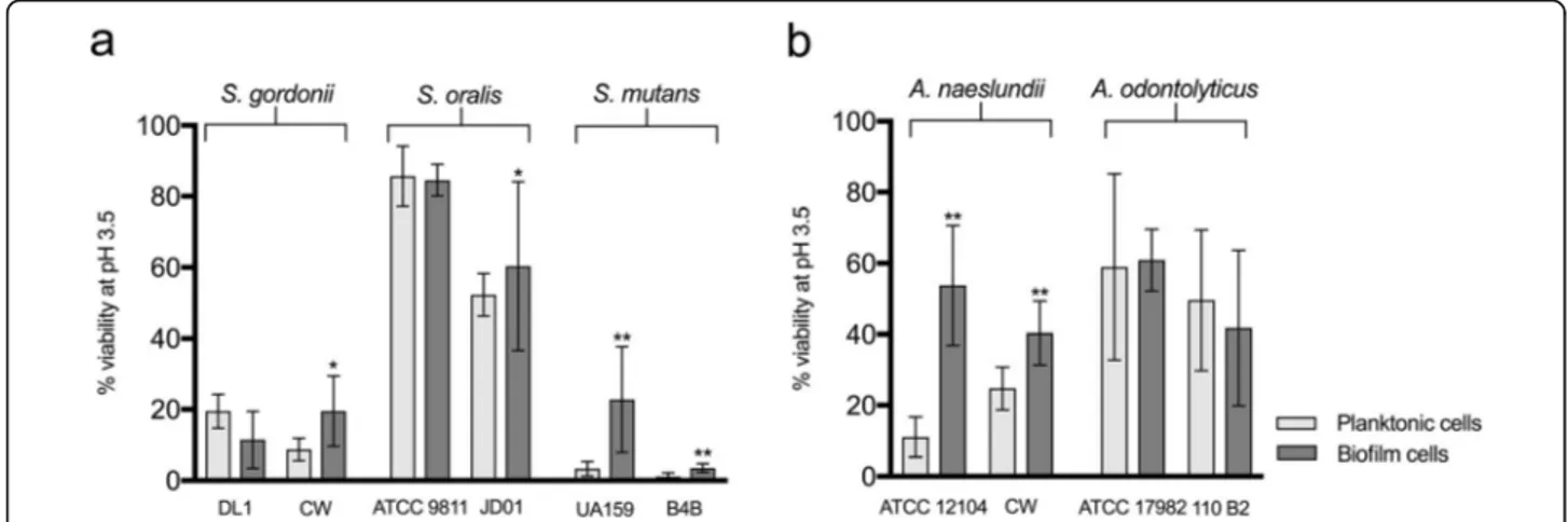
Effect of early biofilm formation on acid tolerance The effect of biofilm formation on the acid tolerance of different early oral colonizing bacteria was assessed by determining the level of survival following a low pH challenge for cells adhered to an uncoated surface and comparing with their planktonic counterparts. No differ-ences in the level of surface coverage of biofilm cells were seen between the biofilm control cells (kept at pH 7.5) and those exposed to the acid challenge used to dif-ferentiate acid tolerant from non-acid tolerant cells [i.e. exposure to acid did not remove the cells from the sur-face (data not shown)]. The clinical strains of S. gordonii (CW), S. mutans (B4B), and S. oralis (JD01) showed small but significant increases in acid tolerance in the biofilms compared to planktonic culture (2.1-fold, 4-fold, and 1.1-fold increase respectively, P < 0.05, Fig.1a). For the laboratory strains, the picture was variable, with the S. mutans strain (UA159) showing a significant in-crease in acid tolerance in the biofilm (7.7-fold, P < 0.001) while no increase was seen for either S. gordonii (DL1) or S. oralis (ATCC9811). For all species, with the
exception of S. gordonii, the biofilm acid tolerance of the laboratory strain was greater than that of the clinical one, although the differences were generally small and not significant. Comparison between the different streptococci showed that both the clinical and laboratory strains of S. oralis had much higher levels of biofilm acid tolerance than the S. gordonii and S. mutans strains (Fig. 1a). Interestingly, the clinical strain of S. mutans (B4B) showed the lowest acid tolerance of all the strains tested. Both the clinical and the laboratory strain of A. nae-slundii showed significantly greater acid tolerance in biofilms compared to their planktonic counterparts (P < 0.001, Fig. 1b), although the most pronounced increase was seen for A. naeslundii ATCC 12104, with a 4.9-fold increase compared with a 1.6-fold increase for A. nae-slundii CW. In contrast, for the two A. odontolyticus strains, there was no increase in acid tolerance on bio-film formation. The overall levels of acid tolerance in biofilms were quite similar for A. odontolyticus and A. naeslundii (Fig. 1b). Thus, for the bacteria with a rela-tively high degree of acid tolerance in planktonic culture, biofilm formation had little or no additive effect whereas varying degrees of increased acid tolerance were seen for the other strains after biofilm formation.
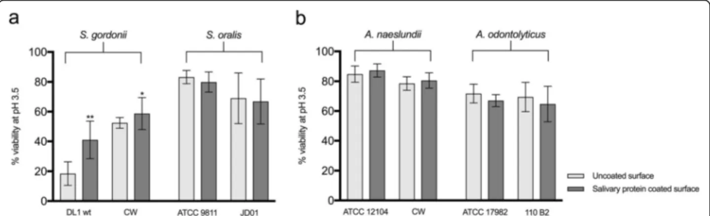
Effect of adherence to a salivary pellicle on acid tolerance The surfaces to which bacteria adhere in the oral cavity are coated with a pellicle of saliva and therefore, we in-vestigated whether salivary proteins could further en-hance the surface-induced acid tolerance of the strains used in this study. Due to the fact that strains of S. mutansadhered poorly to salivary proteins (giving a sur-face area coverage of less than 1%), these were not inves-tigated further. Neither of the strains of S. oralis showed an enhanced acid tolerance on adherence to a saliva-coating compared to uncoated surfaces. For S. gordonii
however, the laboratory (DL1) and clinical strain (CW) showed 1.5-fold and 1.3-fold increases in acid tolerance respectively (Fig.2a), and this change was significant for DL1 (P < 0.001). A similar picture was seen for the Acti-nomycesspecies where no enhancement was seen for the A. odontolyticus strains while a significant increase in acid tolerance was seen for the clinical strain of A. nae-slundii(1.3-fold increase, P < 0.001), but not the labora-tory strain (Fig. 2b). Thus, surface associated salivary proteins enhanced acid tolerance during biofilm forma-tion for some strains of oral bacteria but not others. Development of an ATR
The ability of the bacterial strains to adjust to an acidic environment through induction of an ATR was deter-mined by allowing them to adapt to pH 5.5 prior to the low pH challenge. Exposure to pH 5.5 did not in itself affect the viability of any of the strains as shown by staining with Baclight LIVE/DEAD (data not shown). With the exception of S. oralis, which was already highly acid tolerant, the acid tolerance increased significantly for all the strains of streptococci after a pre-exposure to pH 5.5 (p < 0.001, Fig.3a). The largest difference was ob-served for the S. mutans strains, UA159 and B4B, which showed 3.7-fold and 18-fold increases respectively com-pared to non-adapted cells. Similarly, both strains of S. gordoniialso showed enhanced acid tolerance after pre-exposure to pH 5.5 but the increase was greater for strain CW than DL1 (2.7-fold and 1.6-fold increases, respectively).
All the strains of Actinomyces showed small but sig-nificant (P < 0.001) increases in acid tolerance after pre-exposure to pH 5.5 (Fig.3b). For both A. naeslundii and A. odontolyticus, the change in acid tolerance of the clin-ical strains (CW, 2-fold and 110B2, 1.7-fold increases) was somewhat higher than that for the laboratory strains
Fig. 1 Comparison of the acid tolerance of different streptococcal (a) and Actinomyces (b) strains in planktonic culture and biofilms on uncoated surfaces. Acid tolerance was evaluated after an acid challenge (pH 3.5) followed by LIVE/DEAD® BacLight™ staining and CLSM, and expressed as % viability. The graphs show the mean and standard deviation of three independent biological replicates. *P < 0.05, **P < 0.001
(ATCC12104, 1.6-fold and NCTC17982, 1.2-fold in-creases). In summary, although S. gordonii, A. naeslundii and A. odontolyticus developed an ATR during biofilm formation, the ability of S. mutans to become acid toler-ant through an ATR was greater than for the other strains tested.
Effect of adhesion to salivary proteins on the ATR
To investigate the role of a salivary pellicle in develop-ment of an ATR, viability in response to the low pH challenge after adaptation at pH 5.5 was compared for each strain on an uncoated and a salivary protein-coated surface. For both the strains of S. gordonii, the presence of a salivary pellicle significantly increased ATR develop-ment in the biofilms compared to the uncoated surface (DL1, 2.3-fold increase, P < 0.001 and CW, 1.1-fold in-crease, P < 0.05). This effect was not seen for any of the other strains investigated (Fig.4).
Discussion
The ecological plaque hypothesis and its modifications state that in response to low pH, biofilm communities shift from a normal microbiota, with the majority of bac-teria preferring to grow at around neutral pH, to a dys-biotic state associated with the selection and enrichment of acidogenic and aciduric species [13]. Since levels of S. mutansare often low, particularly at the early stages of a shift towards dysbiosis, other more abundant biofilm species including non-mutans streptococci and Actino-myces spp must play a significant role in initiating the development of an acid tolerant microbiota in caries [20]. Previously the acid tolerance and ability of some early colonizers to induce an ATR have been explored in planktonic cultures [21, 22], but how these bacteria be-have in biofilms, and the effect of salivary proteins on acid tolerance development, has not been well character-ized. Our data showed that the baseline acid tolerance
Fig. 2 Comparison of the acid tolerance of biofilm cells of different streptococcal (a) and Actinomyces (b) strains on uncoated and salivary protein coated surfaces. Acid tolerance was evaluated after an acid challenge (pH 3.5) followed by LIVE/DEAD® BacLight™ staining and CLSM, and expressed as % viability. The graphs show the mean and standard deviation of three independent biological replicates. **P < 0.001
Fig. 3 Ability of different streptococcal (a) and Actinomyces (b) strains to induce an ATR during exposure to an adaptation pH (pH 5.5). Control cells were kept at pH 7.5. Increased acid tolerance (indicating induction of an ATR) was evaluated after an acid challenge (pH 3.5) followed by LIVE/DEAD® BacLight™ staining and CLSM, and expressed as % viability. The graphs show the mean and standard deviation of three independent biological replicates. **P < 0.001
(i.e. acid tolerance in the planktonic state) varied be-tween the different species with, for instance, S. oralis showing high levels of survival following an acid chal-lenge and S. mutans showing a rather low level. These data are in line with previous work where S. sanguinis, S. oralis, S. mitis and S. mutans showed differing degrees of acid tolerance [21], although in that study, the acid tolerance levels were reversed, with S. mutans being highly acid tolerant compared to S. oralis. Consistent with previous reports [29], all the A. odontolyticus and A. naeslundii strains showed some degree of acid toler-ance, although the strains of A. odontolyticus showed a higher level of survival after acid challenge than those of A. naeslundii. The data revealed varying degrees of dif-ferences between the laboratory and the clinical strains for each species which is in agreement with published data showing heterogeneity in acid tolerance on the strain level [21,28].
In the oral cavity bacteria are largely found as multi-species biofilms attached to saliva coated surfaces and it is generally accepted that biofilm cells are different to their planktonic counterparts [26,30]. In S. mutans sur-face contact is known to enhance acid tolerance, and acid tolerance is further increased over time during bio-film maturation [28,30]. It is therefore also possible that the acid tolerance in the species included in this study would increase as the biofilm grows older. In addition, genes involved in S. mutans response to acid stress are also known to be involved in biofilm formation suggest-ing a relation between these properties [31, 32]. In this study, for the strains exhibiting a low degree of inherent acid tolerance (including S. mutans UA 159), we ob-served a significant enhancement associated with adher-ence to a surface. During the early stages of biofilm formation, bacterial cells are known to be affected by electrochemical forces associated with the substrate and
once adhered, they undergo physiological changes in gene and protein expression as well as morphology [33]. In addition, the metabolic state of the bacteria may change, with enhanced expression of glycolytic enzymes and increased ATP production [34, 35]. Studies on Escherichia colihave shown that the pH close to the sur-face may be lower than that of the surrounding liquid suggesting that local adaptation to a low pH environ-ment may also contribute to an increase in acid toler-ance [36]. Although the exact processes underlying the increase in acid tolerance seen on surface adherence of both the streptococci and Actinomyces strains are not known, some of the effect may, at least in part, be ex-plained by the mechanisms mentioned above.
Since the surfaces to which bacteria adhere in the oral cavity are always covered with absorbed salivary pro-teins, the effect of adherence to a salivary pellicle was also investigated. In keeping with previous studies, the S. mutans strains adhered poorly to the salivary pellicle suggesting that although S. mutans is aggregated by sal-ivary proteins in solution the binding epitopes are not exposed on these proteins when bound to a surface [37]. For the majority of the other bacteria which did bind, no additional effect was seen when salivary proteins were present compared to the uncoated surface. For S. gordo-nii (DL1) and A. naeslundii (CW) however, a significant increase in acid tolerance was seen on the saliva-coated surfaces suggesting that interaction between salivary proteins and bacterial adhesins may enhance the effect in these species. S. gordonii is known to express a range of surface adhesins with binding affinity for salivary pro-teins, including amylase and sialylated glycoproteins [38] and previously, binding to salivary amylase via the amylase-binding protein A (AbpA) resulted in an in-creased tolerance to low pH in this species [39]. Little is known about interactions between salivary proteins and
Fig. 4 Effect of adhesion to salivary proteins on the ATR of different streptococcal (a) and Actinomyces (b) strains. Biofilm cells adhered to salivary protein coated or uncoated surfaces were exposed to an adaptation pH (pH 5.5). Increased acid tolerance (indicating induction of an ATR) was evaluated after an acid challenge (pH 3.5) followed by LIVE/DEAD® BacLight™ staining and CLSM, and expressed as % viability. The graphs show the mean and standard deviation of three independent biological replicates. *P < 0.05, **P < 0.001
terized in the model organism S. mutans and includes alterations in bacterial physiology including changes in membrane permeability, increased ATPase activity and a lower pH optimum for glycolytic enzymes [41]. Overall, these changes better equip the bacteria to thrive in a lower pH environment. However, how the biofilm state affects the development of an ATR in bacteria other than S. mutans has not been fully investigated. In this study, the exposure to pH 5.5 for 2 h induced an ATR in all species except S. oralis. As expected, S. mutans UA159 displayed a substantial ATR and the most pro-nounced ATR was seen in clinical isolate S. mutans B4B with an 18-fold increase in acid tolerance. The ability to induce an ATR has been shown to differ between strains of oral bacteria and they have been categorized as “non-responders” which are unable to induce an ATR through to “strong responders”, which develop a robust ATR [42]. In biofilms, the S. gordonii strains behaved in a similar fashion to S. mutans, showing that these bacteria are able to induce an ATR at early stages of biofilm for-mation. In contrast, S. oralis which has been shown to induce an ATR in planktonic culture did not become more acid tolerant after adaptation in this study. How-ever, a large population of the cells of the two strains used here were already inherently acid tolerant, a factor which may have masked their ability to induce an ATR. A similar phenomenon has been described previously by Takahashi and Yamada, where S. sanguinis displayed a high degree of inherent acid tolerance which did not in-crease further after adaptation [21]. A. naeslundii has previously been described as a non-responder but our findings show that both the strains used here have the ability to induce an ATR in biofilms. This phenomenon could be due to strain differences but may also be attrib-utable to biofilm formation per se. A. odontolyticus responded in a similar manner and to the best of our knowledge this is the first time an ATR has been dem-onstrated for this species.
Surface associated salivary proteins did not appear to play a significant role in the development of an ATR for most of the species investigated, with the exception of S. gordonii where ATR development in biofilms was
In this study, the acid tolerance of a range of early oral colonizers varied and no consistent difference between the laboratory and clinical strains was observed. Many of the tested strains showed an increase in acid tolerance during biofilm formation and were able to induce an ATR in biofilms. For S. gordonii, the presence of a saliv-ary pellicle enhanced both acid tolerance development and ATR induction. Overall, these findings suggest that colonizers of early oral biofilms can attain high levels of acid tolerance and that this can be reached in different ways. Physiological factors such as surface contact, the presence of a salivary pellicle and sensing of environ-mental pH, appear to contribute to the development of acid tolerance in early oral colonizers and thus be of im-portance for their role in the early stages of disease de-velopment in caries. However, while the studies presented here contribute significantly to our under-standing of the role of early colonizers in acid tolerance development in biofilms, further investigations are needed to determine whether the phenomena identified here occur in the complex, multispecies communities found on the tooth surfaces in the oral cavity.
Materials and methods Bacterial strains and media
The laboratory and clinical strains of the different bac-terial species, as well as the media used for exponential phase growth are listed in Table 1. S. mutans UA159 previously shown to induce an ATR, was included for comparison. The acid tolerance and ATR experiments were performed using MM4 minimal medium contain-ing 20 mM glucose, buffered with 40 mM phosphate/cit-rate buffer to pH 7.5, 5.5 or 3.5 [42]. Each strain was inoculated from blood agar to growth medium and incu-bated overnight at 37 °C in 5% CO2in air. Aliquots of
the overnight cultures were inoculated into growth medium and allowed to reach exponential growth phase (OD600= 0.5–0.8) at 37 °C in 5% CO2 in air. The cells
were then centrifuged at 5000 rpm, 5 °C for 5 min, washed twice with MM4 minimal medium buffered with 40 mM phosphate/citrate buffer at pH 7.5 and then re-suspended in MM4 pH 7.5.
Preparation of salivary proteins and coating of flow-cells Unstimulated whole saliva was collected from 10 healthy volunteers aged 25–65, with no signs of active oral dis-ease. Collections were made in the early morning and subjects asked to avoid eating and drinking for at least an hour prior to the saliva sampling. All volunteers gave written, informed consent and ethical approval was ob-tained from the human subject review committee at The Faculty of Odontology, Malmö University, Sweden (registration number OD2013/111). Subjects drooled for 30 min into a tube kept on ice after which the samples were pooled, mixed with an equal volume of ice-cold 0.2 M NaCl solution, and gently stirred overnight at 4 °C. The whole saliva sample was then centrifuged at 4400 g for 30 min at 4 °C in a Beckman Coulter Avanti J-E cen-trifuge in order to remove large particulate matter and the supernatant then subjected to cesium chloride (CsCl) density gradient centrifugation [7]. Briefly, CsCl was added to give a starting density of 1.45 g/ml and the sample centrifuged at 36000 rpm for 96 h at 15 °C, in a Beckman Coulter Optima LE-80 K equipped with a 50.2Ti rotor. After centrifugation, the fraction with a density greater than 1.59 g/ml (containing primarily DNA and bacteria) was discarded and the remaining sample dialyzed to equilibrium against artificial saliva buffer, ASB (15.6 mM KCl, 2.6 mM KH2PO4, 0.2 mM
MgCl2, 2.6 mM Na2HPO4, 10 mM NaCl, 4.4 mM NH4Cl,
pH 6.8) [44] using Spectra/Por dialysis membrane with a cut-off of 3500 Da. The sample of whole “native” saliva with the bacteria removed, containing all the major sal-ivary proteins, was kept at− 20 °C until use. To obtain a salivary protein coated surface for biofilm formation, the channels of Ibidi® μ-slide VI Ibi-treat flow-cells (Ibidi GmbH, Gräelfing, Germany) were coated overnight in a humid chamber at room temperature with whole native saliva in the presence of 1 mM CaCl2. The channels of
the flow cells were rinsed with 10 mM PBS pH 7.2 before use.
Acid tolerance of different early oral colonizers
The acid tolerance of the bacteria used in this study was assessed by measuring their viability after exposure to a low pH challenge (pH 3.5) in planktonic culture [28]. Cells in exponential growth phase were suspended in MM4, pH 7.5 and incubated at 37 °C in 5% CO2for 2 h.
The cells were then washed with MM4 pH 3.5, centri-fuged at 5 °C, 5000 rpm for 5 min and the pellets resus-pended in MM4 pH 3.5 prior to incubation at 37 °C in 5% CO2 for 30 min. Control cells were treated in the
same manner, but washed and resuspended in MM4, pH 7.5. Finally, the cells were centrifuged at 5 °C, 5000 rpm for 5 min and pellets resuspended in LIVE/DEAD® BacLight™ solution (Molecular Probes, Eugene, Oreg., USA) prepared according to the supplier’s instructions. Aliquots were introduced into Ibidi® μ-slide VI Ibi-treat flow-cells which were then centrifuged gently at 5 °C, 1000 rpm for 1 min. Flow-cells were then viewed with confocal laser scanning microscopy (CLSM) using a Nikon Eclipse TE2000 microscope (Nikon Corp., Tokyo, Japan) with an Ar laser (488 nm laser excitation). Images were acquired with a Photometrics Prime 95B camera using Nikon NIS-Elements software. Ten randomly se-lected images from each experimental condition were saved for image analysis.
Effect of early biofilm formation on acid tolerance Biofilms were prepared by introducing aliquots of expo-nential growth phase cells of each bacterial strain in MM4 pH 7.5 into μ-slide VI Ibi-treat flow-cells, either uncoated or coated with salivary proteins (see above). The flow-cells were then incubated in a humid chamber in 5% CO2in air at 37 °C for 2 h in order for the cells to
adhere to the surface. The channels of the flow-cells were then washed three times with MM4 pH 7.5 and the biofilm cells incubated in MM4 pH 7.5 as described above for an additional 2 h. Finally, the cells were washed three times with MM4 pH 3.5 and exposed to Table 1 Bacterial strains and exponential phase growth media used in this study
Bacterial species Strain Origin & Source Growth medium
Streptococcus gordonii DL1 ATCC 35105 Tryptone Yeast Extract
CW Dental plaque isolate Tryptone Yeast Extract
Streptococcus oralis 9811 ATCC Tryptone Yeast Extract
JD01 Dental plaque isolate Tryptone Yeast Extract
Streptococcus mutans UA159 ATCC 700610 Todd-Hewitt Yeast Extract
B4B Dental plaque isolate Todd-Hewitt Yeast Extract
Actinomyces naeslundii 12104 ATCC Todd-Hewitt Broth
CW Dental plaque isolate Todd-Hewitt Broth
Actinomyces odontolyticus 17982 NCTC Todd-Hewitt Broth
biofilm formation in the μ-slide VI Ibi-treat flow-cells, the bacteria were washed three times with MM4 pH 5.5 and then incubated in MM4 pH 5.5 in a humid chamber in CO2at 37 °C for 2 h. The flow-cells were then washed
with MM4 pH 3.5 three times and incubated for 30 min. Excess fluid was removed before LIVE/DEAD® BacLight™ solution was added to each channel. Control cells were kept at pH 7.5 and 5.5 respectively, but otherwise treated in the same manner. The flow cells were examined using CLSM and 10 images from each experimental condition saved for image analysis (Fig.5).
Image analysis
Image analysis determining the surface coverage and percentage viability was performed using software bio-Image_L [45]. Briefly, bioImage_L applies a colour seg-mentation routine that automatically segments the colour image into individual pseudo channels, and the areas and percentages of each identified colour
Authors’ contributions
GB, JD and JN conceived and designed the study. GB carried out the experiments. GB, JD and JN analysed the data and prepared the manuscript. The author(s) read and approved the final manuscript.
Funding
This study was funded by The Swedish Research Council and The Knowledge Foundation, Sweden. Open Access funding provided by Malmö University.
Availability of data and materials
The datasets used and/or analysed during the current study are available from the corresponding author on reasonable request.
Ethics approval and consent to participate
Saliva was collected from volunteers, who gave their written informed consent, in compliance with the regulations of the human subject review committee at The Faculty of Odontology, Malmö University, Sweden (Registration number OD-2013/111).
Consent for publication Not applicable.
Fig. 5 Example images of LIVE/DEAD® BacLight™ stained cells of S. gordonii CW and A. naeslundii CW viewed with CLSM. Control cells were kept at pH 7.5 and then exposed to acid challenge (pH 3.5). For ATR induction, cells were exposed to an adaptation pH (pH 5.5) for 2 h prior to exposure to pH 3.5. Viable cells (acid tolerant) appear green while dead cells (non-acid tolerant) appear red. Scale bar shows 20μm
Competing interests
The authors declare that they have no competing interests.
Received: 10 September 2020 Accepted: 3 December 2020
References
1. Dewhirst FE, Chen T, Izard J, Paster BJ, Tanner AC, Yu WH, et al. The human oral microbiome. J Bacteriol. 2010;192(19):5002–17.
2. Sanz M, Beighton D, Curtis MA, Cury JA, Dige I, Dommisch H, et al. Role of microbial biofilms in the maintenance of oral health and in the
development of dental caries and periodontal diseases. Consensus report of group 1 of the Joint EFP/ORCA workshop on the boundaries between caries and periodontal disease. J Clin Periodontol. 2017;44(Suppl 18):S5–s11. 3. Lee YH, Zimmerman JN, Custodio W, Xiao Y, Basiri T, Hatibovic-Kofman S,
et al. Proteomic evaluation of acquired enamel pellicle during in vivo formation. PLoS One. 2013;8(7):e67919.
4. Diaz PI, Chalmers NI, Rickard AH, Kong C, Milburn CL, Palmer RJ Jr, et al. Molecular characterization of subject-specific oral microflora during initial colonization of enamel. Appl Environ Microbiol. 2006;72(4):2837–48. 5. Nobbs AH, Lamont RJ, Jenkinson HF. Streptococcus adherence and
colonization. Microbiol Mol Biol Rev. 2009;73(3):407–50.
6. Kolenbrander PE, Andersen RN, Blehert DS, Egland PG, Foster JS, Palmer RJ Jr. Communication among oral bacteria. Microbiol Mol Biol Rev. 2002;66(3): 486–505.
7. Wickström C, Svensäter G. Salivary gel-forming mucin MUC5B - a nutrient for dental plaque bacteria. Oral Microbiol Immunol. 2008;23(3):177–82. 8. Wickström C, Herzberg MC, Beighton D, Svensäter G. Proteolytic
degradation of human salivary MUC5B by dental biofilms. Microbiology. 2009;155(Pt 9):2866–72.
9. de Soet JJ, Nyvad B, Kilian M. Strain-related acid production by oral streptococci. Caries Res. 2000;34(6):486–90.
10. Burne RA, Marquis RE. Alkali production by oral bacteria and protection against dental caries. FEMS Microbiol Lett. 2000;193(1):1–6.
11. Nyvad B, Takahashi N. Integrated hypothesis of dental caries and periodontal diseases. J Oral Microbiol. 2020;12(1):1710953.
12. Rosier BT, Marsh PD, Mira A. Resilience of the Oral microbiota in health: mechanisms that prevent Dysbiosis. J Dent Res. 2018;97(4):371–80. 13. Marsh PD. Are dental diseases example of ecological catastrophes?
Microbiol. 2003;149(Pt 2):279–94.
14. Meier T, Deumelandt P, Christen O, Stangl GI, Riedel K, Langer M. Global burden of sugar-related dental diseases in 168 countries and corresponding health care costs. J Dent Res. 2017;96(8):845–54.
15. Listl S, Galloway J, Mossey PA, Marcenes W. Global economic impact of dental diseases. J Dent Res. 2015;94(10):1355–61.
16. Loesche WJ. Role of Streptococcus mutans in human dental decay. Microbiol Rev. 1986;50:353–80.
17. van Houte J, Sansone C, Joshipura K, Kent R. Mutans streptococci and non-mutans streptococci acidogenic at low pH, and in vitro acidogenic potential of dental plaque in two different areas of the human dentition. J Dent Res. 1991;70(12):1503–7.
18. Svensäter G, Borgström M, Bowden GH, Edwardsson S. The acid-tolerant microbiota associated with plaque from initial caries and healthy tooth surfaces. Caries Res. 2003;37(6):395–403.
19. Aas JA, Griffen AL, Dardis SR, Lee AM, Olsen I, Dewhirst FE, et al. Bacteria of dental caries in primary and permanent teeth in children and young adults. J Clin Microbiol. 2008;46(4):1407–17.
20. Beighton D. The complex oral microflora of high-risk individuals and groups and its role in the caries process. Community Dent Oral Epidemiol. 2005;33: 248–55.
21. Takahashi N, Yamada T. Acid-induced acid tolerance and acidogenicity of non-mutans streptococci. Oral Microbiol Immunol. 1999;14(1):43–8. 22. Svensäter G, Larsson UB, Greif EC, Cvitkovitch DG, Hamilton IR. Acid
tolerance response and survival by oral bacteria. Oral Microbiol Immunol. 1997;12(5):266–73.
23. Baker JL, Faustoferri RC, Quivey RG Jr. Acid-adaptive mechanisms of Streptococcus mutans-the more we know, the more we don't. Mol Oral Microbiol. 2017;32(2):107–17.
24. Casiano-Colon A, Marquis RE. Role of the arginine deiminase system in protecting oral bacteria and an enzymatic basis for acid tolerance. Appl Environ Microbiol. 1988;54(6):1318–24.
25. Fozo EM, Kajfasz JK, Quivey RG Jr. Low pH-induced membrane fatty acid alterations in oral bacteria. FEMS Microbiol Lett. 2004;238(2):291–5. 26. Costerton JW, Lewandowski Z, Caldwell DE, Korber DR, Lappin- Scott HM.
Microbial biofilms. Annu Rev Microbiol. 1995;49:711–45. 27. McNeill K, Hamilton IR. Acid tolerance response of biofilm cells of
Streptococcus mutans. FEMS Microbiol Lett. 2003;221(1):25–30. 28. Welin-Neilands J, Svensater G. Acid tolerance of biofilm cells of
Streptococcus mutans. Appl Environ Microbiol. 2007;73(17):5633–8. 29. Tanner AC, Mathney JM, Kent RL, et al. Cultivable anaerobic microbiota of
severe early childhood caries. J Clin Microbiol. 2011;49(4):1464–74. 30. Svensäter G, Welin J, Wilkins JC, Beighton D, Hamilton IR. Protein expression
by planktonic and biofilm cells of Streptococcus mutans. FEMS Microbiol Lett. 2001;205(1):139–46.
31. Li YH, Lau PC, Tang N, Svensäter G, Ellen RP, Cvitkovitch DG. Novel two-component regulatory system involved in biofilm formation and acid resistance in Streptococcus mutans. J Bacteriol. 2002;184(22):6333–42. 32. Lemos JA, Brown TA Jr, Burne RA. Effects of RelA on key virulence
properties of planktonic and biofilm populations of Streptococcus mutans. Infect Immun. 2004;72(3):1431–40.
33. Kimkes TEP, Heinemann M. How bacteria recognise and respond to surface contact. FEMS Microbiol Rev. 2020;44(1):106–22.
34. Welin J, Wilkins JC, Beighton D, Svensäter G. Protein expression by Streptococcus mutans during initial stage of biofilm formation. Appl Environ Microbiol. 2004;70(6):3736–41.
35. Hong Y, Brown DG. Variation in bacterial ATP level and proton motive force due to adhesion to a solid surface. Appl Environ Microbiol. 2009;75:2346–53. 36. Ponsonnet L, Boureanu M, Jaffrezic N, Othmane A, Dorel C, Lejeune P. Local
pH variation as an initial step in bacterial surface-sensing and biofilm formation. Mater Sci Eng C. 2008;28(5):896–900.
37. Kindblom C, Davies JR, Herzberg MC, Svensäter G, Wickström C. Salivary proteins promote proteolytic activity in Streptococcus mitis biovar 2 and Streptococcus mutans. Mol Oral Microbiol. 2012;27(5):362–72. 38. Schachtele CF, Nobbs A, Zhang Y, Costalonga M, Herzberg MC. Oral
streptococci: commensals and opportunistic pathogens. In: Hakenback RM, Chhatwal S, editors. Molecular biology of streptococci. Norfolk: Horizon Bioscience; 2007. p. 411–62.
39. Nikitkova AE, Haase EM, Vickerman M, Gill SR, Scannapieco FA. Response of fatty acid synthesis genes to the binding of human salivary amylase by Streptococcus gordonii. Appl Environ Micribiol. 2012;78(6):1865–75. 40. Carlsson J, Hamilton IR. Metabolic activities of oral bacteria. In: Thylstrup A,
Fejerskov O, editors. Textbook of clinical cariology. 2nd ed. Copenhagen: Munksgaard; 1994. p. 71–110.
41. Matsui R, Cvitkovitch D. Acid tolerance mechanisms utilized by Streptococcus mutans. Future Microbiol. 2010;5(3):403–17.
42. Hamilton IR, Svensater G. Acid-regulated proteins induced by Streptococcus mutans and other oral bacteria during acid shock. Oral Microbiol Immunol. 1998;13(5):292–300.
43. Quivey RG Jr, Faustoferri R, Monahan K, Marquis R. Shifts in fatty acid profiles associated with acid adaptation of Streptococcus mutans. FEMS Microbiol Lett. 2000;189:89–92.
44. Bjorklund M, Ouwehand AC, Forssten SD. Improved artificial saliva for studying the cariogenic effect of carbohydrates. Curr Microbiol. 2011;63(1): 46–9.
45. Chavez de Paz LE. Image analysis software based on color segmentation for characterization of viability and physiological activity of biofilms. Appl Environ Microbiol. 2009;75(6):1734–9.
Publisher’s Note
Springer Nature remains neutral with regard to jurisdictional claims in published maps and institutional affiliations.