␣1-Antitrypsin inhibits Moraxella catarrhalis MID
protein-induced tonsillar B cell proliferation and IL-6 release
Radinka Hadzic
a, Izabela Nita
b, Helena Tassidis
a,
Kristian Riesbeck
a, Anette Gj¨orloff Wingren
a, Sabina Janciauskiene
b,∗aDepartment of Medical Microbiology, Wallenberg Laboratory, Malm¨o University Hospital, SE-205 02 Malm¨o, Sweden bDepartment of Medicine, Wallenberg Laboratory, Malm¨o University Hospital, SE-205 02 Malm¨o, Sweden
Received 5 May 2005; received in revised form 5 July 2005; accepted 20 August 2005 Available online 9 September 2005
Abstract
␣1-Antitrypsin (AAT) is a major circulating and tissues inhibitor of serine proteinases implicated in the regulation of inflammation and host defence. There is now increasing evidence that AAT may also exhibit anti-inflammatory activities independent of its protease inhibitor function. This study was undertaken to investigate the effects of native (inhibitory) and polymerized (non-inhibitory) forms of AAT on MID (Moraxella IgD binding protein)-induced human tonsillar B cell activation in vitro. We found that 0.5g/ml MID induces B cell proliferation and stimulates IL-6 release (p < 0.001) relative to non-stimulated controls. Both native and polymerized AAT (0.5 mg/ml) inhibited MID-stimulated B cell proliferation in a similar manner (by 70%, p < 0.001), whereas MID-induced IL-6 release was more strongly suppressed by polymerized (9.9-fold, p < 0.001) as compared to native AAT (2.8-fold, p < 0.01). Electrophoretic analysis of cell culture media did not indicate any interaction between AAT and MID, and flow cytometry data showed no competition for the same receptor. The effects of AATs were observed whether added together with MID or 2 h after MID-addition to cell cultures. Thus, our data demonstrate that AAT inhibits MID-induced B cell activation in vitro that is unrelated to its protease inhibitory activity and is not dependent on MID binding to the cell surface.
© 2005 Elsevier B.V. All rights reserved.
Keywords: ␣1-Antitrypsin; tonsils; B cells; Inflammation; MID
1. Introduction
The inflammatory response to infection or tissue injury is characterized by an influx of inflammatory cells, followed by destruction and/or removal of foreign material. During the acute phase response, which accompanies the ongoing inflammation, a rapid rise in levels of serine protease inhibitors, such as ␣1-antitrypsin (AAT) (three- to four-fold above normal), is observed
[1]. This constitutes an important host defence mechanism, which limits the degradative activity of serine proteases released from inflammatory cells and controls damage to host tissue. The primary function of AAT is to neutralize serine protease activ-ity, particularly neutrophil elastase and proteinase 3, which are up-regulated in the inflammatory response. Alterations of the
∗Corresponding author. Tel.: +46 40331414; fax: +46 40337041.
E-mail addresses: sabina.janciauskiene@medforsk.mas.lu.se,
sabina.janciauskiene@med.lu.se (S. Janciauskiene).
AAT molecule that compromise its activity and thereby reduce its functional level, may result in diseases. The most convinc-ing example of this phenomenon is the inherited, severe Z AAT deficiency, which predisposes individuals to liver and lung dis-eases, not because of reduced synthesis of the AAT, but because of its polymerization and blockage of secretion which results in dramatically reduced circulating levels of the serpin[2].
There is now considerable evidence that AAT may exhibit biological activity independent of its protease inhibitory activity [3–7]. For instance AAT has been shown to inhibit neutrophil superoxide and chemokine (IL-8) production in vitro[7], to induce macrophage-derived interleukin-1 receptor antagonist release [8], and to reduce bacterial endotoxin and TNF␣-induced lethality in vivo [9,10]. Recent studies pro-vide epro-vidence that AAT may also exert antibacterial activities. Knappstein et al. recently reported that AAT binds to the secreted enteropathogenic Escherichia coli proteins (EspB, EspD) and strongly reduces their mediated hemolysis of red blood cells
[11]. Moreover, an interaction between AAT and
Cryptosporid-0165-2478/$ – see front matter © 2005 Elsevier B.V. All rights reserved. doi:10.1016/j.imlet.2005.08.006
ium parvum, the protozoan parasite, has been found to inhibit C. parvum infection suggesting a potential role for AAT in
con-trolling cryptosporidiosis[12]. Recent work in our laboratory has demonstrated that both native (inhibitory) and polymerised or oxidised (non-inhibitory) forms of AAT strongly inhibit lipopolysaccharide (LPS)-induced human monocyte activation in vitro[13]. Moreover, we found that Prolastin, a preparation of purified pooled human AAT used for augmentation therapy, not only inhibits neutrophil elastase, but also exhibits other anti-inflammatory effects; e.g., it inhibits LPS-activated neu-trophil and monocyte cytokine and chemokine release in vitro and inhibits LPS-induced nasal IL-8 release in vivo[14].
After Haemophilus influenzae and Streptococcus
pneumo-niae, Moraxella catarrhalis is the third most common bacterial
agent in acute otitis media in children. M. catarrhalis is a gram-negative human mucosal pathogen causing middle ear infections in infants and children and lower respiratory tract infections in adults, particularly in those with predisposing conditions, such as chronic obstructive pulmonary disease (COPD)[15,16]. Both H. influenzae and M. catarrhalis express outer membrane surface proteins that activate immunoglobulin D (IgD+) B lym-phocytes by cross-linking IgD [17–19]. MID (Moraxella IgD binding protein) is an IgD-binding surface protein which is known to induce a strong proliferative response in human periph-eral IgD+B lymphocytes[19]. In addition to its specific affinity for IgD, MID functions as an adhesin and binds to human epithe-lium[20,21].
In the present study, we aimed to investigate B cell responses elicited by MID in the presence or absence of native (inhibitory, active) or polymeric (temperature inactivated, non-inhibitory) form of AAT. In some experiments, the C-terminal fragment (C-36 peptide) of AAT was used as a negative control. Our findings demonstrate that both native and polymerized AAT sig-nificantly inhibit MID-induced tonsillar B cell proliferation and IL-6 release, in vitro.
2. Materials and methods 2.1. Reagents
The purification of the M. catarrhalis outer membrane pro-tein MID has been described previously [22]. Monoclonal mouse-anti human HLA-DR-FITC and CD3-RPE antibodies (Immunotech, Marseille, France) were used to determine B cell purity.
2.2. α1-Antitrypsin (AAT) preparations
Purified human AAT (Mw= 52 000, purity by
SDS-PAGE > 98%, containing minor contamination of human plasma albumin, inhibitory activity > 85%) was obtained from the Department of Clinical Chemistry, Malm¨o University Hospi-tal, Sweden. Native AAT was diluted in phosphate buffered saline (PBS), pH 7.4. To ensure the removal of endotoxins, AAT was subjected to Detoxi-Gel AffinityPak columns accord-ing to instructions from the manufacturer (Pierce, IL, USA). Purified batches of AAT were then tested for endotoxin
con-tamination with the Limulus amebocyte lysate endochrome kit (Charles River Endosafe, SC, USA). Endotoxin levels were less than 0.1 enzyme units/mg protein in all preparations used. The concentrations of AAT in the endotoxin-purified batches were determined according to the Lowry method. Polymeric AAT was produced by incubation at 60◦C for 10 h. Polymers were confirmed on non-denaturing 7.5% PAGE gels. The polymer-ized AAT was tested for ability to form complex with pancreatic elastase (EC 3.4.21.36) (Sigma, USA). Samples of polymer-ized or native AAT were digested with pancreatic elastase at a 1.2:1 molar ratio for 15 min at room temperature. The reaction was stopped by adding SDS sample buffer, mixtures were anal-ysed by 7.5% SDS-PAGE and stained with Coomassie Blue. Synthetic C-terminal peptide of AAT (C-36) corresponding to residues 358–394, greater than 98% purity, was obtained from Saveen Biotech AB (Sweden). The peptide was reconstituted in sterile medium at a concentration of 2 mg/ml and then diluted immediately, prior to use. In all experiments peptide was used at a concentration of 0.03 or 0.06 mg/ml.
2.3. One percent agarose electrophoresis
Cell supernatants with and without AAT and MID alone or their combinations were analysed by a 1% agarose electrophore-sis as described[23]. Briefly, a 1% (w/v) suspension of agarose in barbital buffer, pH 8.6, containing 2 mM calcium lactate, was heated and then spread on a glass plate. After a firm gel was formed, the samples were applied and the electrophoretic sep-aration was performed with a potential gradient of 20 V/cm for about 50 min (samples migrated towards the anode). After the separation, gels were fixed in picric–acetic acid solution, dried and stained with Coomassie Blue.
2.4. Cell preparations
Human tonsils were obtained from children and adults under-going tonsillectomy at Malm¨o University Hospital (approved by Ethical Committee Lund University, LU 486-01). Briefly, tonsils were minced and cell suspension filtered through 70m nylon cell strainer (Becton Dickinson, NJ, USA) before Lymphoprep (Nycomed, Oslo, Norway) density gradient centrifugation. B lymphocytes were isolated by a negative selection using human B Cell isolation kit II and thereafter anti-CD3-conjugated mag-netic beads (Miltenyi Biotec, Bergisch Gladbach, Germany) to eliminate remaining CD3 positive T lymphocytes. A Vari-oMACS magnetic cell sorter (Miltenyi Biotec) was used for B cell isolation according to the manufacturer’s instructions. Neg-ative selection of B cells allows a purification protocol without the addition of B cell specific mAbs. The purity of negatively selected B cells was routinely >97% HLA-DR+.
2.5. Cell proliferation assay
Purified tonsillar B cells (2× 10⁵) were incubated in RPMI 1640 medium (Life Technologies, Paisley, UK) supplemented with 10% FCS, 50g/ml gentamicin and 200 U/ml penicillin (culture medium) and cultured in 96-well round-bottom plates
(Nunc, Roskilde, Denmark) in triplicates in a final volume of 200l culture medium. Cell proliferation was measured routinely after 3 days by [methyl-3H]thymidine incorporation (5Ci/well; Amersham Biosciences) using an 18 h pulse period.
2.6. Biotinylation of MID
MID was conjugated with biotin using a FluoReporter® Mini-Biotin-XX protein labeling kit (Molecular Probes, USA) according to the manufacturer’s instructions. For analysis of MID-binding to membrane IgD on tonsillar B cells, 25g/ml of MID-biotin was incubated with 4× 10⁵cells for 20 min on ice. The B cells were washed twice with 0.5% PBS-BSA and thereafter incubated for 20 min with streptavidin-RPE as a sec-ondary step. After two washes in 0.5% PBS-BSA, the binding of MID-biotin to the B cells was analysed by flow cytometry (FACSCalibur, BD Biosciences, San Jose, CA).
2.7. Detection of IL-6 by ELISA
IL-6 production was measured from cell supernatants har-vested after 48 h of incubation. In brief, ELISA plates (Maxisorp, Nunc) were coated with 50l of a solution containing rat anti-IL-6 Ab (2g/ml; BD Pharmingen) diluted 1/1500 in 0.1 M Na2HPO4, pH 9.0. Standards and supernatants were diluted in
phosphate buffered saline (PBS)/Tween 20 (0.05%). Biotiny-lated rat anti-IL-6 Ab (1g/ml; BD PharMingen) was used as the secondary antibody, and thereafter HRP-conjugated avidin was added. Finally, plates were developed and absorbance mea-sured at 405 nm.
2.8. Statistical analysis
The differences in the means of experimental results were analysed for their statistical significance with the one-way ANOVA combined with a multiple-comparisons procedure (Scheffe multiple range test), with an overall significance level of
α = 0.05. Statistical Package (SPSS for Windows, release 12.0)
was used for the statistical calculations.
3. Results
3.1. MID binds and activates tonsillar B cells
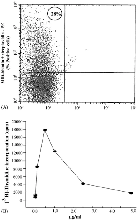
Flow cytometry analysis was performed on eight donors to demonstrate the binding of MID to purified tonsillar B cells. Our observations show that on average 30–40% of the tonsillar B cells express IgD (data not shown). B cells were incubated with MID-biotin and streptavidin-PE mAb as a secondary step. The mean value for the MID binding to the cells was calcu-lated to be 19% (n = 8). In one of the analyzed donors, MID was found to bind to 28% of the tonsillar B cells (Fig. 1A). We have previously shown that MID induces B cell proliferation in vitro[19,21]. Here we used MID over a concentration range of 0.1–5.0g/ml in order to investigate whether MID induces B cell proliferation in a concentration-dependent manner. As illustrated in Fig. 1B, MID induces maximal tonsillar B cell
Fig. 1. Tonsillar B cells bind and proliferate in response to the M. catarrhalis MID protein. Purified tonsillar B cells (4× 10⁵cells/ml) were incubated with biotinylated MID (25g/ml) and streptavidin-PE as a secondary step, and anal-ysed by flow cytometry. The figure shows % of MID-binding cells from one donor out of eight performed. Purified tonsillar B cells (1× 10⁶cells/ml) were incubated in culture medium with or without various concentrations of MID (0.1–5.0g/ml). Cell proliferation was measured by [methyl-3H]-thymidine (5Ci/well) uptake after 3 days of culture. Results are presented as one rep-resentative experiment out of two performed.
proliferation at a concentration of 0.5g/ml compared to con-trols.
3.2. MID-induced B cell proliferation is inhibited by native and polymeric AAT
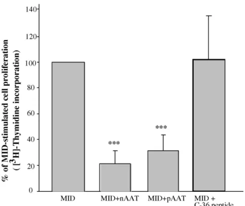
Next, we aimed to investigate whether MID-induced B cell proliferation can be affected by native or polymerized AAT. B cells were cultured with MID (0.5g/ml) either alone or in combination with AATs (0.5 mg/ml) for 72 h. Five individual donors were analysed, and as shown in Fig. 2MID strongly increased the B cell proliferation. Combinations of MID and AAT, however, significantly suppressed B cell proliferation com-pared to MID alone (p < 0.001). The C-terminal fragment of AAT (C-36) was used as a negative control and showed no sig-nificant effects (Fig. 2). Native or polymerized AAT alone had no effect on B cell proliferation compared to basal levels (data not shown).
Fig. 2. MID-induced proliferation of tonsillar B cells is inhibited by native and polymeric AAT. Purified tonsillar B cells (1× 10⁶cells/ml) were incubated in culture medium alone or with MID (0.5g/ml), nAAT (0.5 mg/ml), pAAT (0.5 mg/ml) or C-36 peptide of AAT (0.06 mg/ml) or MID–AATs combinations. Cell proliferation was measured by [methyl-3H]-thymidine (5Ci/well) uptake after 3 days of culture. Results are presented as % of max response as measured from MID-stimulated cells, and show mean values±S.D. from five donors.
3.3. MID-induced IL-6 release is inhibited by AATs
To further investigate the effects of AATs on MID-induced B cell activation, IL-6 release was measured in cell supernatants after exposure for 48 h to MID alone or in combination with AATs. In our earlier studies, using B cell isolation method based on a positive selection with anti-CD19 conjugated beads, we found that MID on its own does not stimulate IL-6 release but rather enhances the IL-6 release induced by CD40L, IL-2 and IL-4[19]. Recently we used negative selection for the isolation of B cells[21]and show that, indeed, MID on its own stimu-lates IL-6 secretion from tonsillar B cells (Fig. 3). In contrast, co-incubation of B cells with MID and native or polymerized
Fig. 3. MID-induced IL-6 secretion from tonsillar B cells is inhibited by native and polymeric AAT. Purified tonsillar B cells (1× 10⁶cells/ml) were stimulated in culture medium with or without MID (0.5g/ml), or in combinations of MID with nAAT, pAAT or C-36 peptide. Supernatants were harvested after 48 h and analysed for IL-6 release by ELISA. Results are presented as the mean value ±S.D. of IL-6 secretion from three experiments performed.
AAT inhibited the release of IL-6 compared to cells treated with MID alone. Interestingly, MID-induced IL-6 release was more strongly suppressed by the polymerized form (9.9-fold,
p < 0.001) compared to native AAT (2.8-fold, p < 0.01). The
C-36 fragment of AAT had no significant effect on MID-induced IL-6 secretion (Fig. 3).
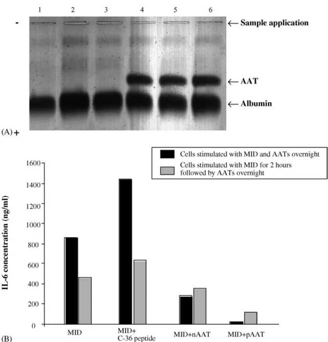
3.4. Analysis of MID and AAT interaction
For investigating a possible binding between MID and AATs, tonsillar B cells were incubated for 24 h with or without MID (0.1 and 0.5g/ml), or in different combinations of MID and native AAT. Cell supernatants were analysed for AAT-MID interaction on 1% agarose gel electrophoresis, at pH 8.6.Fig. 4A compares the electrophoretic pattern produced by AAT alone and in combi-nation with MID. It is evident that AAT alone or in combicombi-nation with MID shows the same electrophoretic mobility towards the anode, therefore, indicating no direct interaction between MID and AAT.
Furthermore, the inhibition of IL-6 secretion from B cells stimulated with combinations of MID and AATs was observed when AATs were added simultaneously with MID or 2 h after the MID protein (Fig. 4B). This suggests that direct AAT–MID interaction is unlikely to explain the anti-inflammatory effects of AATs.
4. Discussion
MID, a protein from M. catarrhalis, is a strong B cell stim-ulator by its unique ability to target IgD[19,21,22]. No binding of MID was detected to IgG, -M, -A or -E and MID was not found to bind or activate human peripheral blood T cells and monocytes[19,24]. These properties of MID make it an impor-tant tool to study IgD-targeted activation of B cells. In this study, we have addressed the question of whether serine pro-tease inhibitor, AAT, could affect the MID-induced tonsillar B cell responses. Our data indicate that both native (inhibitory) and heat inactivated (polymerized) forms of AAT strongly sup-press MID-stimulated B cell proliferation and IL-6 secretion. It is important to point out, that polymerized AAT, a protein with-out antiproteinase activity, not only retained but also enhanced its ability to inhibit MID-induced B cell activation. Thus, the data presented here clearly show that inhibitory activity of AAT is not required for its anti-MID effects. Similarly, studies by She et al. have shown that heating of AAT at 65◦C for 30 min markedly increases its stimulatory effects on human fibrob-last DNA synthesis and phosphorylation of p42/44 mitogen-activating protein kinases, compared to native (inhibitory) form of AAT[25]. It is presently unclear why AAT is more active after treatment at 60◦C, however, one can speculate that temperature-induced conformational changes of AAT molecule may favor the optimal interaction with B cell surface and triggering of anti-inflammatory mechanisms. To prove this idea further studies are needed.
The specificity of AAT effects was demonstrated by failure of a C-terminal fragment of AAT (C-36 peptide) to elicit any biological activity in our experimental model and the failure of
Fig. 4. No interaction between MID and AATs can be demonstrated. (A) Analysis of cell culture medium by 1% agarose electrophoresis, at pH 8.6. The anode is at the bottom of the figure. B cells were cultured alone or in the presence of 0.1 and 0.5g/ml MID or 0.5 mg/ml nAAT or their combinations for 24 h. The cell supernatants were collected and analysed by electrophoresis. Samples were loaded as follows: lane 1, cell medium alone; lanes 2 and 3, cell medium + 0.1 and 0.5g/ml MID, respectively; lane 4, cell medium + nAAT; lanes 5 and 6, cell medium + 0.1 and 0.5 g/ml MID and 0.5 mg/ml nAAT, respectively. (B) Purified tonsillar B cells (1× 10⁶cells/ml) were stimulated in culture medium with MID (0.5g/ml), or MID in combinations with nAAT, pAAT or C-36 peptide. Cells were either exposed to MID and AATs simultaneously overnight, or exposed first for 2 h to MID and after ttreated with MID–AATs combinations overnight. Supernatants were harvested and analysed for IL-6 production by ELISA. Results are presented as one experiment out of two performed.
AATs to inhibit CD3/CD28 stimulated IL-2 receptor expression or proliferation in T-cells which lack TLR4 and CD14 (unpub-lished data). Furthermore, the significant effects of AATs were achieved at concentrations below those reported in vivo (i.e., at 0.5 mg/ml which is equivalent to about 50% of plasma AAT levels). Together these data imply that AAT is a potent and rather specific inhibitor of MID-induced tonsillar B cell acti-vation. Moreover, our data indicate that full length AAT either in its inhibitory active or inactive, polymerized form is needed to express the anti-inflammatory activity.
To exclude the possibility that the inhibitory effects of AAT on MID-stimulated B cell responses are due to a sequestration of the MID, two experimental protocols were followed. First, cells were incubated with AAT or MID separately or in combi-nation and cell culture medium was subjected to electrophoretic analysis. However, no changes in the electrophoretic mobility of the AAT/MID mixture compared to AAT alone were observed. Second, B cells were pre-incubated with MID prior to addition of AATs, but again this made no specific contribution to anti-inflammatory effects of AATs. These results led us to conclusion that the inhibitory effects of AAT towards MID-induced B cell
activation do not appear to involve a direct AAT–MID interac-tion.
These latter findings prompted us to investigate whether AAT interferes with MID binding to the B cell receptors. However, flow cytometer analysis of cells stained with MID-biotin did not show reduced MID-binding when cells were pre-incubated with AATs, indicating that there is no competition for the receptor binding between MID and AAT (data not shown). Therefore, we conclude that neither direct AAT–MID interaction nor AAT interference with the MID cellular receptor(s) binding are likely mechanisms for the observed anti-inflammatory effects of AATs. Further studies are needed to clarify the mechanisms responsible for the observed anti-inflammatory effects of AAT.
Finally, it should be noted that both native (inhibitory) and temperature inactivated, polymeric (non-inhibitory) forms of AAT are effective modulators of MID-stimulated B cell acti-vation suggesting that observed properties of AAT are indepen-dent on its serine protease inhibitory activity. This strengthens the idea that in vivo AAT may not only limit the destructive capacity of serine proteases released from inflammatory cells, but also may act as a protein with broader anti-inflammatory
properties and constitute an important part of host defensive mechanisms.
A strong association between inherited severe Z AAT defi-ciency (basal levels of AAT are reduced by 90%) and COPD led to the hypothesis that a protease–antiprotease imbalance is an important factor for disease development[26]. It became widely accepted that the primary role of AAT in vivo is to minimize pro-teolytic injury to host tissue at sites of inflammation, infection and injury by inhibiting the activity of neutrophil-released serine proteases. However, current studies demonstrate that a short-term regimen of AAT augmentation therapy not only restores airway concentrations of AAT to normal, but also reduces the incidence of lung infections in patients with AAT deficiency and reduces levels of chemoattractants, such as leukotriene B4
[27,28]. Cantin and Woods have also reported that aerosolized AAT suppresses bacterial proliferation in a rat model of chronic
Pseudomonas aeruginosa lung infection[29].
It is known that individuals with inherited AAT deficiency are more susceptible to bacterial infections [30,31], and that combinations of environmental factors, such as smoking and air pollution, bacterial infections and AAT-deficiency may result in COPD development in the early years of life [32,33]. M.
catarrhalis, a common and important human respiratory tract
pathogen, is one of the leading causes of exacerbations in patients with COPD[34]. Thus, our findings that AAT inhibits
M. catarrhalis MID protein-induced B cell activation in vitro,
further support the notion that AAT is a multifunctional protein and provide a basis for exploring as yet uninvestigated anti-inflammatory properties of AAT in vitro and in vivo.
Acknowledgments
This work was supported by grants from the Alfred ¨Osterlund Foundation, the Crafoord Foundation, the Greta and Johan Kock Foundation, the Magnus Bergvall Foundation, the Medical Fac-ulty of Lund, the Swedish Society of Medicine, the Swedish Medical Research Council and the Cancer Foundation at Malm¨o University Hospital.
References
[1] Travis J, Shieh BH, Potempa J. The functional role of acute phase plasma proteinase inhibitors. Tokai J Exp Clin Med 1988;13:313–20. [2] Forney JR, Yang S, Healey MC. Interaction of the human serine protease
inhibitor alpha-1-antitrypsin with Cryptosporidium parvum. J Parasitol 1996;82:496–502.
[3] Dabbagh K, Laurent GJ, Shock A, Leoni P, Papakrivopoulou J, Cham-bers RC. Alpha-1-antitrypsin stimulates fibroblast proliferation and pro-collagen production and activates classical MAP kinase signaling path-ways. J Cell Physiol 2001;186:73–81.
[4] Jeannin P, Lecoanet-Henchoz S, Delneste Y, Gauchat JF, Bonnefoy JY. Alpha-1-antitrypsin up-regulates human B cell differentiation selectively into IgE- and IgG4-secreting cells. Eur J Immunol 1998;28:1815–22. [5] Ikari Y, Mulvihill E, Schwartz SM. Alpha 1-proteinase inhibitor, alpha
1-antichymotrypsin, and alpha 2-macroglobulin are the antiapoptotic fac-tors of vascular smooth muscle cells. Biol Chem 2001;276:11798–803. [6] Weiss G, Goossen B, Doppler W, Fuchs D, Pantopoulos K, Werner-Felmayer G, et al. Translational regulation via iron-responsive elements by the nitric oxide/NO-synthase pathway. EMBO 1993;12:3651–7.
[7] Bucurenci N, Blake DR, Chidwick K, Winyard PG. Inhibition of neu-trophil superoxide production by human plasma alpha 1-antitrypsin. FEBS Lett 1992;300:21–4.
[8] Churg A, Dai J, Zay K, Karsan A, Hendricks R, Yee C, et al. Alpha-1-antitrypsin and a broad spectrum metalloprotease inhibitor, RS113456, have a similar acute-inflammatory effects. Lab Invest 2001;81: 119–31.
[9] Jie Z, Cai Y, Yang W, Jin M, Zhu W, Zhu C. Protective effects of alpha 1-antitrypsin on acute lung injury in rabbits induced by endotoxin. Chin Med J (Engl) 2003;116:1678–82.
[10] Libert C, Van Molle W, Brouckaert P, Fiers W. Alpha 1-antitrypsin inhibits the lethal response to TNF in mice. J Immunol 1996;157:5126–9.
[11] Knappstein S, Ide T, Schmidt MA, Heusipp G. Alpha 1-antitrypsin binds to and interferes with functionality of EspB from atypical and typical enteropathogenic Escherichia coli strains. Infect Immun 2004;72:4344–50.
[12] Forney JR, Yang S, Healey MC. Synergistic anticryptosporidial potential of the combination alpha-1-antitrypsin and paromomycin. Antimicrob Agents Chemother 1997;41:2006–8.
[13] Janciauskiene S, Larsson S, Larsson P, Virtala R, Jansson L, Stevens T. Inhibition of lipopolysaccharide-mediated human monocyte activa-tion, in vitro, by alpha 1-antitrypsin. Biochem Biophys Res Commun 2004;321:592–600.
[14] Nita I, Hollander C, Westin U, Janciauskiene SM. Prolastin, a pharma-ceutical preparation of purified human␣1-antitrypsin, blocks endotoxin-mediated cytokine release. Resp Res 2005;6:12–23.
[15] Catlin BW. Branhamella catarrhalis: an organism gaining respect as a pathogen. Clin Microbiol Rev 1990;3:293–320.
[16] Karalus R, Campagnari A. Moraxella catarrhalis: a review of an impor-tant human mucosal pathogen. Microb Infect 2000;5:547–59. [17] Forsgren A, Grubb A. Many bacterial species bind human IgD. J
Immunol 1979;122:1468–72.
[18] Forsgren A, Penta A, Schlossman SF, Tedder TF. Branhamella
catarrhalis activates human B lymphocytes following interactions with
surface IgD and class I major histocompatibility complex antigens. Cell Immunol 1988;122:78–88.
[19] Gj¨orloff Wingren A, Hadzic R, Forsgren A, Riesbeck K. The novel IgD binding protein from Moraxella catarrhalis induces human B lym-phocyte activation and Ig secretion in the presence of Th2 cytokines. J Immunol 2002;168:5582–8.
[20] Forsgren A, Brant M, Karamehmedovic M, Riesbeck K. The immunoglobulin D-binding protein MID from Moraxella catarrhalis is also an adhesin. Infect Immun 2003;71:3302–9.
[21] Hadzic R, Forsgren A, Cardell LO, Riesbeck K, Gj¨orloff-Wingren A. The CD19 molecule is crucial for MID-dependent activation of tonsillar B cells from children. Scand J Immunol 2005;61:165–72.
[22] Forsgren A, Brant M, M¨ollenkvist A, Muyombwe A, Janson H, Woin N, et al. Isolation and characterization of a novel IgD-binding protein from Moraxella catarrhalis. J Immunol 2001;167:2112–20.
[23] Laurell C-B. Electrophoretic and electro-immunochemical analysis of proteins. Scand J Clin Lab Invest 1972;29(Suppl):124.
[24] Nordstr¨om T, Forsgren A, Riesbeck K. The immunoglobulin D-binding part of the outer membrane protein MID from Moraxella catarrhalis comprises 238 amino acids and a tetrameric structure. J Biol Chem 2002;277:34692–9.
[25] She QB, Mukherjee JJ, Crilly KS, Kiss Z. Alpha(1)-antitrypsin can increase insulin-induced mitogenesis in various fibroblast and epithelial cell lines. FEBS Lett 2000;473:33–6.
[26] Sandford AJ, Silverman EK. Chronic obstructive pulmonary disease. I. Susceptibility factors for COPD the genotype-environment interaction. Thorax 2002;57:736–41.
[27] Stockley RA, Bayley DL, Unsal I, Dowson LJ. The effect of augmenta-tion therapy on bronchial inflammaaugmenta-tion in␣1-antitrypsin deficiency. Am J Respir Crit Care Med 2002;165:1494–8.
[28] Stockley RA, Hill AT, Hill SL, Campell EJ. Bronchial inflammation: its relationship to colonizing microbial load and alpha(1)-antitrypsin defi-ciency. Chest 2000;117:291S.
[29] Cantin AM, Woods DE. Aerosolized prolastin suppresses bacterial pro-liferation in a model of chronic Pseudomonas aeruginosa lung infection. Am J Respir Crit Care Med 1999;160:1130–5.
[30] Chow CK. Cigarette smoking and oxidative damage in the lung. Ann NY Acad Sci 1993;686:289–98.
[31] Needham M, Stockley RA. Alpa 1-antitrypsin deficiency. 3. Clinical manifestations and natural history. Thorax 2004;59:441–5.
[32] Luisetti M, Seersholm N. Alpha 1-antitrypsin deficiency. I. Epidemiol-ogy of alpha 1-antitrypsin deficiency. Thorax 2004;59:164–9. [33] Carrell RW, Lomas DA. Alpha 1-antitrypsin deficiency—a model for
conformational diseases. N Engl J Med 2002;346:45–53.
[34] Furano K, Campagnari AA. Identification of a Hemin utilization pro-tein of Moraxella catarrhalis (HumA). Infect Immun 2004;72:6426– 32.