DISSERTATION
ANALYSIS OF EQUINE ZYGOTE DEVELOPMENT AFTER INTRACYTOPLASMIC SPERM INJECTION
Submitted by Elena Ruggeri
Department of Biomedical Sciences
In partial fulfillment of the requirements For the Degree of Doctor of Philosophy
Colorado State University Fort Collins, Colorado
Spring 2016
Doctoral Committee:
Advisor: Elaine Carnevale Co-Advisor: Colin Clay David Albertini
Copyright by Elena Ruggeri 2016 All Rights Reserved
ABSTRACT
ANALYSIS OF EQUINE ZYGOTE DEVELOPMENT AFTER INTRACYTOPLASMIC SPERM INJECTION
Intracytoplasmic sperm injection (ICSI) is an established and widely used method to achieve oocyte fertilization in equine reproductive assisted technologies. However, not all the oocytes fertilized by ICSI undergo cleavage and develop into viable embryos. Limited
knowledge on equine zygote development after ICSI is available, and reasons why developmental failure occurs after ICSI have been only partially studied and need further investigation. Fertility decline and early embryo loss is associated with maternal aging in the mare, and it is concomitant with reduced oocyte quality. Relatively little is known about the effect of maternal aging and zygote developmental failure or success in the mare. Effects of in vitro maturation of the oocyte or zygote development in the mare still need to be clarified and further studied. The overall objective of this dissertation was to study equine zygote development after ICSI using confocal microscopy. Objectives were to: (1) compare cytoskeletal and nuclear changes during progression of equine zygote development after ICSI for in vivo versus in vitro matured oocytes; (2) compare changes in cytoskeletal and chromosomal configurations after ICSI between oocytes from young and old mares to define maternal-aging related alterations; (3) determine cytoskeletal and nuclear alterations associated with fertilization failure in
donor-Immunostaining and confocal imaging of the equine zygotes was performed using a spinning disk confocal microscope.
After ICSI, five distinct events of development were observed with no major differences over time whether oocytes matured in vivo or in vitro. Oocytes matured in vivo appeared to reach the pronucleus stage earlier after ICSI compared to in vitro matured oocytes. Abnormal phenotypes associated with fertilization failure were more significant in oocytes matured in vitro than in vivo. When ICSI was performed in oocytes from young and old mares, similar stages of zygote development were observed, and the number of zygotes reaching the pronucleus stage was similar between the two age groups. Nucleolus like bodies, sites of ribosomal RNA involved in embryonic genome activation, were observed only in zygotes at the pronucleus stage from young mares; no nucleolus-like bodies were observed in pronuclei of zygotes from old mares. Pronuclei morphology, based on CREST staining, and DNA localization, also differed between pronuclei of young and old mares. Actin vesicles were observed significantly more often within zygotes from old mares compared to young mares during all stages of zygote developmental progression. When potential zygotes were analyzed after failure of cleavage after ICSI, actin vesicles were greater in area, perimeter and number in oocytes from old mares than those from young mares. Tubulin cytoskeletal multiasters were associated with cell aging and with increased interval after ICSI for young mares but not old mares.
In conclusion, zygotes produced from oocytes matured in vivo versus in vitro or collected from young and old mares went through similar stages of development, with pronuclei
attainment appearing to be a crucial event in zygote development. Actin vesicles were a major cytoskeletal difference associated with oocyte origin and a potential factor involved in
methods used to describe the equine zygote development and allowed us to elucidate the cytoskeletal and nuclear remodeling events that follow fertilization after ICSI in the mare.
ACKNOWLEDGEMENTS
I would like to thank my two advisors, Drs. Carnevale and Clay, for enabling me to conduct my research and pursue this challenging and satisfying adventure! Also thank you to Joanne Stokes and the ERL group for oocyte collections and ICSI procedures.
To Dr. Clay, you have been like a father for me in these last five years. To Dr. Seidel, thank you from the bottom of my heart for always having your door open to me, answering my questions and providing me with wise, knowledgeable advice and being such an inspiring mentor to me. To Dr. Jennifer (Jake) and Keith, thank you for hosting me in your incredible laboratory and making me feel like part of your brilliant, amazing crew! To Dr. Albertini, you have been such an inspiring scientist and a role model for me since the beginning. To Cesare and Giovanna, you have been a constant, incredible inspiration, empowering force, and wonderful mentors since I first met you both.
To my best friend Kristin/Kristina, you have been my family here. Molly, you have been such a groovy, wonderful friend! Michela and Viola, thank you for having been my life long best friends, no matter how far away we are.
Most importantly, to my Mom (Giovanna) and Dad (Cesare), my sister (Francesca/Luij), and my incredible grandma (Angiolina/Rinda). Your support, your love, and your strength is what made me who I am today. You were there for me, even from far away, every single day. I hope I make you all proud of me! You are everything for me. I love you so much.
To Fort Collins and the colorful Colorado, you have been a home for me and you always are going to be. See you soon! Arrivederci!!
TABLE OF CONTENTS
ABSTRACT ... ii
ACKNOWLEDGEMENTS ... v
LIST OF TABLES ... viii
LIST OF FIGURES ... ix
LIST OF APPENDICES ... x
CHAPTER I: REVIEW OF LITERATURE... 1
INTRODUCTION ... 1
IN VIVO AND IN VITRO EQUINE OOCYTE MATURATION ... 2
FERTILIZATION OF EQUINE OOCYTES USING INTRACYTOPLASMIC SPERM INJECTION (ICSI) AND ZYGOTE DEVELOPMENT ... 7
INFERTILITY AND MATERNAL AGING... 10
CHAPTER II: USE OF CONFOCAL MICROSCOPY TO EVALUATE EQUINE ZYGOTE DEVELOPMENT AFTER SPERM INJECTION OF OOCYTES MATURED IN VIVO OR IN VITRO ... 13
SUMMARY ... 13
INTRODUCTION ... 14
MATERIALS AND METHODS ... 15
Oocyte Collections ... 15
ICSI and Zygote Culture ... 16
Samples Fixation and Immunostaining ... 17
Determination of Zygote Development Stages and Abnormalities after ICSI ... 18
Statistical Analysis ... 19
RESULTS ... 19
Presumptive Zygote Developmental Events and Pronuclei Assessment ... 19
Abnormalities Observed in Presumptive Zygotes after ICSI ... 24
DISCUSSION ... 26
CONCLUSIONS ... 31
CHAPTER III: EFFECT OF MATERNAL AGING ON EQUINE ZYGOTE DEVELOPMENT AFTER ICSI ... 32
SUMMARY ... 32
INTRODUCTION ... 33
MATERIALS AND METHODS ... 34
Oocyte Collection... 34
Sperm Injection and Culture after ICSI ... 34
Fixation and Immunostaining of Presumptive Zygotes ... 35
Confocal Imaging and Morphometric Analysis ... 36
Statistical Analysis ... 37
RESULTS ... 38
Zygote Development in Young and Old Mares ... 38
Pronuclear Area and Constitutive Heterochromatin Localization in the Equine Zygote .... 43
DISCUSSION ... 47
CHAPTER IV: CYTOSKELETAL ALTERATIONS ASSOCIATED WITH DONOR AGE AND CULTURE INTERVAL FOR EQUINE OOCYTES AND POTENTIAL ZYGOTES THAT FAILED TO CLEAVE AFTER ICSI ... 53
SUMMARY ... 53
INTRODUCTION ... 53
MATERIALS AND METHODS ... 55
Oocyte Collection and Manipulation ... 56
Experiment 1 ... 56
Experiment 2 ... 57
Immunostaining and Confocal Analysis ... 58
Actin and α/β-Tubulin Morphometric Analysis of Oocyte Measurements ... 58
Statistical Analysis ... 59
RESULTS ... 60
Experiment 1 ... 60
Experiment II ... 61
Sperm Head or Tail ... 62
Spindle measurements, number and polarity ... 64
Chromosome configurations and presence of kinetochore proteins... 67
Multiasters ... 68
Actin vesicles: presence, position, dimensions, number and intensity ... 70
DISCUSSION ... 72
CHAPTER V: CONCLUDING REMARKS... 81
CHAPTER VI: LIST OF REFERENCES ... 86
LIST OF TABLES
Table 2.1. Numbers of potential equine zygotes at different points of development at 4, 6, 8, 12 and 16 h from all oocytes matured in vivo (IVO) or in vitro (IVM) and fertilized by ICSI. The total numbers of potential zygotes at the different points include those determined to have abnormal morphologies (see Table 2.2)... 21 Table 2.2. Number of potential equine zygotes with abnormal morphologies at 4, 6, 8, 12 and 16 h after ICSI per total injected oocytes matured in vivo (IVO) or in vitro (IVM). ... 26 Table 3.1. Numbers of potential equine zygotes in normal stages of development and with abnormal developmental characteristics after injection of sperm into oocytes from young mares (Y) and old mares (O) at 4, 8, 16 and 20 h after ICSI, total number per all zygotes, and overall P-values among times. ... 40 Table 3.2. Parameters observed and quantified (mean ± SEM) for pronuclei from zygotes of young and old mares. ... 44 Table 4.1: Number of oocytes with specific morphologies after maturation and culture in vitro to Time 0 (expected time of MII), Time 24 (culture for 24 h after MII) and Time 48 (culture for 48 h after MII) ... 60 Table 4.2: Comparison of mean donor age (years) and hours from intracytoplasmic sperm injection to fixation on the presence or absence of observed morphological end-points (n = 52) 63 Table 4.3: Number of injected oocytes in which different morphological end-points were observed according to donor age group and time of fixation after intracytoplasmic sperm injection... 64 Table 4.4: Spindle and actin vesicle parameters according to donor age group and time of fixation after intracytoplasmic sperm injection ... 67 Table 4.5: Number of injected oocytes in each category of chromatin localization according to donor age group and time of fixation after intracytoplasmic sperm injection ... 68 Table 4.6: Number of actin vesicles and tubulin multiasters and oocyte measurements according to donor age group and time of fixation after intracytoplasmic sperm injection ... 69
LIST OF FIGURES Fig 1.1 ... 10 Fig 2.1 ... 20 Fig 2.2 ... 22 Fig 2.3 ... 23 Fig 2.4 ... 25 Fig 3.1 ... 41 Fig 3.2 ... 42 Fig 3.3 ... 45 Fig 3.4 ... 46 Fig. 4.1 ... 61 Fig. 4.2 ... 65 Fig. 4.3 ... 66 Fig. 4.4 ... 70 Fig. 4.5 ... 71
LIST OF APPENDICES
APPENDIX 1: PROTOCOL TO FIX AND EXTRACT EQUINE OOCYTES AND ZYGOTES ... 105 APPENDIX 2: PROTOCOL TO PERFORM IMMUNOSTAINING OF EQUINE OOCYTES AND ZYGOTES ... 107
CHAPTER I: REVIEW OF LITERATURE
Introduction
Equine oocyte meiotic maturation followed by fertilization and zygote development is a crucial sequence of nuclear and cytoskeletal modification events that determine whether normal embryonic growth and development occur. The oocyte goes through two major meiotic events prior to and after fertilization, leading to possible errors and delays in chromatin and
cytoskeleton events for diverse reasons. The age of the oocyte donor can drastically affect quality of the oocyte during oocyte maturation in both animal and human models as well as the zygote’s to develop into a viable embryo ability after fertilization. Whether or not in vivo versus in vitro maturation of the oocyte can affect success of fertilization needs further investigation. In animal models in vitro maturation may affect meiotic competence and the capability of the oocyte to develop into an embryo. Due to the lack of success of regular in vitro fertilization of the equine oocyte, intracytoplasmic sperm injection (ICSI) still remains the only successful and efficient assisted reproduction technology to evaluate fertilizability of equine oocytes in vitro, with cleavage rates between 60 and 70%. Along with the drop of quality of the oocyte and mare infertility due to advanced maternal age and the possible low quality and viability of sperm, additional questions regarding the effect of ICSI on early embryo development still need to be answered. In the equine clinical setting, all of the above-described conditions are part of the challenge to improve technologies and apply them in diverse scenarios to optimize retrieval of gametes from high quality animals and determine success of pregnancy. In women, assisted reproductive technologies are applied, as in the mare, to overcome infertility due to age or clinical conditions of the patients. ICSI is frequently applied successfully in human settings to
optimize the success of fertilization when female or male gametes quality is problematic. In both equine and human clinical cases, the necessity of studying early events of chromatin and
cytoskeleton remodeling after sperm introduction in the oocyte is warranted. Early embryo development is a complex series of remodeling steps and combining of two sets of DNA are required to successfully approach meiosis II before the first mitotic event happens and the embryo is formed. Due to the decline of embryo development and increase of pregnancy loss rates in both women and mares of advanced maternal age and sub-fertile conditions, it is crucial to determine at what stage of zygote development cell events lead to embryo failure. Causes of early embryo arrest during zygote progression after ICSI need to be investigated and are decisive for success of the pregnancy.
In Vivo and In Vitro Equine Oocyte Maturation
Oocyte maturation is a crucial, although poorly understood process, necessary for successful fertilization and attainment of developmental competence of the embryo. Meiotic progression during maturation is a sequence of nuclear and cytoskeletal modifications that determine the fate of embryo growth, implantation and pregnancy success. Molecular and structural events culminate in arrest of the oocyte at metaphase II in preparation for sperm penetration and consequent activation at fertilization.
In monovular species, recruitment of primordial follicles for an extended period of follicular growth is followed by the selection of a dominant follicle that will subsequently ovulate and release the arrested oocyte, which will resume meiosis I. The nuclear arrest of the oocyte at this time inactivates the female gamete DNA, which decreases its vulnerability,
In the mare, final oocyte maturation occurs within the preovulatory dominant follicle 24 to 36 h preceding ovulation, due to the luteinizing hormone (LH) stimulus (Hinrichs et al. 1993). During the estrous cycle, a progressive rise of the concentration of LH results in rupturing the dominant follicle and expulsion of the oocyte (Evans & Irvine 1975, Bézard et al. 1989, Hinrichs et al. 1993). During the rise of LH, the oocyte, which was arrested in prophase of meiosis I at the germinal vesicle (GV) stage, undergoes germinal vesicle breakdown (GVBD) and subsequently proceeds to chromosome condensation and meiotic spindle formation, resulting in metaphase I. The metaphase I oocyte transitions from anaphase I to telophase I and will arrest at metaphase II, awaiting fertilization (Chen et al. 2010). The granulosa cells and cumulus cells, surrounding the oocyte undergoing meiotic maturation, simultaneously transition from compact and granular to expanded and mucoid during oocyte maturation (Carnevale & Maclellan 2006).
The transition from metaphase I to metaphase II is characterized by numerous molecular processes and an increase in activity of various kinases (Dell'Aquila et al. 2003). Maturation promoting factor (MPF) is a key factor involved in oocyte maturation and acquisition of competence. MPF appears shortly before germinal vesicle breakdown, peaks at metaphase I, decreases dramatically in the transition from metaphase I to metaphase II and re-establishes a peak at attainment of metaphase II (Trounson et al. 2001, Dell'Aquila et al. 2003, Mrazek & Fulka Jr 2003). In addition, the organelles and cytoskeleton are reorganized and redistributed during the asymmetric division of the oocyte as meiosis progresses and the collaborative relationship between the cumulus cells and the oocyte change during the meiotic transitions (Coticchio et al. 2015).
Equine oocytes can be collected from immature follicles undergoing maturation (Hinrichs et al. 1990, Carnevale & Ginther 1993, Cook et al. 1993). Oocyte retrieval from preovulatory
follicles results in a higher collection rate per follicle than from immature follicles and allows the oocyte to undergo a more natural maturation. In the mare, to collect preovulatory oocytes,
maturation is triggered with human chorionic gonadotropin (hCG) and/or a gonadotropin-releasing hormone (GnRH) analog, such as deslorelin (Carnevale & Maclellan 2006, Carnevale & Sessions 2012). Oocytes collected at least 30 h after hCG administration will be
predominantly at metaphase II and ready to be fertilized by the sperm, compared to oocytes collected prior to 30 h after administration of hCG, which need to be cultured in vitro for the completion of meiosis I (Bezard et al. 1997). To obtain oocytes for commercial ICSI or oocyte transfer in the mare, preovulatory follicles are usually targeted. Transvaginal, ultrasound-guided follicular aspiration is the primary method to collect oocytes, with collection rates varying between labs, but around 75% (Carnevale & Ginther 1993). When oocytes need to be obtained from smaller and/or non ovulatory follicles, for example, from ovaries of a mare post mortem, it is not feasible to collect in vivo-matured oocytes (Carnevale et al. 2004).
The collection of immature equine oocytes and in vitro maturation is used to overcome the limited number of mature oocytes that can be obtained from dominant preovulatory follicles. The technology of oocyte maturation in vitro has been established, partly due to use of ICSI for in vitro fertilization (Galli et al. 2013). When collected from the ovary of the live mare,
immature oocytes are harder to retrieve due to the tight adherence of the oocyte to the follicle wall; therefore, the recovery rate is lower (Hawley et al. 1995). Rigorous flushing and scraping of the follicle wall during follicular aspiration is needed to recover immature oocytes at the germinal vesicle stage (Galli et al. 2013). After recovery, oocytes need to complete maturation
In vitro maturation primarily involves the addition of gonadotropins and serum to complex culture media (Carnevale & Sessions 2012). Evaluation of oocyte maturity, done by imaging of the extruded first polar body, is needed to determine whether or not the oocyte is ready to be fertilized (Dell'Aquila et al. 1996, Dell'Aquila et al. 2003). The first successful report of equine oocytes matured in vitro was in 1981, followed by the first embryo produced from an in vitro matured equine oocyte in 1989 (Fulka & Okolski 1981, Zhang et al. 1989). Whether or not in vitro maturation can adversely affect the oocyte’s ability to complete meiotic maturation and be fertilized successfully still needs to be fully evaluated.
Recent studies have focused on the possible interference of in vitro maturation with establishment of post-translational histone H4 modifications, which are implicated in control of gene expression and determine chromatin remodeling that anticipates resumption of meiosis (De La Fuente et al. 2004, Kageyama et al. 2007, Franciosi et al. 2012). In vitro maturation
conditions impact the pattern of H4 acetylation in a residue–specific manner compared to in vivo matured equine oocytes (Franciosi et al. 2012). The disruption of histone acetylation of in vitro matured oocytes could lead to defects in chromosomes segregation and alignment, necessary for completion of meiotic maturation, but further investigation is needed (van den Berg et al. 2011, Franciosi et al. 2012). Possible negative effects of in vitro maturation on epigenetic regulation have been reported in large animal-assisted reproduction where instability of genomic imprinting is represented by large offspring syndrome in ovine and bovine models (Young et al. 1998, Young et al. 2001). Previous studies confirmed successful in vitro maturation of equine oocytes used for ICSI and nuclear transfer, but also suggested the need for improving in vitro maturation conditions of equine oocytes to improve assisted reproduction technology success in the horse (Squires et al. 1996, Galli et al. 2013).
The structure of the equine oocyte after maturation in vitro has been described during nuclear maturation by labeling chromatin (Zhang et al. 1989, Palmer et al. 1990, Shabpareh et al. 1993). The first study that focused on using confocal microscopy to describe meiotic spindle organization during in vitro maturation of equine oocytes was in 1997 (Goudet et al. 1997). This study first described and defined the normal equine meiotic spindle as barrel-shaped with two distinct poles. It was also determined that the percentage of oocytes with a normal metaphase I spindle is low (63%) compared to the rate for normal metaphase II and telophase I spindles (74%) (Goudet et al. 1997). Abnormal spindles were characterized by disorganized microtubules of the two poles (Goudet et al. 1997). Spindles from in vitro matured oocytes were significantly wider and longer then spindles obtained from in vivo oocyte recovery (Goudet et al. 1997). In a following study, equine oocytes both matured in vivo and vitro were analyzed for chromatin and microtubule conformation at different stages of maturation. The reorganization of the
cytoskeleton and chromatin involved in oocyte maturation was described using confocal imaging, but no differences between in vivo or in vitro matured oocytes were reported
(Tremoleda et al. 2001). This study was the first detailed description of the cytoskeleton of the equine oocyte and its changes across maturation stages. Actin filaments were evenly distributed throughout the ooplasm during the germinal vesicle stage and then were concentrated
subcortically during germinal vesicle breakdown (Tremoleda et al. 2001). When the spindle was formed, the actin filaments were concentrated around it and around the polar body. No obvious spindle abnormalities or chromosome mis-segregation was observed (Tremoleda et al. 2001). In the most recent paper on equine oocyte maturation using confocal microscopy, the focus was on
Normal and abnormal spindle and cytoskeletal configurations were reported in this study, and abnormal/detrimental microtubule organization was observed in arrested oocytes that did not complete meiotic maturation correctly (Siddiqui et al. 2009).
The analysis and understanding of nuclear and cytoskeletal configuration throughout meiotic maturation of the equine oocyte in vivo or in vitro can help to understand regulation of the cell cycle during meiosis and improve the ability to select highly competent oocytes for fertilization. Therefore, it is important to develop specific immunostaining and confocal imaging approaches for the maturation process of the equine oocyte to address developmental
reproductive questions and determine the impact of assisted technologies on the quality of meiotic progression of the oocyte.
Fertilization of Equine Oocytes using Intracytoplasmic Sperm Injection (ICSI) and Zygote Development
Intracytoplasmic sperm injection (ICSI) is the most common technique used to fertilize the equine oocyte; a selected sperm is injected directly in the cytoplasm of the oocyte.
Conventional in vitro fertilization in the horse is inefficient and inconsistent due to the failure of equine sperm to efficiently penetrate the zona pellucida of the oocyte in vitro (Choi et al. 1994, Li et al. 1995). Only a few laboratories successfully applied in vitro fertilization (IVF) in horse oocytes (Palmer et al. 1990, Zhang et al. 1990). Only one laboratory reported the production of offspring from the use of IVF (Palmer et al. 1990, Zhang et al. 1990). Even in these studies, fertilization may actually have occurred in vivo because the eggs were returned to the oviducts shortly after “in vitro” fertilization so the site where sperm penetration occurred is uncertain. For these reasons, to successfully fertilize equine oocytes in vitro, ICSI is the only consistently repeatable technique (Carnevale & Sessions 2012). The first pregnancy derived from in vitro
maturation of equine oocytes and ICSI fertilization was in 1996 (Squires et al. 1996). Four fertilized oocytes were produced using sperm injection, and one pregnancy was produced (Squires et al. 1996). This first successful result was followed by a series of variable results that had cleavage rates ranging from 20% to 65% (Squires et al. 2003a). The introduction of the piezo drill (Primetech, Japan) in 2002 improved ICSI significantly. The Piezo drill is an
instrument that causes minute vibrations in the injection pipette facilitating the penetration of the sperm through the zona pellucida and into the cytoplasm. The piezo drill has been used for ICSI with fresh, cooled, and frozen semen, and the cleavage rates reported range from 69 to 89% (Galli et al. 2002, Choi et al. 2004). The use of different sources of sperm, fresh or frozen, showed no differences in pronucleus formation and cleavage rates (Galli et al. 2002).
The procedure of ICSI involves the selection of a highly viable oocyte, usually derived from the preovulatory follicle, which should be associated with high rates of embryonic development. The oocyte needs to be at metaphase II to be competent and to be successfully fertilized; therefore, oocytes having a visible polar body are selected for manipulation. The selection of the sperm for ICSI is performed using different methods and depends upon the quality of the sperm and the protocol of different laboratories (Carnevale & Sessions 2012). A morphologically normal, progressively motile sperm is selected for injection of the oocyte. Prior to injection, and while semen preparation occurs, the oocyte is denuded from the cumulus cells using hyaluronidase and gently pipetting (Carnevale & Sessions 2012). At injection, the sperm is positioned at the tip of the injection pipette that will be inserted through the zona pellucida and into the cytoplasm of the oocyte. The zona is drilled with the piezo device, and once the piezo
cleavage and produce viable blastocysts. To culture the injected presumptive zygotes, the most commonly used system is the medium DMEM/F12 with 10% Fetal Bovine Serum (FBS) (Hinrichs 2005, Altermatt et al. 2009, Galli et al. 2013). Between day 5 and 7 after ICSI, blastocoel formation is observed in those embryos developing normally (Carnevale & Sessions 2012). Comparison of cell counts between in vivo and in vitro cultured embryos at day 7 after fertilization showed a lower number of cells in in vitro-produced embryos (Tremoleda et al. 2003b, Galli et al. 2013). The delay in blastocyst development after ICSI is most likely caused by membrane damage, nuclei fragmentation and possible blastomere apoptosis (Tremoleda et al. 2003b, Pomar et al. 2005, Carnevale & Sessions 2012).
The most significant parameter to determine embryo development in the horse is the time to cleavage, which occurs between 12 and 24 h after ICSI (Carnevale & Sessions 2012).
Embryos that cleave faster are more prone to result in a pregnancy than embryos that cleave at a slower pace, which is also true in other species (Lundin et al. 2001, Carnevale & Sessions 2012). Limited knowledge is available regarding the first 24 h of equine zygote development, and the changes in the oocyte that occur when the female and male genomes meet. Errors in the apposition of the two sets of chromosomes, asynchrony of the nuclear and cytoplasmic
remodeling, and zygotic epigenetic modifications occur before the first mitotic cleavage, and can cause early embryo loss and pregnancy failure.
A complete description of the parental genome movements in horse zygotes after ICSI has been conducted using confocal microscopy (Tremoleda et al. 2003b). A series of
cytoskeleton-mediated events is required for correct progression to the first mitotic division, and failure of this result in developmental arrest. This study characterized nuclear and cytoskeletal events after ICSI in the mare, particularly looking at morphological changes in the first 48 h after
sperm injection. The correct succession of events during zygote development and the abnormal morphological stages of arrest when fertilization failed were determined. Oocyte activation failure was the main cause of abnormal cytoskeletal morphology, and zygote arrest and asynchrony between male and female gametes during fertilization can lead to early embryo arrest. Beyond this study from 2003, there has not been substantial improvement in studying chromatin and cytoskeletal factors involved in equine zygote development prior to the first mitotic cleavage.
Fig 1.1
A summary cartoon of oocyte maturation, fertilization and zygote development. (A) oocyte at the LH surge when a germinal vesicle is present; (B) germinal vesicle breakdown as a result of the LH surge; (C) resumption of the first meiosis due to
ovulation; (D) extrusion of the first polar body; (E) fertilization resulting in the expulsion of the second polar body; (F) formation of the male and female pronuclei in a distanced position; (G) male and female pronuclei in close apposition; (H) breakdown of the pronuclei membranes; (I) fusion of DNA from male and female pronuclei; (J) first cleavage event resulting in a 2 cell embryo.
of assisted reproductive technologies (ARTs) has been overpowering the limit of reduced fertility due to advancing age. Infertility due to advanced maternal age is one of the primary reasons ARTs have been improved and applied both in human and livestock species. In general, the age of the oocyte donor is a primary factor influencing developmental competence of the oocyte (Armstrong 2001). Abnormalities associated with donor age include meiotic incompetence of the oocyte, causing failure of fertilization, and errors in meiosis that allow fertilization but cause genetic abnormalities and cytoplasmic deficiencies at different developmental stages before or after fertilization (Armstrong 2001). Therefore, oocyte competence is crucial for the oocyte to undergo fertilization and develop into an embryo.
In women the age of the oocyte donor is a significant factor influencing oocyte meiotic competence (Boudoures & Moley 2015). Women’s fertility declines with age, and aneuploidies have been shown to increase as women age (Battaglia et al. 1996, Chiang et al. 2012, Boudoures & Moley 2015). Within the human oocyte, reactive oxygen species (ROS) have been found to increase as women age, causing mitochondrial damage that affects oocyte maturation and fertilization success (Boudoures & Moley 2015). Reduction of oocyte mitochondrial competence in older women therefore adversely affects fertility and embryo development (Murakoshi et al. 2013).
In the mare, oocyte morphology changes with increased donor age, and a decline in fertility is associated with a high incidence of early embryo loss in old mares (Carnevale & Ginther 1992, Carnevale 2008). The primary factor that causes reduced fertility in old mares is oocyte developmental quality (Carnevale & Ginther 1995). Mare aging is associated with a higher susceptibility to mitochondrial damage, primarily during in vitro maturation (Rambags et al. 2014). Also, the lower mitochondrial DNA (mtDNA) number could be an effect of mare
aging (Hendriks et al. 2015), and suboptimal coenzyme Q10 availability can cause age-associated deficits leading to infertility in women (Ben-Meir et al. 2015).
Analysis of chromatin and cytoskeletal changes in oocytes from young versus old donors before and after fertilization has been only minimally studied. Also, the effects of donor aging on microtubule and actin cytoskeleton remodeling, as well as chromatin patterns during meiosis and after the sperm is introduced have been poorly considered in both human and animal fields (Coticchio et al. 2014). This shows the necessity for further research on the impact of donor age on oocytes after fertilization and their potential to result in a successful pregnancy.
Oocyte aging and the decreased quality appear to be the main reason of infertility in both women and the mare, but the molecular causes of reproductive aging and decline in oocyte-cell quality still remain undefined. The aging female uterus doesn`t impact pregnancy success when oocytes are transferred into old or young females in both species (Navot et al. 1991, Carnevale et al. 2000b). Oocyte quality and donor age are independent variables from the quality of the uterine environment in younger or older individuals in women and mares (Navot et al. 1991, Carnevale et al. 2000b). These findings further validate that the aging oocyte, in particular the cytoskeleton and chromatin changes, is the main reason behind the fertility decline in aging individuals and is an area of research that needs to be investigated further.
CHAPTER II: USE OF CONFOCAL MICROSCOPY TO EVALUATE EQUINE ZYGOTE DEVELOPMENT AFTER SPERM INJECTION OF OOCYTES MATURED IN
VIVO OR IN VITRO1
Summary
The progression of zygote development has not been well defined in the horse. We used confocal microscopy to investigate zygote development at timed intervals after intracytoplasmic sperm injection (ICSI) of equine oocytes that were either matured in vivo (IVO) or in vitro (IVM). After fixation at 4, 6, 8, 12, or 16 h after ICSI, zygotes were incubated with α/β tubulin
antibodies and human anticentromere antibody (CREST/ACA) and washed in Alexa 488, 647, 561-Phalloidin and Hoechst 33258. Images were acquired using an Olympus IX81 spinning disk confocal microscope. Chi-Square analysis and Fisher’s exact test were used to analyze data. Five different events of zygote development were observed, with only minor differences in
developmental phases over time for IVO and IVM. Oocytes after IVO appeared to form pronuclei earlier (67% and 80% at 6 and 8 h, respectively) than oocytes after IVM (13% and 13% at 6 and 8 h, respectively); 80% of IVM zygotes formed pronuclei by 12 h. More (P=0.04)
1 Authors include: Elena RuggeriA,, Keith F DeLucaB, Cesare GalliCD, Giovanna LazzariD,
Jennifer G DeLucaB, Joanne E StokesA and Elaine M CarnevaleA
A Department of Biomedical Sciences, Colorado State University, Equine Reproduction
Laboratory, 1693 Campus Delivery, Fort Collins, Colorado, 80523-1693, USA.
B Department of Biochemistry and Molecular Biology, Colorado State University, 1870 Campus
Delivery, Fort Collins, Colorado, 80523, USA.
C Department of Veterinary Medical Sciences, University of Bologna, Via Tolara di sopra, 50,
40064 Ozzano Emilia (Bologna) Italy.
zygotes from IVM (30%) than IVO (11%) had abnormal phenotypes, suggesting a failure of normal zygote development after ICSI. Some potential zygotes from IVO had normal phenotypes, although development appeared to be delayed or arrested. Confocal microscopy provided a feasible method to assess equine zygote development using limited samples.
Introduction
In the horse, intracytoplasmic sperm injection (ICSI) is a successful clinical procedure, which has helped to compensate for the failure of standard in vitro fertilization (IVF) techniques (Dell'Aquila et al. 1997, Hinrichs 2005, Carnevale et al. 2007, Galli et al. 2007). In part, because of the failure of IVF, limited numbers of equine zygotes have been available to study early embryo development. Some ICSI-produced equine zygotes have been evaluated with confocal microscopy (Tremoleda et al. 2003b). However, most of the research was conducted before reliable results were obtained using ICSI for the horse, and normal zygote development and fertilization failure have since received minimal attention.
In animal models, in vitro maturation (IVM) of oocytes has been successful with assisted breeding technologies (Binor & Wolf 1979, Hinrichs et al. 1993, Chian et al. 1994, Eppig 1996, Bing et al. 2002, Galli et al. 2007). In the horse, protocols for in vitro maturation of oocytes have improved in recent years, thus allowing the successful use of in vitro-matured oocytes in ART programs (Hinrichs et al. 1993, Galli et al. 2007). Further study is required to assess the
developmental competence of oocytes matured in vitro. One potential risk of maturation in vitro is that nuclear and cytoplasmic maturation are not synchronized and can result in collateral effects on embryonic development (Smitz et al. 2011, Sanfins et al. 2015). Transcriptional
& Schultz 2004, Mamo et al. 2011, El Hajj & Haaf 2013). An understanding of developmental differences for oocytes matured in vivo and in vitro would help further our knowledge of cytoskeletal and nuclear maturation prior to the first mitotic division. In our study, we used confocal microscopy to examine equine zygote development at timed intervals after ICSI of oocytes matured in vivo or in vitro.
Materials and Methods
Oocyte Collections
In vivo-matured oocytes (IVO) were collected from April to August in Fort Collins, CO (40° latitude) from light-horse mares between 4 and 16 years (yr) (mean ± s.e.m. of 10.82 ± 0.69 yr). Reproductive tracts were imaged using ultrasonography to evaluate follicular growth.
Oocytes were collected from dominant follicle(s) during the follicular phase between 18 and 25 h (21 ± 0.3 h) after administration of hCG (human chorionic gonadotropin, 1500 IU, iv; Intervet Inc, Millsboro, DE, USA) and deslorelin acetate (SucroMate™, 0.75 mg, im; Bioniche Life Sciences Inc., Belleville, Ontario, CAN). Oocytes were retrieved by ultrasound-guided,
transvaginal, follicle aspirations (Carnevale et al. 2000a) and cultured for 19.5 to 27 h (22 ± 0.3 h) in TCM-199 with Earle’s salts (Gibco, Life Technologies, Grand Island, NY, USA) with additions of 10% fetal calf serum (FCS, Cell Generation LLC, Fort Collins, CO), 0.2 mM sodium pyruvate, and 25 µg/ml gentamicin sulfate (Sigma Aldrich, St. Louis, MO, USA)] at 38°C or 38.5°C in an atmosphere of 6% CO2 and air.
Oocytes for in vitro maturation (IVM) were collected from excised ovaries in Cremona, Italy (45° latitude) during the natural breeding season (March 2014). Ovaries were obtained from mares of diverse breeds and unknown ages from a local abattoir. After removal, ovaries
were transported at 24°C and arrived at the laboratory within 4 h for collection of cumulus-oocyte complexes (COCs). Retrieved COCs were placed in culture medium [Dulbecco`s modified Eagle`s medium (DMEM)/F12 (D8900; Sigma Aldrich Milan, Italy) with 10% serum replacement (Life Technologies, Monza, Italy) and 0.1 IU/ml of human menopausal
gonadotropin (HMG; Menopur 75, Ferring, Milan, Italy)] at 38.5°C in humidified atmosphere of 5% CO2 and air (Galli et al. 2007).
ICSI and Zygote Culture
Oocytes matured in vivo were denuded of cumulus cells and injected with a frozen-thawed sperm from a single stallion as previously described (Carnevale et al. 2000a) at 49 to 49.5 h (43 ± 0.4 h) after administration of deslorelin/hCG to donor mares. Once oocytes collected had an extruded polar body, a motile sperm with normal morphology was selected for ICSI, which was performed using a piezo drill (Carnevale et al. 2000a). Potential zygotes were cultured in 30-µl drops of medium [DMEM/F12 (Sigma Aldrich, St. Louis, MO, USA) with 10% FCS] under mineral oil at 38.5 °C and in 5% CO2, 5% O2 and 90% N2.
After culture for 28 h, oocytes were denuded of cumulus cells, and oocytes with an extruded first polar body were used for ICSI. Sperm injections were performed using a piezo drill and selecting motile sperm with normal morphology from frozen-thawed semen from one stallion of proven fertility (Galli et al. 2007). After sperm injection, oocytes were placed as a group in a 300-µl drop of mSOF medium with bovine serum albumin (BSA; Sigma-Aldrich, Milan Italy) and MEM amino acids (Sigma-Aldrich, Milan Italy) under mineral oil at 38.5 °C and in 5% CO , 5% O until fixation (Tervit et al. 1972, Colleoni et al. 2011).
Samples Fixation and Immunostaining
Presumptive zygotes were fixed at room temperature in solution containing 2%
formaldehyde and 0.1% Triton X-100 MTSB-XF [(Microtubule Stabilization Buffer Extraction Fix; (Messinger & Albertini 1991)] at 4 (n=5), 6 (n=6), 8 (n=5), 12 (n=5) and 16 (n=7) h
(ICSI=0h) for in vivo-matured samples and at the same time points, 4 (n=8), 6 (n=8), 8 (n=8), 12 (n=10) and 16 (n=10) h for in vitro-matured samples. After fixation, oocytes were rinsed in a wash solution [phosphate-buffered saline (PBS) containing 1% BSA and 0.1% Triton X-100] and stored at 4°C until immunostaining.
Oocytes were incubated with diluted primary antibodies in 2% normal goat serum and positioned in four-well plates on rotating platform shaker for 4 h at 37°C at the following concentrations: α/β tubulin cocktail (1:100, mouse; Sigma Aldrich, St. Louis, MO, USA) and human-anti centromere antibody-CREST/ACA (1:100; Life Technologies, Grand Island, NY, USA). After primary incubation, oocytes were rinsed in 2% normal goat serum for a minimum of 12 h at 4°C, then incubated with secondary antibodies conjugated to either Alexa 488 or Alexa 647 (1:100; Jackson ImmunoResearch Laboratories, West Grove, PA, USA) diluted in 2% normal goat serum for 4 h. When secondary incubation was complete, the oocytes were washed in 2% normal goat serum for 5 h and then incubated with phalloidin (Alexa 561; Life
Technologies) and Hoechst 33258-(1µg/ml; Life Technologies) for another 5 h at 37°C. For confocal imaging, samples were mounted onto coverslips (50% glycerol in PBS with 25 mg/ml sodium azide and 1µg/ml of Hoechst 33258) (Barrett & Albertini 2007).
Confocal images were acquired on an Olympus IX81 microscope (Waltham, MA, USA) fitted with a Yokogawa spinning disk (CSU22 head) using either a 60x/1.42 NA, DIC
image the entire oocyte using z-steps of 1-µm through the entire sample. The 60x objective was then used to image the same sample at 0.2-µm intervals throughout the oocyte. Samples were imaged with a Photometrics (Tucson, AZ, USA) Cascade II EM CCD camera and analyzed using SlideBook software (Intelligent Imaging Innovations, Denver, CO, USA).
Determination of Zygote Development Stages and Abnormalities after ICSI
Five different developmental events were observed in the presumptive equine zygotes: (1) condensed sperm chromatin (CSC), maternal chromosomes (MC) and extruded first polar body (PB1); (2) anaphase to telophase transition, including anaphase/telophase shift of MC, CSC, PB1, and second polar body (PB2) in process of extrusion or fully extruded; (3) formation of the male pronuclei and MC, PB1 and PB2; (4) male and female pronuclei, at distant positions; (5) male and female pronuclei in close apposition.
Male and female pronuclei were identified by characteristics of DNA and human-anti centromere antibody-CREST/ACA localization. Pericentric satellites were observed as ring structures found around the nucleolus-like structures of the two parental pronuclei.
Abnormalities of oocytes after ICSI were classified into five categories based on
observations: (1) premature chromosome condensation (PCC), sperm head undergoing premature chromosome condensation with male chromosomes flanked by a bipolar spindle, caused by inactive/delayed activation of the oocyte; (2) multiple pronuclei (MPN), an abnormal phenotype caused by the possible failure of PB2 extrusion; (3) sperm chromatin induced ectopic-polar body (EPB), caused by PCC and leading to paternal chromatin loss; (4) multipolar spindles, with incorrect separation of chromosomes and presence of multiple centrosomes, a sign of
failure of oocyte activation and potentially causing abnormal chromosome separation or aneuploidy.
Statistical Analysis
Chi-Square analysis was used to determine overall effects across all time points for IVO versus IVM, and Fisher’s exact test was used within each category for comparisons among time points if the overall difference was significant at P<0.05.
Results
Presumptive Zygote Developmental Events and Pronuclei Assessment
Similar events of zygote development were observed after ICSI of oocytes matured in vivo (IVO) and in vitro (IVM) (Fig 2.1, Table 2.1). The number of potential zygotes differed in developmental stage over time only for IVO when condensed sperm chromatin, maternal chromosomes and PB1 were observed (P=0.02, Fig 2.1A-C) and when distant pronuclei were present (P=0.06, Fig 2.2B), and IVM when pronuclei were apposed (P=0.008, Fig 2.2A) (Table 2.1). The number of potential zygotes with pronuclei (distant or apposed) was significantly elevated at 12 h for IVM (80%); although not different over time, the highest numerical percentages were observed at 6 h (67%) and 8 h (80%) for IVO.
Specific centromere-chromatin signatures were imaged in the pronuclei of equine zygotes as ring structures around nucleolus-like bodies in both maternal and paternal pronuclei (Fig 2.2). Pericentric heterochromatic organization around the nucleolar-like bodies were defined by CREST antibody localization; the number of ring structures in both parental pronuclei varied from three to seven. Pericentric heterochromatin patterns were imaged in all pronuclei when PB2 was not extruded and three pronuclei were present (Fig 2.3).
Fig 2.1
Events during zygote development observed between 4 and 16 h after ICSI. (A,B) condensed sperm chromatin (CSC, lower right), maternal chromosomes (MC, left) and first polar body (PB1, just to right of MC); (C) representation of the kinetochores localized on MC and PB1; (D,E) anaphase to telophase transition with CSC with acrosome portion of the sperm (lower left), anaphase/telophase of MC, extruding the second polar body; (F) actin cap surrounding the set of chromosomes being extruded in the second polar body, sperm tail visible next to CSC; (G,H) formation of the male pronucleus (indicated by elongated tubulin structure), MC (left insert); (I) tubulin aster formation around the sperm head at the time of sperm DNA decondensation (Blue, DNA; Red, centromeres and kinetochores, except panel F- actin; Green, tubulin).
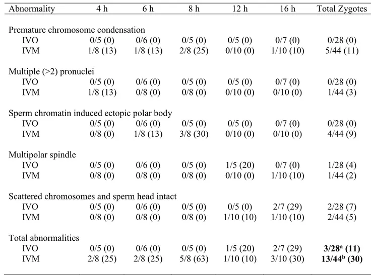
Table 2.1. Numbers of potential equine zygotes at different points of development at 4, 6, 8, 12 and 16 h from all oocytes matured in vivo (IVO) or in vitro (IVM) and fertilized by ICSI. The total numbers of potential zygotes at the different points include those determined to have abnormal morphologies (see Table 2.2).
Overall P-values were not different for points of development over time for IVO and IVM. AB Values within rows with different superscripts differ at P<0.05.
Stages of
development 4 h 6 h 8 h 12 h 16 h P-Value
Condensed sperm chromatin, maternal chromosomes and first polar body
IVO 4/5 (80)a 1/6 (17)ab 0/5 (0)b 0/5 (0)b 2/7 (29)ab 0.02 IVM 3/8 (38) 1/8 (13) 1/8 (13) 1/10 (10) 0/10 (0) 0.23 Anaphase to telophase transition, condensed sperm chromatin, first polar body
IVO 1/5 (20) 1/6 (17) 1/5 (20) 1/5 (20) 0/7 (0) 0.81 IVM 2/8 (25) 2/8 (25) 1/8 (13) 0/10 (0) 0/10 (0) 0.24 Male pronuclei, maternal chromosomes, first and second polar bodies
IVO 0/5 (0) 0/6 (0) 0/5 (0) 1/5 (20) 0/7 (0) 0.31
IVM 1/8 (13) 2/8 (25) 0/8 (0) 0/10 (0) 0/10 (0) 0.16 Distant male and female pronuclei
IVO 0/5 (0) 3/6 (50) 1/5 (20) 0/5 (0) 0/7 (0) 0.06
IVM 0/8 (0) 0/8 (0) 0/8 (0) 2/10 (20) 1/10 (10) 0.32 Apposed male and female pronuclei
IVO 0/5 (0) 1/6 (17) 3/5 (60) 2/5 (40) 3/7 (43) 0.26 IVM 0/8 (0)a 1/8 (13)ab 1/8 (13)ab 6/10 (60)b 6/10 (60)b 0.008
Pronuclei stages (combined distant and apposed)
IVO 0/5 (0) 4/6 (67) 4/5 (80) 2/5 (40) 3/7 (43) 0.103
Fig 2.2
Centromeric chromatin signatures observed in equine zygotes at the pronuclei stage. Ring structures were apparent around nucleolus-like bodies within the ACA-CREST antibody localization for maternal and paternal pronuclei at apposed (A) or distant (B) locations, as a sign of pericentric heterochromatic organization. (Red, centromere; Green, tubulin).
Fig 2.3
Abnormal morphologies of potential equine zygotes after ICSI. (A) premature chromosome condensation, female spindle top left, male chromosome flanked by a spindle structure lower right; (B) multiple pronuclei; (C) sperm chromatin induced ectopic polar body, represented by the two sets of chromosomes undergoing anaphase;
(D) multipolar spindle; (E) scattered maternal chromosomes (Blue, DNA; Red, centromeres and kinetochores; Green, tubulin).
Abnormalities Observed in Presumptive Zygotes after ICSI
Failure of zygote development was observed for IVO and IVM and was associated with abnormal cytoskeletal and chromatin configurations (Fig 2.3, 2.4). In vivo matured oocytes (IVO) had an incidence of abnormalities during zygote development of 3/28 (11%), with no significant differences for the various phenotypes across time (Table 2.2). Of the 44 potential zygotes from IVM, 13 (30%) had abnormal phenotypes, with PCC and EPB as the main, post-injection abnormalities. Overall, potential zygotes from IVM had a higher number of abnormal phenotypes per total injected oocytes than IVO (P=0.04, Table 2.2).
Fig 2.4
Actin cytoskeleton localization during ectopic polar body extrusion. Left panel cortical actin concentrated around the two points of extrusion of DNA. Top right and bottom right panels are magnified images of the two points of extrusion. (Blue, DNA; Red, actin; Green, tubulin).
Table 2.2. Number of potential equine zygotes with abnormal morphologies at 4, 6, 8, 12 and 16 h after ICSI per total injected oocytes matured in vivo (IVO) or in vitro (IVM).
Overall P-values were not different for specific abnormalities over time for IVO and IVM; however, the total number of zygotes with morphologic abnormalities was higher (P=0.04) for IVM when compared to IVO (as denoted by bold numbers and different superscripts in the table).
Abnormality 4 h 6 h 8 h 12 h 16 h Total Zygotes
Premature chromosome condensation
IVO 0/5 (0) 0/6 (0) 0/5 (0) 0/5 (0) 0/7 (0) 0/28 (0)
IVM 1/8 (13) 1/8 (13) 2/8 (25) 0/10 (0) 1/10 (10) 5/44 (11) Multiple (>2) pronuclei
IVO 0/5 (0) 0/6 (0) 0/5 (0) 0/5 (0) 0/7 (0) 0/28 (0)
IVM 1/8 (13) 0/8 (0) 0/8 (0) 0/10 (0) 0/10 (0) 1/44 (3) Sperm chromatin induced ectopic polar body
IVO 0/5 (0) 0/6 (0) 0/5 (0) 0/5 (0) 0/7 (0) 0/28 (0)
IVM 0/8 (0) 1/8 (13) 3/8 (30) 0/10 (0) 0/10 (0) 4/44 (9) Multipolar spindle
IVO 0/5 (0) 0/6 (0) 0/5 (0) 1/5 (20) 0/7 (0) 1/28 (4) IVM 0/8 (0) 0/8 (0) 0/8 (0) 0/10 (0) 1/10 (10) 1/44 (2) Scattered chromosomes and sperm head intact
IVO 0/5 (0) 0/6 (0) 0/5 (0) 0/5 (0) 2/7 (29) 2/28 (7) IVM 0/8 (0) 0/8 (0) 0/8 (0) 1/10 (10) 1/10 (10) 2/44 (5) Total abnormalities IVO 0/5 (0) 0/6 (0) 0/5 (0) 1/5 (20) 2/7 (29) 3/28a (11) IVM 2/8 (25) 2/8 (25) 5/8 (63) 1/10 (10) 3/10 (30) 13/44b (30) Discussion
The equine oocyte is a large cell, approximately 140 µm in diameter, with a high lipid content for metabolic energy (Ambruosi et al. 2009). These properties of the equine oocyte affect
difficult and expensive to obtain, confocal microscopy provides an efficient method to image limited samples. Although relatively few antibodies have been tested for use with the equine oocyte and zygote, we were able to use antibodies that were confirmed in other species.
The first detailed imaging of equine early embryo progression after ICSI of oocytes matured in vitro was published in 2003 (Tremoleda et al. 2003b). No studies have been conducted using zygotes developing from in vivo matured oocytes. Since the initial study, procedures for oocyte maturation, ICSI and embryo culture have progressed, and the procedures have been proven to be successful in producing embryos and offspring. In our study, we detailed development of the equine zygote and described abnormalities after ICSI of oocytes matured IVO or IVM using confocal microscopy. In this study, two systems (CSU and Italy) with consistent embryo production for oocytes matured IVO and IVM were utilized to evaluate early zygote development.
A time line of zygote development has not been established in the horse. Other studies have focused on cow and monkey zygotes, but these studies are very limited and are primarily centered around tubulin remodeling and oocytes that failed to fertilize after IVF or ICSI (Navara et al. 1994, Hewitson et al. 1996). In our study, five main events of early embryo development were observed in equine presumptive zygotes after ICSI of IVO and IVM oocytes. The first event observed during early embryo development was the female spindle arrested at metaphase II with aligned and compact chromosomes, first polar body and paternal DNA identified by the presence of the sperm head and, in some samples, the tail (Fig 2.1A, B). Kinetochores were associated with the maternal chromosomes at the metaphase plate and chromosomes of the extruded first polar body (Fig 2.1C). This early phase of development was predominantly
This stage was also observed in six presumptive zygotes (IVO, n=3, and IVM, n=3) at later time points, suggesting a delay or arrest in development.
The second phase of zygote development was defined as the anaphase to telophase transition in maternal chromatin (Fig 2.1D-F). Maternal chromosomes were approaching anaphase or extruding the second polar body during telophase, and a sperm head was observed. This stage was imaged after ICSI of IVO and IVM, with some presumptive zygotes appearing to be arrested at this point in later hours after ICSI.
The third event observed during zygote development was male chromatin
decondensation, with a microtubule array around the sperm head nuclei, female chromosomes aligned at the metaphase plate, and the first polar body and, in some cases, the second polar body (Fig 2.1G-I). This stage was only observed in three oocytes, including oocytes (IVO) that
appeared to be delayed in development at 12 h after ICSI.
The presence of male and female pronuclei in distant positions next observed (Fig 2.2B). Pronuclei were surrounded by a complex tubulin net, which was diffused throughout the
developing zygote. The entire area of pronuclei was denoted by CREST-anticentromere staining, in which distinct areas were noted with no stain, representing the nucleolus-like bodies, which represent specific heterochromatin signatures at pericentric domains (Fig 2.2A). Pericentric heterochromatin domains are specific zygote transcriptional and epigenetic signatures involved in early zygote development prior to the first mitotic division and embryo genome activation (Probst & Almouzni 2011). When the two pronuclei were apposed (Fig 2.2A), the nucleolus-like bodies were still present within the pronuclei. The tubulin net was now concentrated and
Oocyte maturation in vivo requires resumption and completion of meiotic divisions and epigenetic reprogramming of the oocyte genome (Bromfield et al. 2007). Specific genes needed for correct oocyte maturation are altered subsequent to in vitro maturation and possibly affect early embryo development and genome reprogramming in many species, including humans, cows and mice (Gremeau et al. 2012, El Hajj & Haaf 2013, Salhab et al. 2013, Sanfins et al. 2015). The need to understand the impact of in vitro maturation on early fertilization events and embryonic progression is of utmost importance. Meiotic maturation prior to fertilization
proceeds faster when mouse and bovine oocytes are matured in vitro than in vivo, compromising later oocyte developmental competence due to its unphysiological temporal progression (Hyttel et al. 1997, Gilchrist et al. 2001). Incorrect nuclear and cytoplasmic maturation are possible alterations of in vitro oocyte maturation (Smitz et al. 2011). Chromosome abnormalities and epigenetic changes, including histone incorporation and elevated transcriptional activity during the first hours after sperm entrance, can impact the organization of the zygote at specific
developmental steps (Probst & Almouzni 2011, Smitz et al. 2011). In our study, in vivo matured oocytes seemed to develop to pronuclei sooner after ICSI than in vitro matured oocytes, although some of the oocytes appeared to be delayed or arrested at an early stage of development.
Abnormal morphologies were observed in some potential zygotes. Sperm-transmitted DNA damage leads to diverse abnormal reproductive outcomes and paternal genome loss, and it can be caused by different sperm chromatin defects, including premature sperm condensation and ectopic polar body extrusion after ICSI (Schmiday & Tandler-Schneider 1996, Marchetti & Wyrobek 2005, Deng & Li 2009, Marchetti et al. 2015). After aneuploidy, the most common cause of fertilization failure in human IVF and ICSI is premature sperm condensation
the oocyte to help avoid sperm head premature condensation, which leads to DNA damage and aneuploidy in human oocytes (Manandhar & Toshimori 2003). In our study, premature sperm condensation was only found in oocytes matured in vitro; no oocytes matured in vivo had this abnormal phenotype. Another sperm related abnormality, specific to IVM in our study, was sperm chromatin-induced ectopic polar body extrusion, another possible cause of paternal genome loss in mammalian oocytes (Deng & Li 2009). This process causes the sperm chromatin to form a spindle in the oocyte, leading to failure of fertilization and zygote development.
Premature sperm chromatin condensation and sperm chromatin induced ectopic polar bodies were only observed in oocytes matured in vitro, suggesting that progression to the pronuclei stage was interrupted due to activation delay in oocytes matured in vitro or due to alterations of sperm DNA specific packaging and protamine deficiency, both needed for successful
fertilization. However, we cannot exclude the potential that intrinsic oocyte quality or the stallion affected these results, as these variables were also different in the two ICSI systems.
Additional zygote abnormalities included multiple pronuclei and multipolar spindle or presence of scattered maternal chromosomes and intact sperm head (chromosome
fragmentation). Multiple pronuclei suggest the failure of extrusion of the second polar body, possibly due to low oocyte quality or sperm chromatin defects (Rosenbusch 2001). Multipolar spindles are a possible sign of failure in spindle assembly checkpoints, and they were observed in one oocyte matured in vivo and one oocyte matured in vitro (Sluder et al. 1997, Courtois et al. 2012). Scattered chromosomes and a sperm head were also observed in two potential zygotes from IVO and two from IVM. Overall, we observed more morphological abnormalities in
Conclusions
In conclusion, we used confocal microscopy to observe equine zygote progression after ICSI of oocytes matured in vivo or in vitro. In this study there were differences in systems for oocytes injected in vivo and in vitro varied; therefore, our ability to make direct comparisons was limited. However, we observed a similar progression through the major events of maturation of potential zygotes regardless of type of oocyte maturation. Oocytes matured in vivo appeared to have a more rapid progression to pronuclei, although some of these potential zygotes appeared to be delayed or arrested in development. Abnormal zygote morphologies were observed more frequently in oocytes matured in vitro than in vivo. Confocal microscopy provided a feasible method to assess zygote development after in vivo or in vitro oocyte maturation using a limited number of samples available.
CHAPTER III: EFFECT OF MATERNAL AGING ON EQUINE ZYGOTE DEVELOPMENT AFTER ICSI2
Summary
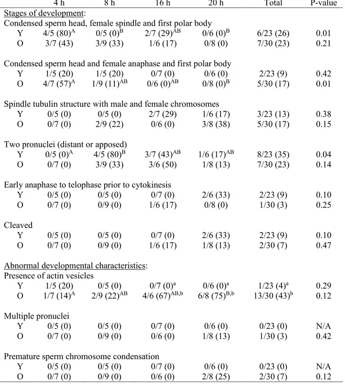
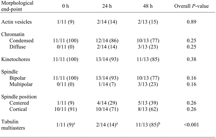
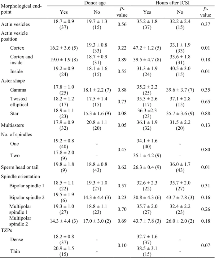
The effect of the oocyte donor age on equine zygote development has not been studied. The aim of our study was to compare early zygote development after intracytoplasmic sperm injection (ICSI) of oocytes from young and old mares, using confocal microscopy to analyze cytoskeletal and chromatin configurations. We evaluated zygote development at 4, 8, 16 and 20 h after ICSI of oocytes collected from young mares (4-16 yr) and old mares (20-29 yr). After fixation, zygotes were incubated with α/β tubulin antibodies and human anticentromere antibody (CREST/ACA) and washed in Alexa 488, 647, 561-Phalloidin, and Hoechst 33258. An Olympus IX81 spinning disk confocal microscope was used to collect images. Chi-Square analysis, Fisher’s exact test, and student’s t-test were used to analyze data. Similar stages of zygote development were observed in young and old mares, although actin vesicles were observed more often (P=0.001) in zygotes of old than young mares. The number of zygotes that reached the pronuclear stage was similar between groups; however, marked differences were observed in the pronuclei from young and old zygotes. Although present in all pronuclei from young mares’ zygotes, no nucleolus like bodies were observed in the pronuclei of old mares’ zygotes, and CREST and DNA localization also differed with age group. Our study is the first to describe the effect of maternal aging on equine zygote development after ICSI.
Introduction
Fertility declines with maternal aging in the mare, and the decline in fertility is associated with an increase in early embryo loss (Ginther 1979, Carnevale & Ginther 1995, Carnevale 2008). Early embryo loss in old mares is potentially accentuated by decreased oocyte developmental quality in old mares (Carnevale & Ginther 1995, Carnevale 2008). Problems associated with oocytes from old mares include chromosomal misalignment at metaphase II and alterations of the actin cytoskeleton in sperm-injected oocytes that failed to cleave (Carnevale et al. 2012, Carnevale & Sessions 2012, Ruggeri et al. 2015). Therefore, maternal aging in the mare affects oocyte competence acquisition and early embryo development. As with horses, fertility in women also declines with increasing age and is associated with a loss in oocyte quality and a consequent increase in chromosomal abnormalities, including aneuploidy (Battaglia et al. 1996, Kuliev et al. 2003).
Intracytoplasmic sperm injection (ICSI) is the primary method used for assisted fertilization of equine oocytes (Choi et al. 2002, Galli et al. 2002). Donor age does not impact the cleavage rates after ICSI (Altermatt et al. 2007). However, not all presumptive zygotes undergo the first mitotic division after ICSI. Limited information is available regarding equine zygote development before the first cleavage and cytoskeletal and nuclear remodeling after ICSI (Goudet et al. 1997, Tremoleda et al. 2003b). Zygote development has not been compared between young and old mares using confocal microscopy.
We hypothesized that cytoskeletal and chromosomal configurations will be altered in ICSI-produced zygotes with maternal aging. Objectives of the present research were to compare potential zygotes from sperm-injected oocytes of young or old mares at 4, 8, 16 and 20 h after
ICSI for: 1) stage of development, 2) nuclear remodeling, 3) cytoskeletal structure, 4) morphology of pronuclei, and 5) abnormal zygote morphology.
Materials and Methods Oocyte Collection
Mares were housed at Colorado State University’s Equine Reproduction Laboratory (Fort Collins, CO, USA), Nonlactating mares of light-horse breeds and between 400 and 550 kg were classified as young, 4 to16 yr (11.00 ± 0.80 yr, mean ± SEM, n=23) or old, 20-29 yr (23.53 ± 0.48 yr, n=30). Reproductive tracts were monitored by ultrasonography to determine stage of the estrous cycle and follicle development. Oocytes were retrieved by ultrasound-guided,
transvaginal follicular aspirations (Carnevale et al. 2000a). Oocytes were collected from follicular-phase, dominant follicles. When follicles were approximately 35 mm in diameter, human chorionic gonadotropin (hCG; 1500 IU, i.v.; Intervet, Millsboro, DE, USA) and
deslorelin acetate (Sucromate; 0.72 mg, i.m.; Bioniche Life Sciences, Belleville, Canada) were administered to donors, and oocytes were collected 18 to 24 h later. After collection, oocytes were cultured for 19 to 24 h in TCM-199 with Earle’s salts (Gibco, Life Technologies, Grand Island, NY, USA) with additions of 10% fetal calf serum (FCS, Cell Generation LLC, Fort Collins, CO), 0.2 mM sodium pyruvate, and 25 µg/ml gentamicin sulfate (Sigma Aldrich, St. Louis, MO, USA)] at 38°C to 38.5°C in an atmosphere of 6% CO2 and air. Oocytes were
injected with a sperm 40 to 45 h after administration of deslorelin/hCG.
selected and injected using a piezo drill (Carnevale et al. 2000a). Potential equine zygotes were cultured individually in drops of 30-µl of medium [DMEM/F12 (Sigma Aldrich, St. Louis, MO, USA) with 10% FCS] under mineral oil at 38.5 °C and in 5% CO2, 5% O2 and 90% N2.
Fixation and Immunostaining of Presumptive Zygotes
Presumptive zygote fixation was performed at room temperature in a solution containing 2% formaldehyde and 0.1% Triton X-100 MTSB-XF [(Microtubule Stabilization Buffer
Extraction Fix (Messinger & Albertini 1991)]. Potential zygotes from sperm-injected oocytes from young mares (Young) and old mares (Old), respectively, were fixed at 4 h (n=5 and 7), 8 h (n=5 and 9), 16 h (n=7 and 6) and 20 h (n=6 and 8) after ICSI. After fixation, oocytes were rinsed in a wash solution [phosphate-buffered saline (PBS) containing 1% BSA and 0.1% Triton X-100)] and stored at 4°C.
Oocytes were incubated in primary antibodies [(α/β tubulin cocktail, 1:100, mouse; Sigma Aldrich, St. Louis, MO, USA) and human-anti centromere antibody-CREST/ACA, 1:100; Life Technologies, Grand Island, NY, USA)] diluted in 2% normal goat serum and distributed in four-well plates on a rotating platform shaker for 4 h at 37°C. After incubation with the primary antibodies, oocytes were rinsed in 2% normal goat serum for a minimum of 12 h at 4°C and then incubated with secondary antibodies conjugated to either Alexa 488 or Alexa 647 (1:100;
Jackson ImmunoResearch Laboratories, West Grove, PA, USA) diluted in 2% normal goat serum for 4 h. Oocytes were washed for 5 h in 2% normal goat serum and incubated for another 5 h with phalloidin (Alexa 561; Life Technologies) and Hoechst 33258-(1µg/ml; Life
mg/ml sodium azide and 1µg/ml of Hoechst-33258) for confocal imaging (Barrett & Albertini 2007).
Confocal Imaging and Morphometric Analysis
Confocal images were acquired on an Olympus IX81 microscope (Waltham, MA, USA) fitted with a Yokogawa spinning disk (CSU22 head) using either a 60x/1.42 NA, DIC
planapochromatic or a 40x/1.35NA planapochromatic oil lens. The 40x objective was used to image the entire oocyte using z-steps of 1 µm through the sample. The 60x objective was then used to image the same sample at 0.1-µm intervals. Images were acquired with a Photometrics (Tucson, AZ, USA) Cascade II CCD camera and analyzed using SlideBook software (Intelligent Imaging Innovations, Denver, CO, USA).
Presumptive zygotes were classified in different developmental stages based on male and female chromatin contribution and remodeling at the different time points of fixation. Zygote development was classified into five progressive stages. Zygotes in Stage 1 had a condensed sperm head, a female metaphase II spindle, and the first polar body. In Stage 2, a condensed sperm head and first polar body were again imaged; but the female chromosomes had progressed to anaphase. In Stage 3, female and male complements of DNA were image with diffusion of associated tubulin. In Stage 4, pronuclei were formed and positioned either distant or apposed. Stage 5 included the early anaphase to telophase transition prior to the first mitotic cleavage. In the final stage, Stage 6, cleavage had occurred with complete cell separation and actin borders between the two cells.
their largest diameter from compressed stacks collected at 60x magnification at 0.1-µm intervals of the top to the bottom using the “mask-draw function” on Slidebook software. The male and female pronuclei were presumptively differentiated based upon relative size, as the literature in other species states that the male pronucleus is larger than the female pronucleus (Scott 2012). The location of the male and female pronuclei could not be definitively confirmed by the location of the polar body and/or sperm tail using methods in this experiment.
Chromosome alignment at the female meiotic spindle was determined at stage 1 of zygote development, as well as the presence of a symmetrical, barrel-shaped microtubule-based spindle containing the chromosomes. Presence or absence of tubulin multiasters within the zygote cytoplasm and tubulin net at the pronuclei stage was determined.
When actin vesicles were observed in the presumptive zygotes, the total integrated fluorescence intensity of actin was determined from compressed images selecting the stacks occupied by the vesicles at 40x magnification at 1-µm intervals, using the “mask-draw function” on SlideBook software. The area and perimeter of the actin vesicles was determined from compressed stacks collected at 40x.
Statistical Analysis
Chi-Square analyses were used to determine differences across the time points for developmental stages of Young and Old. If there was a significant (P<0.05) overall difference, Fisher’s exact test was used within each stage of development and abnormality for comparisons among time points. Student’s t-test was used to compare Young and Old for area per pronucleus, difference in pronuclei area, area of presumptive male and female pronuclei, and the number of nucleolus like bodies (NLBs) per pronucleus.