birpublications.org/dmfr
ReseaRch aRticle
Measurability and reliability of assessments of root length and
marginal bone level in cone beam ct and intraoral radiography: a
study of adolescents
1,2Ayman Al-Okshi, 1Liselotte Paulsson, 1Madeleine Rohlin, 1,3Eman Ebrahim and 1Christina Lindh
1Faculty of Odontology, Malmö University, Malmö, Sweden; 2Department of Oral Medicine and Radiology, Faculty of Dentistry, Sebha University, Sebha, Libya; 3Department of Orthodontics, Faculty of Dentistry, Sebha University, Sebha, Libya
Objectives: To evaluate measurability and reliability of measurements of root length and marginal bone level in CBCT, periapical (PA) and bitewing (BW) radiographs.
Methods: CBCT of both jaws, PA of maxillary incisors and posterior BW radiographs of 10 adolescents (mean age 13.4) were selected. The radiographs comprised part of the base-line examinations of a trial of orthodontic treatment. Six raters assessed measurability and measured root length and marginal bone level. Three raters repeated their assessments. Measurability was expressed as frequency of interpretable sites and reliability as intraclass correlation coefficient (ICC).
Results: Measurability was 100 % in CBCT and 95 % in PA of maxillary incisors for root length measurements. For marginal bone level, measurability was 100 % in CBCT, 76 % in PA and 86 % in posterior BW. Mean ICC for interrater reliability for root length measurements in CBCT was 0.88 (range 0.27–0.96 among different teeth) and 0.69 in PA of maxillary incisors. For marginal bone level measurements, mean ICC was 0.4 in CBCT, 0.38 in PA of maxillary incisors and 0.4 in posterior BW. Intrarater reliability varied among methods, root length or marginal bone level and among raters, except for root length measurements in CBCT, which presented high reliability (above 0.8) for all raters.
conclusions: As measurability and reliability were high for root length measurements in CBCT, this may be the method of choice for scientific analyses in orthodontics. For clinical praxis, we recommend PA following the “as low as diagnostically acceptable” principle, as clinical decisions seem to be influenced only when severe root resorption occurs.
Dentomaxillofacial Radiology (2019) 48, 20180368. doi: 10.1259/dmfr.20180368
cite this article as: Al-Okshi A, Paulsson L, Rohlin M, Ebrahim E, Lindh C. Measurability and reliability of assessments of root length and marginal bone level in cone beam CT and intraoral radiography: a study of adolescents. Dentomaxillofac Radiol 2019; 48: 20180368. Keywords: radiography, dental; cone-beam computed tomography; humans; orthodontics; reproducibility of results
introduction
Therapeutic interventions, such as orthodontic treat-ment, can be associated with adverse effects of varying severity. Most research on adverse effects of ortho-dontic treatment has focused on external root resorp-tion,1–4 which is commonly expressed as a change of root
or tooth length over time. In most clinical studies, this
change has been assessed by means of periapical (PA) radiography of the maxillary incisors. However, this imaging method has certain shortcomings regarding image distortion and tissue overlapping, even when efforts are made to ensure accurate technique concerning receptor positioning and tube angulation. With the introduction of CBCT during the 1990s, a possibility to overcome these shortcomings opened up and over the past decades, CBCT has become a frequently used Correspondence to: DR Ayman Al-Okshi, E-mail: ayman. al- okshi@ mau. se
imaging method in orthodontics. With regard to root length measurements, CBCT images underestimated root lengths of porcine teeth by less than 0.3 mm as
compared to an average of 2.6 mm for PA radiographs.5
For repeated measurements of root length of a dry skull by one rater, errors were small in CBCT, ranging
between 0.19 and 0.32 mm.6
There has not been a great deal of research focused on the adverse effects on the marginal bone tissue after orthodontic treatment. According to a systematic review
of orthodontics,7 “orthodontic treatment can cause a
reduction of the bone level between teeth; the scope of this reduction, however, is so small that it lacks clinical relevance”. This conclusion was based on studies using bitewing (BW) radiographs and limited to what occurs at the mesial and distal sites of the roots. Using CBCT,8
it was found that bone height decreases on the buccal and lingual surfaces of incisors after orthodontic treat-ment indicating the usefulness of CBCT imaging for analysis of marginal bone tissue.
A diagnostic test produces interpretable/uninterpre-table results with varying frequencies depending on the test as well as on the diagnostic task. Frequencies of such results are reported to a limited extent although
these will have an effect on test performance.9 This is
pertinent to measurements of the tooth/root length and the marginal bone level, where failure to identify the reference points may cause uninterpretable results. One crucial reference point for these measurements is the cemento-enamel junction (CEJ), which has been reported to be uninterpretable in BW radiographs used
for linear measurements of the marginal bone tissue.10
In a study of root length measurements of porcine teeth made in PA radiographs and CBCT images, difference between techniques was attributed to difficulties in
iden-tifying the CEJs in the PA radiographs.5 Not only the
diagnostic task but also characteristics of the examined sample affect the interpretation of images. In the main group of patients in orthodontic treatment, the root development and tooth eruption is ongoing. This means that the apical part of the root and marginal bone crest may be incompletely mineralized, which may cause problems when identifying reference points for measure-ments of root length and marginal bone level.
When assessing dental and bone tissues, the validity of the imaging method is dependent on accuracy and
reliability.11 Investigating the agreement between and
within raters provides information about the amount of error inherent in a diagnosis or score and the rater agreement may represent an “upper boundary” for
diagnostic accuracy efficacy.12 Knowledge concerning
reliability is a prerequisite when analysing results of baseline and follow-up examinations of orthodontic treatment. To the best of our knowledge, the intra- and interrater reliability of root length and marginal bone level measurements has not been assessed for intraoral radiography and CBCT of adolescents. Furthermore, the frequency of interpretable or uninterpretable test
results is an important indicator of the usefulness of an imaging method in a targeted clinical context
(condi-tion).13 The aim of this study was therefore to analyse
the frequency of interpretable results, i.e. measurability and the reliability of measurement of root length and marginal bone level in CBCT, PA radiographs of ante-rior incisors, and posteante-rior BW radiographs performed in a clinical trial of adolescents.
Methods and materials
This is a prospective study of measurability and reli-ability of root length and marginal bone level measure-ments in CBCT, PA and BW images in adolescents. The study comprises part of the baseline examinations of a clinical trial of orthodontic treatment and no addi-tional radiographic examination was performed for the present study. The Regional Ethical Review Board, Lund, Sweden, gave ethical approval ( D. no: 2014/647) to the clinical trial with the radiographic examinations, included in the present study. This study was conducted, analysed and reported in accordance with the
Guide-lines for Reporting Reliability and Agreement Studies.12
Sample
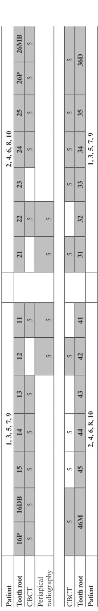
Adolescents with permanent teeth, crowding and tooth displacement were examined with one CBCT scan 8 x 8 cm presenting both jaws, three PA radiographs of maxillary incisors and two posterior BW radiographs in each side of the mouth. For the present study, the radiographic examinations of 10 adolescents (mean age 13.4; range 12–17) with a male-to-female ratio of 1:1, examined during March 2016 and March 2017, were selected. Table 1 presents the patient distribution, teeth and sites selected for measurement of root lengths. In the case of a premolar with two apices, the buccal root was measured. In Table 2, the patients, teeth, and sites selected for measurements of the marginal bone level are shown.
Radiographic equipment and data processing
CBCT images were obtained with 3D Accuitomo® 170
(J. Morita, Kyoto, Japan) units in two departments, using the same scanning protocol and operating at 80 kV and 3 mA. The option of 360 revolution of the X-ray source and standard acquisition mode with a field of view (FOV) of 8 cm (diameter) x 8 cm (height) and 160 µm voxel size were used to examine the upper and lower jaw together. The patients were oriented with the same plane setting controlled by the first author as a part of the study protocol.
PA and BW radiographs were obtained with a rect-angular positioning device and paralleling technique in four clinics according to written instructions. The dental X-ray units, exposure parameters and imaging systems
are shown in Table 3. The X-ray units were equipped
t able 1 P atient distrib ution and n umber of sites a vaila ble f or measur ement of r oot lengths f or CBCT and peria pical r adio gr aph y f or each of six r aters P atient 1, 3, 5, 7, 9 2, 4, 6, 8, 10 tooth r oot 16 P 16 DB 15 14 13 12 11 21 22 23 24 25 26 P 26 MB CBCT 5 5 5 5 5 5 5 5 5 5 5 P eria pical radio gr aph y 5 5 5 5 CBCT 5 5 5 5 5 5 5 5 5 tooth r oot 46 M 45 44 43 42 41 31 32 33 34 35 36D P atient 2, 4, 6, 8, 10 1, 3, 5, 7, 9 p = pala tal, DB = disto-b uccal, MB = mesio-b
uccal, D = distal, M = mesial
t able 2 P atient distrib ution and n umber of sites a vaila ble f or measur ement of mar ginal bone le vel f or CBCT , peria
pical, and bite
wing r adio gr aph y f or each of six r aters P atient 1, 3, 5, 7, 9 2, 4, 6, 8, 10 tooth r oot 16 P 16 DB 15 14 13 12 11 21 22 23 24 25 26 P 26 MB CBCT 14 9 18 19 20 20 19 19 20 15 10 Bite wing radio gr aph y 10 10 10 5 5 10 10 10 P eria pical radio gr aph y 10 10 10 10 Bite wing radio gr aph y 10 10 10 5 5 10 10 10 CBCT 15 20 20 19 20 20 20 20 15 tooth r oot 46 M 45 44 43 42 41 31 32 33 34 35 36D P atient 2, 4, 6, 8, 10 1, 3, 5, 7, 9 p = pala tal, DB = disto-b uccal, MB = mesio-b
examinations, one of the authors checked the radio-graphic equipment parameters as described by Senneby et al.14 Intraoral radiographs were assessed by one of the
authors to be of acceptable quality.
10 CBCT volumes were stored in Digital Imaging and Communications in Medicine file format and prepared with i-Dixel software on a workstation. All intraoral radiographs were imported from the worksta-tions of the four clinics to Image J software (National Institute of Health, Bethesda, MD). For all images a BARCO (MFGD 1318; BARCO, Kortrijk, Belgium) 18.10 greyscale liquid crystal display monitor with a
luminance of 400 cd/m2 and resolution of 1280 × 1024
pixels was used. Linear measurement calibration of intraoral radiographs was performed by calculating the ratio between a defined distance and the pixel number of each of the four receptors, later used in Image J soft-ware for measurements.
Raters and ratings
An initial study protocol, including data collection, raters and statistical analyses was prepared. Six raters were selected to ensure a diverse level of professional experience: two were specialists in dental and maxillo-facial radiology with 25 and 30 years of experience in radiology, respectively, two were specialists in orthodon-tics with 10 years of experience each, one was trainee in orthodontics and one rater was a general dental prac-titioner. The raters were aware of the purpose of the study and blinded to each other´s measurements.
A radiologist with experience of CBCT selected cross-sectional CBCT images and sites to be measured (Tables 1 and 2) using i-Dixel software. Sites for measurements were selected in PA of maxillary incisors
and posterior BW radiographs (Tables 1 and 2).
Measurements were derived from the CBCT, PA and BW using Image J software. The reading room illumination was dimmed (below 50 lux as recom-mended by American Association of Physicists in
Medicine Task Group 18)15 and kept constant. The
reading distance was approximately 50–60 cm. There were no restrictions on reading time and zooming was allowed. All images were assessed in the same order. First, the CBCT images were assessed, after 10 days the PA images and finally after further 10 days the BW images were assessed.
First, all raters attended a 10 min educational presen-tation given by the first author demonstrating the proce-dure using examples of root length and marginal bone level measurements on CBCT and intraoral images. During the session, the raters were given examples of a procedure similar to what they were expected to perform. The aim was to familiarise the raters with the Image J software and measurement procedure. Firstly, the raters were asked to identify the reference points. Secondly, when reference points were interpretable, they were asked to measure the root length and marginal bone level distance. Regarding marginal bone level, both mesial and distal sides of selected teeth were measured except for the maxillary canine where only the distal side was measured. All measurements recorded in millime-tres were rounded to one decimal. The patient informa-tion was masked on all images.
The following definitions were applied:
• Measurability: frequency of sites with interpretable reference points for measurements of root length and marginal bone level, respectively
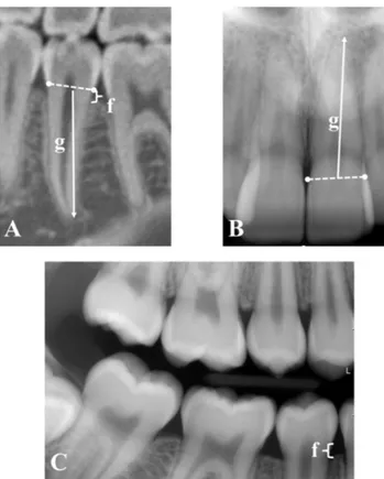
• Root length: distance between the mid-point between
the CEJ and root apex (Figure 1). When a root was
curved the measurement could be performed in two steps: first measure was the distance from the mid-point between the CEJ and the mid-point where the root start to curve and secondly from this point to the root apex. The distances were then summed up.
• Marginal bone level: distance between CEJ and alve-olar bone crest (Figure 1).
In order to calculate intrarater reliability, three raters (two dental and maxillofacial radiologists and one orthodontist) performed replicate measurements of selected sites in CBCT images (48% of available sites), in PA (73% of available sites) and in BW (51% of available sites). There were 3 weeks between the first and second sessions in order to minimise rater recall bias.
All results were collected in a computer database for analysis. For measurability, the frequency of sites where reference points were possible to identify was calculated. For analysing the reliability of each method, Intraclass correlation coefficient (ICC 2.1) with 95% confidence intervals (CI) was calculated. All statistical
analyses were performed using IBM SPSS® Statistics v.
22.0 (IBM Corp., New York, NY; formerly SPSS Inc., Chicago, IL).
table 3 Dental X-ray units, exposure parameters, and imaging
systems used in four clinics for periapical and bitewing radiography
Clinic X-ray unit (name), exposure parameters Imaging system (name) pixel size
Dental school Planmeca ProX (Planmeca; Helsinki, Finland) 60kV, 7mA, 0.125 s ProSensor® (Planmeca; Helsinki, Finland) Pixel size 30 × 30 µm2
Hospital 1 Kavo, Gendex 765 DC (Kavo; Biberach/Riss, Germany)
65kV, 7 mA, 0.25 s for periapical and 0.125 s for bitewing radiography
ProSensor® (Planmeca;
Helsinki, Finland) Pixel size 30 × 30 µm2
Hospital 2 Planmeca Intra (Planmeca; Helsinki, Finland)
60kV, 8mA, 0.160 s
Schick 33 (Sirona Dental, Salzburg, Austria) Pixel size 15 × 15 µm2
Orthodontic
clinic Sirona – HELIODENT DS (Sirona Dental Systems, Bernsheim, Germany) 60kV, 7 mA, 0.16 s Sigma CCD (GE/ Instrumentarium Imaging, Tuusula, Finland) Pixel size 39 × 39 µm2
Results Measurability
All rater assessments were included for final analysis.
As presented in Table 4, the number of sites that were
interpreted and measured varied among (i) imaging methods, (ii) measured object (root length or marginal bone level) and (iii) rater. For root length all avail-able sites were interpreted and measured by all raters in CBCT and all but one site in PA of maxillary inci-sors. For marginal bone level, all available sites were measured in CBCT, 79% of sites (189 of 240) in PA of maxillary incisors and 86% of sites (719 of 840)
in posterior BW (Table 4). Considering the six raters,
measurability for root length and marginal bone level was 100% in CBCT and for root length measurements of maxillary incisors in PA except for one rater being 95%. For marginal bone level, frequencies varied among the raters for PA (range 67–95%) and for BW
(range 82–88%) (Table 4).
Root length measurements
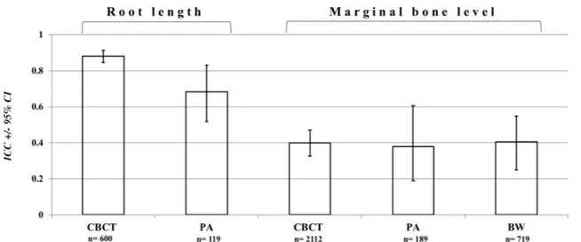
Interrater reliability: For all raters and roots, ICC between measurements performed in CBCT was 0.88
(CI 0.85–0.98) (Figure 2). ICC for roots of different
teeth, presented in Figure 3, ranged between 0.27 and
0.96, being the highest for mandibular left second premolars and the lowest for maxillary right canines. ICCs for measurements of maxillary right central incisor and left lateral incisor in CBCT were 0.88 (CI 0.67–0.98) and 0.76 (CI 0.45–0.97), respectively (Figure 3). Corresponding ICCs for measurements of these teeth in PA were 0.64 (CI 0.28–0.94) and 0.68
(CI 0.35–0.95), respectively (Figure 3). ICC between
measurements performed in PA of all four maxillary
incisors was 0.69 (CI 0.52–0.83) (Figure 2). For
pair-wise interrater reliability, ICC of all measured roots in CBCT was above 0.85 (range 0.85–0.91). For measure-ments of the maxillary incisors in PA, ICC was above 0.44 (range 0.44–0.86).
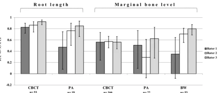
Intrarater reliability: ICCs among the three raters for measurements performed in CBCT were comparable,
ranging between 0.82 and 0.92 (Figure 4). For
measure-ments of the maxillary anterior incisors performed in
PA, ICCs ranged between 0.47 and 0.84 (Figure 4).
Marginal bone level measurements
Interrater reliability: For all raters and all measured sites, ICCs were 0.4 (CI 0.32–0.47) in CBCT, 0.38 (CI 0.19–0.6) in PA of the maxillary incisors and 0.4 (CI
0.25–0.55) in posterior BW images (Figure 2). For
pair-wise reliability, ICCs varied among the pairs of raters being between 0.34 and 0.68 for CBCT, between 0.12 and 0.66 for PA of maxillary incisors and between 0.20 and 0.66 for posterior BW images.
Intrarater reliability: ICC varied depending on
imaging method among the three raters (Figure 4). For
measurements in CBCT, ICC was comparable for the raters, ranging between 0.56 and 0.57. For PA of maxil-lary anterior incisors, ICCs ranged between 0.29 and 0.62. ICCs for posterior BW were above 0.7 for two raters, but lower for the third rater (Figure 4).
Figure 1 Cross-sectional CBCT image (A), periapical (B), and
bite-wing (C) images used for measurement of root length (g) and marginal bone level (f).
table 4 Measurability
Rater
Root lengths Marginal bone level
CBCT n = 100 Periapical radiography n = 20 n = 352CBCT Periapical radiography n = 40 Bitewing radiography n = 140 Measurability (%) 1 100 95 100 70 82 2 100 100 100 77 88 3 100 100 100 77 87 4 100 100 100 67 84 5 100 100 100 95 85 6 100 100 100 85 85 Mean (%) 100 99 100 79 85 N 600 119 2112 189 719
n = numbers of available sites for each rater, N = total number of
measured sites of six ratersBW, bitewing; PA, periapical.
Frequency(%) of measured sites related to number of available sites for each rater (n)for CBCT, PA and BW radiography for measurement of rootlength and marginal bone level.N = totalnumber of sites measured by six raters.
Discussion
The findings of the present study demonstrated that the frequency of sites with interpretable reference points for root length measurements in CBCT and PA of maxillary incisors was high. Measurability and reliability was lower for measurement of the marginal bone level than for that of root length, which was high. Intrarater reliability varied among methods,
object measured and raters, except for the root length measurements in CBCT, which presented a high reli-ability for all raters.
The sample was selected from images, which were obtained for a clinical trial of fixed orthodontic appli-ance. No additional radiographs were performed for this study resulting in a limited number of sites for intra-oral images. CBCT and intraintra-oral radiography were not compared on measurability and reliability.
Figure 2 Interrater reliability expressed as intra-class correlation coefficient with confidence interval for six raters’ measurements of root length
and marginal bone level in CBCT, PA of maxillary incisors and posterior BW images. n= total number of measurements by six raters. BW, bite-wing; PA, periapical.
Figure 3 Interrater reliability expressed as intra-class correlation coefficient and confidence intervals for six raters’ measurements of root length
of different teeth in CBCT and in PA images of maxillary incisors by six raters. P, palatal; DB, distobuccal; MB, mesiobuccal; D, distal; M, mesial root.
A meticulous assessment of the roots and marginal bone tissue prior to orthodontic treatment provides an important baseline to evaluate changes that may occur during orthodontic treatment and may guide treatment
approaches.1,2 Therefore, the diagnostic tasks chosen
were measurement of root lengths and marginal bone levels of adolescents.
The spatial resolution of CBCT mainly depends on receptor physical pixel size. Other factors such as projec-tion geometry, scatter radiaprojec-tion, detector moprojec-tion blur, focal spot size, number of basis projections and
recon-struction algorithm may affect the image resolution.16
Additionally, contrast resolution is important in order to distinct between structures with minor differences in attenuation and to present them with different grey levels. The CBCT unit used offered four predefined scan modes with possibilities to change the mA- and kV-settings and dimension of FOV. As recommended by
Al-Okshi et al,17 full rotation with 80 kV and modified
mA to 3 according to patient size was used to obtain optimal subjective image quality for assessment of peri-odontal structures. As a low exposure (3 mA) was used, full rotation was chosen in order to improve the signal-to-noise ratio. The size of FOV influences the image as a large FOV may reduce contrast-to-noise ratio resulting
in decreased visibility of anatomical structures.16,18
Although the root apex and the marginal bone level can be subtle structures in adolescents, we selected an 8 × 8 cm FOV to capture the upper and lower jaw during the same scanning in order to reduce patient radiation dose. Another influential factor when imaging subtle anatom-ical structures is the voxel size—the smaller the voxel size, the higher the spatial resolution18 but with higher
image noise, which may have influenced the reliability of the measurements of the marginal bone level.
Measurability is one way of simultaneously expressing image quality and the usefulness of an imaging method. Measurability adds to visual grading analysis of the images, which is commonly used to eval-uate image quality, as it demands the rater to perform a clinically relevant task. The included patients comprised adolescents, who will have fixed orthodontic appli-ances and undergo several radiographic examinations during their treatment. Although all raters stated that all measurements were possible to perform in CBCT, it may be expected that this will not be the case in subse-quent radiographic examinations. Then, the patients will have metal/radiopaque material on their teeth, which increases the risk that a case will be scored as not
interpretable in CBCT.18 Thus, a lower measurability is
to be expected in some the follow-up examinations of orthodontic treatment performed with CBCT. All but one root length was measured in PA of the maxillary incisors whilst the frequency of measurable sites of the marginal bone level in PA was lower and varied consid-erably among the raters. The frequency of uninterpre-table sites for measurement of the marginal bone level in PA of the maxillary incisors (about 20%) corresponds
to that found in a study of adult patients.19 A common
reason for uninterpretability is said to be the difficulty in identifying the CEJ.5,10,20 This did not seem to be the
case for PA in the present study as all but one measure-ment of root length, which were based on the CEJ, were performed. Instead it may be attributed to difficulties in identifying the other reference point, i.e. the marginal bone level. One explanation for this may be that some PA images were slightly distorted as it is difficult to perform PA of the maxillary incisors with optimal quality in adolescents with crowded teeth. For posterior BW the lower measurability may be due to the fact that
Figure 4 Intrarater reliability of three raters expressed as intraclass correlation coefficient with confidence intervals for measurements of root
length and marginal bone level in CBCT, PA images of maxillary incisors and posterior BW images. n, number of measured sites for each rater; BW, bitewing; PA, periapical.
the marginal bone septa were incompletely mineralized. The reported frequency of unacceptable image quality for assessment of the marginal bone tissue in posterior BW varies between 6 and 29% in previous studies of adult patients.10,20 As far as we know, there is no study of
measurability of intraoral radiographs of adolescents. When planning clinical trials and analysing treatment outcomes, results on measurability are important to take into account. If results on measurability are reported to a limited extent and simply removed from the analysis, it may lead to biased analyses of the diagnostic modalities and to incorrect reports of treatment outcomes.
Accuracy is a keystone in assessing the efficacy of diagnostic methods. When a reference standard is not available, reliability studies can be used to indicate the
upper bound of accuracy.21 In other words, the
reli-ability estimations are useful in determining the extent to which the inaccuracy of a system is due to deci-sion-making errors. Unfortunately, reliability studies are generally neglected and do not appear in the different
stages of evaluating studies of diagnostic methods12 or
in studies where diagnostic methods are used to
eval-uate treatment outcomes.21 Changes in dental or bone
tissues caused by different treatment methods are often assessed by comparing measurement results from the baseline examination with those of follow-up examina-tions. Then, accuracy may not be the issue but rather the reliability of the method used to assess any change. In studies of orthodontic treatment, the measurement errors of baseline and follow-up examinations should fall short of the assessed change of root length and marginal bone level. Furthermore, it is important to be aware of several raters’ performance.
In the present study, interrater reliability for six raters’ measurements of root length in CBCT was high (ICC 0. 88) and so was intrarater reliability (ICC above 0.8). Although direct comparison with previous studies
on reliability of root length measurements in CBCT22,23
is not pertinent, our results are broadly in agreement. The fact that interrater reliability varied among roots of different teeth, should be considered when selecting roots for analysis of treatment outcomes. In the previous
studies of root length measurements in CBCT,22,23 the
cusp/incisal edge represented one of the reference
points. As the cusp/incisal edge may change over time due to attrition, we preferred to use the CEJ as the refer-ence point as it is more stable when comparing root lengths. Interrater reliability of root length measure-ment performed in PA of anterior incisors was rather high (ICC 0.69) but lower than that reported for two raters (ICC above 0.90) by Lago et al22 for inter and
intr-arater reliability.
Overall, also in CBCT, reliability for measurement of the marginal bone level was low. The results for poste-rior BW were somewhat unexpected as BW is recom-mended for assessment of the marginal bone tissue. The low reliability may be due to the sample, which consisted of young adolescents. The delineation of the marginal bone is therefore imaged more vaguely, especially around second premolars and second molars.
conclusions
Our results on high measurability in CBCT for measure-ments of root lengths and marginal bone level as well as high reliability for root length measurements further add to the results of previous clinical studies indicating that CBCT may be the best choice for scientific analyses. But for clinical praxis, we recommend intraoral radiog-raphy as preventive measures such as the use of lower forces, resting periods, and shortening of treatment time are not taken until root resorption from 2 mm up to 1/3
of the root length is diagnosed.4 This is in accordance
with the “as low as diagnostically acceptable” prin-ciple,24 as the radiation dose for intraoral radiography is
lower than that of a CBCT examination.25 Thus, a
radio-graphic method must be justified for each patient, both regarding radiation dose and diagnostic image quality, which is emphasised in the SEDENTEXCT guidelines
on CBCT for Dental and Maxillofacial Radiology.26
acknowledgement
This study project was supported by research funds for Oral Health Related Research by Region Skåne, Sweden [OFRS512901].
References
1. Brezniak N, Wasserstein A. Root resorption after orthodontic treatment: Part 1. literature review. Am J Orthod Dentofacial
Orthop 1993; 103: 62–6. doi: https:// doi. org/ 10. 1016/ 0889- 5406( 93) 70106-X
2. Brezniak N, Wasserstein A. Root resorption after orthodontic treatment: Part 2. literature review. Am J Orthod Dentofacial
Orthop 1993; 103: 138–46. doi: https:// doi. org/ 10. 1016/ S0889- 5406( 05) 81763-9
3. Killiany DM. Root resorption caused by orthodontic treatment: an evidence-based review of literature. Semin Orthod 1999; 5: 128– 33. doi: https:// doi. org/ 10. 1016/ S1073- 8746( 99) 80032-2
4. Weltman B, Vig KWL, Fields HW, Shanker S, Kaizar EE. Root resorption associated with orthodontic tooth movement: a systematic review. Am J Orthod Dentofacial Orthop 2010; 137: 462–76. doi: https:// doi. org/ 10. 1016/ j. ajodo. 2009. 06. 021
5. Sherrard JF, Rossouw PE, Benson BW, Carrillo R, Buschang PH. Accuracy and reliability of tooth and root lengths measured on cone-beam computed tomographs. Am J Orthod Dentofacial
Orthop 2010; 137(Suppl): S100–S108. doi: https:// doi. org/ 10. 1016/ j. ajodo. 2009. 03. 040
6. Lund H, Gröndahl K, Gröndahl H-G. Cone beam computed tomography for assessment of root length and marginal bone level
during orthodontic treatment. Angle Orthod 2010; 80: 466–73. doi: https:// doi. org/ 10. 2319/ 072909- 427.1
7. Malocclusions and orthodontic treatment in a health perspective.
Systematic review. ISBN: 91-85413-06-2 • ISSN: 1400-1403. The Swedish Council on Technology Assessment in Health Care (SBU) Report no: 176. (In Swedish) Summary in English 2005;.
8. Lund H, Gröndahl K, Gröndahl H-G. Cone beam computed tomography evaluations of marginal alveolar bone before and after orthodontic treatment combined with premolar extractions.
Eur J Oral Sci 2012; 120: 201–11. doi: https:// doi. org/ 10. 1111/ j. 1600- 0722. 2012. 00964.x
9. Whiting P, Rutjes AWS, Reitsma JB, Bossuyt PMM, Kleijnen J. The development of QUADAS: a tool for the quality assessment of studies of diagnostic accuracy included in systematic reviews.
BMC Med Res Methodol 2003; 3: 25. doi: https:// doi. org/ 10. 1186/ 1471- 2288- 3- 25
10. Ivanauskaite D, Lindh C, Rohlin M. Observer performance based on marginal bone tissue visibility in Scanora panoramic radiog-raphy and posterior bitewing radiogradiog-raphy. Stomatologija 2008; 10: 36–43.
11. Fryback DG, Thornbury JR. The efficacy of diagnostic imaging.
Med Decis Making 1991; 11: 88–94. doi: https:// doi. org/ 10. 1177/ 0272989X9101100203
12. Kottner J, Audigé L, Brorson S, Donner A, Gajewski BJ, Hróbjartsson A, et al. Guidelines for reporting reliability and agreement studies (GRRAS) were proposed. Int J Nurs Stud 2011;
48: 661–71. doi: https:// doi. org/ 10. 1016/ j. ijnurstu. 2011. 01. 016
13. Bossuyt PM, Reitsma JB, Bruns DE, Gatsonis CA, Glasziou PP, Irwig LM, et al. The STARD statement for reporting studies of diagnostic accuracy: explanation and elaboration. Clin Chem 2003; 49: 7–18. doi: https:// doi. org/ 10. 1373/ 49. 1.7
14. Senneby A, Elfvin M, Stebring-Franzon C, Rohlin M. A novel classification system for assessment of approximal caries lesion progression in bitewing radiographs. Dentomaxillofac Radiol 2016; 45: 20160039: 20160039: . doi: https:// doi. org/ 10. 1259/ dmfr. 20160039
15. Samei E, Badano A, Chakraborty D, Compton K, Cornelius C, Corrigan K, et al. Assessment of display performance for medical imaging systems: Executive summary of AAPM TG18 report.
Med Phys 2005; 32: 1205–25. doi: https:// doi. org/ 10. 1118/ 1. 1861159
16. Pauwels R, Araki K, Siewerdsen JH, Thongvigitmanee SS. Tech-nical aspects of dental CBCT: state of the art. Dentomaxillofac
Radiol 2015; 44: 20140224. doi: https:// doi. org/ 10. 1259/ dmfr. 20140224
17. Al-Okshi A, Theodorakou C, Lindh C. Dose optimization for assessment of periodontal structures in cone beam CT exami-nations. Dentomaxillofacial Radiology 2017; 46: 20160311. doi:
https:// doi. org/ 10. 1259/ dmfr. 20160311
18. Spin-Neto R, Costa C, Salgado DM, Zambrana NR, Gotfredsen E, Wenzel A. Patient movement characteristics and the impact on CBCT image quality and interpretability.
Dento-maxillofac Radiol 2018; 47: 20170216. doi: https:// doi. org/ 10. 1259/ dmfr. 20170216
19. Åkesson L, Rohlin M, Håkansson J. Marginal bone in perio-dontal disease: an evaluation of image quality in panoramic and intra-oral radiography. Dentomaxillofac Radiol 1989; 18: 105–12. doi: https:// doi. org/ 10. 1259/ dmfr. 18. 3. 2637876
20. Persson RE, Tzannetou S, Feloutzis AG, Brägger U, Persson GR, Lang NP. Comparison between panoramic and intra-oral radio-graphs for the assessment of alveolar bone levels in a periodontal maintenance population. J Clin Periodontol 2003; 30: 833–9. doi:
https:// doi. org/ 10. 1034/ j. 1600- 051X. 2003. 00379.x
21. Swets JA, Pickett RM. Evaluation of diagnostic systems: methods
from signal detection theory. New York: Academic Press; 1982.
22. Lago GV, Fernandes TMF, Oltramari-Navarro PVP, Almeida MR, Ladewig VdeM, Conti ACdeCF. Reliability of CBCT and periapical radiography methods to evaluate external apical root resorption during early phase of orthodontic treat-ment. J. Health Scie. 2018; 20: 2–7. doi: https:// doi. org/ 10. 17921/ 2447- 8938. 2018v20n1p2-7
23. Schwartz JP, Raveli TB, Almeida KCdeM, Schwartz-Filho HO, Raveli DB. Cone beam computed tomography study of apical root resorption induced by Herbst appliance. J Appl Oral Sci 2015; 23: 479–85. doi: https:// doi. org/ 10. 1590/ 1678- 775720150224
24. Bushberg JT. Science, radiation protection, and the NCRP: building on the past, looking to the future. In: NCRP Fiftieth
Annual Meeting Program. Bethesda, MD: National Council on
Radiation Protection and Measurements; 2014. pp. 5–7. 25. Al-Okshi A, Lindh C, Salé H, Gunnarsson M, Rohlin M.
Effec-tive dose of cone beam CT (CBCT) of the facial skeleton: a systematic review. Br J Radiol 2015; 88: 20140658. doi: https:// doi. org/ 10. 1259/ bjr. 20140658
26. European Commission. Radiation protection No 172: cone beam
CT for dental and maxillofacial radiology. Evidence-based guide-lines. Luxembourg: Directorate-General for Energy; 2012.