Faculty of Veterinary Medicine and Animal Science
Prevalence of Antibodies Against Rift Valley
Fever and Sheep and Goat Pox in Small
Ruminants in Northern Zambia
Prevalens av antikroppar mot Rift Valley
fever och får- och getkoppor hos små
idisslare i norra Zambia
Siri Linde
Uppsala 2019
Prevalence of Antibodies Against Rift Valley
Fever and Sheep and Goat Pox in Small
Ruminants in Northern Zambia
Prevalens av antikroppar mot Rift Valley fever och får- och
getkoppor hos små idisslare i norra Zambia
Siri Linde
Supervisor: Karin Alvåsen, Department of Clinical Sciences
Assistant Supervisor: Sara Lysholm, Department of Clinical Sciences, Musso Munyeme,
University of Zambia
Examiner: Johanna Lindahl, Department of Medical Biochemistry and Microbiology, Uppsala
University & Department of Clinical Sciences, SLU
Degree Project in Veterinary Medicine
Credits: 30
Level: Second cycle, A2E Course code: EX0869 Place of publication: Uppsala Year of publication: 2019
Online publication: https://stud.epsilon.slu.se
Key words: Rift Valley Fever, Sheep and Goat Pox, epidemiology, prevalence Nyckelord: Rift Valley fever, får- och get-koppor, epidemiologi, prevalens
Sveriges Lantbruksuniversitet
Swedish University of Agricultural Sciences Faculty of Veterinary Medicine and Animal Science
SUMMARY
There are many transboundary diseases that could be of importance to livestock owners in Zambia, among them are diseases such as Sheep and Goat Pox (SGP) as well as Rift Valley Fever (RVF). Both of these diseases are on the OIEs list of diseases of serious socio-economic and public health significance and are important threats to trade of animals and animal products. Sheep and Goat Pox (SGP) is caused by Sheep Pox Virus (SPPV) and Goat Pox Virus (GTPV), which affects the animals by giving them symptoms like fever, rhinitis and papules on the body and in the organs, usually the lungs. Indigenous breeds are usually less susceptible to the disease than for example European breeds. If a herd gets infected with the disease the outcome will vary partially depending on the severity of the isolate.
RVF is caused by Rift Valley Fever Virus, an arbovirus belonging to the Bunyaviridae family. Since the first isolation in 1931 it has caused several outbreaks in Africa, and also been proven to be able to override large geographical barriers, for example the first time it emerged in Egypt north of the Sahara, and the first time it was found outside of Africa in Saudi Arabia and Yemen. The disease is characterized by abortion storms and neonatal mortality in affected herds, as well as fever, diarrhoea and vomiting. RVF is also a zoonosis that in humans give symptoms that resembles the flu, in more rare cases it can progress to haemorrhagic fever, retinitis and encephalitis. RVF is transmitted by mosquitoes, with outbreaks being closely related to preceding heavy rainfalls.
The study performed for this thesis project consisted of two parts. One being sampling of 480 goats and sheep in two districts along the border to Tanzania in northern Zambia, the other being questionnaire interviews with the 160 owners of these animals. The questions used in this thesis were focused on management routines such as grazing system and contact with other ruminants. Of the sampled animals, none were positive for antibodies against SGP. Eleven animals (2.3%) were positive for antibodies against RVF.
Even though it was a long time since the last outbreak of RVF in Zambia, this study gives evidence that the virus is still circulating at low levels among the animals in this region. It is important to maintain surveillance for RVF to prevent outbreaks in the future.
TABLE OF CONTENT
Introduction ... 1
Literature Review ... 2
The importance of transboundary diseases ... 2
Veterinary services in Zambia ... 2
Sheep and goat pox ... 2
The virus ... 2
Clinical Signs ... 3
Epidemiology and transmission ... 4
Effect on the community ... 4
Rift Valley Fever ... 5
The virus ... 6
Clinical signs ... 6
Epidemiology and transmission ... 6
Seroprevalences in Africa ... 8
Rift Valley Fever in Zambia ... 8
Effect on the community/society ... 8
Vaccination ... 9
Material and Methods ... 10
Data collection ... 10
Blood sampling ... 10
Questionnaire ... 10
Rift Valley Fever and Capripox ELISA ... 11
Statistical analysis ... 11
Results ... 12
Seroprevalence ... 13
Questionnaire ... 13
Discussion ... 15
Sheep and Goat Pox ... 15
Rift Valley Fever ... 15
Methodological considerations ... 16 Conclusion ... 18 Populärvetenskaplig sammanfattning ... 19 Acknowledgements ... 22 References ... 23 Appendix 1 ... 1
ABBREVIATIONS
SGP: Sheep and Goat Pox SPPV: Sheep Pox Virus SPP: Sheep Pox
GTPV: Goat Pox Virus GTP: Goat Pox
RVF: Rift Valley Fever
RVFV: Rift Valley Fever Virus IEP: Inter-Epizootic Period
INTRODUCTION
The aim of this study was to investigate the prevalence of antibodies against Rift Valley Fever (RVF) and Sheep and Goat Pox (SGP) in an area in northern Zambia along the border to Tanzania. Another objective was to study the existing literature to get a better understanding of the importance on economy and animal welfare of these two diseases. To evaluate the prevalence of antibodies ELISA tests were used.
Zambia is a landlocked country in eastern Africa bordering Tanzania to the northeast, Malawi to the east, Mozambique to the southeast, Zimbabwe to the south, Botswana and Namibia to the southwest, Angola to the west and the Democratic Republic of the Congo to the north. The country is divided into ten provinces and in this study goats and sheep from Muchinga province and Northern province along the border to Tanzania and Malawi were sampled. The climate in Zambia is generally characterized by three seasons, hot-dry from August to October, warm-wet from November to April and cool-dry from May to July.
In Zambia there are about 500 000 households keeping goats and 20 300 households keeping sheep, in total there are about 3.5 million goats and 150 000 sheep. In the Muchinga province lives 4.4% of the country’s goats and 2.3% of the country’s sheep, and in the Northern province lives 6.1% of all goats and 2.3% of all sheep (Central Statistical Office, 2017).
Both RVF and SGP are defined as diseases of serious socio-economic importance with great impact on the international trade of animals and animal products (OIE, 2018c). In an analysis made by the World Bank in cooperation with FAO and OIE in 2011, SGP was named as one of the top ten diseases that affect sheep and goats around the world, and the number one disease by number of outbreaks (World Bank & TAFS Forum, 2011).
LITERATURE REVIEW
The importance of transboundary diseases
For livestock producers, especially small-scale producers, there is a threat of transboundary diseases having an impact on their livelihood. The impact that the different diseases can have depend on the disease, the number of animals at risk and the owners’ dependency on livestock for their livelihood. The threat of transboundary diseases is not only restricted to livestock producers, as trade is a big part of today’s society. Exporting countries want to maintain their market, while importing countries are concerned with the protection of their domestic livestock populations. The protection of trade is therefore of great interest for many countries and a major incentive for investigating prevalence, prevention and control of transboundary diseases (Domenech et al., 2006).
Both RVF and SGP are OIE-listed diseases, in other words diseases that are of serious socio-economic and public health importance, and that are of importance to the international trade of animals and animal products (OIE, 2018c). The diseases are also on the select agent list of the United States Department of Agriculture and the US National Select Agent Registry, since these viruses are considered to have the potential to pose a severe threat to both human and animal health (Federal Select Agent Program, 2018).
Veterinary services in Zambia
Historically all veterinary services in Zambia have been free of charge. There was a deterioration in coverage and quality of the veterinary services due to a shrinking budget over the years. To address the problems the veterinary system was reorganized in 1996, which redefined the public and private sectors. The role of the public sector was henceforth limited to regulation, control and monitoring, while the private sector was responsible for implementing most field activities (Chilonda et al., 1999).
Livestock diseases continue to be among the main constraints limiting livestock development in Zambia for small-scale farmers in the traditional sector, partly because of underutilization of veterinary inputs by the farmers, and a general reluctance to pay for veterinary services following the end of free veterinary services (Chilonda & Van Huylenbroeck, 2001). The traditional sector with small-scale farmers own the majority of both goats and sheep, 99% and 87% respectively, with the rest belonging to the commercial sector (Central Statistical Office, 2017).
Sheep and goat pox
SGP is endemic in Africa north of the equator, the Middle East, Turkey, India, Nepal, parts of the People’s Republic of China and Bangladesh to name some regions and countries. There has recently also been an outbreak in Greece (OIE, 2018b).
The virus
Sheep pox virus (SPPV) and goat pox virus (GTPV) both belong to the family Poxviridae in the genus Capripox together with lumpy skin disease which affects cattle (OIE, 2017). The viruses that causes sheep pox (SPP) and goat pox (GTP) are very similar and have been shown
to share 96% of their genes (Tulman et al., 2002). There are no serotypes of SPPV and GTPV as the viruses are antigenically indistinguishable (Kitching & Taylor, 1985; Kitching, 1986). The similarity has previously led to the belief that they are strains of a single virus, that is however incorrect according to more recent studies of the genome (Tulman et al., 2002; Hosamani et al., 2004). Some strains of the viruses have been shown to be able to cause clinical disease in both sheep and goats (Yan et al., 2012; OIE, 2013), though sheep usually only show mild clinical signs when infected with GTPV (Kitching et al., 1986; Babiuk et al., 2009) and goats only show mild clinical signs when infected with SPPV (Bhanuprakash et al., 2010). Some isolates give similar symptoms regardless which species is the source and which it infects (Kitching & Taylor, 1985).
Recent immunohistochemical studies in skin and lung of naturally infected lambs have confirmed that the majority of infected cells in the inflammatory infiltrates are virus-infected monocytes and macrophages (Gulbahar et al., 2006). Bowden et al (2008) suggested that dissemination of SPPV and GTPV from primary lesions to draining lymph nodes and to the systemic circulation is through infected monocytes and macrophages.
Clinical Signs
After an incubation period of 8-13 days the clinical picture in sheep and goats are similar (OIE, 2017). The animals get an increased rectal temperature of 40-41.5 °C, develop rhinitis with mucous discharge from the nostrils and the superficial lymph nodes become grossly enlarged (Kitching et al., 1986; Bhanuprakash et al., 2010). Hard intradermal papules about 1-2 cm in diameter develop over the face and body, predominantly in areas with less hair or wool e.g. around the eyes and mouth, along the abdomen, under the tail and in groin and axilla (Kitching
et al., 1986; Bhanuprakash et al., 2010). The papules can coalesce to form larger swellings and
the skin covering the papules can become necrotic and be rubbed off. The scabs after the papules can remain on the body for 3-4 weeks if the animal survives (Kitching et al., 1986).
Death usually occurs 7-10 days after onset of disease, but some individuals may survive longer before perishing (Mondal et al., 2004). Post mortem examination shows papules in lungs (Bhanuprakash et al., 2010), and frequently also in the tongue, pharynx and the kidneys. Lymph nodes, both internal and superficial, are grossly enlarged and oedematous (Kitching et al., 1986). Though the lymph nodes become enlarged, one study has shown that the lymphadenopathy is not associated with high viral replication in the nodules (Bowden et al., 2008).
Some indigenous breeds in Africa are less susceptible to disease whereas the European breeds are more susceptible and case fatality rate among some European breeds may reach 100% (Kitching & Taylor, 1985; OIE, 2013). In endemic areas the morbidity rate is about 70-90% and the mortality rate 5-10% (OIE, 2013), but morbidity and case fatality in young individuals can reach up to 100% and 70%, respectively (Hailat et al., 1994). Though animals of all ages can get sick young animals are more susceptible (Mondal et al., 2004).
Epidemiology and transmission
Transmission of the virus usually occurs through aerosols infecting the epithelial lining of the airways when infected and susceptible animals are in close proximity of one another (Kitching & Taylor, 1985; Carn, 1993). Infection can also happen through other mucous membranes or abraded skin (OIE, 2013). One study from India showed that SPP can be transmitted when there is a shortage of fodder and the sheep nibble the leaves of thorny plants such as Acacia, therefore damaging the skin on face and lips and infection thus spreads from infected to susceptible sheep (Bhanuprakash et al., 2005). The animals also frequently scratch themselves where the skin papules develop, and in this manner manage to contaminate the area around them (Kitching & Taylor, 1985). Different isolates of capripoxvirus can have different kinetics of viral shedding depending on isolate, species and route of virus inoculation (Babiuk et al., 2009). Viral shedding from nasal, oral and conjunctival secretions correlate with the appearance of lesions on the mucosal surfaces, but viral shedding has also been shown to occur from mucosal surfaces without visible lesions present. Some animals have been seen to shed virus for up to two months after the onset of fever (Bowden et al., 2008).
Vectors are not an important factor for the transmission of this disease, but it has been shown under experimental conditions that Stomoxys calcitrans (the common European stable fly) is capable of transmitting the virus mechanically (Mellor et al., 1987) which is promoted by the high titres of virus in the skin papules of infected animals (Bowden et al., 2008; Babiuk et al., 2009).
A study in India described disease with SPP being most prevalent during November-May, with most outbreaks in March (Bhanuprakash et al., 2005). During that time of the year the climate conditions are the least favourable, which is stressful for the sheep and that makes them more susceptible to diseases. Another study done in the Arabian Peninsula during a three-year period describes the major outbreaks occurring during February and March in one of the provinces, but there was no obvious seasonal incidence overall (Kitching et al., 1986).
Effect on the community
Shepherds as well as owners know sheep and goats as “moving banks”. The income from the animals is very important, but it is often reduced due to infectious diseases, including SGP. Outbreaks of SGP have a big impact on the economy of farmers. One study looked at the effects of an outbreak of sheep pox on a dairy farm in Israel. There was a decrease in milk production during several weeks, as well as an increase in somatic cell count during the same period. The morbidity was calculated to 20%, and the case fatality 20%, with the highest case fatality in lambs (23 of 24 infected lambs died). The fertility of the herd was also studied, the conception rate before the outbreak was about 80%, during the following year this rate had decreased to 48%, which the authors believe may be a non-specific effect of the inflammatory nature of the disease (Yeruham et al., 2007).
Another study showed that it on average took 6 years for a sheep flock in India to recover after an outbreak. Owners familiar with the disease would sell their affected animals for a reduced price (for slaughter), while the ones not familiar with the disease would keep the animals in
hope of their recovery. When the affected animals died the only salvageable product were the hides, damaged from the skin papules. Pregnant goats are likely to abort, and the ones who have young will cease their milk production regardless whether they recover or not, leading to neonates dying from lack of milk from their mothers (Garner et al., 2000).
The spread of capripoxvirus into new areas is predominately associated with the increase of illegal animal movement through trade (Domenech et al., 2006), as well as inadequate or breakdown of veterinary services (Rweyemamu et al., 2006). In countries where capripoxdisease is exotic the cost of disease eradication and trade restrictions would be substantial and comparable to Foot and Mouth Disease-outbreaks, as shown in a modelling study made in Australia (Garner & Lack, 1995). Veterinary authorities of countries free from sheep and goat pox may prohibit import or transit of animals through their territory from countries considered infected (OIE, 2018a). There is a great potential of capripoxviruses being spread; GTPV isolated in Vietnam was identical to that of several isolates from China (Babiuk
et al., 2009). Countries surrounding infected areas should be prepared to respond to outbreaks.
Vaccination
Vaccination may be an important tool to hinder disease outbreaks, as was shown in India where there was a low vaccination rate in 1988, which may have contributed to the high number of outbreaks in 1989 (Bhanuprakash et al., 2005). One developed vaccine demonstrated immunity for 12 months with no transmission of vaccine either horizontally or vertically (Kitching et al., 1987).
It is possible to use a single live attenuated vaccine to control capripoxvirus disease in endemic countries. It is however not a viable option in countries free of the disease due to trade restrictions and instead slaughtering infected animals is used to control the spread of the disease (Kitching et al., 1987; Carn, 1993). There is also currently not possible to differentiate infected from vaccinated animals (Tuppurainen et al., 2017).
Rift Valley Fever
RVF has only caused outbreaks in Africa and the Arabian Peninsula (OIE, 2018b; Nanyingi et
al., 2015) since it was first isolated in Kenya in the Great Rift Valley in 1931 (Daubney et al.,
1931 see Bird et al., 2009). For many years the disease was restricted to sub-Saharan countries, but in 1977-79 there was an outbreak in Egypt north of the Sahara (Nanyingi et al., 2015). In 2000, the virus was reported for the first time outside of Africa in Saudi Arabia and Yemen, which again showed that the virus has a capability to override large geographical barriers. The isolates implicated at the time showed high similarities to isolates from an outbreak in Kenya and Somalia 1997-98, which suggests that the virus was introduced from East Africa (Shoemaker et al., 2002; Bird et al., 2007). It is possible that the virus was circulating in low levels during the years preceding the outbreak in 2000 in Yemen and Saudi Arabia, until the heavy rains before the outbreak made the perfect climactic conditions for a large outbreak to occur.
The virus
Rift Valley Fever Virus (RVFV) is a zoonotic arbovirus that belongs to the family Bunyaviridae in the genus Phlebovirus. There is only one serotype of RVFV known to date, but strains of different virulence exists (OIE, 2009).
Clinical signs
New-born lambs and kids follow a peracute course of disease with fever, abdominal pain, collapse and then death, with mortalities varying from 70-100% (Gerdes, 2004). Adult sheep and goats are less susceptible and may show varying clinical signs, from very acute to those that are inapparent. The symptoms may include an elevated temperature, lymphadenitis, diarrhoea and vomiting. The animals may become recumbent, have haemorrhagic diarrhoea, develop icterus and abort. Abortion is common in all stages of gestation (Gerdes, 2004), an experimental inoculation of pregnant ewes with RVFV gave abortion after 4-12 days together with a retained placenta and a prolonged puerperial period (Wasel et al., 1989). Virus can be found in both foetal and placental tissues (Gerdes, 2004). Another symptom is anaemia with widespread haemorrhages in most organs due to vascular damage, consumption of clotting factors or thrombocytopenia (Kamal, 2009). Loss of more than 70-80% of the functional hepatic mass is sufficient to cause a clinical coagulopathy. Post mortem examination shows that the liver is the most affected organ and that lesions are more severe in young than in adults (Kamal, 2009). The virus causes hepatic discoloration, haemorrhage and necrotic changes in foetal livers.
In humans the disease is usually self-limiting, with flu-like symptoms such as fever, malaise, myalgia, anorexia, dizziness and abdominal pain (Madani et al., 2003; OIE, 2009). Complications such as retinitis, blindness, meningo-encephalitis and haemorrhagic fever may also occur (Madani et al., 2003). Case-fatality rates are generally around 1-3%, but cases associated with the complications stated above usually correlate with a higher case-fatality rate (Madani et al., 2003; Pepin et al., 2010). It has long been known that RVFV causes abortions in ruminants and there is also a study that shows a connection between RVF infection and pregnant women miscarrying (Baudin et al., 2016).
Epidemiology and transmission
RVF has two cycles: the epizootic cycle and the enzootic cycle (also known as the inter-epizootic period, IEP). During the IEP there are no outbreaks and no clinical signs, but seroconversions occur (Roger et al., 2014).
Outbreaks in Africa have been found to occur in irregular intervals of 5-15 years in the savannah grasslands, and in the semi-arid regions every 25-35 years (Dautu et al., 2012).
Epizootic outbreaks are often linked to persistent heavy rainfalls and flooding which causes the
Aedes mosquito to emerge in large numbers (Davies et al., 1985). The flooding is especially
important in areas called “dambos”, seasonally waterlogged shallow depressions that serve as breeding grounds for the Aedes mosquitoes (Howard, 2005). In the dambos the Aedes lay its drought-resistant eggs which hatch when the area is flooded (Mondet et al., 2005). Virus can also be found in other species of mosquito such as Culex and Anopheles (Linthicum et al., 1985;
Tantely et al., 2015). These other mosquito species colonize the newly flooded areas soon after the flooding and act as secondary vectors to amplify the spread of RVFV (Logan et al., 1991). During IEPs it is believed that the virus is being maintained through transovarian transmission in the Aedes mosquitoes (Linthicum et al., 1985).
There are some different opinions about the role mosquitoes play in the transmission of the virus during outbreaks. Bird et al (2009) means that mosquitoes are the predominant mode of transmission whatever the circumstances or part of the outbreak, while Pepin et al (2010) explains that mosquitoes are important during the early stages of an outbreak, but later during the amplification stage of the epizootic the predominant mode of transmission is direct contact with infected animals or tissues. During the IEPs when the virus is circulating but no clinical symptoms are seen the most important mode of transmission is by mosquitoes (Pepin et al., 2010).
Most arthropod-borne viral infections persist in nature because of maintenance of the virus in an animal reservoir, but a predominant animal reservoir of RVFV has not been identified. (LaBeaud et al., 2011). Wildlife species are probably acting as reservoirs between outbreaks, which is supported by studies that show a high seroprevalence during IEPs (Clark et al., 2018). Seropositivity has been found in several different species, such as rhinoceros, waterbuck, African buffalo, kudu and impala (Anderson & Rowe, 1998; Evans et al., 2008; LaBeaud et al., 2011). However, it is unlikely that rhinoceros could play a significant role in RVFV maintenance during IEPs due to their low population density in nature (Evans et al., 2008). It is also unlikely that RVFV causes severe disease in wild animals since there were no reported abortions, haemorrhagic disease or massive deaths during the outbreak 2007-2008 in Kenya. It is more likely that there is subclinical disease with low viraemia that enables transmission from wildlife to wildlife and sometimes from wildlife to livestock (Evans et al., 2008).
Rodents have also been found to be sero-positive for anti-RVFV antibodies. In Senegal one study demonstrated that 4 species of rodents were carriers of antibodies and the overall prevalence of RVFV antibodies was 3.8 % in the sampled rodents. The low seroprevalence could be the result of significant turnover in the rodent population. Experimental infection also showed that one of the species demonstrated a transient viraemia sufficient to transmit the virus via the mosquito (Gora et al., 2000).
Since the reservoir for RVFV has not yet been identified other models have been proposed, one being that animals are not at all necessary as a reservoir for RVFV, and that mosquitoes are enough to persist in nature (Favier et al., 2006).
There are several risk factors associated with human infection of RVFV, the most important being consuming and/or handling products of sick animals. Other risk factors identified were e.g. being a herdsperson, slaughtering or skinning animals and milking animals (Anyangu et
al., 2010). A risk factor for humans identified by several studies were exposure to infected
tissues, such as blood (Anyangu et al., 2010; Lernout et al., 2013; Clark et al., 2018). Lernout
et al. (2013) also found that exposure to infected tissues was a risk factor for ruminants and
Another identified risk factor associated with seropositivity in both humans and animals were increasing age, which most likely reflects a longer risk period of exposure and not because of age-dependent susceptibility (Clark et al., 2018).
Seroprevalences in Africa
The median seroprevalence of RVFV in Africa has been reported to be 12.9% (range 0-100%) and 10.1% (range 0-69.9%) in sheep and goats, respectively. Seroprevalence in goats and sheep was significantly higher during outbreaks than during IEPs – but that is likely because of faster population turnover (bigger pool of susceptible individuals between IEPs and outbreaks) (Clark
et al., 2018).
Rift Valley Fever in Zambia
RVF was first reported in Zambia in 1974 when an epizooty affected sheep and cattle in the Central Province (Hussein et al., 1985). A serological survey in the area was performed in 1981-1983 which concluded that the virus was still circulating in both sheep and cattle. Serum collected in 1990-1991 from cattle in nine districts in Zambia during a sero-epidemiological study indicated that the virus could be spread in the whole country; of all the cattle tested the prevalence of virus was 10.5% (Samui et al., 1997). All positive herds had a history of abortions and stillbirths despite yearly vaccinations against brucellosis.
Most years there are sufficient rain falls in Zambia to allow some RVFV activity to occur. A sentinel herd studied in 1982-86 of indigenous cattle showed a low level of seasonal RVFV activity with 3-8% of the exposed cattle each year seroconverting, but no abortions or deaths associated with RVF had occurred. This implies that affected livestock developed no clinical signs or that it only was a mild febrile illness with no obvious clinical disease (Davies et al., 1992).
The last outbreak of RVFV was in 1985, which was indicated by increased seroprevalences (Davies et al., 1992; Hussein, 1987 see Davies et al., 1992). Another study took samples two years after that outbreak, and still found sero-positive calves, which indicates that the virus was still circulating in the Kafue flats in Southern Province (Ghirotti et al., 1991).
Effect on the community/society
The impact of RVFV on the community is great, both economically with affected animals who may abort or die, but also because it is a zoonosis that causes disease in humans as well. The virus has been able to override geographic barriers several times and cause large epidemics among humans and animals alike. In Egypt there was an outbreak in 1977-79 in which approximately 200 000 people were affected, in Kenya 1997-98 approximately 27 000 people were affected, and in Saudi Arabia and Yemen in 2000 approximately 2000 people were severely affected or hospitalized (Bird et al., 2009).
Animal trade has been implicated as the main route of virus dissemination in outbreaks on Mayotte and the Comoros (Cêtre-Sossah et al., 2012; Lernout et al., 2013; Roger et al., 2014). Cêtre-Sossah et al. (2012) observed a high seropositivity among animals illegally imported
from the Comoros to Mayotte, suggesting that illegal animal movements were a likely source of introduction of RVFV to Mayotte.
After the outbreak in Kenya and Somalia during 1997-98 a ban of trade from East Africa to the Middle East was initiated. The ban was lifted, but then implemented again after the outbreak in Saudi Arabia and Yemen in 2000. It has had an immense impact on Somaliland’s economy as livestock trade is a big part of their economy. In 1997, the year before the ban was implemented, 2.8 million heads valued at US$120 million were exported (Holleman, 2002).
After the outbreak of RVF in Kenya 2006-2007 there was a study made to evaluate the regional and national socio-economic impact. On the producers, the main negative impact was the loss of animals due to the infection, and the loss of future income and food security due to the animal abortions. The traders and the butchers tried to cope with the outbreak by drawing from their savings, many exhausted their capital and therefore had a hard time resuming operations once the outbreak was contained. Many slaughterhouses closed for a time, which affected both the workers there and many people who depend indirectly on them for their livelihood – tea sales, cart transport of meat and scrap sales. One short-lived positive effect of the outbreak was the increased sale of inspected meat. On a national level the market value of goat/sheep production declined with 1%, and a 1% overall decline in the market value for meat. There was also an impact of the value of crops, due in part to lower demand for feed crops. Wanyoike & Rich (2010) discuss that many downstream actors were particularly affected by the outbreak since they had a hard time starting up their operations after the outbreak. Public investment, once the outbreak had subsided, tended to focus on impacts on the producer end and neglected downstream impacts, which had a negative impact on the community level. The possibility of the public sector assisting with loans to affected traders, slaughterhouses and butchers to refinance their operations would dampen the negative effect on the community of the disease, and also provide an incentive for the impacted groups to not engage in activities that might compromise disease control (e.g. illegal trading during periods of quarantine and animal movement controls) (Wanyoike & Rich, 2010).
Vaccination
Vaccination with live attenuated vaccines for animals has previously proven dangerous. A Smithburn strain was tested on animals in Egypt which is an endemic area. The use of such vaccine could cause wider spread of the RVFV instead of its eradication (Kamal, 2009). There are so far no vaccines that are compliant with tests to differentiate between vaccinated and infected animals (DIVA-vaccines), therefore vaccination history is an important factor in serological surveys (Clark et al., 2018).
Heavy rainfalls in Eastern Africa linked to El Niño/Southern Oscillation events usually precede RVF outbreaks. These events can be forecasted almost a year in advance, and the areas that are likely to be impacted thereby have the ability to prepare by vaccinating domestic animals and treating mosquito habitats close to livestock and humans with insecticides (Linthicum, 1999; Anyamba et al., 2001).
MATERIAL AND METHODS Data collection
The study was performed in 40 randomly chosen villages, of which 30 were located in the Nakonde district in the Muchinga province and 10 in the Mbala district in the Northern Province. In each village the goal was to sample 3 goats and/or sheep from 4 different households to achieve a total number of 480 sampled animals. The different households were targeted through snowball sampling, which is a chain referral method where an initial contact is made and thereafter asked to provide introductions to further associates, who in turn refer to others (Wright & Stein, 2005). Some of the randomized villages were not accessible due to bad roads and some villages simply had too few households with goats. If that were the case a new village was either chosen randomly again, or the village closest to the unavailable one was sampled instead. In some instances where sampling had already started in one village but there were too few households with goats or sheep, an extra household was sampled in another village on the list, or an extra household was sampled in a neighbouring village. To be able to plot out the households on a map, a GPS was used to mark the coordinates.
Blood sampling
The study was approved by an ethical committee (ILRI-IREC2018-04).
The goats and sheep were required to be 4 months of age or older to be sampled. The blood was sampled from the jugular vein using a syringe and vacutainer. During the sampling information about sex, age, breed, origin and disease history for the past year for each animal was recorded. After collection the blood was stored in a cooler box with an ice pack until the end of the day when the serum was separated into cryogenic vials. The samples from Mbala were centrifuged before separation. Due to field conditions some samples were not separated until the day after sampling, the samples were then stored in a refrigerator until separation. After separation the samples were stored in a freezer until they could be analysed for antibodies.
Questionnaire
The interviews were carried out with the help of an interpreter, usually the veterinary or livestock assistant in the district, the answers were written down either by the interpreter or by the author herself. The questions that were used in this thesis (see below) were focused on management e.g. grazing systems and contact with other ruminants, and if the farmer ever had bought sheep or goats from another county. The full questionnaire consisted of 40 questions and can be found in appendix 1.
- What grazing systems are you utilizing?
- How often are your animals in contact with sheep and/or goats from other herds? - How often are your animals in contact with cattle from other herds?
- How often are your animals in contact with wild ruminants?
- Have you ever bought sheep and/or goats from other countries? If yes, which countries? - Herd size
Rift Valley Fever and Capripox ELISA
To detect antibodies against RVFV a competitive ELISA (enzyme-linked immunosorbent assay) from ID-vet was used called “Rift Valley Fever Competition Multi-species” (IDvet, 2018a). This ELISA detects antibodies directed against the RVFV nucleoprotein in multiple species. The sensitivity and specificity of the RVF ELISA according to preliminary validation were both 100% (Comtet et al., 2010). In a ring trial to evaluate several different RVF ELISAs the one used in this study got a sensitivity of 91-100% and a specificity of 100% (Kortekaas et
al., 2013).
To detect antibodies against capripoxviruses the “Capripox Double Antigen Multi-species” from IDvet was used (IDvet, 2018b). This ELISA is designed to detect antibodies against both lumpy skin disease virus as well as GTPV and SPPV. According to the manufacturer the specificity in capripox-free regions for the ELISA test is >99.7%.
All tests were performed according to the instructions from the manufacturer and all the samples were run only once.
Statistical analysis
The results were organized in Microsoft Excel in Microsoft Office 365. The prevalence of antibodies against RVFV in the total study population and in the sheep and goat populations were calculated. A confidence interval of 95% was calculated for all prevalences using the Clopper-Pearson method (Ausvet, 2019).
Fisher’s exact test was used to evaluate if there were any significant associations between the serological status for RVFV and the following management routines: communal grazing during dry season, tethering during rainy season, contact at least weekly with other sheep/goat herds, contact at least weekly with other cattle herds, contact with wild ruminants.
Analysis for association between serological status for RVFV and species and gender were also made. The Fisher’s exact test was performed in Easy Fisher Exact Test Calculator (Social Science Statistics, 2019). A cut off p-value of <0.05 was used to determine if an association was considered significant.
RESULTS
In total, 480 animals from 160 different households were sampled (40 households in Mbala and 120 households in Nakonde). Table 1 shows the number of sampled sheep and goats in each district.
Table 1. Distribution of goats and sheep in the two districts
Goats Sheep Total
Nakonde 345 15 360
Mbala 117 3 120
Total 462 18 480
The map in figure 1 shows the distribution of the households sampled.
Figure 1. Black stars are villages in Mbala, black dots are villages in Nakonde, and the red dots are the households positive for RVF in Nakonde.
Seroprevalence
None of the animals were positive for antibodies against capripoxviruses. Of the 160 households sampled, 8 had seropositive animals for RVFV antibodies. Table 2 shows the overall seroprevalence of antibodies for RVFV in the sampled population. No households in Mbala were positive for RVFV.
Table 2. Frequency of seroprevalence for RVFV in sheep and goats
Species Positive Doubtful Negative Prevalence
(%) 95% Confidence interval Goat 10 2 450 2.16 1.04;3.94 Sheep 1 0 17 5.56 0.14;27.29 Total 11 2 467 2.29 1.15;4.06
According to the livestock owners, none of the seropositive animals had aborted during the last 12 months. Only 2 of the seropositive households reported that they had had abortions among their animals during the last 12 months. Of the seropositive goats, one had diarrhoea during the last 12 months.
Questionnaire
Of the 160 livestock owners the majority kept only goats. Two livestock owners kept only sheep, and 7 kept both sheep and goats. The herd sizes varied from 2-36 animals, with the mean number being 8.9 animals per herd. No significant association could be found between serological status for RVFV and gender or species.
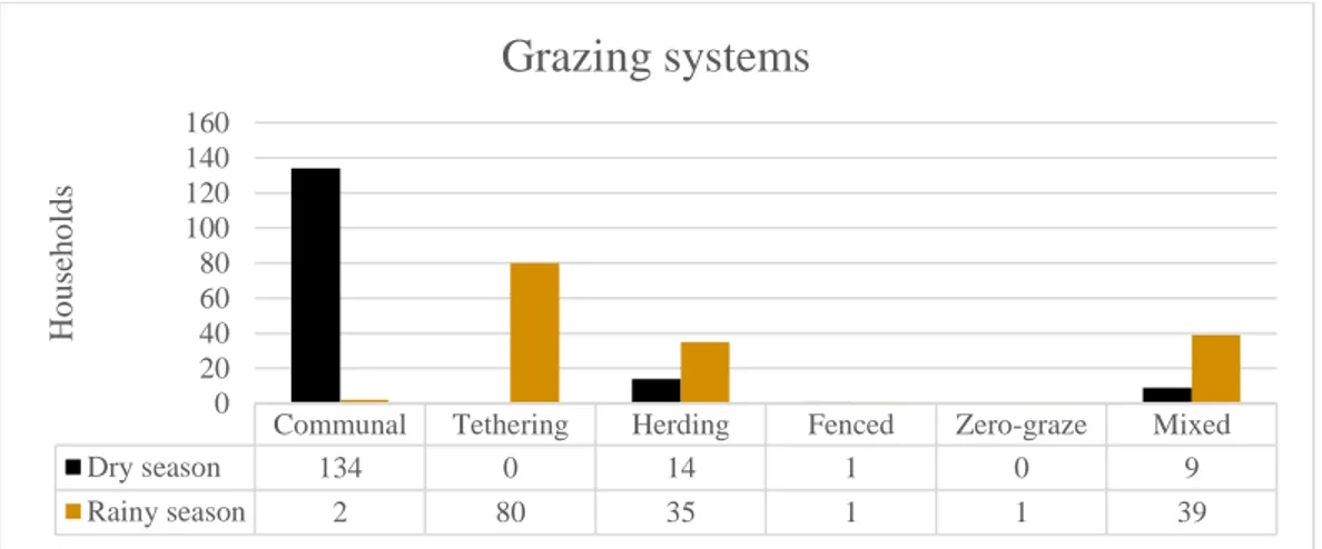
The origin of the sampled animals varied slightly, the most common being born at the farm they were sampled at (75%, n=359), or that they came from the village or a nearby village in the same district (21%, n=103). Seven of the animals were from the government, and one was from a charity organization called Self Help Africa. Ten animals (2%) were of unknown origin. The most common grazing system during the dry season is communal grazing, which means that the goats are let out to graze freely on their own in the community. Most animals are locked in during the nights, but some farmers let the goats roam around freely during both day and night mainly because there have been problems in the communities with thieves stealing the goats if they are locked up. No significant association could be found between serological status for RVFV and communal grazing during the dry season.
During the rainy season tethering the animals is the most common grazing system. Herding and a mix of herding and tethering is also very common. The reason for tethering or herding the animals in the rainy season is to protect the crops in the fields from being eaten by the animals. The grazing systems used are summarized in figure 2. Two households did not specify the
grazing systems and are therefore not included. No significant association could be found between serologicalstatus for RVFV and tethering during the rainy season.
Figure 2 Grazing systems during dry season and rainy season.
The other part of the questionnaire that was analysed was the contact with other herds of goats and sheep, other herds of cattle and contact with wild animals. As for contact with other herds of sheep and/or goats the most common was contact at least weekly during the dry season, since most goats and sheep are let out to graze freely during the dry season the chance of them meeting other herds in the community is big. About 23% (n=37) of the herds had no contact at all with other herds of sheep and/or goats.
The most common answer to the question about contact with other herds of cattle was no contact (70%, n=113). Fifteen percent (n=24) of farmers said that their herds had contact at least weekly during the dry season. Ten (6.3%) of the farmers answered that their animals had contact with cattle at least weekly during wither the rainy season or all year round. Twelve (7.5%) of the farmers said their animals had contact with cattle less often during the year.
Most households had no contact with wild ruminants during any time of the year (83%, n=134). Twelve percent (n=19) of farmers answered variations of contact less often during the year. The remaining 5% of farmers mentioned contact at least weekly with wild ruminants during the dry season or all year. No significant association could be found between serological status for RVFV and any frequency of contact with other herds of sheep/goats or cattle or contact with wild ruminants.
Another factor that might be important is trading between countries. One question was whether the farmer had ever bought sheep or goats from another country, 5 of the 160 farmers who answered the questionnaire said that they had bought animals from Tanzania, and 3 said they had bought from Malawi.
Communal Tethering Herding Fenced Zero-graze Mixed
Dry season 134 0 14 1 0 9 Rainy season 2 80 35 1 1 39 0 20 40 60 80 100 120 140 160 Ho u seh o ld s
Grazing systems
DISCUSSION
The aim of this study was to investigate the seroprevalence of RVFV and SPPV/GTPV among sheep and goats in two districts in Northern Zambia. The overall seroprevalence of antibodies against RVFV was 2.29% (2.16% in goats and 5.56% in sheep). There were no animals positive for antibodies against SPPV or GTPV.
Sheep and Goat Pox
SGP is endemic in areas in Africa north of the equator (OIE, 2013), therefore it would be a rare occurrence for an animal to be seropositive in Zambia. As far as I know there are no previous studies done to estimate the prevalence of SGP in Zambia.
Rift Valley Fever
There have been other studies in Zambia made to evaluate the seroprevalence of antibodies against RVFV, but all of them are old. The most recent serological study collected serum during 1990-91 from cattle in Zambia’s all nine districts and showed that the virus could be spread in the whole country (Samui et al., 1997). Only one study included sheep to evaluate seroprevalence (Hussein et al., 1985). The four farms in the Central Province had varying prevalence from 2-60% in different age categories. As far as I know this is the only sero-prevalence study made in the area on both sheep and goats.
Whereas Zambia has been free of outbreaks with RVF for a long time the neighbouring country of Tanzania has not been. In 2006-07 there was an outbreak in Tanzania (OIE, 2018b). Trading (including illegal trade) has been implicated as an important way of spreading the disease (Cêtre-Sossah et al., 2012; Lernout et al., 2013; Roger et al., 2014). This implies that it is possible for the disease to spread also to Zambia, since the border between Tanzania and Zambia (in Nakonde) is not controlled due to the lack of physical barriers and that people can freely walk over the border without passing through the border control (Personal communic-tion, Owen Malambo, 2018).
One of the goals of the study was to see if there were any connection between management routines the farmers have and seroprevalence in the sampled herds. Since only 11 animals in 8 different households were positive for antibodies against RVFV it is hard to draw any conclusions.
One of the main symptoms of RVF infection is abortions (Gerdes, 2004). Of the seropositive animals none had experienced abortions during the last 12 months, and only 2 of the households with seropositive animals reported having had an abortion in the herd during the last 12 months. Another common symptom of RVF is diarrhoea, which one of the goats reported having had during the last 12 months. It is possible that the affected animals experienced symptoms because of RVF, but it is also possible that the symptoms are the result of other causes. The tests used in this study analyses antibodies which may be found in the bloodstream long after the infection has taken place, which means that an infection could have occurred long before the twelve months the symptoms were inquired for.
To be included in the study the animals had to be of 4 months of age or older, since there is a risk of misinterpretation of the results due to the existence of maternal antibodies if the animals are younger.
In a study made by Hussein et al. (1985) there were a significantly higher sero-prevalence rate in animals less than a year old, and this age group had a lower titre than those from the older animals. The authors of the article say that this can, at least in part, be accounted for by the presence of maternally derived antibodies. This has relevance to this study since three of the 11 animals positive for antibodies against RVFV came from individuals less than a year old. It is therefore possible that these three individuals were in fact not infected but got the antibodies from their mothers. One of the two samples that were classified as “doubtful” was a 6 months old goat, it is also possible that that goat had maternally derived antibodies with a lower titre of antibodies and therefore was classified as doubtful. Since this study only analysed the serum with ELISA and no titration was made to estimate the amount of antibodies it is impossible to know if the antibodies were of maternal origin or not.
Since animals during IEPs only show mild clinical disease or no disease at all it is hard to detect disease. To find affected animals there has to be a well-focused and active surveillance, which is hard in countries such as Zambia where there are limited resources to carry out such a surveillance during IEPs (Dautu et al., 2012). Outbreaks are usually first discovered in humans before detection of the disease in animals (Bird et al., 2009). This means that if there would be an outbreak in Zambia of RVF the spread among livestock would probably be quite vast before it would be noticed, both since it has been such a long time since the last outbreak which could make the disease more difficult for officials to recognize, but also because of the limited veterinary resources as stated above.
Antonis et al. (2013) showed that 4 out of 7 inoculated pregnant ewes did not show any symptoms or any detectable viraemia, they were also negative on three standard ELISA tests. However, viral DNA could be found in the liver and brains of all sheep, and in the liver and/or brains of the foetuses of the serologically negative sheep. This is of importance since one of the ELISA tests used in the study by Antonis et al. (2013) was the same as the one used in this study. It is therefore possible that the seroprevalence could be underestimated.
Rainfall is implemented as a risk factor for occurrence of RVF. The regions sampled in this study are located in a region in Zambia that gets more than 1000 mm rainfall per season, which makes it prone to flooding (Dautu et al., 2012).
Methodological considerations
There is a risk of selection bias, since we often took samples of the goats that were brought to us instead of randomizing the selection. The reason for this were because the animals were not kept in an enclosure and therefore had to be caught to be sampled, in that case it is possible that the caught animals were a little bit slower than the rest because of illnesses. This is however not very likely since the goats and sheep were generally very hard to catch. If the owners were the ones to bring the animals out of their house or enclosure there is also a possibility of selection bias – either because it was an animal that had been sick previously that they wanted
us to look at, or the opposite, with the animal being one of the healthy ones out of fear for us finding a contagious disease in one of the animals.
An observation bias which is possible is the “Clever Hans”-effect, which means that the farmers might have changed their answers according to what they thought we as the interviewers wanted to hear.
Since we were asking for information during the last 12 months there is also a risk of recall bias, with the farmers e.g. not remembering exactly when an animal was sick.
It is possible that the questionnaire was not understood in the correct way and that information was lost along the way since English is not our first language, and neither the first language of the interpreter. It is also possible that the questions were asked in a way that the farmer did not understand it the way it was anticipated.
When out in the field sampling the animals, each farmer had to say how old the animals we sampled were. Most farmers had very precise knowledge of how old each animal was, but there is the possibility of them being wrong about the age of their animals. The age is especially important for the younger animals, if they were younger than 4 months of age they were not supposed to be included in the study due to the existence of maternal antibodies, and according to the study made by Hussein et al. (1985) there is a possibility of interfering maternal antibodies even in older animals.
Furthermore, it is possible that we could have gotten skewed answers due to the snowball sampling e.g. if neighbours are likely to keeping the goats in similar ways. Though that is not likely since there were only four households in each village to be sampled, and often we had to go around asking for the goats since some goat owners were not aware of the other farmers in the same village.
Several things could have gone wrong to make the result of the ELISAs wrong: samples being handled the wrong way, wrong incubation time for the ELISA plates. However, all plates were validated according to the manufacturer’s instruction, which makes it less likely that that should be the case.
Since the prevalence of RVF in the tested region is very low there is a possibility of false positive tests results, since the positive predictive value (PPV) then is very low. Even a very good test in sense of sensitivity and specificity may get wrong results if the prevalence is very low. Methods to try and get a better PPV is to get a better specificity or a higher prevalence. A better specificity could be achieved by doing other tests to try and find the truly positive animals. To get a higher prevalence testing in high-risk populations is a solution. Since both the ELISA tests for RVF and capripox have very good specificity, >99.7% for the capripox (IDvet, 2018b) and 100% for the RVF (Comtet et al., 2010; Kortekaas et al., 2013) it would be better to try and get a higher prevalence. That is also a challenge, since the disease is so rare it is hard to know which animals are in the high-risk populations.
CONCLUSION
The results of this study indicate that RVFV is circulating among the population of sheep and goats in the provinces near the border to Tanzania. There have been no outbreaks of RVF in Zambia for the past 30 years, but since the virus is still circulating it is still possible for there to be an outbreak in the future. It is therefore advisable to continue serological studies in Zambia as well as in nearby regions. No evidence of the existence of SGP in the area has been found.
POPULÄRVETENSKAPLIG SAMMANFATTNING Transboundary diseases
In the world many transboundary diseases are of importance to small-scale livestock owners since these diseases pose a threat to their livelihood. The diseases are not only a threat to the people keeping animals but also to the society at large, since international trade has become such an important part of today’s society. Both the countries exporting and importing have high stakes at risk; the exporters want to maintain their market, while the importing countries are concerned with the protection of their own domestic livestock population. It is therefore important to keep surveillance as well as control of these diseases to protect the trade.
The two of these transboundary diseases discussed in this article is Rift Valley Fever (RVF) and Sheep and Goat Pox (SGP).
Sheep and Goat pox
SGP is caused by two different viruses, Sheep pox virus (SPPV) and Goat pox virus (GTPV). They are very similar to each other, so far as to have led to the previous belief that they were different strains of a single virus, that theory was debunked when the viruses’ genomes were studied more closely. Some of the different strains that exist can however cause disease in both species, though it is usually more severe in the species that has given it its name.
Clinical signs showed are fever, rhinitis with mucous discharge from the nostrils and hard papules in the skin developing on the face and body, more so where there is less hair or wool. The papules can coalesce to form larger swellings, the skin covering them can become necrotic and be rubbed off. If the animal is affected with a more severe strain death usually occurs 7-10 days after onset of disease. Animals of indigenous breeds in endemic areas are more resistant to disease than for example European breeds.
The virus is transmitted when infected and susceptible animals are in close contact with each other, either by inhaling the virus or by virus getting in through wounds in the skin. The papules on the skin are irritating to the animals which causes them to scratch themselves, thereby contaminating the area around them with viral particles. It is also a possibility of virus being transmitted with insects such as the European stable fly, but that has only been shown in experimental studies and is probably not a very prominent mode of transmission in nature. The disease has a high impact on the economy of farmers since the animals can get sick and die. The milk production may decrease, which mostly affects dairy farmers, but also farmers keeping the animals for other reasons than dairy since lack of milk production also affects the new-borns who can die from lack of food intake. The fertility in the herd is also affected, with animals aborting and having a harder time conceiving, which might be an effect of the inflammatory nature of the disease.
Rift Valley Fever
RVF is a virus that was first isolated in Kenya in 1931 and has caused many outbreaks in sub-Saharan Africa, until there was an outbreak in Egypt during 1977-79. In 2000 it was first
The clinical picture is varied but the most common sign is abortions in a majority of the pregnant animals in the affected herd. New-borns follow a rapid course of disease with fever, abdominal pain and a large proportion succumbing to death. The liver is the most affected organ and if a large part of it becomes damaged it affects clotting in the body, making the animals have anaemia with haemorrhages in most organs. Other symptoms may include fever, diarrhoea and vomiting.
The virus also affects humans as it is a zoonotic disease. The symptoms in humans is flu-like with fever, nausea, vomiting, abdominal pain and diarrhoea. Usually recovery follows after 2-7 days, but in rare cases it may progress to haemorrhagic fever, inflammation in the retina and brain and death. Risk factors for humans associated with human infection with RVFV are for example consuming and/or handling products of sick animals, exposure to blood and assisting animals during birth, since there are many viral particles in the placenta and aborted foetuses. The virus has two cycles, called the enzootic and epizootic cycle. During the enzootic cycle the virus is circulating in low levels in wild and domestic animals without causing considerable disease. This enzootic phase can last very long periods, in the savannah it has been seen to last 5-15 years and in the drier regions as long as 25-35 years. This is because the epizootic cycle, when outbreaks occur, is highly correlated with abnormally heavy rainfalls prior to the event. The heavy rainfalls cause large numbers of mosquitoes of the species Aedes to emerge in large numbers from their breeding grounds in so-called “dambos”, shallow depressions at the headwaters of drainage systems. The drought-resistant eggs of the Aedes mosquitoes may be infected with the virus when they hatch since these mosquitoes can transmit the virus directly to the eggs. The virus is then transmitted to the animals when the mosquitoes bite them. After a while other mosquito species also emerge and can promote spread of the virus further. Most viruses that are transmitted with the help of insects persist in nature with the help of an animal reservoir, such an animal has however not been found for RVFV. Several wild animals have been found to have antibodies against the virus, such as African buffalo, waterbuck, kudu, impala, rhinoceros and even rodents. Though it is very unlikely that rhinoceros have a big impact on the maintenance of the virus in nature due to their low population density in nature. Wild animals are also not thought to get sick from the virus, since no abortion of haemorrhagic disease of deaths were reported during an outbreak in 2007-08 in Kenya, they are more likely to enable transmission of the virus by getting a low viral load in the blood.
Veterinary services and seroprevalences in Zambia
In Zambia the veterinary services have historically all been free of charge from the government. Due to a shrinking budget over the years the coverage and quality slowly began to deteriorate, which led to actions being taken with a reorganization in 1996. Since then the public sector has been in charge of regulation, control and monitoring, while the private sector is in charge of implementing most field activities. In Zambia there is still an underutilization of veterinary inputs and a reluctance to pay for veterinary services, which makes livestock diseases one of the main constraints limiting livestock development in the country.
SGP has never been reported in Zambia, but RVF has. The last outbreak was in 1985, and since then a few serological studies have been made to estimate the spread of disease in the country, the last being in 1990-91 where cattle from each of the nine districts were sampled. The conclusion from that study was that RVFV could be spread in the whole country.
Seroprevalence in Nakonde and Mbala
The two districts in this study were Nakonde and Mbala in the Muchinga and Northern Provinces in Zambia, bordering to Tanzania. Forty villages were randomly chosen, 30 in Nakonde and 10 in Mbala. In each village 4 households were visited where 3 goats and/or sheep were sampled for serum. A questionnaire was also carried out with the help of an interpreter to get information about the sampled animals and about management in the herds. The serum was analysed with ELISA, with the result being that none of the animals were positive for antibodies against SGP, and 11 individuals being positive for antibodies against RVFV.
Conclusion
It is a long time since the last outbreak of RVF in Zambia, but the study shows that virus is still circulating in low levels in the sheep and goat herds in Northern Zambia. It is therefore important to continue the serological surveillance to prevent outbreaks in the future.
ACKNOWLEDGEMENTS
I would like to extend my gratitude towards everyone who made this project possible. For the funding I thank the Linnaeus-Palme exchange program scholarship, the Elsa Paulsson memory foundation and the International committee of Veterinärmedicinska Föreningen. Without the funding this project would not have been possible.
I am also thankful for my supervisor Karin Alvåsen, for helping with the preparations and planning as well as reading through my thesis and coming with helpful comments. I am grateful to Sara Lysholm, my assistant supervisor, for helping in getting us started with the sampling in Zambia as well as helping with the writing process. I want to thank Johanna Lindahl, my examiner, for coming with helpful comments. Thank you also to Jonas Wensman for helping in all preparations for this project, and for being of great assistance with questions regarding all things prior to and during the trip.
I would also like to thank Musso Munyeme, my assistant supervisor at the University of Zambia for assisting in getting us settled on site in Zambia, and thank you to Owen Malambo, Perry Nyambe and Stephen Mbala for assisting in the field work.
Last but not least I want to thank my friend and colleague, Lydia Mitternacht, for sharing this experience with me, through both the good and the less good times.
REFERENCES
Anderson, E. C. & Rowe, L. W. (1998). The prevalence of antibody to the viruses of bovine virus diarrhoea, bovine herpes virus 1, Rift Valley fever, ephemeral fever and bluetongue and to Leptospira sp in free-ranging wildlife in Zimbabwe. Epidemiology and Infection [online], 121(2), pp 441–449. Available from: DOI: http://dx.doi.org/10.1017/S0950268898001289.
Antonis, A. F. G., Kortekaas, J., Kant, J., Vloet, R. P. M., Vogel-Brink, A., Stockhofe, N. & Moormann, R. J. M. (2013). Vertical transmission of Rift Valley fever virus without detectable maternal viremia. Vector-Borne and Zoonotic Diseases [online], 13(8), pp 601–606. Available from: DOI: https://doi.org/10.1089/vbz.2012.1160.
Anyamba, A., Linthicum, K. J. & Tucker, C. J. (2001). Climate-disease connections: Rift Valley fever in Kenya. Cadernos de Saúde Pública [online], 17(Suppl), pp 133–140. Available from: DOI: https://doi.org/10.1590/S0102-311X2001000700022.
Anyangu, A. S., Hannah Gould, L., Sharif, S. K., Nguku, P. M., Omolo, J. O., Mutonga, D., Rao, C. Y., Lederman, E. R., Schnabel, D., Paweska, J. T., Katz, M., Hightower, A., Kariuki Njenga, M., Feikin, D. R. & Breiman, R. F. (2010). Risk factors for severe Rift Valley fever infection in Kenya, 2007. The American Journal of Tropical Medicine and Hygiene [online], 83(2 Suppl), pp 14–21. Available from: DOI: https://doi.org/10.4269/ajtmh.2010.09-0293.
Ausvet. Calculate confidence limits for a sample proportion. [online] (2019). Available from: http://epitools.ausvet.com.au/content.php?page=CIProportion. [Accessed 2019-01-06].
Babiuk, S., Bowden, T. R., Parkyn, G., Dalman, B., Hoa, D. M., Long, N. T., Vu, P. P., Bieu, D. X., Copps, J. & Boyle, D. B. (2009). Yemen and Vietnam capripoxviruses demonstrate a distinct host preference for goats compared with sheep. Journal of General Virology [online], 90(1), pp 105– 114. Available from: DOI: https://doi.org/10.1099/vir.0.004507-0.
Baudin, M., Jumaa, A. M., Jomma, H. J. E., Karsany, M. S., Bucht, G., Näslund, J., Ahlm, C., Evander, M. & Mohamed, N. (2016). Association of Rift Valley fever virus infection with miscarriage in Sudanese women: a cross-sectional study. The Lancet Global Health [online], 4(11), pp 864–871. Available from: https://doi.org/10.1016/S2214-109X(16)30176-0.
Bhanuprakash, V., Moorthy, A. R. S., Krishnappa, G., Gowda, R. N. S. & Indrani, B. K. (2005). An epidemiological study of sheep pox infection in Karnataka state, India. Revue Scientifique et Technique (International Office of Epizootics) [online], 24(3), pp 909–920. Available from: https://pdfs.semanticscholar.org/3739/879163d331e82fcd75b761c4c81d855d5c20.pdf. [Accessed 2018-09-14].
Bhanuprakash, V., Venkatesan, G., Balamurugan, V., Hosamani, M., Yogisharadhya, R., Chauhan, R. S., Pande, A., Mondal, B. & Singh, R. K. (2010). Pox outbreaks in sheep and goats at Makhdoom (Uttar Pradesh), India: Evidence of sheeppox virus infection in goats. Transboundary and
Emerging Diseases [online], 57(5), pp 375–382. Available from: DOI: https://doi.org/10.1111/j.1865-1682.2010.01158.x.
Bird, B. H., Khristova, M. L., Rollin, P. E., Ksiazek, T. G. & Nichol, S. T. (2007). Complete genome analysis of 33 ecologically and biologically diverse Rift Valley fever virus strains reveals
widespread virus movement and low genetic diversity due to recent common ancestry. Journal of Virology [online], 81(6), pp 2805–2816. Available from: DOI: https://doi.org/10.1128/JVI.02095-06.
Bird, B. H., Ksiazek, T. G., Nichol, S. T. & Maclachlan, N. J. (2009). Rift Valley fever virus. Journal of the American Veterinary Medical Association [online], 237(7), pp 883–893. Available from: DOI: https://doi.org/10.2460/javma.234.7.883.
Bowden, T. R., Babiuk, S. L., Parkyn, G. R., Copps, J. S. & Boyle, D. B. (2008). Capripoxvirus tissue tropism and shedding: A quantitative study in experimentally infected sheep and goats. Virology [online], 371(2), pp 380–393. Available from: DOI: https://doi.org/10.1016/j.virol.2007.10.002. [Accessed 2018-10-26].
Carn, V. M. (1993). Control of capripox infections. Vaccine [online], 11(13), pp 1275–1279. Available from: DOI: https://doi.org/10.1016/0264-410X(93)90094-E.
Central Statistical Office (2017). Preliminary Livestock and Aquaculture Census Results. Available from:
https://www.zamstats.gov.zm/phocadownload/Agriculture/Preliminary%202017%20Livestock%2 0and%20Aquaculture%20Census%20Results.pdf. [Accessed 2018-09-13].
Cêtre-Sossah, C., Pédarrieu, A., Guis, H., Defernez, C., Bouloy, M., Favre, J., Girard, S., Cardinale, E. & Albina, E. (2012). Prevalence of Rift Valley fever among ruminants, Mayotte. Emerging Infectious Diseases [online], 18(6). Available from: DOI: https://doi.org/10.3201/eid1806.111165. Chilonda, P. & Van Huylenbroeck, G. (2001). Attitude towards and uptake of veterinary services by
small-scale cattle farmers in Eastern Province, Zambia. Outlook on Agriculture [online], 30(3), pp 213–218. Available from: DOI: https://doi.org/10.5367/000000001101293670.
Chilonda, P., Van Huylenbroeck, G., D’Haese, L., Samui, K. L., Musaba, E. C., Imakando, M. & Ahmadu, B. (1999). Cattle production and veterinary care systems in Zambia. Outlook on Agriculture [online], 28(2), pp 109–116. Available from: DOI:
https://doi.org/10.1177/003072709902800208.
Clark, M. H. A., Warimwe, G. M., Di Nardo, A., Lyons, N. A. & Gubbins, S. (2018). Systematic literature review of Rift Valley fever virus seroprevalence in livestock, wildlife and humans in Africa from 1968 to 2016. PLoS Neglected Tropical Diseases [online], 12(7). Available from: DOI: https://doi.org/10.1371/journal.pntd.0006627.
Comtet, L., Marié, J.-L., Davoust, B., Cêtre-Sossah, C. & Pourquier, P. (2010). Preliminary Validation of the ID Screen® Rift Valley Fever Competition Multi-Species ELISA. Available from:
https://www.id-vet.com/pdfs/newsletter1/poster_riftc.pdf. [Accessed 2018-11-19].
Daubney, R., Hudson, J. R. & Garnham, P. C. (1931). Enzootic hepatitis or Rift Valley fever, an undescribed virus disease of sheep, cattle and man from East Africa. Journal of Pathology and Bacteriology, 34, pp 545–579.
Dautu, G., Sindato, C., Mweene, A. S., Samui, K. L., Roy, P., Noad, R., Paweska, J., Majiwa, P. A. O. & Musoke, A. (2012). Rift Valley fever: Real or perceived threat for Zambia? Onderstepoort Journal of Veterinary Research [online], 79(2). Available from: DOI:
https://doi.org/10.4102/ojvr.v79i2.466.
Davies, F. G., Kilelu, E., Linthicum, K. J. & Pegram, R. G. (1992). Patterns of Rift Valley fever activity in Zambia. Epidemiology and Infection [online], 108(1), pp 185–191. Available from: DOI: https://doi.org/10.1017/S0950268800049633.
Davies, F. G., Linthicum, K. J. & James, A. D. (1985). Rainfall and epizootic Rift Valley fever. Bulletin of the World Health Organization [online], 63(5), pp 941–943. Available from: https://www.ncbi.nlm.nih.gov/pmc/articles/PMC2536443/. [Accessed 2018-09-13].