Original Article 1
Evaluation of Panoramic Radiographs in Relation
to the Mandibular Third Molar and to Incidental
Findings in an Adult Population
Josefine Cederhag
1Nina Lundegren
2Per Alstergren
3,4Xie-Qi Shi
1,5Kristina Hellén-Halme
1 1Department of Oral and Maxillofacial Radiology, Faculty ofOdontology, Malmö University, Malmö, Sweden
2Department of Oral Diagnostics, Faculty of Odontology, Malmö University, Malmö, Sweden
3Department of Orofacial Pain and Jaw Function, Faculty of Odontology, Malmö University, Malmö, Sweden
4Specialized Pain Rehabilitation, Skåne University Hospital, Lund, Sweden
5Section of Oral and Maxillofacial Radiology, Department of Clinical Dentistry, University of Bergen, Bergen, Norway
Address for correspondence Josefine Cederhag, DDS, Department of Oral and Maxillofacial Radiology, Faculty of Odontology, Malmö University, Malmö SE-205 06, Sweden
(e-mail: josefine.cederhag@mau.se).
Objectives The aim was to evaluate the characteristics of the mandibular third
molars, especially in relation to the inferior alveolar nerve. Further aims were to
inves-tigate incidental findings in panoramic radiographs in an adult population, and to
investigate image quality related to patient positioning.
Materials and Methods From a previous study with 451 randomly selected adult
participants who lived in Sweden, 442 panoramic radiographs from four dental public
health clinics were used. The third molars’ characteristics and relation to inferior
alve-olar nerve were evaluated. Incidental findings and patient positioning were recorded.
Statistical Analysis Frequency analysis was used to investigate the occurrence of all
findings and their possible interconnections. Whether the patients’ age or gender had
an impact or not was also analyzed.
Results The third molars were erupted and in vertical position among 73%
regard-less of age. When retained or semi-retained, they were most commonly in
mesioan-gular positions. The inferior alveolar nerve was located inferior to the roots in 52%,
whereas an overlapped position was most common if the third molar was retained
(90%), semi-retained (83%) or the age was less than 30 years (66%). Common
inciden-tal findings were apical radiolucencies, idiopathic osteosclerosis, and tooth fragments.
Suboptimal patient positioning was found in one-third of the radiographs.
Conclusions Panoramic radiography is a useful method to evaluate third molar
prior to surgical removal and may be the only image required. Most incidental
find-ings on panoramic radiographs does not seem to require any further odontological
management.
Abstract
Keywords ►third molar
►panoramic
radiography
►incidental findings
►adult
DOI https://doi.org/ 10.1055/s-0040-1721294 ISSN 1305-7456.© 2020. European Journal of Dentistry.
This is an open access article published by Thieme under the terms of the Creative Commons Attribution-NonDerivative-NonCommercial-License, permitting copying and reproduction so long as the original work is given appropriate credit. Contents may not be used for commercial purposes, or adapted, remixed, transformed or built upon. (https://creativecommons.org/licenses/by-nc-nd/4.0/)
Thieme Medical and Scientific Publishers Pvt. Ltd., A-12, 2nd Floor, Sector 2, Noida-201301 UP, India
Introduction
Dental panoramic imaging is a radiological method com-monly used in dentistry since 1950s. It is a useful diagnostic tool owing to its broad coverage of teeth, bone, and surround-ing anatomical structures in the maxillofacial region. Together with a clinical examination, a panoramic radiograph facili-tates the clinician’s diagnosis and treatment plan for several diagnostic tasks. It is a two-dimensional radiograph based on the tomographic technique where structures within the image layer are sharply depicted; however, the resolution is lower and less detailed compared with intraoral radiographs. Previous studies1-3 have concluded that a majority of exposed panoramic radiographs had certain flaws, of which the most common was related to patient positioning and head align-ment. Studies have also shown that a panoramic radiograph has many limitations in terms of diagnostic accuracy.4-6
Surgical removal of the third mandibular molars is one of the most common surgical procedures in dentistry.7,8 To make a proper assessment of the relationship/proximity between inferior alveolar nerve (IAN) and the roots, a pan-oramic radiograph is commonly justified.5,9,10 However, the possibility to determine a true close relationship between structures has been questioned.4,5 The radiograph has proven to be diagnostically acceptable in most cases to show the location of the third molar and its location to IAN.6 Previous studies5,6,11 conclude that panoramic radiographs, in many cases, despite the lower resolution, are considered accept-able for a third molar presurgical investigation. The pan-oramic X-ray unit is well used, the examination is considered convenient by the patients, and importantly, the radiation dose to the patient is considerably lower compared with cone beam computed tomography.10 Therefore, the method is considered cost-effective12 because the equipment is reasonably inexpensive and the examination time for the patients and dental office is relatively short.
In panoramic radiographs, certain incidental findings of pathology and abnormality may be seen.13 Studies have reported the frequency of incidental findings in children, often related to orthodontic treatment.14-16 In adults, a few studies have investi-gated specific incidental findings, such as idiopathic osteoscle-rosis17 and radiolucencies according to apical periodontitis.18 A new study by MacDonald and Yu19 concluded that in a group of new patients, mixed to permanent dentition, coming for a den-tal examination, 32.1% had at least one incidenden-tal finding when investigated by panoramic imaging. In a previous study of oral health, panoramic radiographs were exposed in an adult popu-lation.20 In accordance with and in reference to the radiological principle of As Low As Reasonably Achievable (ALARA),21 we believe that the additional evaluation of the already-exposed panoramic radiographs could contribute to further knowledge. Accordingly, a few previous studies17-19 evaluated the incidental findings of panoramic radiographs; however, incidental find-ings in a cross section of adult patients participating for research purpose have not been evaluated.
The aims of this study were to evaluate the characteris-tics of the mandibular third molars, especially in relation to IAN. Further aims were to investigate the number and
kind of incidental findings in panoramic radiographs in an adult population, and to investigate image quality related to patient positioning.
Materials and Methods
The study was approved by the Ethical Committee for Human Research in Lund, Sweden (Dnr 513/2006).
Study Design
This observational study included 442 digital panoramic radiographs of 224 (51%) women and 218 (49%) men, which were randomly selected from an adult popula-tion (age 20–89) living in a south county of Sweden with approximately 1.3 million inhabitants, in 2007. The radio-graphs were a part of a previous study20 and were exposed between 2007 and 2008 at four different dental clinics in a south county of Sweden. All panoramic radiographs were captured by storage phosphor plates then exported in DICOM format22 and imported into the viewing program Romexis (Planmeca, Helsinki, Finland). All exposed radiographs were included in the present study.
Evaluation Conditions
All evaluations were performed under standardized condi-tions using a display monitor (BARCO; Kortrijk, Belgium) cal-ibrated for the evaluation of radiographic radiographs, with ambient light less than 50 lux.23,24 The first 50 radiographs were evaluated together by two observers, one junior radiol-ogist (J.C.), and one senior radiolradiol-ogist in oral and maxillo-facial radiology (K.H.H.). The remaining radiographs were evaluated by the first observer (J.C.). If there was any doubt, a consensus was reached by a discussion with the senior radiologist. When needed, the display of the radiographs was adjusted according to personal preferences.
The radiographs were evaluated according to a protocol (►Table 1). In order for the mandibular third molars to be defined as erupted, the main part of the occlusal surface had to be in level with the occlusal surface of the neighboring second molar; semi retained: the crown of the third molar had to be partially covered by bone and for retained: the crown of the third molar had to be completely covered by bone. Incidental findings of pathology and abnormality in the whole radiographs were recorded. Image quality accord-ing to patient positionaccord-ing duraccord-ing exposure was evaluated in coronal plane and to head alignment in relation to the occlusal plane.
Statistical Methods
Frequency analysis was used to investigate the occurrence of different radiographic findings and their possible interconnec-tions. Whether the patients’ age or gender had an impact or not were analyzed using cross-tabulations. To test if any sta-tistically significant difference existed, a chi-square test was used. To be considered as significant the p-value was set to 0.05. Data analyses were performed using the Statistical pack-age for the social sciences (IBM SPSS, version 23 for Windows. Armonk, NY: IBM Corp).
Results
Mandibular Third Molar
Of the participants, 58% had at least one mandibular third molar, and 40% had both mandibular third molars. The total number was 435 (i.e., 210 participants had tooth 38 and 225 participants had tooth 48). The distribution of man-dibular third molars in the different age groups is shown in ►Fig. 1. A total of 98% of the teeth showed complete root development. ►Fig. 2 shows how the mandibular third molars were erupted in relation to age. In total, 73% had an erupted location, 12% had a retained location, and 15% had a semi-retained location. A vertical position (73%) was most frequent, but it differed if the tooth was erupted or retained and semi-retained (►Fig. 3).
In 52% of the teeth, IAN was “located inferior to the roots,” followed by “overlapped” and “not assessable,” in 40 and 7%, respectively. In the erupted group, 68% had an infe-rior location, which was less common in the retained and semi-retained group, where the majority had an overlapped position on the roots (90 and 83%). The most common loca-tion of IAN was inferior in all age groups (40–69%), apart from the youngest group, where it was in an overlapped loca-tion (66%).
In 10 participants, 11 third molars had apical radiolu-cencies, 10 of which were found in the erupted group. Eight out of these 10 participants had other pathological findings (such as apical radiolucencies on other teeth or/and tooth fragments). In 20% of the teeth, a distal periodontal pocket was present, 76% of those teeth were erupted, and 19% were semi-retained.
There was no difference between genders in any of the results.
Incidental Findings of Pathology and Abnormality
The total number of incidental findings were 357 and their distribution are shown in ►Fig. 4 (A, B). Incidental findings were present in 57% (n = 252) of the participants. There was no difference between genders, and incidental findings
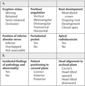
Table 1 Parameters from examination protocol regarding
(A) findings of mandibular third molars and (B) general radiographic findings and image quality parameters
A. Eruption status Missing Retained Semi-retained Occlusion Position/ angulation Vertical Mesioangular Distoangular Transversal Horizontal Root development Mineralized crown Ongoing root Development Closed apex Position of inferior alveolar nerve Inferior Overlapped Not assessable Periodontal pocket Yes No Apical radiolucencies Yes No B. Incidental findings of pathology and abnormality Yes No Patient positioning in coronal plane Correct Anterior Posterior Head alignment in occlusal plane Correct Head tilted upward Head tilted downward
Fig. 1 The distribution of mandibular third molars in the differ-ent age groups. Total number of participants (n = 442) and third molars (n = 435).
Fig. 2 Mandibular third molars eruption status: erupted/semi- retained/retained in the different age groups.
Fig. 3 Mandibular third molars position/angulation in relation to eruption status: erupted/retained/semi-retained, in total and respectively.
were present in all age groups but more commonly seen in the older age groups. The most common incidental findings among the participants (n = 442) at the individual level were apical radiolucencies in 25%, idiopathic osteosclerosis in 20%, tooth fragments in 9%, and torus mandibularis in 7%.
Image Quality Related to Patient Positioning and Head Alignment
Patient positioning in coronal plane was correct in 62% of the patients and head alignment in occlusal plane was correct in 65%. Regarding coronal positioning errors, patients were placed posteriorly of the image layer in 28% while anteriorly placed in 10%. The corresponding values with respect to head alignment in occlusal plane were 26 and 9% for head tilted upward and downward, respectively. The periapical status could be evaluated in most of the panorama radiographs, apart from 12 cases where the apical part of the maxillary teeth could not be evaluated properly due to tongue displace-ment, as the tongue was not in contact with the hard palate during the exposure.
Discussion
Our results indicate that, in about half of third molars, the IAN is located inferior to the roots of third molars. This was an interesting and important finding since this suggests that in many cases a panoramic radiograph can be enough when investigating a third molar prior to surgical removal. However, if the clinical question asked only is a question about the mandibular third molar, two intraoral radiographs provide a sharp and distinct depiction of anatomical structures with high resolution, but for the patient, it can be challenging to manage a correct receptor positioning in the region. If so, a cropped panoramic radiograph can be a suitable option. Considering the well-known principle ALARA21 and radiolog-ical European guidelines,10 as well as radiation dose, a choice of two-dimensional radiographs can be recommended to exclude a true close relationship between IAN and the third molar. Indeed, panoramic radiographs have been sug-gested to be justified as the method of choice for presurgical
examination to exclude a true close relationship,5 i.e., an infe-rior position of the IAN. This suggestion is in agreement with the results of the present study.
Our study population was randomly selected from adult individuals living in a south county of Sweden in 2007. In the two oldest age groups, considerably fewer individuals chose to participate.20 Those groups were included, but when analyzing the result, this factor needs to be viewed accord-ingly. Nevertheless, apart from the oldest age group, the study sample corresponds well to the age distribution in the county, both at that time and today.20,25 In addition, the study sample was selected from a region, taking not only demo-graphic distribution but also representability of ethnicity and educational level into account, and those were, and still are, considered to be adequate.25 One limitation, in terms of the radiography, was that the panoramic radiographs were exposed in four different public dental clinics for research purposes about oral health. No specific justifications were made ahead of the exposure of the panoramic radiographs. The exposure was in relation to the aims of that study20 and was ethically approved at that time. The different technical settings and supporting devices for patient positioning of each X-ray unit were not known, and thus, the evaluation of image quality was restricted to include patient positioning in coronal plane and head alignment in occlusal plane The image quality was something flawed in accordance to previ-ous studies,2,26 yet, a panoramic radiograph provides a useable overview. More specifically, although many of the evaluated panoramic radiographs were not considered optimal, the mandibular third molar could easily be properly examined. However, the evaluation of apical lesions, especially of max-illary teeth, was impaired in a few radiographs; therefore, the distribution of apical lesions has to be interpreted with caution. Another possible limitation was that only one junior radiologist evaluated all the radiographs. Nevertheless, the evaluation parameters were well defined and did not include diagnostic difficulties. Moreover, when in doubt, a consensus was reached together with a senior radiologist. We therefore do not consider that our examination methodology influ-ences our results.
This study mapped out the characteristics of mandibular third molars in an adult population, from young adults to pen-sioners, and found that most third molars expectedly, were erupted, had a vertical position, and that IAN was located inferior to the roots. More than half of the study population had one or both mandibular third molars, and not surpris-ingly, most of those were found in age groups of 50 years and younger. As the dental record was not available, the reasons for a missing mandibular third molar, i.e., previous extraction or absence from birth, are unknown. However, it was often assumed that some in the older age groups had a history of tooth extraction. A study from Finland concluded that only a few third molars survived to old age. However, these findings where only based on clinical assessments.27
Erupted third molars seem to be the most common erup-tion status in all age groups but even more so in those who are in the age group of 30 to 49 years. At this age, the teeth have had time to erupt but may not yet have acquired any
Fig. 4 (A) Incidental findings of pathology and abnormality in per-centages of all incidental findings (n = 357); (B) Other uncommon incidental findings of pathology and abnormality (6%) according to ►Fig. 4A, in numbers. *These 5% are shown in relation to specific findings and number in ►Fig. 4B.
pathology that would trigger extraction. The participants in the youngest age group had fewer erupted teeth in compar-ison with the other age groups. In accordance, a relatively large proportion of the semi-retained teeth belonged to the youngest age group, which were teeth under eruption. In the other age groups, the distribution of semi-retained teeth was equally low. In the retained group, the age distribution was quite equal, leading to the conclusion that no matter what age, some teeth are retained and remain so throughout life.
It is difficult to compare our results with other studies, as classification models of impaction, position/angulation, and included age span vary between studies. Nevertheless, Wowern and Nielsen28 reported in a 4-year follow-up study, that young people (i.e., 18 years), especially if their tooth was semi-retained, it had a possibility of being erupted as time goes by. Several studies have shown similar findings: depend-ing on erupted status, there was a low to moderate chance for a tooth to become erupted during one’s twenties.28-30 Another explanation for the equally low number of semi-retained teeth in participants older than 30 years could be that semi-retained teeth in young adults, are at greater risk of pericoronitis, and therefore, had been removed.31
The position/angulation of mandibular third molars in relation to eruption status found in this study was as expected. A vertical position was most frequent in the erupted group. However, for the two other groups (retained and semi-re-tained) mesioangular position was seen the most, even if the differences between mesioangular and vertical position were small among the semi-retained teeth. This equal proportion could be because semi-retained teeth were more likely to be found in the youngest age group with teeth in vertical posi-tion, but probably would end in an erupted state. The litera-ture reports that vertically impacted teeth were more likely to erupt in time and also those in the mesioangular and distoan-gular position, whereas horizontally retained teeth remained unerupted.28,29 Ryalat et al32 and Quek et al33 investigated the position/angulation of both retained and semi-retained teeth in a population aged 18 to 26 and 20 to 40, respectively, and found that mesioangular position/angulation was most com-mon, which is consistent with the results of this study.
If a tooth was erupted or the participants’ age was more than 30 years, most had an inferior position of the IAN; how-ever, when a tooth was retained or semi-retained, or the age was less than 30 years, it was most likely that the IAN had an overlapped position. The fact that the youngest age group was the only one where an overlapped position was more common probably because of the large proportion of semi-retained teeth in this age group, with a greater prob-ability for the IAN to have an overlapped position. This is in agreement with Miloro and DaBell,34 who concluded that unerupted (both retained and semi-retained) third molars were closer to the IAN than erupted third molars.
The IAN was only evaluated as “not assessable” in consid-erably few participants. Almost all those teeth were erupted and in vertical position. Previous studies35,36 have reported that the best visibility of the IAN in a panoramic radiograph was in the most posterior part of the mandible and dimin-ished with the further anterior location it was; the region
of third molars was found to be second best. Furthermore, it was pointed out that the visibility may vary in the same indi-vidual, probably because the IAN is situated more buccally in the anterior region.36
Ten out of 11 third molars that had apical radiolucen-cies were erupted and corresponded well to a recent study by Ventä et al,37 who found that third molars in an occlu-sal level were more likely to be diseased. However, that study had more criteria, including clinical criteria, for being classed as diseased. Hence, the comparison and support for the findings but also the modest number of apical radiolu-cencies in the present study should be viewed accordingly. An interesting, even though not surprising finding in the present study, was that a majority of the participants had other pathological findings that were preferably found in the older age groups, which was in line with the study by Lundegren et al.20
Several incidental findings of pathology and abnormality were present in more than half of the study population, espe-cially in older age groups. However, many of those findings did not require further odontological management. The location in northern Europe could be one possible explanation for this result since Sweden has a national dental insurance system to attract patients to go regularly for a dental examination. According to recent statistics from National Board of Health and Welfare, 69% of Sweden’s adult population did have a regular examination during a 3-year period.38 Nevertheless, the most common finding, apical radiolucency, often needs management, indicating that the clinician should be aware of and evaluate the whole radiograph irrespective of question asked, i.e., initial indication for exposure. Even though it has been a while since the radiographs were exposed, it can be assumed that the number and kind of incidental findings in an adult population do not differ extensively over time.
Image quality in panoramic radiographs is dependent on correct patient positioning and head alignment. Several studies1-3,39 have concluded that operators’ careful handling of technique and training experience, but also patients’ coop-erating and physical stature, are of great importance to obtain an acceptable panoramic radiograph. The limited dimensions of the focal trough in panoramic imaging has been identified as the weak link.1,3 Besides, digital image processing enables image enhancements that were not possible with the ana-logue technique2 and improvements of digital systems have also been made since the panoramic radiographs included in this study were exposed. A notable finding in this study was the poor result of patient positioning and head align-ment, where especially coronal posterior and vertical head tilted upward errors dominated. Previous studies3,39,40 have reported similar findings, thus confirming the importance of proper handling and correct patient positioning in pan-oramic radiography.
Conclusion
This study indicates that panoramic radiography is a useful method for evaluating the relation between the third molar and IAN prior to surgical removal. It may be the only image
required. In the case of an overlapped position of IAN, other radiological modalities should be considered.
Most incidental findings on panoramic radiographs do not seem to require any further odontological management. However, all radiographic examinations should be fully eval-uated regardless of the indication.
Patient positioning seems to be flawed in about one-third of panoramic radiographs.
Conflict of Interest
None declared.
References
1 Rondon RH, Pereira YC, do Nascimento GC. Common position-ing errors in panoramic radiography: a review. Imagposition-ing Sci Dent 2014;44(1):1–6
2 Gross H, Nilsson M, Hellén-Halme K. Detectability of nor-mal anatomy in digital panoramic radiographs. Swed Dent J 2014;38(4):179–185
3 Dhillon M, Raju SM, Verma S, et al. Positioning errors and quality assessment in panoramic radiography. Imaging Sci Dent 2012;42(4):207–212
4 Huang CK, Lui MT, Cheng DH. Use of panoramic radiogra-phy to predict postsurgical sensory impairment following extraction of impacted mandibular third molars. J Chin Med Assoc 2015;78(10):617–622
5 Atieh MA. Diagnostic accuracy of panoramic radiogra-phy in determining relationship between inferior alveo-lar nerve and mandibualveo-lar third moalveo-lar. J Oral Maxillofac Surg 2010;68(1):74–82
6 Bell GW, Rodgers JM, Grime RJ, et al. The accuracy of dental panoramic tomographs in determining the root morphology of mandibular third molar teeth before sur-gery. Oral Surg Oral Med Oral Pathol Oral Radiol Endod 2003;95(1):119–125
7 Coulthard P, Bailey E, Esposito M, Furness S, Renton TF, Worthington HV. Surgical techniques for the removal of mandibular wisdom teeth. Cochrane Database Syst Rev 2014;(7):CD004345
8 Cheung LK, Leung YY, Chow LK, Wong MC, Chan EK, Fok YH. Incidence of neurosensory deficits and recovery after lower third molar surgery: a prospective clinical study of 4338 cases. Int J Oral Maxillofac Surg 2010;39(4):320–326
9 Korkmaz YT, Kayıpmaz S, Senel FC, Atasoy KT, Gumrukcu Z. Does additional cone beam computed tomography decrease the risk of inferior alveolar nerve injury in high-risk cases undergoing third molar surgery? Does CBCT decrease the risk of IAN injury? Int J Oral Maxillofac Surg 2017;46(5):628–635 10 European Commission Radiation Protection N° 172 Cone Beam
CT for Dental and Maxillofacial Radiology. Evidence-Based Guidelines. Directorate-General for Energy, Directorate D— Nuclear Energy, Unit D4—Radiation Protection; 2012:1681–6803. Available at: Available at: http://www.sedentexct.eu/files/radia-tion_protection_172.pdf. Accessed June 30, 2020
11 Bell GW. Use of dental panoramic tomographs to predict the relation between mandibular third molar teeth and the infe-rior alveolar nerve. Radiological and surgical findings, and clinical outcome. Br J Oral Maxillofac Surg 2004;42(1):21–27 12 Petersen LB, Olsen KR, Christensen J, Wenzel A. Image and
surgery-related costs comparing cone beam CT and panoramic imaging before removal of impacted mandibular third molars. Dentomaxillofac Radiol 2014;43(6):20140001
13 Rushton VE, Horner K. The use of panoramic radiology in den-tal practice. J Dent 1996;24(3):185–201
14 Bekiroglu N, Mete S, Ozbay G, Yalcinkaya S, Kargul B. Evaluation of panoramic radiographs taken from 1,056 Turkish children. Niger J Clin Pract 2015;18(1):8–12
15 Asaumi JI, Hisatomi M, Yanagi Y, et al. Evaluation of pan-oramic radiographs taken at the initial visit at a depart-ment of paediatric dentistry. Dentomaxillofac Radiol 2008;37(6):340–343
16 Bondemark L, Jeppsson M, Lindh-Ingildsen L, Rangne K. Incidental findings of pathology and abnormality in pre-treatment orthodontic panoramic radiographs. Angle Orthod 2006;76(1):98–102
17 Yonetsu K, Yuasa K, Kanda S. Idiopathic osteosclerosis of the jaws: panoramic radiographic and computed tomo-graphic findings. Oral Surg Oral Med Oral Pathol Oral Radiol Endod 1997;83(4):517–521
18 Chala S, Abouqal R, Abdallaoui F. Prevalence of apical peri-odontitis and factors associated with the periradicular status. Acta Odontol Scand 2011;69(6):355–359
19 MacDonald D, Yu W. Incidental findings in a consecutive series of digital panoramic radiographs. Imaging Sci Dent 2020;50(1):53–64
20 Lundegren N, Axtelius B, Akerman S. Oral health in the adult population of Skåne, Sweden: a clinical study. Acta Odontol Scand 2012;70(6):511–519
21 ICRP. The 2007 Recommendations of the International Commission on Radiological Protection. ICRP publication 103. Ann ICRP 2007;37(2-4):1–332
22 DICOM. Digital Imaging and Communication in medicine. DICOM Grayscale Standard Display Function; 2011. Available at: http://medical.nema.org. Accessed June 30, 2020
23 Hellén-Halme K, Petersson A, Warfvinge G, Nilsson M. Effect of ambient light and monitor brightness and con-trast settings on the detection of approximal caries in dig-ital radiographs: an in vitro study. Dentomaxillofac Radiol 2008;37(7):380–384
24 American Association of Physicists in Medicine (AAPM). Task Group 18. Assessment of Display Performance for Medical Imaging Systems. 2006 (updated July 23, 2018). Available at: http://deckard.mc.duke.edu/~samei/samei_tg18/index.html. Accessed June 30, 2020
25 Statistics Sweden (Statistiska centralbyrån). Statistical data-base. Available at: http://www.statistikdatabasen.scb.se/ pxweb/en/ssd/. Accessed June 30, 2020
26 Akesson L. Panoramic radiography in the assessment of the marginal bone level. Swed Dent J Suppl 1991;78:1–129 27 Suominen-Taipale LNA, Vehkalahti M, Aromaa A. Oral Health
in the Finnish Adult Population: Health 2000 Survey. National Public Health Institute; 2008 Kansanterveyslaitos, Finland 28 von Wowern N, Nielsen HO. The fate of impacted lower third
molars after the age of 20. A four-year clinical follow-up. Int J Oral Maxillofac Surg 1989;18(5):277–280
29 Kruger E, Thomson WM, Konthasinghe P. Third molar out-comes from age 18 to 26: findings from a population-based New Zealand longitudinal study. Oral Surg Oral Med Oral Pathol Oral Radiol Endod 2001;92(2):150–155
30 Ventä I. Predictive model for impaction of lower third molars. Oral Surg Oral Med Oral Pathol 1993;76(6):699–703
31 Knutsson K, Brehmer B, Lysell L, Rohlin M. Pathoses associated with mandibular third molars subjected to removal. Oral Surg Oral Med Oral Pathol Oral Radiol Endod 1996;82(1):10–17 32 Ryalat S, AlRyalat SA, Kassob Z, Hassona Y, Al-Shayyab
MH, Sawair F. Impaction of lower third molars and their association with age: radiological perspectives. BMC Oral Health 2018;18(1):58
33 Quek SL, Tay CK, Tay KH, Toh SL, Lim KC. Pattern of third molar impaction in a Singapore Chinese population: a retrospective radiographic survey. Int J Oral Maxillofac Surg 2003;32(5):548–552
34 Miloro M, DaBell J. Radiographic proximity of the mandibular third molar to the inferior alveolar canal. Oral Surg Oral Med Oral Pathol Oral Radiol Endod 2005;100(5):545–549
35 Kamrun N, Tetsumura A, Nomura Y, et al. Visualization of the superior and inferior borders of the mandibular canal: a comparative study using digital panoramic radiographs and cross-sectional computed tomography images. Oral Surg Oral Med Oral Pathol Oral Radiol 2013;115(4):550–557
36 Angelopoulos C, Thomas SL, Hechler S, Parissis N, Hlavacek M. Comparison between digital panoramic radiog-raphy and cone-beam computed tomogradiog-raphy for the iden-tification of the mandibular canal as part of presurgical dental implant assessment. J Oral Maxillofac Surg 2008;66(10): 2130–2135
37 Ventä I, Vehkalahti MM, Suominen AL. What kind of third molars are disease-free in a population aged 30 to 93 years? Clin Oral Investig 2019;23(3):1015–1022
38 The National Board of Health and Welfare (Socialstyrelsen). Statistik om tandhälsa 2018; 2019. Available at: https://www. socialstyrelsen.se/statistik-och-data/statistik/statistikamnen/ tandhalsa/. Accessed June 30, 2020
39 Rushton VE, Horner K, Worthington HV. The quality of pan-oramic radiographs in a sample of general dental practices. Br Dent J 1999;186(12):630–633
40 Ekströmer K, Hjalmarsson L. Positioning errors in panoramic images in general dentistry in Sörmland County, Sweden. Swed Dent J 2014;38(1):31–38