RESEARCH ARTICLE
Changes in the vaginal microbiota following
antibiotic treatment for Mycoplasma
genitalium, Chlamydia trachomatis and
bacterial vaginosis
Peter AhrensID1*, Lee O’Brien Andersen1, Berit Lilje1, Thor Bech Johannesen1, Ebba Gomez Dahl1,2,3¤, Sharmin Baig1, Jørgen Skov JensenID1*, Lars Falk2,3
*
1 Department of Bacteriology, Parasitology and Mycology, Statens Serum Institut, Copenhagen, Denmark, 2 Department of Dermatovenereology, Linko¨ping University Hospital, Linko¨ping, Sweden, 3 Department of
Biomedical and Clinical Sciences, Linko¨ping University, Linko¨ping, Sweden
¤ Current address: Health Center Gullviksborg, Malmo¨, Sweden
*pae@ssi.dk(PA);jsj@ssi.dk(JSJ);lars.falk@regionostergotland.se(LF)
Abstract
The human vagina harbor a rich microbiota. The optimal state is dominated by lactobacilli that help to maintain health and prevent various diseases. However, the microbiota may rap-idly change to a polymicrobial state that has been linked to a number of diseases. In the present study, the temporal changes of the vaginal microbiota in patients treated for sexually transmitted diseases or bacterial vaginosis (BV) and in untreated controls were studied for 26 days. The patients included 52 women treated with azithromycin, tetracyclines or moxi-floxacin for present or suspected infection with Chlamydia trachomatis or Mycoplasma geni-talium. Women with concurrent BV were also treated with metronidazole. The controls were 10 healthy women of matching age. The microbiota was analyzed by 16S rRNA gene deep sequencing, specific qPCRs and microscopy. There was generally good correlation between Nugent score and community state type (CST) and qPCR confirmed the sequenc-ing results. By sequencsequenc-ing, more than 600 different taxa were found, but only 33 constituted more than 1 ‰ of the sequences. In both patients and controls the microbiota could be divided into three different community state types, CST-I, CST-III and CST-IV. Without met-ronidazole, the microbiota remained relatively stable regarding CST although changes were seen during menstrual periods. Administration of metronidazole changed the microbiota from CST-IV to CST-III in approximately 50% of the treated patients. In contrast, the CST was generally unaffected by azithromycin or tetracyclines. In 30% of the BV patients, Gard-nerella vaginalis was not eradicated by metronidazole. The majority of women colonized with Ureaplasma parvum remained positive after azithromycin while U. urealyticum was eradicated. a1111111111 a1111111111 a1111111111 a1111111111 a1111111111 OPEN ACCESS
Citation: Ahrens P, Andersen LO, Lilje B,
Johannesen TB, Dahl EG, Baig S, et al. (2020) Changes in the vaginal microbiota following antibiotic treatment for Mycoplasma genitalium, Chlamydia trachomatis and bacterial vaginosis. PLoS ONE 15(7): e0236036.https://doi.org/ 10.1371/journal.pone.0236036
Editor: Deborah Dean, University of California, San
Francisco, Universit of California, Berkeley and the Childrens Hospital Oakland Research Institute, UNITED STATES
Received: February 12, 2020 Accepted: June 28, 2020 Published: July 28, 2020
Copyright:© 2020 Ahrens et al. This is an open access article distributed under the terms of the
Creative Commons Attribution License, which permits unrestricted use, distribution, and reproduction in any medium, provided the original author and source are credited.
Data Availability Statement: The sequence data
are available from the NCBI Sequence Read Archive (accession no. PRJNA638549).
Funding: The authors received no specific funding
for this work.
Competing interests: The authors have declared
Introduction
The vaginal microbiota plays an important role in health and disease in reproductive age women. It is widely accepted that lactic acid producingLactobacillus species is a major factor
for maintenance of an environment unfavorable for various pathogenic bacteria, and thereby of importance for vaginal health. Depletion of lactobacilli may predispose to adverse condi-tions as preterm birth, acquisition of sexually transmitted infeccondi-tions and pelvic inflammatory disease. Further, depletion of Lactobacilli may lead to bacterial vaginosis (BV), a condition that is characterized by a more diverse microbiota consisting ofGardnerella vaginalis, Atopobium vaginae, Megasphaera and number of other anaerobe or facultative anaerobe bacterial species
[1].
To characterize the vaginal microbiota Ravelet al introduced the term community state
types (CSTs) where CST-I; CST-II, CST-III and CST-V were dominated by lactobacilli whereas CST-IV showed a higher diversity and was dominated by a mix of mainly anaerobic bacteria [2] and related to BV.
The prevalence of BV varies in different populations. American studies have found the highest prevalence in women of African descent and lower in women of European and Asian origin [3]. Whether these observed differences are caused mainly by genetic factors or also related to socio-economic factors, is not fully understood. Recent studies have found BV related to birth control methods and other factors as menstrual hygiene practices [4], obesity [5] and level of education [6]. A CST-IV vaginal microbiota has also been linked to preterm birth [7] and decreased embryo implantation success in assisted reproduction [8,9].
Earlier longitudinal studies have shown the vaginal microbiota to be highly variable, fluctu-ating with menstrual period, pregnancy and sexual activity, but also remarkably stable in a number of women [2,10,11]
In Western countries,Chlamydia trachomatis and Mycoplasma genitalium are the most
common sexually transmitted infections (STIs); BV has been linked to gonorrhea and chla-mydia in earlier studies [12] and has also been associated withM. genitalium acquisition [13]. More recent studies have associated Lactobacilli with protection against chlamydia [14], and immune mediators appears to be of importance [15].
Few longitudinal studies of the vaginal microbiota during treatment with antibiotics have been conducted.
Here we studied the dynamics of the vaginal microbiota for 26 days in patients treated for
C. trachomatis or M. genitalium infection with and without BV. In addition, matched healthy
untreated controls were included. The purpose was to estimate the changes in the vaginal microbiota caused by antibiotics for treatment of STIs and BV.
Materials and methods
Patients
Patients in this study were participants in a prospective longitudinal cohort study comprising an observational study and a randomized treatment trial aimed at determining the time to eradication ofM. genitalium [16]. The present study includes additional women not reported in the primary study. A total of 52 patients were selected from women attending two sexually transmitted disease clinics between April 2010 and July 2015. The patients were attending two STD-clinics (Norrko¨ping or Linko¨ping, Sweden) and asked for participation if they were con-tactsM. genitalium infected patients or if they had symptoms requiring antibiotic treatment.
Group 1 comprised patients with a high risk ofM. genitalium infection, i.e. with a current
genitalium test. These patients (n = 30) were treated with extended azithromycin (500 mg day
1 followed by 250 mg on days 2–5) which is the recommended treatment forM. genitalium in
Sweden. It was deemed unethical to randomize these patients. If there was evidence of macro-lide resistance, moxifloxacin (400 mg x 1 for 7 days) was prescribed (n = 2). Group 2 com-prised symptomatic patients with no known exposure toM. genitalium. These patients were
randomized to receive tetracyclines, the standard of care in Sweden (doxycycline 200 mg on day 1 and 100 mg once daily on days 2–9; a total of 1 g (n = 5) or alternatively, during summer, to decrease the risk of photosensitization lymecycline 300 mg twice daily for 10 days (n = 1)) or to a 1 g single dose of azithromycin (used for syndromic treatment of urethritis/cervicitis), on the day of the first visit (n = 14). A substantial proportion of these patients had a chlamydial infection or idiopathic urethritis and/or cervicitis. Furthermore, 10 healthy controls of match-ing age (recruited among university students in the same area, Linko¨ping, Sweden) were included.
In the clinic, patients underwent a genital examination, including evaluation of bacterial vaginosis (BV) by Amsel criteria [17] with microscopy of vaginal wet smears. Methylene blue stained smears from the endocervix and urethra were examined by microscopy for cervicitis and urethritis. An additional endocervical/vaginal swab was collected in GeneLock DNA pre-serving transport medium (Sierra Diagnostics, Sonora CA, USA). Women fulfilling Amsel’s criteria for BV, were prescribed treatment with oral metronidazole (400 mg bid for 7 days (n = 16) or a 2g dose at the day of the gynecological examination (day 0) and at day 2(n = 6)). Two women did not receive metronidazole despite clinical BV; the reason was not noted. Six women only collected self-obtained vaginal swab samples at the day of inclusion, and no geni-tal examination was performed. The 10 untreated women participating as controls did not undergo genital examination either.
All participants were provided with 2x12 Copan flocked swabs (Copan, Brescia, Italy), 12 tubes with GeneLock transport medium, and the patients also 12 glass slides with transport covers and given instructions on how to collect vaginal smears properly by inserting two sterile flocked swabs 3–4 cm into the vagina, using the breaking score as a guide. At home, vaginal swab samples were collected at day 1, 3, 5, 8, 10, 12, 15, 17, 19, 22, 24 and 26. One swab was used for DNA preparation, the other rolled on a glass slide for microscopy. Samples were sent to Statens Serum Institut (SSI) in Copenhagen, Denmark at weekly intervals. The glass slides were heat fixed and Gram-stained at SSI and BV was scored according to Nugent [18].
From the 10 controls, all 12 samplings were used for sequencing, from patients, only the samples from days 0, 1, 3, 5, 8, 12, 19 and 26 were sequenced due to limited sequencing capacity.
The patient’s age, symptoms, treatment, anticonception, number of sexual partners (life-time and during the last 12 months) were recorded (shown inTable 1).
Extraction of DNA
From 100μL of transport medium, vaginal swab DNA was extracted with the FastDNA™ spin kit for soil as recommended by the supplier (MP biomedicals, Santa Ana, Ca, USA) and subse-quently eluted in 100μL DNAse free water.
qPCR
The total bacterial load was determined by qPCR using the same primers as used for sequenc-ing of 16S rRNA genes (341F and 806R) as previously described [19]. Further, the load of selected bacterial species was determined by specific qPCR tests as previously described forM.
genitalium [20]C. trachomatis [21]U. urealyticum and U. parvum [22],A. vaginae and G. vagi-nalis [23].
Sequencing
The V3-V4 regions of the 16S rRNA gene, were PCR amplified and sequenced on the Illumina MiSeq platform using primers and the v2 reagent kit as earlier described [24,25]. All PCR amplifications included dilutions ofLegionella pneumophila as positive control and, as
nega-tive controls, pure water samples (blanks) were included. All PCR preparations were per-formed in laminar flow hoods in dedicated laboratories to minimize environmental contamination.
Data analysis
Sequences were mapped using BION, a k-mer-based software as earlier described [26]. All sequence runs included positive and negative controls) that went through DNA purification, PCR amplification and sequencing along with the clinical samples. Sequences from 10 of these blanks that contained a sufficient number of sequences were used for contaminant analysis. Contaminating bacterial sequences is a common problem in microbiota analysis, especially in low biomass samples [27]. To address this, species that were significantly negatively correlated
Table 1. Characteristics of the included patients and controls.
Characteristic Controls Patients p
(n = 10) (n = 52)
Age in years 23 (22–30) 23(16–53) ns
No. sex partners, past 12 months 1(0–8) 3 (1–20) 0.041
No. life-time sex partners 12.5 (2–30) 10.5 (1–150) ns
Sexual preference
WSM 10 50 ns
WSW 0 2
Symptoms and signs (n = 46)
Bacterial vaginosis - 24 (52%) Cervicitis – 27 (59%) Urethritis – 12 (26%) Urethritis Intermediate – 10 (22%) Treatment (n = 52) Azithromycin - 44 (85%) Tetracycline - 6 (12%) Moxifloxacin - 2 (4%) Metronidazole - 22 (42%) Infection C. trachomatis - 10 (19%) M. genitalium - 32 (62%)
M. genitalium and C. trachomatis - 2 (4%)
M. genitalium and C. trachomatis negative Contraception 10 8 (15%)
Hormone, combined 3 (30%) 18 (35%) ns
Hormone, gestagen only 3 (30%) 13 (25%) ns
Other (copper intra uterine device) 1 (10%) 1 (2%) ns
None, condom 3 (30%) 19 (37%) ns
NA 0 1 (2%) ns
to bacterial load and species that were dominant in blanks were considered contaminants and removed. A list of these contaminant species is shown in supportiveS1 Table.
The cut off level for sequences for inclusion in the analysis was determined subjectively by balancing the number of samples versus sequencing depth. To enable direct comparison of samples with variation in number of sequences, these were rarefied to 5431 sequences per sam-ple (the lowest sequencing depth of any included samsam-ple). The sequence data has been depos-ited in Genbank, Sequence Read Archive under SRA accession: PRJNA638549.
Statistics
Fisher’s exact test, Mann-Whitney test, Pearson’s Correlation and Wilcoxon signed-rank test were done using the calculator atwww.soscistatistics.com.
Pearson correlation (calculated in Excel) using Benjamini-Hochberg adjustment for false discovery rate [28] was used for analysis of possible contaminants.
PCoA plots, Shannon diversity calculation and plots, heatmaps and barplots were generated in R version 3.5.0 [29]. For the heat map Bray-Curtis dissimilarity and Ward clustering was used.
A significance level of 5% and two-sided results was used in all tests.
Ethics statement
All participants provided verbal consent after having received written and oral information regarding the study. The verbal consent was noted in their medical record by the researcher. The regional research ethics committee of Linko¨ping, Sweden, approved the study (M 134–09, T126-09) and (Dnr 2016/87-31) including the verbal consent procedure (patients treated with antibiotics (2009 M134-09). All controls provided written informed consent (2016/87-31). According to the Biobanks in Medicine Care Act (2002:297) a biobank was registered in the Department of Dermatology and Venereology, Region O¨ stergo¨tland. The study was described in Clinical Trials Registration # NCT01661985.
Results
Patients and samples
A total of 52 patients and 10 controls were included in the study. Patient demographics are summarized inTable 1.
For treatment of diagnosed or presumedM. genitalium or C. trachomatis infection, all
patients were prescribed either azithromycin (n = 44; 14 received 1g single dose, 30 extended 1.5g dose), tetracyclines (n = 6; 5 doxycycline, 1 lymecycline), or moxifloxacin (n = 2). For treatment of diagnosed BV, 22 patients were prescribed metronidazole.
There was no difference in the median age or number of lifetime sexual partners between patients and controls. Patients had, however, a significantly higher number of sex partners in the previous 12 months.
We observed no significant differences in contraceptive methods in patients or controls. A number of samples were lost in the course of analysis: eight samples were missing or without biological material at arrival in the analyzing laboratory. Following quantification of the bacterial load by broad-range 16S qPCR, a cut off was set to 9.0E5 copies per mL of the original sample. Samples with lower DNA load were considered low biomass samples and dis-carded (n = 32).
A minimum sequencing depth of 5000 sequences per sample was chosen for samples included in the analysis. This requirement was not met by two patient samples and three
samples from controls. These filtering steps left 374 samples from 52 patients and 117 samples from 10 controls with a median load of 16S rRNA genes of 3.3E+08 (range 9.8E+5–6.7E+10) for analysis (Fig 1).
The 32 discarded samples were from 21 different patients. These were from the following samplings: Day 0: 5; day 1: 2; day 3: 3; day 5: 2; day 8: 7; day 12: 5; day 19: 4 and day 26: 4. The three discarded samples from controls were from day 3, 5 and 15 of three different controls.
Sequences
A total of 29,249,395 sequences was obtained from the included samples. The samples had a median of 58,263 sequences (range 5,431–124,303), and all samples were rarefied to the mini-mum number of sequences (5431).
After filtering, the total number of species/taxa was 615 in the 491 patient and control sam-ples. In patients, the median number of species was 23 (range 9–44) at day zero before treat-ment and 17 (range 3–77) for the days after treattreat-ment. In the control samples the median number of species was 19 (range 6–52).
The microbiota was diverse: 370 (58%) of the bacterial taxa were only present in three or fewer of the 491 samples and only 33 constituted more than 1 ‰ of all sequences. Furthermore, 340 of the 615 species (55%) were present with fewer than 10 copies in the complete rarefied dataset.
A number of sequences could not be classified to species, these were analyzed at a higher taxonomic level. In particular,U. urealyticum and U. parvum sequences were identical and
shown asUreaplasma spp, Veillonellaceae unclass included Megasphaera type 1, and all
mem-bers of the familyEnterobacteriaceae were combined into one taxon.
The distribution of species in patients and controls is shown inS2 Table.
Abundant species before treatment with antibiotics
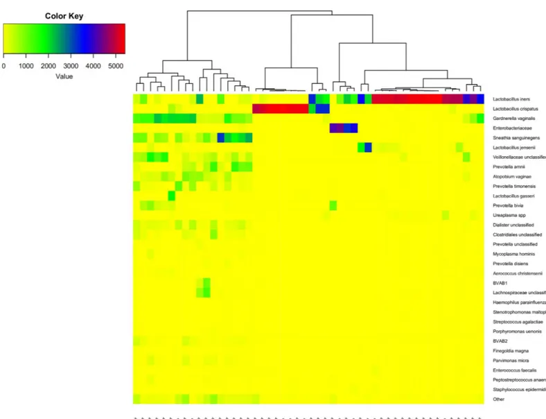
The distribution of 31 of the 33 taxa each constituting more than 1‰ of the sequences (and the rest combined as “other”) for sample 0 from the patients and sample 1 from the controls is shown in the heat map inFig 2.Two of the 33 dominant species,Haemophilus haemolyticus
andAchromobacter ruhlandii, were not present in the first samples.
Fig 1. Flowchart of samples from 52 patients and 10 controls.
These 31 dominant taxa constituted 98.1% of all sequences in these first samples. The sam-ples formed three main clusters corresponding to the common CSTs: CST-I, CST-III and CST-IV (Fig 2) [2]. One cluster of 11 samples dominated byL. crispatus, another of 22
domi-nated byL. iners, this cluster also included samples dominated by Enterobacteriaceae. Finally, a
third cluster of 17 samples containing a mix of different species includingG. vaginalis, A. vagi-nae, Prevotella and other BV associated bacteria. The median Shannon diversity index for the
three clusters was 0.15, 0.34 and 1.93 respectively (p-values CST-I vs CST-III: NS, CST-I vs CST-IV: 0.00008, CST-III vs CST-IV: 0.00001, Mann-Whitney).
Dominant species per patient
Only 10 taxa constituted more than 1% of the sequences:L. iners (41.0%), L. crispatus (26.0%), G. vaginalis (6.5%), Enterobacteriaceae (3.5%), Sneathia sanguinegens (3.1%), L. jensenii
(2.5%),Veillonellaceae unclass (1.9%), Prevotella amnii (1.7%), A. vaginae (1.7%) and P. timo-nensis (1.3%). In total, these ten taxa constituted 91.5% of all sequences.
Fig 2. Heatmap of 31 dominant OTUs and the rest combined as “other” of sample 0 from the patients and sample 1 from the controls. The dendrogram show a
hierarchical clustering of samples based on Bray-Curtis dissimilarity. The patient number and group (con = control, pt = patient) is shown on the x-axis.
To study the temporal changes of the microbiota, the presence of these 10 most prevalent species in vaginal swabs is shown per patient inFig 3, where the patients are sorted according to the most dominant CST.
The controls and patients could be divided into three different profiles. 1) Those dominated (i. e. > 50% of sequences) byL. crispatus (corresponding to CST-I), 2) those dominated by L. iners (CST-III) and 3) those dominated by other bacteria (CST-IV) [2]. By this definition, a microbiota dominated by e.g.Enterobacteriaceae was classified as CST-IV.
In the controls, the microbiota was found to be relatively stable over time as shown inFig 3A. Two of the three CST-I controls showed a stable,L. crispatus dominated microbiota that
briefly changed during a menstrual period but rapidly returned to CST-I after the bleeding. In the five CST-III controls, two of these also showed a change in the microbiota related to bleed-ing, but otherwise showed a stableL. iners microbiota, but also supplemented with L. jensenii.
Finally two controls had a microbiota initially dominated by mixed bacteria (CST-IV). In one of these controls (#219) the mixed microbiota completely shifted to one dominated byL. cris-patus after a menstrual period. Interestingly, L. criscris-patus was present at low abundance in the
CST IV-type microbiota before the menstrual bleeding, but absent from two samples during the bleeding.
In the patients (Fig 3B–3F), 14 (27%) were dominated byL. crispatus (Fig 3B). None of these patients were diagnosed with BV and most of the samples (71%) had a normal Nugent score. In three patients, the majority of samples had an intermediate Nugent score, and five other patients had one or more samplings showing an intermediate Nugent score. A number of patients hadL. iners dominated (#103, #39, #190) or diverse microbiota (#27, #110) in their
initial samples, but went on toL. crispatus dominated microbiota after treatment for their STI
without metronidazole. In two other patients (#159, #24) there was an increase ofL. iners in
the late samples. Two patients (#190 and #196) had no gynecological examination and no ini-tial evaluation; one patient (#187) had no follow-up evaluation. Only three patients (#61, #166 and #110) showed a normal Nugent score in all samples.
For 22 patients (42%), the microbiota was dominated byL. iners, and 11 showed no sign of
BV at day 0 (Fig 3C), but all had one or more samplings with an intermediate Nugent score although most of the samples (58%) were scored as normal. In many of these patients,L. jense-nii was also prominent. In three patients (#25, #37, #141) L. crispatus was present in all
sam-ples, but without a clear change in CST during the observation period. Two patients were diagnosed with BV, but not treated with metronidazole (#22 and #135). Despite no treatment, both of these patients changed to anL. iners dominated microbiota before the end of the study
period (Fig 3C). In another 9 patients, the vaginal microbiota was also dominated byL. iners,
but these patients were initially diagnosed with BV and treated for the condition with metroni-dazole. In all of these patients,L. iners became the dominant species shortly after treatment
(Fig 3D). All of these patients had one or more BV positive samples and the majority with an intermediate Nugent score and only one patients (#134) had two samples that were Nugent 0–3. One patient (#64) was earlier (day -25) diagnosed with BV but not treated with metroni-dazole before day 0.
Finally, 16 patients (31%) were dominated by a microbiota of mixed bacteria, CST IV. Eleven of these 16 patients (Fig 3E) were treated with metronidazole but in 6/11 there was no change of the Nugent score during the study period. In another five patients, there was some effect of the antibiotic treatment, but no sample from any of the patients reached a Nugent score of 0–3. Five patients with mixed bacteria were not treated with metronidazole (Fig 3F): Three patients had no gynecological examination, another two (#50, #121) were Amsel BV negative in the clinic at day 0, but BV positive by Nugent in the home-collected samples (Fig 3F).
Across the CST groups, short periods dominated byEnterobacteriaceae was observed in
both patients and controls. In a most of the patients, these temporary changes in the micro-biota were seen in conjunction with periods of bleeding (Fig 3). Also the Shannon diversity index was higher during bleeding (0.94 vs 0.44; p = 0.00008 Mann-Whitney).
Antibiotics, load and diversity
A reduction in the bacterial load following treatment with metronidazole and other antibiotics, could be expected. However, there was no significant reduction in the median total bacterial load, as determined by broad range 16S rRNA gene qPCR in patient samples from day zero and any of the following sampling days (S1 Fig, p = 0.66, 0.7, 0.94, 0.12, 0.26, 0.07 and 0.4, respectively, Wilcoxon).
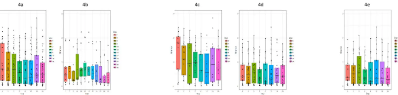
In contrast to the total bacterial load, the diversity before and after treatment differed. When comparing diversity in patients according to sampling time, there was a trend towards a declining Shannon index from day 0 to day 12), followed by an increase at day 19 and 26, shown inFig 4A.
However, only at day 8 and 12 the index was statistically significantly lower than at day 0 (p< 0.002 and p< 0.03, respectively, Wilcoxon).
When comparing controls and patients treated with azithromycin, tetracyclines and moxi-floxacin respectively, there was no significant difference in Shannon index between these groups (not shown).
Patients diagnosed with BV had a more diverse microbiota than patients without this diag-nosis. At days 0–8 and 26 the diversity was higher in metronidazole treated BV patients than in patients without metronidazole treatment, including patients without BV. However, only at days 0, 1, 5 and 26 the difference was statistically significant (p = 0.006, 0.007, 0.02 and 0.005, respectively; Mann-Whitney (Fig 4C and 4D). For the five patients diagnosed with BV but without metronidazole treatment, the Shannon index was between 0.72 and 2.45, i.e. similar to
Fig 3. Bar plot of the ten most common bacterial species shown per patient. For each day the percentage for each of
the 10 most prevalent species is shown. Other species appear combined as white in top of each bar. Above each bar plot is given the patient code and, in parentheses, the antibiotic used (Azi,): azithromycin, (Tetra,): tetracycline, (Moxi,): moxifloxacin. (, Metro): metronidazole. A few patients received metronidazole before day 0, this is indicated as M-11, M-7 etc. Patients without gynecological examination are marked “no G.E.”. Under each plot is given the day and bacterial vaginosis score (BV, intermediate or normal). Further days with bleeding are shown in red. A: Controls. Top panel CST-I, Middle panel: CST-III, bottom panel CST-IV. B: Patients with CST-I. C: Patients with CST-III not treated with metronidazole. D: Patients with CST-III, treated with metronidazole. E: Patients with CST-IV, treated with metronidazole. F: Patients with CST-IV, not treated with metronidazole.
https://doi.org/10.1371/journal.pone.0236036.g003
Fig 4. Boxplot of Shannon diversity index given per sampling. a: All patients. b: All controls. c: Patients treated with metronidazole. d Patients without metronidazole
treatment. e: Patients initially positive forC. trachomatis or M. genitalium and not treated with metronidazole.
day zero for the metronidazole treated patients, but without a reduction over time as seen in the treated patients.
For patients not treated with metronidazole, there was no statistically significant different decrease in diversity in relation to the antimicrobial treatment (Fig 4D). This was true regard-less of the antimicrobial class although too few patients received tetracyclines or moxifloxacin to allow meaningful conclusions.
Nineteen (37%) of the patients were treated for a presumed STI based on symptoms and/or signs of urethritis or cervicitis. The remaining patients (63%) were treated as contacts toM. genitalium or C. trachomatis positive partners or due to a positive diagnostic test for one of the
two STIs. It could be speculated, that the inflammation caused by STIs could promote a shift in the microbial diversity. However, for patients positive forC. trachomatis or M. genitalium,
but without metronidazole treatment, there was no statistically significant difference in diver-sity before or after treatment forM. genitalium and C. trachomatis (Fig 4E).
Bacterial vaginosis and microbiota
In the clinic, 24 of the 52 patients were diagnosed with BV by Amsel criteria. Another two patients (#50 and #121) were negative by Amsel at day 0 but were found BV positive by Nugent all the following days. These two patients had a mixed CST-IV microbiota, also at day 0.
The relation between the microbiota and Nugent score is displayed in the PCoA plot inFig 5, showing a good correlation between the two different methods.
Fig 5. PCoA plot the 277 patient samples with a Nugent score. Clustering based on Bray Curtis dissimilarity
calculated from taxonomic composition and colored according to Nugent score: Green = normal (Nugent 0–3), blue = intermediate (Nugent 4–6) and red = BV (Nugent score 7–10).
Of the 22 patients treated with metronidazole, all were qPCR positive forG. vaginalis.
How-ever, two of the 22 patients were treated one and two weeks respectively before day 0 and their samples had low organism load, so the effect of metronidazole could not be evaluated. In 11 (58%) of the evaluable 19 patients,G. vaginalis was eradicated or significantly reduced (more
than 50 fold), while there was no effect on theG. vaginalis load in 8 (42%) patients. In one
patient there was a reduction day 3 and 5 followed by an increase in load. There was no differ-ence in the treatment effect related to the single-dose therapy versus the 7-day regimen.
In the 11 patients withG. vaginalis eradication or significant reduction, A. vaginae was also
present in 10 and eradicated in all. Of the eight patients without treatment effect onG. vagina-lis, six were also A. vaginae positive and three remained unchanged during treatment while in
three,A. vaginae was eradicated (in two only temporary). Of the eight patients without
treat-ment effect onG. vaginalis, seven were positive and effectively treated for M. genitalium or C. trachomatis.
Specific qPCR compared to 16S sequencing
M. genitalium and C. trachomatis. By qPCR, 57 samples were M. genitalium positive.
However, only 10 (18%) of these were detected in the microbiota analysis. By qPCR, the median organism load was 394 for the sequence-positive and 23 for the sequence–negative (p<0.00001, Mann-Whitney).
ForC. trachomatis, 36 samples were qPCR positive and eight (22%) samples from five
patients were sequence positive. The median organism load for the eight sequence-positive samples was 168000 while it was 854 for the 28 sequence negative samples. All of the sequence-positive samples were from day 0, 1 or 3.
Ureaplasma spp. In total, 53 patient samples were U. urealyticum positive by qPCR, 3/53
were alsoU. parvum positive. Of the 53 U. urealytium qPCR positive, 29 (55%) were sequence
positive. The median load (qPCR) was 274 for the sequence positive and 7.5 for the sequence negative (p<0.00001, Mann-Whitney). A total of 188 samples wereU. parvum positive by
qPCR and of these, 156 (83%) were sequence positive. The median qPCR load was 59 for the sequence positive and 17.5 for the sequence negative (p<0.002, Mann-Whitney).
Specific qPCR and antimicrobial treatment. The preparation of samples for sequencing
at the MiSeq platform includes a number of normalization steps that, potentially, could skew the distribution of species in the sequenced samples. To address this, we compared the bacte-rial load as determined by species-specific qPCR with the total bactebacte-rial load by qPCR multi-plied by the relative abundance of sequences for the individual species. The relation between qPCR and normalized sequences forG. vaginalis, A. vaginae and Ureaplasma is shown inFig 6
(log 10 data).
Fig 6. Correlation between sequences and qPCR forG. vaginalis, A. vaginae and Ureaplasma. The bacterial load determined by rarefied sequence number multiplied
by total bacterial load (sequences) versus the load determined by specific qPCR forGardnerella vaginalis, Atopobium vaginae and Ureaplasma. Log scale. Ureaplasma is U. parvum and U. urealyticum combined.
For the more abundant species,G. vaginalis and A. vaginae, there was a relatively good
cor-relation between the load determined by specific qPCR and the load determined by sequenc-ing. ForUreaplasma, where the load was approximately 100 fold lower, the correlation was
relatively poor, in particular for samples with high bacterial load and low load ofUreaplasma.
For all three species, a considerable number of samples showed a qPCR of 100–1000 copies, but zero sequences, probably reflecting that specific PCR is more sensitive than a broad-range 16S PCR.
Ureaplasma. Twelve patients (23%) were qPCR positive forU. urealyticum at day zero or
one, and all twelve were treated with azithromycin. For eight of these patients (67%),U. urealy-ticum was eradicated, while no effect was observed in three patients. In one patient the load
was too low for evaluation.U. parvum was only eradicated in one of the four patients who
were alsoU. parvum positive.
Twenty-eight patients (54%) wereU. parvum positive at day at 0 or 1 and were treated with
azithromycin. In one of these, the load was too low for evaluation. In 25 of the 27 patients (93%),U. parvum persisted as determined by qPCR and only in two patients (7%) (#9 and #27,
both CST-IV)U. parvum was eradicated by azithromycin.
The six patients treated with tetracyclines were allU. parvum positive at day 0 or 1. Five of
these (83%) demonstrated eradication, while one had a temporary reduction in the organism load.
For 274 of the patient samples, both qPCR results and a Nugent score were obtained. The load ofU. parvum, as determined by qPCR, was significantly related to the Nugent score
(p<0.0045, Pearson), whereas there was no statistically significant relation between Nugent score and load ofU. urealyticum. (p = 0.092).
The 25 patients whereU. parvum was persistent were equally distributed regarding CSTs
(CST-I: 6/14 (43%), CST-III: 12/22 (55%), CST-IV: 7/16 (44%)) or concomitant use of metro-nidazole (10 treated with metrometro-nidazole, 15 without treatment).
G. vaginalis, A. vaginae and Prevotella. Using the concept of conversion from relative
abundance to absolute counts, there was no significant difference in the load ofG. vaginalis or A. vaginae for metronidazole treated patients at any time-point. In contrast, the genus Prevo-tella (combined data for all PrevoPrevo-tella species) showed a considerable reduction following
administration of metronidazole, but only significant for day 5 and day 8, both p<0.01 (Wilcoxon).
M. genitalium, C. trachomatis and BV. At day 0, 32 patients were M. genitalium positive,
10 wereC. trachomatis positive while two patients were positive for both of these species. Eight
patients were negative for both.
Of the 32M. genitalium positive patients, 16 (50%) were BV positive in the clinic by Amsel,
while five (50%) of the 10C. trachomatis positive had BV. None of the two double infected
patients was treated for BV (one negative, one without gynecological examination). Of the eight patients without Ct and Mg, three (38%) had BV.
NeitherC. trachomatis nor M. genitalium were related to any specific community state
type. In CST-I, CST-III and CST-IV 21%, 13% and 31%, respectively wereC. trachomatis
posi-tive, forM. genitalium the corresponding figures were 57%, 50% and 75%.
Discussion
The vaginal microbiota, particularly in women with BV, is relatively complex and with consid-erable differences between individuals. We found 615 different taxa in vaginal swabs from 62 women. Despite this high number of different species, only 33 taxa represented more than 1 ‰ of the sequences and combined covered almost 98% of the sequences. In any environment,
administration of antibiotics may not only eradicate the intended species, but may also affect other bacteria with unknown roles in the microbiota. Thus, we aimed to longitudinally evalu-ate the effect of antimicrobial treatment on the vaginal microbiota to study its resilience. We were particularly interested in the different spectrum of azithromycin and tetracyclines to inform the current discussions recommending to abandon azithromycin treatment for syn-dromic management of non-gonococcal urethritis and cervicitis in favor of doxycycline [30,31].
It is well known that risk of acquisition of several STIs is increased in the presence of BV [12,13] but acquisition of a bacterial STI may also lead to disturbed vaginal microbiota [12]. This was reflected in the high proportion of women in the present study having BV by Amsel criteria at inclusion and made comparison of a “clean” macrolide versus tetracycline treatment difficult, as it was considered unethical to withhold BV treatment.
Despite this limitation, we saw a surprising resilience of the microbiota regardless of the treatment provided. Even though treatment efficiently eradicatedC. trachomatis and M. geni-talium, we were not even able to document a statistically significant decrease in the total
bacte-rial load as measured by qPCR. In women with BV, a temporary decrease in diversity was observed in connection with the metronidazole treatment, but at the end of the 26 day obser-vation period, most women had their pre-treatment level of diversity.
The vaginal microbiota in both controls and patients was in accordance with earlier described community state types (CST-I–CST-V) except that we did not find patients with a microbiota dominated byL. gasseri (CST-II) or by L. jensenii (CST-V), although a few samples
had high abundance ofL. jensenii and a single of L. gasseri.
This is somewhat in contrast to earlier findings; Ravelet al found CST-II in 6% and CST-V
in 5% of patients from the US, and even higher fractions in patients who identified themselves as white[2], while Virtanenet al found both L. gasseri and L. jensenii to be dominant in each
4% of the samples from Nordic women [6].
However, the relatively low number of women in the present study could also explain the inability to identify CST-II and CST-V.
In the controls, the 10 most abundant species were rather stable over time in nine out of ten women and this was the case not only inL. crispatus dominated CST-, but also in CST-III
dominated byL. iners.
In the patients, treatment with azithromycin was very effective in eradicating the intended targetsM. genitalium and C. trachomatis, but had little impact on the vaginal microbiota and
did only change the CSTs in a few patients. In contrast to azithromycin, metronidazole had a significant effect on the diversity of the vaginal microbiota (Fig 4). Still, in approximately 50% of the patients, metronidazole did not change the CST-IV status. In another 10 patients the microbiota rapidly changed to CST-III. Interestingly, two patients without metronidazole treatment also changed from BV to CST-III. Such an increase inL. iners following treatment
with metronidazole has also been seen in other studies [32,33]. Jacobsson and Forsum sug-gested thatL. iners dominance could be a transitional stage between abnormal and normal
microbiota [34]. This was, however, not supported in the present study, at least during the 26 days observation period, as not a single treated BV patient changed from CST-IV over CST-III to CST-I.
Metronidazole is one of the most commonly used antimicrobial treatments for BV, how-ever, the cure rate is rather low [35] and recurrence is frequent [36].G. vaginalis is one of the
prominent vaginal bacteria associated with BV. We found that metronidazole only eradicated or significantly reducedG. vaginalis in 58% of the treated patients, and in these patients, other
BV associated bacteria such asA. vaginae were also eradicated. In those patients where G. vagi-nalis was not affected, other anaerobic bacteria were eradicated in half the patients,
demonstrating that the microbiota was affected to some extent by the metronidazole treat-ment, even ifG. vaginalis did not respond. In the remaining half of the patients, none of these
anaerobic bacteria was eradicated. This could be due to biofilm formation protectingG. vagi-nalis as well as other members of the biofilm, but the persistence of G. vagivagi-nalis could also
reflect true metronidazole resistance which has been reported in >50% of endometrialG. vagi-nalis and A. vaginae isolates [37].
The persistence ofG. vaginalis could also be caused by patients not using the prescribed
metronidazole. However, all patients carefully collected and submitted samples. Further, in most of the patients without effect onG. vaginalis, M. genitalium or C. trachomatis were
eradi-cated, demonstrating that the antibiotics prescribed for STIs were used. We therefore find it likely that most or all patients did comply with the prescribed treatment.
In contrast to the present study, Mayeret al [38] found a rapid and profound shift in the vaginal microbiota after treatment for BV by specific qPCRs. In particularLeptotrichia/ Sneathia and Megasphaera were rapidly cleared but significant reduction in G. vaginalis was
also observed in all patients. However, the majority of the women received local metronidazole which may have produced higher concentrations locally explaining the difference. On the other hand oral and local metronidazole is considered equally clinically efficient [39]. Still, the median load ofGardnerella was not significantly reduced from day 0 before treatment to any
of the following samplings when all patients receiving metronidazole were compared. In the patients whereG. vaginalis was unaffected by metronidazole, the load actually increased from
day zero, probably reflecting that competitor species were reduced or eradicated. The increas-ing load in these samples could mask a decreasincreas-ing trend in patients with metronidazole sus-ceptibleG. vaginalis.
In both controls and patients we saw changes in the microbiota and increasing diversity related to bleeding. In most cases, the microbiota rapidly returned to the CST observed before the bleeding incident. In particular, the taxonEnterobacteriaceae was related to periods of
vag-inal bleeding both in patients and controls. This is consistent with an earlier study that found
E. coli associated with the menstrual phase [40]. Bleedingper se and the following shift in pH
and enrichment of iron and other nutrients could promote such flare up ofEnterobacteriaceae.
Another explanation could be a depletion of lactobacilli. A bactericidal activity of genital tract secretions againstE. coli has earlier been described and Kalyoussef et al found proteins
secreted from different lactobacilli to be associated with this bactericidal activity [41]. A later study found that supernatant ofL. crispatus cultures could inhibit growth of E. coli, though
some effect of supernatant ofL. iners and G. vaginalis also was observed [42]. Other studies have found other bacteria related to menses, Srinivasanet al observed an increase of G. vagina-lis [11]. Interestingly, Gajeret al [10] found no significant difference in Shannon diversity index in relation to menstrual period when analyzing 32 women twice weekly for 16 weeks but found the highest rate of change in the diversity during menses.
Treatment with metronidazole reduced, temporarily, the diversity (Fig 4) as the abundance ofL. iners increased while Prevotella decreased. This is consistent with earlier findings by
Ver-wijset al [35] who found a reduction in BV associated taxa combined with an increase of lacto-bacilli. However, in contrast to the present study, they found almost 1 log reduction in the total bacterial load following treatment with metronidazole.
While we found tetracyclines effective against bothU. parvum and U. urealyticum, the
response to azithromycin differed between the two species.U. urealyticum was eradicated in
67% of the treated patients, whereasU. parvum was eradicated in only 7% of the treated
patients. Interestingly, the load ofU. parvum was positively correlated to the Nugent score, i.e.
related to CST-IV. This is in accordance with our previous findings in men where the load of BV associated bacteria was positively correlated with the ureaplasma load [43]
The acidic environment created by the vaginal lactobacilli, particularlyL. crispatus [44] could diminish the activity of azithromycin. However, we did not see more ureaplasma persis-tence in the CST-I dominated patients, on the contrary, patients with CST-III and CST-IV showed a higher proportion of persistentU. parvum than CST-I dominated patients.
Few studies have used standardized methods forin vitro MIC determination of
azithromy-cin and distinguished between the two species. Valentine-King and Brown foundU. parvum
to have low resistance to azithromycin (MIC of �0.5–2μg/ml) for 60 isolates from the United States and only slightly higher MICs forU. urealyticum (MIC of �0.5–4 μg/ml) [45]. A similar trend towards higher MIC forU. urealyticum was reported from China [46], and a study ana-lyzing ureaplasmas from neonates from the UK found no resistance to macrolides and no dif-ferent activity of azithromycin, but a significantly higher erythromycin MIC forU.
urealyticum [47]. However, thesein vitro studies may not reflect the in vivo activity, where
fac-tors like biofilm or pH may influence the activity of antibiotics. To our knowledge, the differ-ential in vivo response to azithromycin has not previously been investigated.
Strengths and limitations
Strengths of the present study includes repeated sampling of patients and controls over a 26 day period and sequencing data supplemented with qPCR to allow a better quantitation. A limitation is the size of the study population where the 491 sequenced samples represent only 52 patients and 10 controls. With this limited number of participants more subtle changes in the microbiota may have been missed. More participants would possibly have allowed stronger quantitative conclusions.
In conclusion, the more dominant part of the vaginal microbiota was found to be remark-ably resilient after treatment with azithromycin and tetracyclines as treatment only changed the CST in a few patients. Only women receiving metronidazole changed from CST-IV to CST-III, but only in 50% of the treated patients, and the effect on key species asG. vaginalis
andA. vaginae was limited and temporary. A differential activity of azithromycin against Urea-plasma spp. was observed which should be confirmed in future studies
Supporting information
S1 Table. Bacterial species identified as contaminants.
(DOCX)
S2 Table. Spreadsheet showing the distribution of rarefied sequences in patients and con-trols. Column A: Sample name. Column B: Patient code. Patients treated with antibiotics:
8–200. Controls: 207–226. Column C: Sampling number. Column D: Sampling day. Column AL: Sum of all species constituting < 1‰ of the sequences. Column E–AK and AM—WV: Bacterial species in descending order.
(XLSX)
S1 Fig. Bacterial load (log10) in patients for day 0 to day 26.
(DOCX)
Acknowledgments
We thank Susanne Cramer Johansson, Carola Skov Hilby and Martin Ibenholdt Pedersen for laboratory assistance and Anna Ingham for helpful advice on bioinformatics.
Author Contributions
Conceptualization: Peter Ahrens, Jørgen Skov Jensen, Lars Falk.
Data curation: Peter Ahrens, Lee O’Brien Andersen, Berit Lilje, Thor Bech Johannesen, Ebba
Gomez Dahl, Lars Falk.
Formal analysis: Peter Ahrens, Berit Lilje, Thor Bech Johannesen, Sharmin Baig.
Investigation: Peter Ahrens, Lee O’Brien Andersen, Berit Lilje, Thor Bech Johannesen, Ebba
Gomez Dahl, Sharmin Baig, Jørgen Skov Jensen, Lars Falk.
Project administration: Peter Ahrens, Jørgen Skov Jensen, Lars Falk.
Resources: Jørgen Skov Jensen, Lars Falk.
Validation: Berit Lilje, Thor Bech Johannesen. Visualization: Peter Ahrens.
Writing – original draft: Peter Ahrens, Jørgen Skov Jensen.
Writing – review & editing: Peter Ahrens, Lee O’Brien Andersen, Berit Lilje, Thor Bech
Johannesen, Ebba Gomez Dahl, Sharmin Baig, Jørgen Skov Jensen, Lars Falk.
References
1. Onderdonk AB, Delaney ML, Fichorova N. The Human Microbiome during Bacterial Vaginosis. Clin Microbiol Rev. 2016; 29(2):223–38.https://doi.org/10.1128/CMR.00075-15PMID:26864580
2. Ravel J, Gajer P, Abdo Z, Schneider GM, Koenig SSK, McCulle SL, et al. Vaginal microbiome of repro-ductive-age women. Proc Natl Acad Sci [Internet]. 2011; 108(Supplement_1):4680–7. Available from: http://www.pnas.org/cgi/doi/10.1073/pnas.1002611107
3. Kenyon C, Colebunders R, Crucitti T. The global epidemiology of bacterial vaginosis: A systematic review. Am J Obstet Gynecol [Internet]. 2013; 209(6):505–23. Available from:https://doi.org/10.1016/j. ajog.2013.05.006PMID:23659989
4. Noyes N, Cho KC, Ravel J, Forney LJ, Abdo Z. Associations between sexual habits, menstrual hygiene practices, demographics and the vaginal microbiome as revealed by Bayesian network analysis. PLoS One. 2018; 13(1):1–25.
5. Brookheart RT, Lewis WG, Peipert JF, Lewis AL, Allsworth JE. Association between obesity and bacte-rial vaginosis as assessed by Nugent score. Am J Obstet Gynecol [Internet]. 2019; 220(5):476.e1–476. e11. Available from:https://doi.org/10.1016/j.ajog.2019.01.229
6. Virtanen S, Rantsi T, Virtanen A, Kervinen K, Nieminen P, Kalliala I, et al. Vaginal Microbiota Composi-tion Correlates Between Pap Smear Microscopy and Next GeneraComposi-tion Sequencing and Associates to Socioeconomic Status. Sci Rep [Internet]. 2019; 9(1):1–9. Available from:https://doi.org/10.1038/ s41598-018-37186-2PMID:30626917
7. Ralph SG, Rutherford AJ, Wilson JD. Influence of bacterial vaginosis on conception and miscarriage in the first trimester: Cohort study. Br Med J. 1999; 319(7204):220–3.
8. Haahr T, Jensen JS, Thomsen L, Duus L, Rygaard K, Humaidan P. Abnormal vaginal microbiota may be associated with poor reproductive outcomes: A prospective study in IVF patients. Hum Reprod. 2016; 31(4):795–803.https://doi.org/10.1093/humrep/dew026PMID:26911864
9. Koedooder R, Singer M, Schoenmakers S, Savelkoul PHM, Morre´ SA, de Jonge JD, et al. The vaginal microbiome as a predictor for outcome of in vitro fertilization with or without intracytoplasmic sperm injection: a prospective study. Hum Reprod. 2019; 34(6):1042–54.https://doi.org/10.1093/humrep/ dez065PMID:31119299
10. Gajer P, Brotman RM, Bai G, Sakamoto J, Schu¨tte UME, Zhong X, et al. Temporal dynamics of the human vaginal microbiota. Sci Transl Med. 2012; 4(132).
11. Srinivasan S, Liu C, Mitchell CM, Fiedler TL, Thomas KK, Agnew KJ, et al. Temporal variability of human vaginal bacteria and relationship with bacterial vaginosis. PLoS One. 2010; 5(4).
12. Wiesenfeld HC, Hillier SL, Krohn MA, Landers D V, Sweet RL. Bacterial vaginosis is a strong predictor of Neisseria gonorrhoeae and Chlamydia trachomatis infection. Clin Infect Dis. 2003; 36(5):663–8. https://doi.org/10.1086/367658PMID:12594649
13. Lokken EM, Balkus JE, Kiarie J, Hughes JP, Jaoko W, Totten PA, et al. Association of Recent Bacterial Vaginosis with Acquisition of Mycoplasma genitalium. Am J Epidemiol. 2017; 186(2):194–201.https:// doi.org/10.1093/aje/kwx043PMID:28472225
14. Edwards VL, Smith SB, McComb EJ, Tamarelle J, Ma B, Humphrys M, et al. The Cervicovaginal Micro-biota-Host Interaction Modulates Chlamydia trachomatis infection. MBio. 2019; 10(4):1–18.
15. Filardo S, Pietro D, Tranquilli G, Latino A, Recine N, Porpora G. Selected Immunological Mediators and Cervical Microbial Signatures in Women with Chlamydia trachomatis Infection. mSystems. 2019; 4 (4):1–14.
16. Falk L, Enger M, Jensen JS. Time to eradication of Mycoplasma genitalium after antibiotic treatment in men and women. J Antimicrob Chemother [Internet]. 2015 Nov [cited 2015 Dec 10]; 70(11):3134–40. Available from:http://www.ncbi.nlm.nih.gov/pubmed/26283670 https://doi.org/10.1093/jac/dkv246 PMID:26283670
17. Amsel R, Totten PA, Spiegel CA, Chen KCS, Eschenbach D, Holmes KK. Nonspecific vaginitis. Diag-nostic criteria and microbial and epidemiologic associations. Am J Med. 1983; 74(1):14–22.https://doi. org/10.1016/0002-9343(83)91112-9PMID:6600371
18. Nugent RP, Krohn MA, Hillier SL. Reliability of diagnosing bacterial vaginosis is improved by a standard-ized method of gram stain interpretation. J Clin Microbiol [Internet]. 1991; 29(2):297–301. Available from:http://www.ncbi.nlm.nih.gov/pubmed/1706728%0Ahttp://www.pubmedcentral.nih.gov/ articlerender.fcgi?artid=PMC269757PMID:1706728
19. Mortensen C, Karlsen S, Grønbæk H, Nielsen DT, Frevert S, Clemmesen JO, et al. No difference in tal and hepatic venous bacterial DNA in patients with cirrhosis undergoing transjugular intrahepatic por-tosystemic shunt insertion. Liver Int. 2013; 33(9):1309–15.https://doi.org/10.1111/liv.12205PMID: 23763259
20. Jensen JS, Bjornelius E, Dohn B, Lidbrink P. Use of TaqMan 5 ‘ nuclease real-time PCR for quantitative detection of Mycoplasma genitalium DNA in males with and without urethritis who were attendees at a sexually transmitted disease clinic. J Clin Microbiol [Internet]. 2004 Feb; 42(2):683–92. Available from: isi:000189379000032https://doi.org/10.1128/jcm.42.2.683-692.2004PMID:14766837
21. Westh H, Jensen JS. Low prevalence of the new variant of Chlamydia trachomatis in Denmark. Sex Transm Infect [Internet]. 2008 Dec; 84(7):546–7. Available from: isi:000261378800009https://doi.org/ 10.1136/sti.2008.031906PMID:18653564
22. Frølund M, Bjo¨rnelius E, Lidbrink P, Ahrens P, Jensen JS. Comparison between culture and a multiplex quantitative real-time polymerase chain reaction assay detecting Ureaplasma urealyticum and U. par-vum. PLoS One. 2014;9(7).
23. Datcu R, Gesink D, Mulvad G, Montgomery-Andersen R, Rink E, Koch A, et al. Vaginal microbiome in women from Greenland assessed by microscopy and quantitative PCR. BMC Infect Dis [Internet]. 2013 Jan [cited 2014 Jun 12]; 13(1):480. Available from:http://www.pubmedcentral.nih.gov/articlerender. fcgi?artid=3853076&tool=pmcentrez&rendertype=abstract
24. Krogsgaard LR, Andersen LOB, Johannesen TB, Engsbro AL, Stensvold CR, Nielsen HV, et al. Char-acteristics of the bacterial microbiome in association with common intestinal parasites in irritable bowel syndrome. Clin Transl Gastroenterol [Internet]. 2018; 9(6). Available from:http://dx.doi.org/10.1038/ s41424-018-0027-2
25. Ring HC, Thorsen J, Saunte DM, Lilje B, Bay L, Riis PT, et al. The follicular skin microbiome in patients with hidradenitis suppurativa and healthy controls. JAMA Dermatology. 2017; 153(9):897–905.https:// doi.org/10.1001/jamadermatol.2017.0904PMID:28538949
26. McDonald JE, Larsen N, Pennington A, Connolly J, Wallis C, Rooks DJ, et al. Characterising the canine oral microbiome by direct sequencing of reverse-transcribed rRNA molecules. PLoS One. 2016; 11 (6):1–17.
27. Eisenhofer R, Minich JJ, Marotz C, Cooper A, Knight R, Weyrich LS. Contamination in Low Microbial Biomass Microbiome Studies: Issues and Recommendations. Trends Microbiol [Internet]. 2019; 27 (2):105–17. Available from:https://doi.org/10.1016/j.tim.2018.11.003PMID:30497919
28. Benjamini Y, Hochberg Y. Controlling the false discovery rate: a practical and powerful approach to mul-tiple testing [Internet]. Vol. 57, Journal of the Royal Statistical Society. 1995. p. 289–300. Available from:http://www.jstor.org/stable/2346101%5Cnhttp://about.jstor.org/terms
29. R Core Team RF for SC puting. R: A Language and Environment for Statistical Computing [Internet]. Vienna, Austria; 2017. Available from:https://www.r-project.org/
30. Horner PJ, Blee K, Falk L, van der Meijden W, Moi H. 2016 European guideline on the management of non-gonococcal urethritis. Int J STD AIDS. 2016; 27(11):928–37.https://doi.org/10.1177/
31. Singh AE, Manhart L. Is It Time for the United States and Canada to Reconsider Macrolides as the First-line Empiric Treatment for Males With Symptomatic Urethritis? Clin Infect Dis. 2019;(Xx Xxxx):1– 3.
32. Ferris MJ, Norori J, Zozaya-Hinchliffe M, Martin DH. Cultivation-independent analysis of changes in bacterial vaginosis flora following metronidazole treatment. J Clin Microbiol. 2007; 45(3):1016–8. https://doi.org/10.1128/JCM.02085-06PMID:17202272
33. Joag V, Obila O, Gajer P, Scott MC, Dizzell S, Humphrys M, et al. Impact of standard bacterial vaginosis treatment on the genital microbiota, immune milieu, and ex vivo human immunodeficiency virus suscep-tibility. Clin Infect Dis. 2019; 68(10):1675–83.https://doi.org/10.1093/cid/ciy762PMID:30407498
34. Jakobsson T, Forsum U. Lactobacillus iners: A marker of changes in the vaginal flora? [2]. J Clin Micro-biol. 2007; 45(9):3145.https://doi.org/10.1128/JCM.00558-07PMID:17652481
35. Verwijs MC, Agaba SK, Darby AC, van de Wijgert JHHM. Impact of Oral Metronidazole Treatment on the Vaginal Microbiota and Correlates of Treatment Failure. Am J Obstet Gynecol [Internet]. 2019; Available from:https://doi.org/10.1016/j.ajog.2019.08.008
36. Bradshaw CS, Morton AN, Hocking J, Garland SM, Morris MB, Moss LM, et al. High Recurrence Rates of Bacterial Vaginosis over the Course of 12 Months after Oral Metronidazole Therapy and Factors Associated with Recurrence. J Infect Dis. 2006; 193(11):1478–86.https://doi.org/10.1086/503780 PMID:16652274
37. Petrina MAB, Cosentino LA, Wiesenfeld HC, Darville T, Hillier SL. Susceptibility of endometrial isolates recovered from women with clinical pelvic inflammatory disease or histological endometritis to antimi-crobial agents. Anaerobe [Internet]. 2019 Apr; 56:61–5. Available from:https://linkinghub.elsevier.com/ retrieve/pii/S1075996419300289 https://doi.org/10.1016/j.anaerobe.2019.02.005PMID:30753898
38. Mayer BT, Srinivasan S, Fiedler TL, Marrazzo JM, Fredricks DN, Schiffer JT. Rapid and profound shifts in the vaginal microbiota following antibiotic treatment for bacterial vaginosis. J Infect Dis. 2015; 212 (5):793–802.https://doi.org/10.1093/infdis/jiv079PMID:25676470
39. Sherrard J, Wilson J, Donders G, Mendling W, Jensen JS. 2018 European (IUSTI/WHO) International Union against sexually transmitted infections (IUSTI) World Health Organisation (WHO) guideline on the management of vaginal discharge. Int J STD AIDS. 2018; 29(13):1258–72.https://doi.org/10.1177/ 0956462418785451PMID:30049258
40. Chow AW, Percival-Smith R, Bartlett KH, Goldring AM, Morrison BJ. Vaginal colonization with Escheri-chia coli in healthy women: Determination of relative risks by quantitative culture and multivariate statis-tical analysis. Am J Obstet Gynecol [Internet]. 1986 Jan 1 [cited 2019 Nov 7]; 154(1):120–6. Available from:https://www.sciencedirect.com/science/article/abs/pii/0002937886904060 https://doi.org/10. 1016/0002-9378(86)90406-0PMID:3511702
41. Kalyoussef S, Nieves E, Dinerman E, Carpenter C, Shankar V, Oh J, et al. Lactobacillus Proteins Are Associated with the Bactericidal Activity against E. coli of Female Genital Tract Secretions. PLoS One. 2012; 7(11).https://doi.org/10.1371/journal.pone.0049506PMID:23185346
42. Ghartey JP, Smith BC, Chen Z, Buckley N, Lo Y, Ratner AJ, et al. Lactobacillus crispatus dominant vag-inal microbiome is associated with inhibitory activity of female genital tract secretions against Escheri-chia coli. PLoS One. 2014; 9(5):1–8.
43. Frølund M, Falk L, Ahrens P, Jensen JS. Detection of ureaplasmas and bacterial vaginosis associated bacteria and their association with non-gonococcal urethritis in men. PLoS One. 2019; 14(4):1–13.
44. Witkin SS, Mendes-Soares M. LI, Jayaram Aswathi J LW, Forney Larry J. Influence of Vaginal Bacteria and d- and l-Lactic Acid Isomers on Vaginal Extracellular Matrix Metal.pdf. MBio. 2013; 4(4):1–7.
45. Valentine-king MA, Brown MB. Antibacterial Resistance in Ureaplasma species and Mycoplasma homi-nis Isolates from Urine Cultures in College-Aged Females. Antimicrob Agents Chemother. 2017; 61 (10):1–11.
46. Wang N, Liu W, Zhou Y, Liu Y. In vitro activities of nemonoxacin and other antimicrobial agents against human mycoplasma and ureaplasmas isolates and their defined resistance mechanisms. Front Micro-biol. 2019; 10(AUG):1–9.
47. Beeton ML, Chalker VJ, Jones LC, Maxwell NC, Spiller OB. Antibiotic resistance among clinical Urea-plasma isolates recovered from neonates in England and Wales between 2007 and 2013. Antimicrob Agents Chemother. 2016; 60(1):52–6.https://doi.org/10.1128/AAC.00889-15PMID:26459899