Model of the complex of Parathyroid
hormone-2receptor and Tuberoinfundibular peptide of39
residues
Mirna Abraham-Nordling, Bengt Persson and Erik Nordling
Linköping University Post Print
N.B.: When citing this work, cite the original article.
Original Publication:
Mirna Abraham-Nordling, Bengt Persson and Erik Nordling, Model of the complex of
Parathyroid hormone-2receptor and Tuberoinfundibular peptide of39 residues, 2010, BMC
Reseach Notes, (3), 270.
http://dx.doi.org/10.1186/1756-0500-3-270
Copyright: BioMed Central
http://www.biomedcentral.com/
Postprint available at: Linköping University Electronic Press
http://urn.kb.se/resolve?urn=urn:nbn:se:liu:diva-65705
39 residues
Abraham-Nordling et al.
Abraham-Nordlinget al. BMC Research Notes 2010, 3:270 http://www.biomedcentral.com/1756-0500/3/270 (27 October 2010)
S H O R T R E P O R T
Open Access
Model of the complex of Parathyroid hormone-2
receptor and Tuberoinfundibular peptide of
39 residues
Mirna Abraham-Nordling
1*, Bengt Persson
2, Erik Nordling
2Abstract
Background: We aim to propose interactions between the parathyroid hormone-2 receptor (PTH2R) and its ligand the tuberoinfundibular peptide of 39 residues (TIP39) by constructing a homology model of their complex. The two related peptides parathyroid hormone (PTH) and parathyroid hormone related protein (PTHrP) are compared with the complex to examine their interactions.
Findings: In the model, the hydrophobic N-terminus of TIP39 is buried in a hydrophobic part of the central cavity between helices 3 and 7. Comparison of the peptide sequences indicates that the main discriminator between the agonistic peptides TIP39 and PTH and the inactive PTHrP is a tryptophan-phenylalanine replacement. The model indicates that the smaller phenylalanine in PTHrP does not completely occupy the binding site of the larger tryptophan residue in the other peptides. As only TIP39 causes internalisation of the receptor and the primary difference being an aspartic acid in position 7 of TIP39 that interacts with histidine 396 in the receptor, versus isoleucine/histidine residues in the related hormones, this might be a trigger interaction for the events that cause internalisation.
Conclusions: A model is constructed for the complex and a trigger interaction for full agonistic activation between aspartic acid 7 of TIP39 and histidine 396 in the receptor is proposed.
Findings
Background
The recent extension of the structural knowledge of both class A and class B G-protein coupled receptors (GPCRs) with the X-ray determination of the human adrenergic b 2 receptor [1], the turkey adrenergic b 1 receptor [2], the human A2A Adenosine receptor [3] and squid rhodopsin [4,5] coupled with the structures of extra-cellular domains (ECDs) of several class B mone receptors [6-9], including the parathyroid hor-mone-1 receptor (PTH1R), makes it possible to begin a computational exploration of possible interactions of the parathyroid hormone-2 receptor (PTH2R) [10] and its natural ligand, the tuberoinfundibular peptide of 39 resi-dues (TIP39) [11,12].
The aim of the present study is to propose possible interactions between the hormone and its receptor. There are three related peptide hormones that interact with the two PTH receptors. Parathyroid hormone (PTH) binds and stimulates both receptors resulting in intracellular cAMP release and Ca2+ signalling. TIP39 binds to both receptors, but to PTH2R a hundredfold stronger than to PTH1R and only signal through PTH2R. The parathyroid hormone related protein (PTHrP) binds to and signals only through PTH1R. However, only TIP39 induces b-arrestin and protein kinase Cb mobilisation and receptor internalisation of PTH2R [13]. A further objective of the study is to inves-tigate how the related peptides can selectively bind to and activate the receptors. The truncated TIP39(7 - 39) is a high affinity antagonist for the PTH1R, while losing much of its affinity for the PTH2R [14], which indicates that the N-terminal portion is involved in both the selectivity between the two receptors and affinity to PTH2R.
* Correspondence: Mirna.Abraham.Nordling@ki.se
1
Division of Surgery, Department of Clinical Sciences, Danderyd Hospital, Karolinska Institutet, Stockholm, Sweden
Full list of author information is available at the end of the article Abraham-Nordlinget al. BMC Research Notes 2010, 3:270 http://www.biomedcentral.com/1756-0500/3/270
© 2010 Abraham-Nordling et al; licensee BioMed Central Ltd. This is an open access article distributed under the terms of the Creative Commons Attribution License (http://creativecommons.org/licenses/by/2.0), which permits unrestricted use, distribution, and reproduction in any medium, provided the original work is properly cited.
The function of the TIP39 - PTH2R system appears to be diverse with primary sites of expression including the nervous system [15], thyroid gland, pancreas, heart, vas-cular muscle, the reproductive system and lung [16]. An NO-dependent vasodilatory effect of TIP39 was demon-strated if the PTH1R is desensitized by either exogen-ously administered or endogenexogen-ously released PTHrP [17,18]. Knockout mice lacking the gene encoding TIP39 are sterile, examination of the testes shows that they contain no spermatids. By antibody labelling of the chromosome spreads it was shown that spermatogonia do not complete the prophase of meiosis I [19]. They also display a more stress anxiety prone phenotype in behaviour tests than wild type mice [20]. Another study has shown that the TIP39 - PTH2R system is activated in response to acoustic stress [21]. A recent report also points out a connection to neuropathic pain, where PTH2R was shown to be selectively localized on myeli-nated A-fibers. Pharmacological studies showed that TIP39 induced nociceptive responses that were mediated by activation of Gsand cAMP-dependent
pro-tein kinase. It was found that nociceptive responses induced by TIP39 were significantly greater following partial sciatic nerve injury induced neuropathic pain, without changes in PTH2R expression [22].
There is a possibility of using TIP39 as a tool to inves-tigate the function of PTH2R in more detail by defining its interaction sites with PTH2R and it may also be pos-sible to use it as a source of antagonistic peptides as the truncated TIP39(7 - 39) is for the PTH1R. That possibi-lity could be of interest to modulate the pain sensation in neuropathic pain.
A class B GPCR of subfamily B1, a hormone receptor, has an extracellular N-terminal domain (ECD), a trans-membrane (TM) domain and an intracellular C-terminal domain [23]. The fold of the TM region was revealed when the first structure of a GPCR was crystallographi-cally determined (bovine rhodopsin [24]) and it consists of sevena-helices that form an anti-parallel seven-mem-ber helical bundle. As the ECD of PTH1R has been crys-tallized in complex with PTH it is possible to construct a model which captures both the binding of the C-terminal part of the hormone to the ECD and its interactions with the transmembrane region of the receptor. As the current structure of the TM region of GPCRs is from class A and only distantly related to class B, it is reassuring that bio-physical studies indicate that the eighth helix of the class A GPCRs, which is positioned parallel to the membrane, also is present among the class B GPCRs. However, no apparent sequence homology is detected in that part of the receptor [25]. In contrast to the available class A structures, PTH2R has a large extracellular loop 1 (ECL1) between TM helix 2 and 3, which consist of 31 amino acid residues. In the case of ECL1, NMR
experiments of the corresponding part of PTH1R have indicated that it should form a central helix comprising residues 256 - 264, which is shown to contact the hor-mone with a centrally placed Leu261 binding to Lys27 of the hormone [26]. As no structure has been publically made available, the loop has been left to the prediction scheme with the added notion that the prediction accu-racy might be low for this region.
As the intracellular part of the receptor lacks a homo-logue with known structure we do not attempt to incor-porate that domain in the model.
NMR experiments of TIP39 indicate that it has two helical regions comprising the residues 5 19 and 27 -34, and that the two helical regions are tilted in relation to each other [27]. Similar experiments of PTH frag-ments 1 - 34 and 1 - 39 also indicate two helical regions connected by a loop region consisting of His14 to Ser17 [28]. However, X-ray studies of PTH(1 - 34) show a single slightly bent helical region comprising residues 3 -33, with the bend located between residues 12 and 21 [29]. This is in accordance with previous studies of the bioactive conformation of PTH, where lactam bridges were used to trap the hormone in rigid helical confor-mations and an increase of the activity was seen at con-formations that were in compliance with the extended helical model of the hormone [30]. The extended con-formation of PTH was also used to construct a model of PTH’s interaction with the PTH1R TM region, where prominent interactions involve Ser1 and Lys13 of PTH and Met425 and Arg186 of the receptor [29]. As the ECD structures for class B GPCRs were not available at the time of the modelling, the authors could not pro-pose a model of that domain. NMR structures of PTHrP (1 - 36) also show two helical regions, His5 - Leu8 and Gln16 - Leu27, connected by a loop region. Analysis of the 30 conformations deposited in the protein databank entry 1BZG [31] shows that 12 of the conformations have an irregular helical conformation in the loop region which leads us to the conclusion that the extended con-formation shown to be bioactive for PTH might also be active for TIP39 and PTHrP.
Methods
The sequences of TIP39, PTH2R and PTHrP were retrieved from the Uniprot protein sequence database with identifiers TIP39_HUMAN [Swiss-Prot:Q96A98], PTH2R_HUMAN [Swiss-Prot:P49190] and PTHR_HU-MAN [Swiss-Prot:P12272] [32]. The first 24 amino acid residues of PTH2R that form the signal peptide were removed to yield the mature chain of PTH2R. The num-bering of the PTH2R model is based on the full sequence of the Uniprot entry PTH2R_HUMAN [Swiss-Prot:P12272], starting with 1 at the first residue of the signal peptide.
For the extracellular domain of PTH2R and TIP39, the complex between PTH1R and PTH was used as tem-plate [PDB:3C4M] [7]. The alignment of the ECD domains was generated using MUSCLE [33].
The evaluation of the most appropriate template for the TM region is performed by the method presented in Worthet al. 2009 [34], which is based on the existence of certain key structural features in each TM helix and sequence similarity where multiple receptors has the same structural feature. They also performed a struc-tural alignment of the available GPCR X-ray structures which we extracted from their paper for use in the eva-luation process. The subfamily B1 of the human class B GPCRs was aligned based on the alignment presented in Harmar 2001 [23]. As profile - profile alignments are shown to be more sensitive for transmembrane proteins with decent alignment accuracy for proteins with sequence identities of between 10 and 20% [35], a pro-file - propro-file alignment of the two alignments was con-structed using MUSCLE [33]. By following the published scheme the following templates are suitable for the different transmembrane helices, TM1: 2VT4 [PDB: 2VT4], TM2: 2RH1 [PDB: 2RH1], TM3: 2VT4 [PDB: 2VT4] or 2RH1 [PDB: 2RH1], TM4: 2VT4 [PDB: 2VT4], TM5: 1U19 [PDB: 1U19], TM6: 2VT4 [PDB: 2VT4], TM7: 1U19 [PDB: 1U19]. As 2VT4 [PDB: 2VT4] was the highest scoring template for four of the seven helices it was selected as the most suitable template to base a model upon.
The resulting alignment to the most similar template was subsequently manually adjusted in the loop regions to conserve disulfide bridges of the overall GPCR fold. A crude model was built to inspect how the structure was affected by insertions and deletions in the align-ment. In cases were an insertion caused the amino acid chain to create a bulge before continuing in the track of the template, was the insertion extended in order to sample a larger portion of the loop to find a low energy conformation. For deletions, the major concern is that the backbone will assume a strained conformation, thus it may require that a few more residues are left una-ligned at the edges of the deleted region. This procedure is required due to the nature of the modelling procedure where the software aims to preserve structural elements derived from the template. Based on these operations the resulting alignment deviates from the evolutionary alignment for regions that have evolved to contain dif-ferent structural characteristics than in the template. This is evident by the artificially created gaps at posi-tions 32-37, 144-145, 179-180, 244-245, 276-277, 283-285, 322-323, 364-365, 390-392 to allow the modelling software to perform an efficient sampling of loop con-formations to find a low energy conformation for those regions.
The software used for the homology modelling was ICM (Molsoft LLC, San Diego, CA, USA) [36]. The modelling was performed using the Bioinfo module. The procedure includes side-chain optimisation and loop sampling performed according to the ICM scripts.
The structures of TIP39 and PTHrP(1 - 36) were modelled on the X-ray structure of PTH(1 - 34) in 1ET1 [PDB:1ET1] [29], which subsequently were super-imposed on the C-terminal part of PTH in the complex of 3C4M [PDB:3C4M] [7]. Even though the sequence identity is limited to 15%, common themes are evident in the three related hormones TIP39, PTH and PTHrP. The alignment was generated using MUSCLE [33].
The model of the PTH2R - TIP39 complex was con-structed by placing the ECD model in such a manner that TIP39 could reach into the transmembrane region and interact with the top of TM3 and TM7, a region which has been experimentally verified to discriminate binding of PTHrP to PTH2R [37,38]. The sequence of the ECD and the TM region of the PTH2R chain was threaded through the placed parts and a combined model was constructed. The construction of the model in complex with TIP39 required an additional step of refinement of ECL1 due to sterical clashes with the hor-mone. Using the loop sampling feature of ICM, the third highest ranking conformation was selected as it was the most frequently visited conformation during the sampling. The region between the ECD and the TM region was also refined using loop sampling. The most frequently visited conformation which also was the low-est energy conformation was selected. Unfavourable interactions in the complex were relieved by iterative minimizations using restraints placed on the backbone atoms of the TM region of the receptor.
The structural model of the TIP39 - PTH2R complex is available in the Protein Model DataBase [39,40] under the accession number PM0076250.
Complexes with the hormones PTH and PTHrP and PTH2R were built by superimposing them upon the TIP39 - PTH2R complex, followed by energy minimisation.
Results
The scheme used to select template for the TM region resulted in that 2VT4 [PDB:2VT4] was found to be the most appropriate template, with chain b showing elec-tron density for the largest portion of the residues and 2VT4 was therefore used. The overall sequence identity to 2VT4 was 11%, the highest of available GPCRs with known structure. The TM regions showed identities ranging from 7 to 19%.
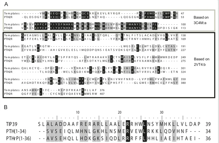
Figure 1A shows the alignment of PTH2R and the templates used in the modelling. The ECD shows 50% residue identity while the TM region shows only 11%.
Abraham-Nordlinget al. BMC Research Notes 2010, 3:270 http://www.biomedcentral.com/1756-0500/3/270
Seven residues form the linker region between the ECD and the TM region. It is noticeable that ECL1 (residues 179 - 205 in the mature chain of PTH2R) is longer than in the template and thereby lacks a structural template. ECL2 (residues 276 - 285) which contains the conserved disulfide in the GPCR super-family is shorter than in b1R, which contain a helical region.
The alignment of the hormones is shown in Figure 1B, and even though the sequence identity is only 15%, sev-eral common themes are evident in the three related hormones TIP39, PTH and PTHrP. The amphipatic nat-ure of the peptides can be traced through the conserved pattern of hydrophobic residues in the C-terminal por-tion of the alignment. Also worth noticing is the con-served basic residues at positions 15 and 22 (using TIP39 numbering).
The structure follows the general fold of the GPCR family (see Figure 2) with the ECD positioned (coloured khaki) above TM helix 1, directing the N-terminus of the hormone into the central cavity of the TM helical bundle (coloured white), making contact with the
extracellular ends of TM helices 3 and 7. The regions without template, the seven residues between the ECD and the TM region and the large ECL1 are coloured blue. The loop sampling of the ECL1 indicated an unor-dered structure with a central helical region flanked by two loops connecting TM helix 2 and 3. A disulfide bond is formed between Cys236 in the top of TM3 and Cys306 in ECL2. As ECL2 is shorter than in the tem-plate no helix can be formed and the loop has an extended conformation.
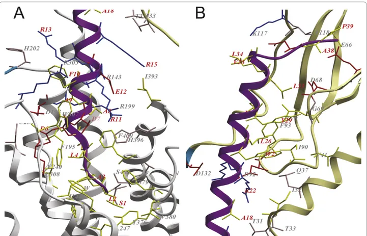
The interactions with TIP39 and the receptor are partly defined by polar interactions of basic, acidic and polar residues (shown in blue, red and pink, respectively, in Figure 3), and partly defined by hydrophobic interac-tions (marked with yellow sticks in Figure 3).
Primary differences between the hormones TIP39 and PTH that bind and at least partially activate PTH2R and PTHrP which lacks binding affinity, include Phe23 in PTHrP which corresponds to a Trp in the agonistic pep-tides, interacting with a mainly hydrophobic environment consisting of residues Ile10, Gln13, Ile14, Val17 and Ile66
Figure 1 Alignments. Black shaded residues are identical and grey shaded residues are similar according to PAM250 similarity matrix. A: Alignment between PTH2R and the templates used for homology modelling; thick border box extracellular domain; thin border box -transmembrane region; no box - intracellular domain. B: Alignment of tuberoinfundibular peptide of 39 residues (TIP39), parathyroid hormone (PTH) and parathyroid hormone related protein (PTHrP). The amphipatic nature of the peptides is visible through the pattern of conserved hydrophobic residues in the C-terminal portion of the alignment.
Figure 2 Structure of predicted complex. The PTH2R model in ribbon representation, regions without template in blue, the extracellular domain in khaki and the TM region in white and the tuberoinfundibular peptide of 39 residues in magenta. TM helix 1 is located to the right and 4 to the far left.
Abraham-Nordlinget al. BMC Research Notes 2010, 3:270 http://www.biomedcentral.com/1756-0500/3/270
in the ECD (see Figure 4B), and His5 corresponding to Asp in TIP39 and Leu in PTH and which is placed in the vicinity of His396 of TM helix 7 (see Figure 4A).
Discussion
We have calculated a model of PTH2R based upon tem-plate structures for the ECD and the TM region. The most reliable region of the model is the ECD, as it is modelled on the related PTH1R. A sequence identity of 50% can reliably give a model of good resolution. The major difference in the alignment of the two parathyroid receptors ECDs originates from that the shorter loop of PTH2R of residue 32 - 37 is unaligned. This allows the modelling software to find a low energy conformation that connects the conserved structure elements prior to, and after the loop. The low sequence identity within the TM region is of course a challenge for the construction of a reliable model. Previous studies of divergent protein families with a common fold show that models can be created for proteins with sequence identity as low as 20% [41]. Within the GPCR super-family, bovine
rhodopsin has 18% sequence identity to the human adrenergicb 2 receptor, while still sharing the same fold with an all-atom RMSD of 3 Å for the TM region [34]. In Vohraet al. 2007 [42], a plant GPCR named GCR1 was used to construct an alignment of the TM region of GPCR class A and B, as it had sequence similarity to both classes. Their published alignment is in accordance with our alignment of the TM regions [42], indicating that the alignment of the TM helices may indeed be correct.
An additional issue with the model is the large ECL1. The loop sampling of ICM can not reliably predict such large loops, although structural characterisation of ECL1 of the PTH1R has shown the existence of a central heli-cal region of the loop that interact with PTH, hinting at that some of the loops characteristics are found by the modelling process [26].
The conformation of TIP39 is likely to be predicted with a fair amount of precision. The template is the parathyroid hormone, where both X-ray [29] and con-strained helical analogues, which are forced to adopt a
Figure 3 Close-up of the binding region between PTH2R and the tuberoinfundibular peptide of 39 residues. The extracellular domain (ECD) in khaki, regions without template in blue, the TM region in white and the tuberoinfundibular peptide of 39 residues (TIP39) in magenta. Residues in the binding interface of TIP39 and the receptor (within 3Å distance of each other) are shown in sticks. Acidic residues are coloured red, polar residues are coloured pink, basic residues are coloured blue and hydrophobic residues are coloured yellow. Residues in TIP39 are labelled in red and residues in the receptor are labelled in gray. A. Interactions of the N-terminus of TIP39 with the TM region. B. Interactions of the C-terminus of TIP39 with the ECD.
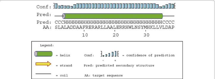
continuous helical conformation, indicate that a slightly twisted single helix is the bioactive conformation of PTH [30]. A previous study of the TIP39 structure using a combined approach based on NMR and molecu-lar dynamics indicates the presence of two helical regions in TIP39, residues 5 - 19 and 27 - 34 with a flexible linker joining them [27]. Similar findings are evi-dent in NMR structures of PTH [28]. However, examin-ing the ensemble of structures generated in the experiments one can find examples of conformations that display a single elongated helical region. In Jin et al. 2000, the authors speculate that the discrepancy between the solution structure and their X-ray structure involves the fact that PTH is a hydrophobic peptide which might need a more hydrophobic environment to adopt its bioactive conformation [29]. A similar situation could be envisioned for TIP39. Another indication that a single helical conformation is plausible is that a PSIPRED secondary structure prediction for TIP39 indi-cates that it is a single helical structure ranging from residue 4 - 35 (Figure 5) [43].
The model of the complex places the N-terminus of the hormone in the central cavity of the TM helical bundle, allowing its residues to be positioned in the TM region and interact directly with the receptor. The importance of the N-terminal part has been highlighted by the fact that deletion of the six first residues makes the remainder a potent antagonist of the PTH1R, while losing much of the strength of the binding to the PTH2R [14,44]. In Figure 3A, one can see that the first residues of TIP39 bind in a hydrophobic site in the cen-tral cavity. The only polar residue in the N-terminus, Ser1 in TIP39 are in position to form a hydrogen bond with Ser403 in TM7. Positions in PTH2R which have been shown to be directly involved in the discrimination between the PTH and PTHrP for PTH2R; Ile244 (inter-acts with Leu2 and Ala3 in TIP39 in the model of the complex) in TM3 and Cys397 (matched to Glu12 in TIP39) and Phe400 (in proximity of Ala5 and Ala8 in TIP39) in TM7 are in close contact with the placed ligand [38]. The major mismatch of the TIP39 residues in the central cavity are Asp6 which is positioned into a
Figure 4 Interactions between PTH2R and the investigated hormones. Tuberoinfundibular peptide of 39 residues in magenta, parathyroid hormone in pink and parathyroid related protein in yellow. A. The central interaction of Asp7, Ile5, His5 with His396 of TM helix 7 of the receptor. B. Trp25, 23 and Phe23 is shown with interacting residues Ile34, Gln37, Ile38, Val41 and Ile90 in the ECD of the receptor. Abraham-Nordlinget al. BMC Research Notes 2010, 3:270
http://www.biomedcentral.com/1756-0500/3/270
cavity between TM helix 3 and 4, its closest match is Asp198 in an otherwise hydrophobic environment. His396 in TM7 are a more suitable interaction partner for both Asp6 and Asp7, which could be accomplished by a slight rotation of the hormone. A further interac-tion is the salt bridge formed by Glu12 of TIP39 and Arg143 and Arg199 of the receptor.
The interaction of the hormone with the ECD (see Figure 3B) is characterised by hydrophobic forces of Trp25, Leu26, Tyr29, Met30 and Leu 33 that interact with a continuous surface made up by Val41, Leu70, Ile90 and Phe93 in the ECD. These interactions are sup-ported by those of Arg22 and Arg23 in TIP39 with Asp92 and Asp132 in the receptor.
The modelling of the complexes of related peptides hormones, TIP39, PTH and PTHrP with PTH2R show that the mutationally verified importance of the Trp at position 25 in TIP39 and at position 23 in PTH in bind-ing to the receptor, while the smaller Phe in PTHrP can not fill the binding site (see Figure 1B and 4B). Further it can be noted that His5 of PTHrP is placed in contact with His396 of the receptor, mutation of that position to Ile, the corresponding residue of PTH, restores activ-ity in combination of mutation of Phe23 to Trp [38]. Lingering on the interaction with His396, TIP39 has a Asp at the corresponding site and as it causes the recep-tor to internalize and activates further signalling path-ways than PTH [13], it is tempting to speculate that TIP39s possibility to evoke those signalling responses lies in that acidic residue, unique for it among the related hormones (see Figure 1B and 4A). The hypoth-esis could be tested by the mutation of the Ile5 of PTH to Asp in order to explore if that mutant can fully acti-vate PTH2R. Of the 43 reported orthologues in the ENSEMBL database to PTH2R, all except the one from
the nine-banded armadillo (Dasypus novemcinctus) have a conserved His at that position, while the armadillo has an Arg which still could interact with an acidic residue. Unfortunately, there is no reported TIP39 sequence for Dasypus novemcinctus, but all 23 reported TIP39 ortho-logues have two aspartic acids at positions 6 and 7 [45].
Conclusions
In this work, we present a hypothesis of how TIP39 could bind to PTH2R and how the receptor may discri-minate among the related peptides TIP39, PTH and PTHrP. Furthermore, we propose that the peptide acti-vates the receptor in a single helical conformation as seems to be the case for PTH. We also propose a mutant of PTH, Ile5Asp, which we speculate could act as a full agonist and cause internalisation of the recep-tor. We hope that this study may provide insight into an interesting system that deserves more attention as its full implications are just beginning to be unravelled. The connection to neuropathic pain suggests a new route to modulate the pain sensation via the TIP39 -PTH2R system, which today is an area of great unmet medical need.
Acknowledgements
Financial support from Linköping University is gratefully acknowledged. Author details
1
Division of Surgery, Department of Clinical Sciences, Danderyd Hospital, Karolinska Institutet, Stockholm, Sweden.2IFM Bioinformatics, Linköping
University, S-581 83 Linköping, Sweden. Authors’ contributions
MAN participated in the design of the study, sequence alignment and draft of the manuscript. BP participated in the design of the study and revision of the manuscript. EN participated in the design of the study, model building and draft of the manuscript. All authors read and approved the final manuscript.
Figure 5 Secondary structure prediction for TIP39. Prediction of the secondary structure of tuberoinfundibular peptide of 39 residues by PSIPRED.
Competing interests
Erik Nordling is, besides being affiliated with IFM Bioinformatics, Linköping University, also employed by Biovitrum AB, which has no interest in the current article. The other authors declare that they have no competing interests.
Received: 4 May 2010 Accepted: 27 October 2010 Published: 27 October 2010
References
1. Cherezov V, Rosenbaum DM, Hanson MA, Rasmussen SG, Thian FS, Kobilka TS, Choi HJ, Kuhn P, Weis WI, Kobilka BK, Stevens RC: High-resolution crystal structure of an engineered human beta2-adrenergic G protein-coupled receptor. Science 2007, 318:1258-1265.
2. Warne T, Serrano-Vega MJ, Baker JG, Moukhametzianov R, Edwards PC, Henderson R, Leslie AG, Tate CG, Schertler GF: Structure of a beta1-adrenergic G-protein-coupled receptor. Nature 2008, 454:486-491. 3. Jaakola VP, Griffith MT, Hanson MA, Cherezov V, Chien EY, Lane JR,
Ijzerman AP, Stevens RC: The 2.6 angstrom crystal structure of a human A2A adenosine receptor bound to an antagonist. Science 2008, 322:1211-1217.
4. Murakami M, Kouyama T: Crystal structure of squid rhodopsin. Nature 2008, 453:363-367.
5. Shimamura T, Hiraki K, Takahashi N, Hori T, Ago H, Masuda K, Takio K, Ishiguro M, Miyano M: Crystal structure of squid rhodopsin with intracellularly extended cytoplasmic region. J Biol Chem 2008, 283:17753-17756.
6. Runge S, Thogersen H, Madsen K, Lau J, Rudolph R: Crystal structure of the ligand-bound glucagon-like peptide-1 receptor extracellular domain. J Biol Chem 2008, 283:11340-11347.
7. Pioszak AA, Xu HE: Molecular recognition of parathyroid hormone by its G protein-coupled receptor. Proc Natl Acad Sci USA 2008, 105:5034-5039. 8. Pioszak AA, Parker NR, Suino-Powell K, Xu HE: Molecular recognition of
corticotropin releasing factor by its G protein-coupled receptor CRFR1. J Biol Chem 2008, 283:32900-32912.
9. Pioszak AA, Parker NR, Gardella TJ, Xu HE: Structural basis for parathyroid hormone-related protein binding to the parathyroid hormone receptor and design of conformation-selective peptides. J Biol Chem 2009, 284:28382-28391.
10. Usdin TB, Gruber C, Bonner TI: Identification and functional expression of a receptor selectively recognizing parathyroid hormone, the PTH2 receptor. J Biol Chem 1995, 270:15455-15458.
11. Hansen IA, Jakob O, Wortmann S, Arzberger T, Allolio B, Blind E: Characterization of the human and mouse genes encoding the tuberoinfundibular peptide of 39 residues, a ligand of the parathyroid hormone receptor family. J Endocrinol 2002, 174:95-102.
12. John MR, Arai M, Rubin DA, Jonsson KB, Juppner H: Identification and characterization of the murine and human gene encoding the tuberoinfundibular peptide of 39 residues. Endocrinology 2002, 143:1047-1057.
13. Bisello A, Manen D, Pierroz DD, Usdin TB, Rizzoli R, Ferrari SL: Agonist-specific regulation of parathyroid hormone (PTH) receptor type 2 activity: structural and functional analysis of PTH- and
tuberoinfundibular peptide (TIP) 39-stimulated desensitization and internalization. Mol Endocrinol 2004, 18:1486-1498.
14. Hoare SR, Usdin TB: Tuberoinfundibular peptide (7-39) [TIP(7-39)], a novel, selective, high-affinity antagonist for the parathyroid hormone-1 receptor with no detectable agonist activity. J Pharmacol Exp Ther 2000, 295:761-770.
15. Usdin TB, Hoare SR, Wang T, Mezey E, Kowalak JA: TIP39: a new neuropeptide and PTH2-receptor agonist from hypothalamus. Nat Neurosci 1999, 2:941-943.
16. Usdin TB, Hilton J, Vertesi T, Harta G, Segre G, Mezey E: Distribution of the parathyroid hormone 2 receptor in rat: immunolocalization reveals expression by several endocrine cells. Endocrinology 1999, 140:3363-3371. 17. Ross G, Engel P, Abdallah Y, Kummer W, Schluter KD: Tuberoinfundibular
peptide of 39 residues: a new mediator of cardiac function via nitric oxide production in the rat heart. Endocrinology 2005, 146:2221-2228. 18. Ross G, Heinemann MP, Schluter KD: Vasodilatory effect of
tuberoinfundibular peptide (TIP39): requirement of receptor
desensitization and its beneficial effect in the post-ischemic heart. Peptides 2007, 28:878-886.
19. Usdin TB, Paciga M, Riordan T, Kuo J, Parmelee A, Petukova G, Camerini-Otero RD, Mezey E: Tuberoinfundibular Peptide of 39 residues is required for germ cell development. Endocrinology 2008, 149:4292-4300.
20. Fegley DB, Holmes A, Riordan T, Faber CA, Weiss JR, Ma S, Batkai S, Pacher P, Dobolyi A, Murphy A, et al: Increased fear and stress-related anxiety-like behavior in mice lacking TIP39. Genes Brain Behav 2008. 21. Palkovits M, Helfferich F, Dobolyi A, Usdin TB: Acoustic stress activates
tuberoinfundibular peptide of 39 residues neurons in the rat brain. Brain Struct Funct 2009, 214:15-23.
22. Matsumoto M, Kondo S, Usdin TB, Ueda H: Parathyroid hormone 2 receptor is a functional marker of nociceptive myelinated fibers responsible for neuropathic pain. J Neurochem 2010, 112:521-530. 23. Harmar AJ: Family-B G-protein-coupled receptors. Genome Biol 2001, 2:
REVIEWS3013.
24. Palczewski K, Kumasaka T, Hori T, Behnke CA, Motoshima H, Fox BA, Le Trong I, Teller DC, Okada T, Stenkamp RE, et al: Crystal structure of rhodopsin: A G protein-coupled receptor. Science 2000, 289:739-745. 25. Conner M, Hicks MR, Dafforn T, Knowles TJ, Ludwig C, Staddon S,
Overduin M, Gunther UL, Thome J, Wheatley M, et al: Functional and biophysical analysis of the C-terminus of the CGRP-receptor; a family B GPCR. Biochemistry 2008, 47:8434-8444.
26. Piserchio A, Bisello A, Rosenblatt M, Chorev M, Mierke DF: Characterization of parathyroid hormone/receptor interactions: structure of the first extracellular loop. Biochemistry 2000, 39:8153-8160.
27. Piserchio A, Usdin T, Mierke DF: Structure of tuberoinfundibular peptide of 39 residues. J Biol Chem 2000, 275:27284-27290.
28. Marx UC, Adermann K, Bayer P, Forssmann WG, Rosch P: Solution structures of human parathyroid hormone fragments hPTH(1-34) and hPTH(1-39) and bovine parathyroid hormone fragment bPTH(1-37). Biochem Biophys Res Commun 2000, 267:213-220.
29. Jin L, Briggs SL, Chandrasekhar S, Chirgadze NY, Clawson DK, Schevitz RW, Smiley DL, Tashjian AH, Zhang F: Crystal structure of human parathyroid hormone 1-34 at 0.9-A resolution. J Biol Chem 2000, 275:27238-27244. 30. Condon SM, Morize I, Darnbrough S, Burns CJ, Miller BE, Uhl J, Burke K, Jariwala N, Locke K, Krolikowski PH, et al: The Bioactive Conformation of Human Parathyroid Hormone. Structural Evidence for the Extended Helix Postulate. J Am Chem Soc 2000, 122:3007-3014.
31. Weidler M, Marx UC, Seidel G, Schafer W, Hoffmann E, Esswein A, Rosch P: The structure of human parathyroid hormone-related protein(1-34) in near-physiological solution. FEBS Lett 1999, 444:239-244.
32. Apweiler R, Bairoch A, Wu CH, Barker WC, Boeckmann B, Ferro S, Gasteiger E, Huang H, Lopez R, Magrane M, et al: UniProt: the Universal Protein knowledgebase. Nucleic Acids Res 2004, 32:D115-119.
33. Edgar RC: MUSCLE: a multiple sequence alignment method with reduced time and space complexity. BMC Bioinformatics 2004, 5:113.
34. Worth CL, Kleinau G, Krause G: Comparative sequence and structural analyses of G-protein-coupled receptor crystal structures and implications for molecular models. PLoS One 2009, 4:e7011. 35. Forrest LR, Tang CL, Honig B: On the accuracy of homology modeling
and sequence alignment methods applied to membrane proteins. Biophys J 2006, 91:508-517.
36. Abagyan R, Batalov S, Cardozo T, Totrov M, Webber J, Zhou Y: Homology modeling with internal coordinate mechanics: deformation zone mapping and improvements of models via conformational search. Proteins 1997, , Suppl 1: 29-37.
37. Bergwitz C, Jusseaume SA, Luck MD, Juppner H, Gardella TJ: Residues in the membrane-spanning and extracellular loop regions of the parathyroid hormone (PTH)-2 receptor determine signaling selectivity for PTH and PTH-related peptide. J Biol Chem 1997, 272:28861-28868. 38. Turner PR, Mefford S, Bambino T, Nissenson RA: Transmembrane residues
together with the amino terminus limit the response of the parathyroid hormone (PTH) 2 receptor to PTH-related peptide. J Biol Chem 1998, 273:3830-3837.
39. Protein Model DataBase. [http://mi.caspur.it/PMDB/main.php]. 40. Castrignano T, De Meo PD, Cozzetto D, Talamo IG, Tramontano A: The
PMDB Protein Model Database. Nucleic Acids Res 2006, 34:D306-309. 41. Nordling E, Oppermann UC, Jornvall H, Persson B: Human type 10 17
beta-hydroxysteroid dehydrogenase: molecular modelling and substrate docking. J Mol Graph Model 2001, 19:514-520, 591-513.
Abraham-Nordlinget al. BMC Research Notes 2010, 3:270 http://www.biomedcentral.com/1756-0500/3/270
42. Vohra S, Chintapalli SV, Illingworth CJ, Reeves PJ, Mullineaux PM, Clark HS, Dean MK, Upton GJ, Reynolds CA: Computational studies of Family A and Family B GPCRs. Biochem Soc Trans 2007, 35:749-754.
43. McGuffin LJ, Bryson K, Jones DT: The PSIPRED protein structure prediction server. Bioinformatics 2000, 16:404-405.
44. Hoare SR, Clark JA, Usdin TB: Molecular determinants of
tuberoinfundibular peptide of 39 residues (TIP39) selectivity for the parathyroid hormone-2 (PTH2) receptor. N-terminal truncation of TIP39 reverses PTH2 receptor/PTH1 receptor binding selectivity. J Biol Chem 2000, 275:27274-27283.
45. Hubbard TJ, Aken BL, Ayling S, Ballester B, Beal K, Bragin E, Brent S, Chen Y, Clapham P, Clarke L, et al: Ensembl 2009. Nucleic Acids Res 2009, 37: D690-697.
doi:10.1186/1756-0500-3-270
Cite this article as: Abraham-Nordling et al.: Model of the complex of Parathyroid hormone-2 receptor and Tuberoinfundibular peptide of 39 residues. BMC Research Notes 2010 3:270.
Submit your next manuscript to BioMed Central and take full advantage of:
• Convenient online submission • Thorough peer review
• No space constraints or color figure charges • Immediate publication on acceptance
• Inclusion in PubMed, CAS, Scopus and Google Scholar • Research which is freely available for redistribution Submit your manuscript at