Does chemomechanical
caries removal affect
restoration survival?
A systematic review
Nicole Stolic
Supervisor: Dan Ericson
Bachelor Thesis (15 ECTS)
Malmö University
Programme in Dentistry
Faculty of Odontology
February, 2015
205 06 Malmö
1
ABSTRACT
Aim
This systematic review aimed to summarize randomized controlled trials (RCTs) that evaluate the survival rates of restorations, comparing the Carisolv system to hand excavation and/or the conventional drilling method. The aim was also to collect all data in one place to be used for further research.
Materials & Methods
A database screening of PubMed and the Cochrane library was performed. One reviewer read all titles and abstracts, those considered of interest were fully scrutinized. A total of 9 articles were included and each was evaluated using the GRADE-system. The studies were appraised to have a low level of evidence.
Results
A total 913 teeth received treatment either with Carisolv, hand excavation and/or drilling. The studies evaluated the success rate of dental restorations at follow-ups between 6 months to two years. The results showed no significant difference in success rate of restorations made either by Carisolv, bur or hand excavation.
Conclusions
The results showed as high success rates for caries removal with Carisolv as with other methods, in terms of restoration survival, and that there was no statistically significant difference between the test group and control group. These results are encouraging for caries removal with Carisolv since it also can be used as an alternative treatment for children and patients with dental fear due to the less painful experience and lesser need for local anesthesia. The present studies on this subject are few and have limited evidence, thus underlining the need for more studies on the subject and studies with higher evidence for further research.
2
Swedish abstract Syfte
Denna systematiska översiktsstudie gjordes i syfte att sammanställa
randomiserade kontrollerade studier som jämför fyllningsöverlevnaden där kaviteten preparerats med Carisolv, borr och/eller hand exkavering. Studien gjordes också för att kunna användas till fortsatta studier inom området.
Material & Metod
Databassökning i PubMed och Cochrane gjordes. Alla titlar och
sammanfattningar lästes, de som var relevanta för denna studie lästes sedan i fulltext. Totalt 9 artiklar inkluderades och varje artikel granskades m h a GRADE modellen för att bestämma studiernas evidensstyrka.
Resultat
913 tänder behandlades med en av metoderna. Studierna undersökte
fyllningsöverlevnaden vid olika uppföljningstillfällen, mellan 6 månader och 2 år. Resultaten visar inga statistiskt signifikanta skillnader för fyllningsöverlevnad mellan de olika metoderna.
Slutsats
Resultaten visar ingen statistisk skillnad i fyllningsöverlevnad mellan Carisolv och andra metoder för att avlägsna karies. Resultaten kan stödja karies exkavering med Carisolv, eftersom metoden dessutom kan användas för karies exkavering på barn och tandvårdsrädda patienter då metoden är mindre obehaglig och patienten behöver således inte lokalanestesi lika ofta. Dock är studierna inom detta område få samt att de har låg evidensstyrka vilket gör att det behövs fler studier på området med högre evidensstyrka.
3
INDEX
ABSTRACT
………...11. INTRODUCTION
………...41.1 Dental caries
………...…..41.2 Hand excavation
………...……51.3 Chemomechanical caries removal
………52. AIM
………73. MATERIALS & METHODS
………..………….73.1 Specification of the problem
………...73.2 Data sources
……….……..73.3 Selection criteria
………..…83.4 Quality assessment
………...8Table 1
………...9Figure 1
………...104. RESULTS
………...114.1 Search results and characteristics of studies included in the
review
………..114.2 Filling survival / Success rate
……….…..11Table 2
………...125. DISCUSSION
………..166. CONCLUSION
………..174
1. INTRODUCTION
Dental caries is one of the most common infectious diseases nowadays and there are many different methods for operative caries removal, for example the
atraumatic restorative treatment (ART), hand excavation, the conventional drilling method, air-abrasion, air-polishing and lasers1, 2. Although these methods have improved over recent years, problems associated with rotary instruments have remained unsolved. The conventional method for caries removal has adverse effects on the pulp owing to heat, pressure and vibration, while also removing sound parts of the dentine in addition to the decayed areas3, 4.
Drilling often causes pain and anxiety amongst patients and requires local
anesthesia, Peric et al3 investigated patients attitudes towards drilling and the need of local anesthesia. 33% of the patients did not like drilling and 13% associated drilling with pain, 58% of the patients preferred local anesthesia because of painless and more comfortable treatment.
Patients response to caries removal with Carisolv has been positive. Less pain, discomfort and shorter perceived treatment times has ben reported, compared to conventional drilling. Caries removal with Carisolv also allows treatment without the need for local anesthesia5. This should be considered when choosing treatment for the patient. Dental procedures that patients fear the most are drilling,
application of local anesthetics and tooth extraction.
These unsolved problems have resulted in a higher interest in using pain-free, minimally invasive (tissue preserving) methods for caries removal3. One
minimally invasive method for caries removal is the chemomechanical method, which involves the application of a solution that softens the carious dentine for easier removal using hand instruments5.
1.1 Dental caries
Caries is the result of demineralization of enamel and/or dentine caused by bacterial acids. The disease is not caused by one single factor; it is a result of the interaction between several factors that causes changes in the bacterial ecology in the biofilm located on the tooth surface. In order for bacteria to play a role in caries they must possess certain attributes: (i) the ability to rapidly transport and convert fermentable sugars, (ii) the ability to maintain sugar metabolism during extreme environmental conditions, (iii) the ability to produce extra- and
intracellular polysaccharides6.
Resident oral bacteria use endogenous nutrients (e.g. salivary proteins and glycoproteins) for growth and acid is produced during the metabolism of these proteins. However, the acid production is relatively slow so there is little risk for enamel and/or dentine demineralization. A carbohydrate-rich diet increases the acid production, thus also increasing the growth rate of specific bacterial species that can adapt and survive in acidic environments6. When the pH level in the plaque-fluid drops below 5,5-5,0, due to the acids produced by bacteria in the biofilm, the minerals in the enamel will start to dissolve7. As this process
progresses the dentinal tubules will eventually allow acids to penetrate the tubules and also allow invasion by bacteria. This causes further acid attack and
5
The dentine consists of minerals, water and an organic matrix mostly consisting of a protein called collagen. When the organic matrix has been demineralized, the collagen and other organic matrix components are degraded by host bacterial proteases and other hydrolases. Two different degraded zones can be
distinguished within a carious lesion, there is an inner layer, which is only partially demineralized and can therefore be remineralized because the collagen fibrils are still intact. Also there is an outer layer where the organic matrix is demineralized and the collagen fibrils are partially degraded and can therefore not be remineralized8.
1.2 Hand excavation
Removal of dental caries by hand requires a dental hatchet (enamel) and different spoon excavators (dentine). One method for caries removal by hand is the
atraumatic restorative treatment (ART) technique. This method was initially developed to provide fast, preventive and restorative treatment to patients in low-income countries but has been widely used over recent years9. The ART technique includes hand instruments and adhesive filling materials, generally
glass-ionomers. The technique is based on removing carious dentine using only hand instruments and has been well accepted in many countries, mostly by children because it is a, more or less, pain-free procedure in comparison to the
conventional drilling method10.
In the ART treatment the cavity is widened with a dental hatchet until the enamel is free from visible demineralization and access to the dentine is established, this procedure replaces the bur. Spoon excavators are used to excavate carious dentine from the walls and floor of the cavity9. When all carious dentine is excavated the cavity is restored with glass ionomer. This material is used due to its chemical adhesion, which reduces the need for mechanical retention, and its leakage of fluoride, which prevents secondary caries development10.
1.3 Chemomechanical caries removal
There have been many different solutions for chemomechanical caries removal on the market, for example Caridex which contains N-monochloro-DL-2-aminobyric acid (NMAB)11. These methods was not adopted by many because of prolonged working time and because a large volume of solution was needed due to its low efficacy in terms of caries removal12.
In the late 90’s a new chemomechanical caries removal named Carisolv
(MediTeam) was introduced to the market. The Carisolv system replaced NMAB with three naturally occurring amino acids; lysine, leucin and glutamic acid. The Carisolv system includes two syringes that are mixed together at room
temperature, one syringe holds sodium hypochlorite (0,5%) and the other syringe holds the three amino acids, a gel substance, sodium chloride, sodium hydroxide, a coloring agent (erythrosine) and saline solution, with a pH of 115, 12.
The action of the Carisolv system is initiated when the contents are mixed together, the amino acids bind chlorine thus forming chloramines. The amino acids have different charges, which allows them to bind electrostatically to different proteins in the carious dentine. The formation of these chloramines decreases the reactivity of chlorine but does not alter its chemical function. The result of these actions is further breakdown of degraded collagen found in carious
6
lesions; this is possible due to the open structure of the partially degraded collagen fibers in the outer layer of the carious lesion4.
The mixed gel is applied onto the surface of the carious dentine and left there for 30 seconds to allow the chemical process of the amino acids to soften the carious dentine. The softened dentine is then scraped off using specially designed hand instruments, this process is repeated until the dentine is free from any “tug-back” sensation and caries-free dentin is obtained. The applied gel should be washed away prior to controlling the hardness of the dentine and more gel should be placed prior to further excavation12.
Ericson et al.13 investigated the time needed for complete caries removal with
Carisolv or conventional drilling. The mean time for complete caries removal with Carisolv was 10.4 min (SD=6.1) and the mean time for complete caries removal with bur was 4,4 min (SD=2,2). Even though the working time was stated to be longer for Carisolv approximately 52% of the patients reported their Carisolv session to have been faster than previous drilling sessions, 25%
estimated it to have lasted the same and 11% estimated it to have lasted longer. The study also investigated patients’ pain experience, 54% reported having felt no pain and 41% reported a little pain.
The need for local anesthesia is also of interest when choosing a method for caries removal. Fure et al.14 compared the need for local anesthesia in patients treated with either Carisolv or the conventional drilling method. Four of the 34 patients in the Carisolv group asked for anesthesia, compared to 6 of 26 patients in the drilling group. Three other studies 3,13,15 also showed that patients treated with Carisolv had less need for local anesthesia. Peric et al.3 showed a statistically significant difference between the two groups, where 7 of 60 patients treated with Carisolv asked for local anesthesia and a total of 36 of 60 patients treated with the conventional drilling method felt a need for local anesthesia.
Another aspect to consider when choosing method is the complete caries removal rate (CCR). Four studies 3,13,14,15 described CCR by presenting the number of caries free teeth. All studies show mixed results but there is no statistically
significant difference between the two groups. There has also been discussed what effect Carisolv gel has on exposed pulpal tissue, Hamama et al.16 presented studies showing that Carisolv has no adverse effects on exposed pulpal tissue and some studies even mentioned that it may have a similar effect on pulpal tissue as Ca(OH)2 pulp capping materials. Even though Carisolv is shown to cause a
minimal superficial necrosis of the pulpal tissues it also has bactericidal and haemostatic effects on exposed pulpal tissue, which comes from the high pH of 11, thus promoting tissue repair.
Though the Carisolv system is considered to be a minimally invasive method and a well-accepted method for caries removal5, certain aspects regarding survival rate of restorations prepared with Carisolv must also be considered, such as dentine adhesion of dental fillings. As the system contains amino acids, sodium
hypochlorite and has a high pH, it might interfere with dentine bonding mechanisms if any remnants were left. Haak R et al.17 studied the shear bond strength and fracture patterns within the adhesive layer when using Carisolv or the conventional method (drilling) for caries removal. The study demonstrated that shear bond strength and fractures of dental fillings did not differ significantly
7
between the two methods which means that the Carisolv system has no adverse effects on bonding strength to caries-affected dentine.
Many studies investigate the survival rates for restorations treated with the
chemomechanical method. In one study, Barata et al.18 evaluated the survival rate of glass ionomer restorations comparing the Carisolv system and ART. The study showed that Carisolv had 100% success rate for restorations at the 12-month follow-up and the ART group had 97,6% success rate.
More recently other chemomechanical caries removal methods has been
introduced as Papacárie and Biosolv, but the literature is rather scarce on clinical information of those systems16.
2. Aim
This systematic review aimed to summarize randomized controlled trials (RCTs) that evaluate the survival rates of restorations, comparing the Carisolv system to the ART-technique and/or the conventional method. The aim was also to collect all data in one place to be used for further research.
3. MATERIALS AND METHODS
3.1 Specification of the problem
The evaluation of the problem was made according to PICO19;
“Patient/Population/Problem”, “Intervention”, “Comparison/Control” and “Outcome”. The following question defines the problem: In children and adolescents with carious teeth, will caries excavation with Carisolv affect the survival rates of restorations, compared to conventional caries removal and/or hand excavation?
The intervention evaluated in this review was chemomechanical caries removal with Carisolv. Studies that did not include Carisolv were excluded. Methods for caries excavation assessed as comparison/control interventions were hand excavation and/or drilling.
3.2 Data sources
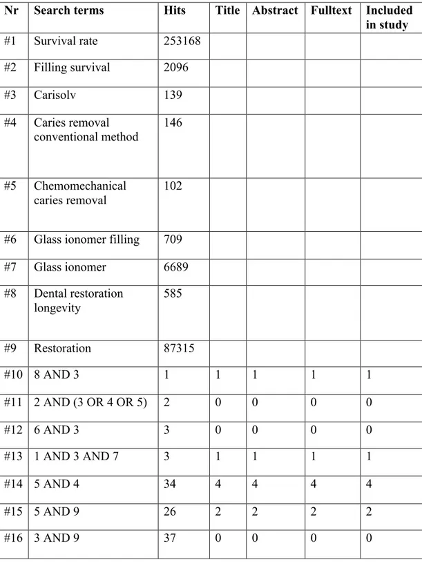
Electronic database screening was performed in PubMed and the Cochrane library for full articles published in English, though all studies included were found in PubMed. Duplicates were excluded manually. Combinations of search terms can be found in table 1.
The screening was performed between February and May 2014. One reviewer read all available titles, titles that met the inclusion criteria was considered
eligible for the next level of the screening process. Abstracts of articles eligible on the title level was read and controlled against the inclusion criteria, abstracts considered eligible were then obtained in full text. The search and study selection, including criteria for inclusion, can be found in figure 1.
Case reports and articles published in other languages than English were excluded. Reference lists of included studies were then hand searched for titles
8
containing one of these phrases:‘chemomechanical caries removal’, ‘Carisolv’, ‘filling survival’. Articles on reference lists considered of interest for this review were then read in full text.
The initial database search in PubMed yielded in 106 articles. Duplicates were excluded and the titles were screened and controlled against the inclusion criteria, this resulted in 8 articles considered relevant for screening on the abstract level. All 8 articles met the inclusion criteria and were read in full text, all 8 articles were included in this review. The reference screening of obtained articles resulted in one additional article, the total number of articles included was 9.
3.3 Selection criteria
Included studies in this review was prospective, randomized controlled trials (RCT). The studies evaluated the survival rates of dental restorations for cavities (primary or secondary caries) prepared with Carisolv, bur and/or ART and compared them.
The studies have a step-by-step description of different working-methods for caries removal and also have described criteria for complete caries removal. The studies included were also in-vivo studies with follow-ups after between three to 12 months. The materials used were only composite or glass ionomer.
3.4 Quality assessment
Each record was thoroughly evaluated using the GRADE-system (Grade Working Group) 20 to sense the methodological reliability of the articles. The GRADE-system consists of a graded scale, high – moderate – low - very low, that describes the scientific evidence. A grade can be lowered or raised for certain reasons, higher grades indicate better reporting. For a study to be considered having high (++++) scientific evidence it should be a randomized controlled trial (RCT). The randomization process, inclusion/exclusion criteria and reasons for dropouts should be described. The sample size should be sufficiently large to detect the effect of the treatment. Operators and evaluators should be blinded to the
intervention and control group. The outcomes should be thoroughly described, the results should be reported in an adequate way and there should be no conflicts of interest.
The study was considered having low scientific evidence if the following criteria were met: The randomization process was not fully described, the study lacked adequate inclusion/exclusion criteria and the total number of dropouts was not described. If less than 80% of the patients included in a study received less than 80% of the treatment the study was considered having low compliance, thus also having lower scientific evidence. If the study was an open investigation it was also considered having lower scientific evidence. The study had a follow up after less than one year.
A study was considered having moderate level of scientific evidence if it was lacking any of the criteria set for high scientific evidence, but without any shortcomings from associated with low level of scientific evidence.
9
Table 1. Combinations of search terms
Nr Search terms Hits Title Abstract Fulltext Included in study #1 Survival rate 253168 #2 Filling survival 2096 #3 Carisolv 139 #4 Caries removal conventional method 146 #5 Chemomechanical caries removal 102
#6 Glass ionomer filling 709
#7 Glass ionomer 6689 #8 Dental restoration longevity 585 #9 Restoration 87315 #10 8 AND 3 1 1 1 1 1 #11 2 AND (3 OR 4 OR 5) 2 0 0 0 0 #12 6 AND 3 3 0 0 0 0 #13 1 AND 3 AND 7 3 1 1 1 1 #14 5 AND 4 34 4 4 4 4 #15 5 AND 9 26 2 2 2 2 #16 3 AND 9 37 0 0 0 0
10
Fig. 1. Flow-chart of search and study selection.
Database screening n = 106
Duplicates excluded
Title and abstract n = 8 Fulltext n = 8
Articles included in
study
n = 9 Additional articles n = 1 Inclusion criteria: - Survival rate - Carisolv Inclusion criteria: - RCT - Follow-up - In-vivo - Composite/glass ionomer materials11
4. RESULTS
4.1 Search results and characteristics of studies included in the review
Table 2 shows the results of the literature search. Six articles were fully reviewed and presented. The articles screened compared the success rate for dental fillings at follow-up appointments between Carisolv, ART and drilling methods. The studies were published between 2005 and 2009; no suitable studies were found before 2005 or after 2009. A total of 788 patients were examined and 913 teeth received treatment either with Carisolv, ART and/or drilling. All studies were randomized and quality assessed by the GRADE scale.
4.2 Filling survival / Success rate
All studies evaluated the success rate of dental restorations at follow-ups between 6 months to two years. The articles included in this study showed no significant difference in success rate of restorations made either by Carisolv, bur or the ART technique.
Three studies9, 15, 21 showed that restorations prepared with bur had a lower
restoration failure rate than restorations prepared with Carisolv. Topaloglu et al.9 showed that the predominant reason for restoration failure was total loss of restoration (73%) followed by marginal integrity (15%) and secondary caries (12%).
Roeveld et al.21 compared Carisolv with both bur and ART. These results also showed that bur has the highest success rate, both after 7 months and after one year. At the first evaluation the success rates were: 50,0% for bur, 38,2% for ART and 34,6% for Carisolv. At the second evaluation the success rates for the groups were 42,3%, 29,8% and 27,6% respectively.
A study made by Bergmann et al.15 evaluated the proportion of lost/fractured restorations and recurrent caries in restorations prepared with either Carisolv or bur after 6 months. At the follow-up they found two cases of secondary caries in the Carisolv group and one case of secondary caries in the bur group. They also recorded one lost/fractured restoration in both groups.
Peric et al.3 and Barata et al.18 both showed the opposite, that Carisolv had higher success rate than bur, ART and traditional hand excavation. In the study made by Peric et al. the success rate for Carisolv was 95% and 90% for bur. They found that three restorations were lost in the Carisolv group and two restorations were lost in the bur group. Also four restorations were with secondary caries. The study made by Barata et al. shows similar success rates to what Peric et al. showed. After 12 months the success rate for Carisolv was 100% and 97,6% for ART. The last article included in this study, by Kirzioglu et al.1, evaluated restoration failures after 3, 6, 9 and 12 months. Criteria for restoration failure were failure due to anatomic form, failure due to marginal adaption and failure due to
secondary caries. The study showed that Carisolv has lower restoration failures in all three groups than hand excavation.
12
Table 2. Details of, and findings from, included studies
Study Method Participants Intervention
Carisolv Intervention Bur/ ART/ hand exc. Follow-up Filling survival GRADE Bergmann et al. 2005 RCT, prospective, open investigation 92 deciduous teeth from 46 consecutive patients. Each patient presented at least two active dentine caries lesions after the routine examination. 46 46 Success criteria: No recurrent caries and intact remaining restorations No statistically significant difference between the two groups in the evaluation of filling survival after 6 months + (low number of patients, no description of criteria for exclusion or number of excluded patients, open investigation) Roeleveld et al 2006 Prospective, RCT 217 children between 6-7 years old. Each patient presented one class II cavity in a primary molar with an untreated tooth adjacent to the cavity. No pulp exposure 68 77 ART 72 Bur Success criteria: Intact remaining restorations, no secondary caries, no defect at the margin or wear of the surface, and no gaps No statistically significant difference between the three groups in terms of success score at the follow-ups + (Low number of patients, no description of the process for randomization, no exclusion criteria or number of excluded patients, open investigation)
13 present. Topaloglu et al. 2008 Prospective, RCT with parallel study design 327 children 53% male 47% female. 568 restorations were made. One class II dentine lesion in a primary molar. No pulp involvement. 163 165 Success criteria: Intact remaining restoration, no marginal integrity and no secondary caries No statistically significant difference in survival percentages of the restorations between the two groups over the 2-year period. Also no statistically significant difference regarding secondary caries development + (Low number of patients, no description of exclusion criteria or number of excluded patients, high number of dropouts) Peric et al.
2009 Prospecitve,RCT. Blinded 120 patients Each patient presented one primary caries lesion with no development disorders, pulp pathology or pulp 60 60 Success criteria: Intact restoration, no loose or missing restoration, no secondary caries and no signs of pulp pathology. No statistically significant difference between the two groups regarding success rates of the restorations + (Low number of patients, no description of number of patients excluded, no description of compliance)
14 necrosis. All patients were consecutive patients. Kirzioglu et
al. 2007 Prospective, RCT, open investigation 28 patients, 56 lesions. All patients presented contralateral primary molars with an occlusal primary decay, positive results on sens-test. 28 28 Success criteria: Mismatch in color, shade, or translucency. Cavosurface marginal discoloration, discontinuous anatomic form, marginal adaptation, secondary caries No statistically significant difference between the two groups regarding cavosurface marginal discoloration, or the marginal adaption restored with Dyract AP. No statistically significant difference was found in the evaluation of secondary caries. The evaluation of the anatomic form showed no statistically + (Low number of patients, open investigation, no description of reason for dropout, no description of randomization)
15 significant difference. Barata et al. 2008 RCT, prospective pilot study. Split-mouth design. Blinded 50 pregnant women, 50 pairs of teeth. Each patient presented two dentinal caries lesions with no pulp exposure, no history of pain and no presence of swelling or fistula. 34 molars 7 premolars 9 incisors Total: 50 32 molars 5 premolars 13 incisors Total: 50 Success criteria: Present restoration in good condition, or with a slight marginal defect or slight wear with no need for repair. No statistically significant difference regarding success rates of the restorations between the two groups was found + (Low number of patients)
16
5. DISCUSSION
Studies have evaluated the success and failure rates for restorations prepared with Carisolv compared to bur excavation and/or hand excavation, but with mixed results and conclusions, thus this systematic review was performed in an attempt to gather information and gain further insight on the subject. The importance of knowing and understanding the affect of chemomechanical caries removal on survival rates of restorations is great, thus this study can be used for further research.
The results from studies included in this review show no statistically significant difference in the test/control group, regarding the survival rates of restorations. However, all articles showed risk of bias. The main shortcomings in papers were: (i) randomization procedures were not always explained, which lowers the articles level of evidence on the GRADE scale. (ii) Co-investigators were not always blinded, (iii) in some studies there were more than one operator, this also lowers the reliability of the article on the GRADE-scale thus increasing risk for bias. All studies tried to reduce this risk by calibrating the operators.
The number of patients included in the studies is also a possible bias, the lowest amount of patients were 28 and the highest 327 (a total of 788 patients). The number of teeth examined (total of 913) can also be considered low, thus possible bias. This amount is not sufficient thus evaluations including a higher number of patients would be convenient. Some studies included in this article did not have inclusion criteria regarding cavity size after caries excavation and therefore evaluated the success rates of restorations made in different sizes. Also, carious lesions were allowed on all sites on the teeth, this can lead to possible bias because larger and deeper cavities can lead to, for example, pulp exposure, damaged pulp due to heat and/or pressure and decreased retention in dentine and enamel which all leads to restoration failure. Difficulties when applying the restoration material can result in marginal adaptation, cervical gaps and/or
marginal discoloration, which also lead to restoration failure. Many small cavities are easier to restore and restorations made on anterior teeth can therefore give a higher success rate at follow-ups.
Follow-up times varied in all studies, this is a problem in terms of restoration longevity because restoration failures may decrease or increase differently over time.
Differences in working methods regarding filling procedures and materials used, among the studies can result in misleading outcomes. Some studies used glass-ionomer as restoration material and one study2 used composite, glass-ionomer or amalgam. Different materials have different properties and behave differently in the patient, it would therefore be of clinical relevance to evaluate restoration survival among studies using the same restoration material.
It would also be of clinical relevance to evaluate survival rates of restorations over a longer time period, since the risk of secondary caries increases with the lifetime of restorations. All studies included caries as a reason for failure, this can affect the outcome because the diagnosis is dependent on the judgment of the evaluators, who were not calibrated among the different studies. According to Opdam et al.22 and Demarco et al.23 caries risk plays a dominant role in restoration survival. High
17
or medium caries risk is associated with a 2 to 3 times higher risk of restoration failure, this risk factor is more important than material factors for restoration survival.
One shortcoming of this article is that there was only one reviewer reading all titles, abstracts and full texts. This could mean that some relevant studies were missed, it is therefore of convenience to be at least two reviewers when a review article is written. Another problem is the low amount of articles included in this review due to the low amount of articles published on this subject. All included studies were assessed to have a low level of scientific evidence for reasons described above and in table 2, underlining the need for more studies on this subject with higher scientific evidence.
This systematic review indicates that caries removal with Carisolv, in comparison to bur and ART, do not differ for the outcome restoration survival. Carisolv can reduce the need for local anesthesia because it is less painful, this is of importance when treating fearful patients and children even though the treatment time is prolonged.
6. CONCLUSION
The results showed as high success rates for caries removal with Carisolv as with other methods, in terms of restoration survival, and that there was no statistically significant difference between the test group and control group. These results are encouraging for caries removal with Carisolv since it also can be used as an alternative treatment for children and patients with dental fear due to the less painfull experience and lesser need for local anesthesia. The present studies on this subject are few and have limited evidence, thus underlining the need for more studies on the subject and studies with higher evidence for further research.
18
7. References
1. Kirzioglu Z, Gurbuz T, Yilmaz Y. Clinical evaluation of chemomechanical and mechanical caries removal: Status of the restorations at 3, 6, 9 and 12 months. Clin Oral Investig. 2007; 11: 69-76.
2. Li R, Zhao Y, Ye L. How to make choice of the carious removal methods, Carisolv or traditional drilling? A meta-analysis. J Oral Rehabil. 2014; 4: 432-42. 3. Peric T, Markovic D, Petrovic B. Clinical evaluation of a chemomechanical method for caries removal in children and adolescents. Acta Odontol Scand. 2009; 67: 277-83.
4. Yazici AR, Atílla P, Özgünaltay G, Müftüoglu S. In vitro comparison of the efficacy of Carisolv and conventional rotary instrument in caries removal. J Oral Rehabil. 2003; 30: 1177-82.
5. Morrow LA, Hassall DC, Watts DC, Wilson NH. A chemomechanical method for caries removal. Dent Update. 2000; 27: 398-401.
6. Fejerskov O, Kidd E. 2008. Dental caries, the disease and its clinical management. Second edition. Blackwell Munksgaard Ltd.
7. Bradshaw DJ, Lynch RJ. Diet and the microbial aetiology of dental caries: new paradigms. Int Dent J. 2013; 63 Suppl 2: 64-72.
8. Beeley JA, Yip HK, Stevenson AG. Chemomechanical caries removal: a review of the techniques and latest developments. Br Dent J. 2000; 22; 188: 427-30.
9. Topaloglu-Ak A, Eden E, Frencken JE, Oncag O. Two years survival rate of class II composite resin restorations prepared by ART with and without a
chemomechanical caries removal gel in primary molars. Clin Oral Investig. 2009; 13: 325-32.
10. Frencken JE, Pilot T, Songpaisan Y, Phantumvanit P. Autramatic restorative treatment (ART): Rationale, Technique, and Development. J Public Health Dent. 1996; 56(3 Spec No): 135-40
11. Maragakis GM, Hahn P, Hellwig E. Chemomechanical caries removal: a comprehensive review of the literature. Int Dent J. 2001; 51: 291-9
12. Ziskind D, Kupietzky A, Beyth N. First-choice treatment alternatives for caries removal using the chemomechanical method. Quintessence Int. 2005; 36: 9-14.
13. Ericson D, Zimmerman M, Raber H, Götrick B, Bornstein R, Thorell J. Clinical evaluation of efficacy and safety of a new method for chemomechanical removal of caries. Caries Res. 1999; 33: 171-7.
19
14. Fure S, Lingström P, Birkhed D. Evaluation of carisolv for the
chemo-mechanical removal of primary root caries in vivo. Caries Res. 2000; 34: 275-80. 15. Bergmann J, Leitao J, Kultje C, Bergmann D, Clode MJ. Removing dentine caries in deciduous teeth with Carisolv: A randomised, Controlled, Prospective study with six-month follow-up, comparing chemomechanical treatment with drilling. Oral Health Prev Dent. 2005; 3: 105-11.
16. Hamama H, Yiu C, Burrow M. Current update of chemomechanical caries removal methods. Aust Dent J. 2014; 59: 446-56.
17. Haak R, Wicht MJ, Noack MJ. Does chemomechanical caries removal affect dentine adhesion? Eur J Oral Sci 2000; 108: 449-455.
18. Barata TJ, Bresciani E, Mattos MC, Lauris JR, Ericson D, Navarro MF. Comparison of two minimally invasive methods on the longevity of glass ionomer cement restorations: Short-term results of a pilot study. J Appl Oral Sci. 2008; 16: 155-60.
19.Springshare. PICO or PICo? 2014. Available at:
http://libguides.library.curtin.edu.au/content.php?pid=395178&sid=3578094. Assessed 29 January, 2015.
20. GRADE working group. List of GRADE working group publications and grants. 2011. Available at:
http://www.gradeworkinggroup.org/publications/index.htm. Assessed 20 February, 2014.
21. Roeleveld AC, van Amerongen WE, Madari GJ. Influence of residual caries and cervical gaps on the survival rate of class II glass ionomer restorations. Eur Arch Paediatr Dent. 2006; 7: 85-91.
22. Opdam NJ, Bronkhorst EM, Loomans BA, Huysmans MC. 12-year survival of composite vs. amalgam restorations. J Dent Res. 2010; 89: 1063-7.
23. Demarco FF, Correa MB, Cenci MS, Moraes RR, Opdam NJ. Longevity of posterior composite restorations: not only a matter of materials. Dent Mater. 2012; 28: 87-101.