Diagnostic Accuracy of
Radiological Methods
Regarding Root Resorption of
Teeth Neighbouring Impacted
Maxillary Canines
- A systematic review
Lisa Granberg
Julia Stevens
Supervisor: Anna Senneby, Helena Christell, Kristina Hellén-Halme
Department of radiology
Master Thesis in Odontology 30 hp
Malmö University
Dentistry Program
Faculty of Odontology
Abstract
Aim: The aim of this study was to systematically review the scientific literature in diagnostic
accuracy of radiographic methods regarding root resorption of teeth neighbouring impacted maxillary canines.
Material and method: In order to get a systematic and transparent approach, the PRISMA
statement and CRD’s guidance was followed. Four databases were searched, PubMed, Cochrane, Web of Science and Scopus by the two authors. The included studies were evaluated by the QUADAS-2 tool for quality assessment.
Result: The search resulted in 2175 studies. Five studies were included in this review and
all of them reported diagnostic accuracy. Intraoral periapical radiograph, panorama, CT and CBCT was used as index test among the studies. Extracted teeth, CT and CBCT was used as reference test. Low risk of bias was shown in approximately half of the domains for risk of bias and for all domains regarding applicability. The accuracy of three-dimensional ranged from 0.85-1.0 and the accuracy of two-dimensional ranged from 0.45-0.78.
Conclusions: According to this systematic review, three-dimensional imaging showed the
highest sensitivity and specificity regarding root resorptions caused by impacted maxillary canine. CBCT showed higher accuracy than two-dimensional imaging. Though, CBCT has higher accuracy it also has a higher radiation dose and might not always be beneficial regarding treatment planning. Therefore, CBCT should be used with respect and not routinely.
Sammanfattning
Syfte: Syftet med denna studie var att systematiskt granska den vetenskapliga litteraturen
gällande diagnostisk tillförlitlighet för röntgenmetoder avseende rotresorption på granntänder till överkäkshörntanden.
Material och metod: För att uppnå en systematisk och ett transparant tillvägagångssätt
användes PRISMA och CRDs handbok. Sökningen gjordes i fyra olika databaser, PubMed, Cochrane, Web of Science och Scopus av de två författarna. De inkluderade studierna kvalitetsgranskades enligt QUADAS-2 dokument.
Resultat: Sökningen resulterade i 2175 studier. Fem studier inkluderades i denna
systematiska litteraturöversikt och alla rapporterade diagnostisk tillförlitlighet. Intraoral periapikal röntgen, panorama, CT och CBCT var olika indextest i studierna. Extraherade tänder, CT och CBCT användes som referensstandard. Låg risk för bias visades i ungefär hälften av domänerna gällande risk för bias och för alla domäner gällande applicerbarhet. Tillförlitligheten hos tredimensionell röntgen varierade mellan 0,85-1,0 och tillförlitligheten hos tvådimensionell röntgen varierade från 0,45 till 0,78.
Konklusion: Denna systematiska litteraturöversikt visade att tredimensionell röntgen hade
högst sensitivitet och specificitet gällande rotresorption på granntänder till överkäkshörntänder. CBCT visade en högre tillförlitlighet än tvådimensionell röntgen. Även om CBCT har högre tillförlitlighet så har den också en högre strålningsdos och är inte alltid fördelaktig gällande terapiplanering. Därför bör CBCT användas med respekt och inte rutinmässigt.
Contents
1. Introduction ... 4
1.1 Background ... 4
1.2 Aim ... 9
1.3 Clinical relevance ... 9
2. Materials and methods ... 9
2.1 Problem specification ... 9
2.2 PIRO ... 9
2.3 Literature search ... 10
2.4 Publication retrieval ... 10
2.5 Data extraction and quality assessment ... 11
3. Results ... 11
3.1 Literature identification ... 11
3.2 Study characteristics ... 11
3.3 Assessment of methodological quality ... 12
4. Discussion ... 16
4.1 Methodological considerations ... 16
4.2 Discussion of results ... 17
5. Conclusion ... 18
6. Reference ... 19
Appendix 1 ... 24
Appendix 2 ... 25
Appendix 3 ... 26
1. Introduction
1.1 Background Diagnostic accuracy
Patient history, symptoms, clinical examination and different diagnostic test are valuable in diagnosing. In dental care the two most commonly used diagnostic tests are visual-tactile examination and imaging procedures (1). Accurate diagnosis is not only important for the patients’ treatment and prognosis, but also for professional communication and clinical research (2). Diagnosis can be difficult and the dentist sometimes struggle with errors in clinical data, variations of interpretation and uncertainties in results of the diagnostic methods that can affect the clinical judgement and subsequent the treatment decision.
"Diagnostic Reliability refers to a test's ability to distinguish between individuals with a disease (or more generally a sought condition) and those without disease" (3) based on negative or positive findings. A diagnostic method can also be used to screen a population in order to diagnose and/or prevent a disease. When reliability or accuracy of a test is evaluated it is referred to an index test which is compared with a reference test or reference standard. Sometimes the term gold standard or criterion standard is used instead of reference standard, indicating that the test is 100% reliable but such tests does not exist and therefore reference standard is a better term (3). Sensitivity and specificity are measures of the accuracy of diagnostic tests. Specificity is the ability of the test to correctly identify patients who are healthy -true negative rate (TNR), while sensitivity is the ability of the test to correctly identify patients who are ill -true positive rate (TPR) (3). The positive predictive value (PPV) is defined as the proportion of patients with a positive test result that is correctly diagnosed and the negative positive value (NPV) as the proportion of patients with negative result that is correctly diagnosed. The measures of accuracy give information on the test’s usefulness in practice but are affected by disease prevalence (4). The diagnostic accuracy is important to evaluate since it affects treatment decisions-, outcomes-, prognosis and patient well-being (3).
Fryback and Thornbury 1991 describe how diagnostic tests contribute to the patient management process (5). They present a six-level hierarchical model of efficacy concerning diagnostic imaging. In summary the subjects are: 1) technical quality, 2) diagnostic accuracy, 3) if the information changes diagnostic thinking, 4) efficacy in helping with treatment-planning, 5) studies in patient outcome and finally 6) benefit of diagnostic imaging technology and societal costs. A diagnostic test may be very good in detecting disease and other conditions, but if the test doesn’t contribute to treatment-planning it cannot be assumed to help the patient.
Disease No disease Positive test True positive (TP) False positive (FP) Negative
test False negative (FN) True negative (TN)
Sensitivity =TP/(TP+FN) Specificity=TN/(TN+FP)
PPV=TP/(TP+FP) NVP= TN/(FN+TN)
A systematic review of good quality is performed according to a well described study design that makes it transparent and reproducible (4). It aims to provide an overview of the scientific evidence of, for example an intervention and to evaluate and summarize available scientific literature within this specific area to find out whether there is a homogeneity regarding the study results that will enable a meta-analysis. If the results of the studies are diverging or if good quality studies in the area are lacking, a knowledge gap is identified that will invite to further research.
Quality Assessment of Diagnostic Accuracy Studies, the QUADAS tool was developed in 2003 for systematic reviews of diagnostic accuracy studies with the purpose to assess the quality of diagnostic accuracy studies. Later the tool was revised and the-QUADAS-2 tool developed (6). The strength of the gathered studies is evaluated and each study is assessed with the international Grading of Recommendations Assessment, Development and Evaluation system (GRADE) (3). The main focus for GRADE is whether the studies is concerning the risk- and benefit perspective for the patient. It is important to analyse whether the improved diagnostic method also is providing patients benefit.
Radiation principles
Electromagnetic radiation can be explained as the movement of energy through space. It is generated when the velocity of an electrically charged particle is altered. X-rays, ultraviolet rays, visible light, microwaves and radio waves are all examples of electromagnetic radiation. If the radiation is ionizing or nonionizing depends on the amount of energy and the wave length of the radiation. The quantum theory considers electromagnetic radiation as a bundle of energy – photons. The photon travels at the speed of light and is carrying a specific amount of energy. The X-ray is an example of ionizing radiation, the photon is carrying enough energy to free electrons from an atom, in a subject, that the photon collides with (7).
Intraoral radiographs -bitewing, periapical and occlusal projection, is the most common imaging method in general dental practice. Bitewing images are the most commonly used radiographic examination and include the crowns of mandibular and maxillary teeth as well as the alveolar crest. It is a valuable examination for detecting early caries lesions and to evaluate periodontal conditions (7). The periapical image includes the crown and the root of the teeth as well as the surrounding alveolar bone. It is used to diagnose changes in the bone or root, such as apical periodontitis, cystic formations and external/internal root resorption. Trauma is also an indication for the periapical projection. Occlusal projection display a large segment of the dental arch. It is useful when a patient isn’t capable of opening the mouth wide or can’t accept the periapical receptor as well as to localise a tooth-root or a supernumerary tooth. Together with a periapical image the occlusal radiograph can provide a three-dimensional conception for the therapist (1).
When maxillary canines are late in eruption with no palpable buccal bulge, periapical images can be useful to determine their location. The parallax technique or Clarks’ rule using one isometric and one eccentric periapical projection gives information of the buccal-palatal location of the canine in relation to the neighbouring teeth (8). The periapical projection is the primary diagnostic tool for detection of impacted canines (9). Studies have, however, shown that 2D-radiographs are limited regarding diagnosis of mild external root resorption and information of the lesion dimension especially when located on the buccal and lingual area of the tooth (10).
Panoramic imaging is a radiographic method that produce a broad single tomographic image of the facial bones and teeth. It has an important diagnostic value when investigating major jaw bone changes, retinal teeth, osseous lesions and other developmental disorders (7,11). Panoramic radiography is an important tool for orthodontists as it provides an overall image of
tooth development, the erupting tooth and its position and eruption angle (9,12). Its major limitation is that the panorama doesn’t give the same detailed reproduction as intraoral images. Overlap in the proximal surfaces in the premolar areas is very common, which make the evaluation of carious lesions as well as minor bone loss uncertain (13). Due to the 2-dimensional limitation of panoramic and intraoral images it is impossible to estimate the extent of the lesion on the buccal and palatal surfaces on the incisors’ roots (7). But when a panoramic image is used properly, it can add valuable information to the patient, for instance, it is a common method to use when detecting maxillary canines with eruption disturbance (9).
In early 1980s Cone-beam computed tomography (CBCT) was developed principally for angiography but in the late 1990s it also became useful to dental- and maxillofacial imaging. CBCT system has since than been used in dentistry, for indication like identification and location of impacted canines and also detect root resorptions (7). CBCT is based on a volumetric tomography, were an extended digital array provides as an area detector combined with a 3D x-ray beam. The X-ray tube rotates in a circular path around the patient with centre located either in the median line or in the area of diagnostic interest. On the opposite side attenuated radiation is projected on a detector. The projection constitutes of raw data that is transmitted to a computer were it is reconstructed by an algorithm to generate an image (7,14).
In the dental practice CBCT provides a number of advantages compared to medical CT. The radiation dose is less by limiting the irradiated area to the area of interest. The CBCT units can scan very small regions compared to CT thus lowering the radiation dose. Image accuracy: The CT voxel size, the “pixel” that resorbs the radiation, is rectangular and can be as small as 1x0.6 mm. The machine produces “slices” compared to the CBCT that uses an isotropic voxel (equal in all 3 dimensions). This gives a resolution in sub-millimetres ranging from 0.4 to 0.125 mm3 depending on the manufacturer. Display mode: a workstation is required when interacting with medical CT. It can be converted to a personal computer but it’s a process that is expensive and requires time which prolong the diagnostic phase. CBCT imaging is reconstructed by a personal computer and it’s possible to do real-time analysis, measurement algorithms and show a multiplanar and 3D reconstruction (7,14,15). Studies show that the CBCT is a useful diagnostic tool in the dentomaxillofacial region and has been suggested for localization of impacted canines and the presence of root resorption on adjacent teeth (15,16).
Risk in Radiography
In dental imaging the x-ray beam enters hard and soft tissue of the face and strikes a digital sensor. The beam consists of photons of varying energy. As it goes through the patient the intensity of the beam is reduced due to the interaction between photons of the beam and atoms of the tissues and the photons are either scattered or absorbed. The scattered interaction occurs when photons interact with atoms and then moves in another direction. In the absorbed interaction the photons interact with the atoms of the tissue by ionizing them. They transfer their energy to electrons that eject and the photon cease to exist. The attenuation of the radiation beam depends on the matter that it passes through, the tissue that is exposed. 9 % of the primary photons will pass through the patient without interaction. The high energy is capable of affecting tissues and their cells in different ways. Direct effects occur when biological molecules absorb energy from the ionizing radiation. Unstable free radicals are formed when interacting with photons. The free radicals quickly reforms by dissociation or cross-linking molecules. The structure of an origin molecule is altered. Indirect effects come from the interaction between ionizing water molecules and organic molecules resulting in organic free radicals which are unstable and have different chemical and biological properties than the original molecule. Both direct and indirect effects are made after 10-5 seconds and the resulting damages take hours to decades before they show (7).
Radiation dose
Due to the effects of ionized radiation on biological structures, it is important to limit radiation exposure. Radiation dose is often compared to the daily background radiation which is the total radiation exposed to humans through cosmic and terrestrial radiation. Background radiation varies in different countries but an average is at about 1 millisievert (mSv) / year/human in Sweden (17). Sieverts is the SI unit for the equivalent dose and effective dose, that is, the biological effect on organs and the estimated risk in humans. It measures the health effect of low levels of ionizing radiation in the human body (7). The effective dose in the maxillofacial region has been reported to 280-1210 uSv for CT and 9.7-1073 uSv for CBCT depending on the different field of view (FOV), type and model (7,14,18). The CBCT values are approximately equivalent 4 to 77 digital panoramic radiographs which is 13.3 uSv/image whereas posterior bitewings have an effective dose around 5 uSv (7).
Tooth eruption
Tooth eruption is a complex mechanism, not fully understood. This is probably due to ethical standards that have limited the number of histological studies on humans. Clinical as well as radiological studies have however, documented normal and pathological conditions in humans regarding eruption. In both primary and permanent dentition the time of tooth eruption, the progress and where they erupt have been studied, but why teeth erupt is still not understood (19,20). It is generally believed that eruption is a biological process with multiple tissue changes, such as root elongation, alveolar bone remodelling and developing of the periodontium (20,21). The mechanisms behind tooth eruption are similar in both deciduous and permanent teeth. Eruption process can be divided into the following staged: pre-eruptive,
intraosseous, mucosal penetration, preocclusal and postocclusal.
When the tooth crown is formed and has a relatively stable position in the jaw it is called
the pre-eruptive phase. Next phase, the intraosseous, is when the root begins to take form and
start to move towards the oral cavity. The tooth’s axial movement is through bone and can also be through the roots of the primary teeth. The third phase is when the erupted tooth root almost has formed and the crown is penetrating the mucosa, mucosal penetration phase. The timespan for the preocclusal phase is short and the tooth is in preocclusal position. The finish phase
postocclusal, can go on for several years (21).
Primary teeth begin to erupt at the age around 8 months. First the mandibular central incisors pop out and ends with the maxillary second molars. Usually all the teeth have erupted around 30 months of age. The primary dentition is a total of 20 teeth (21). The eruption time for permanent dentition can differ between the sexes. Usually teeth erupt 6 months earlier in girls than in boys, but the sequence of the order is the same. On the other hand, the variability of eruption time can be observed, the largest are found for the canines and premolars in both upper and lower jaw. Around the aged of six the first permanent teeth erupt, the mandibular central incisors and first molar in each jaw. The maxillary canines and the second molars finish the eruption at around age 12-13. The finished permanent dentition is a total of 28. Some people also erupt the third molar in both jaws, usually that happens in the age young adult/20 years old. In the permanent dentition it is common with local disturbance during eruption (21,22). When the last erupted permanent second molar is meeting the opposite tooth the axial eruption is finished, usually when the person is around 13 years old. Although the permanent teeth are erupted they will continue to move in a vertical, mesial and transversal direction until adult age. This is because of the continued growth of the jaw and the alveolar process (20,21). It is believed that the eruption path is determined by local environmental, such as crowding, and also genetic factors. Two-dimensional x-ray and computed tomography are helpful techniques to analyse a tooth’s individual eruption path (21).
Impacted maxillary canines
Impacted teeth is an eruption disturbance that occurs to human teeth. The definition of impacted and ectopic teeth can vary, but the most common definition is when the normal eruption path and time is altered. The most common local factors for impaction of the maxillary canines is when the canine is located palatal and/or lack of space in the region (23,24). Impacted maxillary canines have a prevalence that varies from 0.9% to 3% and they are the second most impacted teeth after the third molars. Of these, 85% are palatal and 15% are buccal. Nearly 50% of retained teeth have been shown to cause resorption damage on the roots of adjacent teeth (22,25,26).
Normally, the permanent maxillary canines begin to erupt with an increasing distal inclination at 10-12 years of age. During the dental clinical examination, around the age 9-11, a buccal bulge should be palpated in the region of the maxillary canine (22,27). In cases where canines can’t be palpated, a radiological examination should be indicated as this may suggest eruption disturbance which can lead to damage on adjacent permanent roots (22,28). The angulation of the tooth and the mesio-distal location of the crown have shown to be two different predictors for canine eruption disturbance with risk of impaction (12).
The prevalence of root resorption on adjacent teeth caused by ectopic canines is between 27-67% (29-31). What causes the resorption is still unknown. Pressure and inclination of the erupting canine, follicular activity, orthodontic forces, genetic and trauma is considered as possible reasons of resorption (30,32,33). In a rather recent study, it has been shown that the dental follicle has no significant role in root resorption of teeth neighbouring impacted maxillary canines (34). The major risk factor of root resorption on central and laterals incisors is the existing contact between the erupting canine and effected tooth. One study showed that a physical proximity 1 mm or less between the impacted canines and an adjacent root appears to be a good predictor for root-resorption (28). A close proximity between the maxillary canine and the adjacent tooth is believed to arise a pressure activating a cellular process that initiates the resorption. In patients with severe incisor root resorptions, the chance of bilateral appearance is high. In these cases, the lateral incisors are more affected then the central incisors (35).
Imaging methods for localisation of maxillary canines and root resorption on adjacent teeth
Intraoral and panoramic X-ray and CBCT are various modalities used to determine cuspid position and map the occurrence of resorptions (7,22). According to Schindel R.H. et al., a sector analysis of panoramic radiograph can give the dentist helpful information whether or not a CBCT is indicated (36,37). The use of panoramic to early predict impaction of maxillary canines have in some studies shown to be successful by using geometric measurements (38). 3-dimensional (3D) volumetric imaging is compared to conventional radiographic methods more reliable. 3D imaging, such as CT and CBCT eliminate overlapping of neighbouring teeth, detect possible root resorptions of adjacent teeth, reduce blurring and provides good tissue contrast (29,39,40).
Treatment of impacted canines and root resorptions on adjacent teeth
For maxillary canines with eruption disturbance where there is a resorption damage or risk of resorption on an adjacent tooth, the primary treatment is often to change the direction of the canine’s eruption path. A common treatment is extracting the deciduous canine (22,41-43). Depending on the position and angulation of the canine the outcome of the treatment can vary. According to Ericson et al., 78% of the palatally displaced canines change their eruption path within a year after extracting the deciduous canine (12). The chances are however reduced if
the canine is angled more than 31° to the midline (44). Whether to expose or to remove an impacted upper permanent canine, studies have shown two radiographic factors that affect the orthodontic treatment decision -the bucco-palatal crown position and the angle of the impacted tooth to the central line. If the canine is more palatally located it is more likely to be surgically exposed compared with a more labial position and if the angulation to the midline increases the canine is more likely to be removed (45).
The different imaging methods provide different level of detailed information of the canine location and of any root resorption of the neighbouring teeth. Hence the choice of imaging method is in some cases crucial for the treatment decision and possibly also for patient benefits. A study by Christell et al. 2017 showed that orthodontists’ treatment decision regarding patients with maxillary canines with eruption disturbances in most cases were the same regardless of whether they had access to CBCT and panoramic radiography or intraoral and panoramic radiography. Hence, for most patients CBCT is probably not beneficial. CBCT is superior regarding identification of root-resorption and for patients with space insufficiency that requires a tooth extraction a CBCT examination can result in avoiding extraction of an undamaged tooth (46).
1.2 Aim
The aim of this study was to systematically review the scientific literature in diagnostic accuracy of radiographic methods regarding root resorption of teeth neighbouring impacted maxillary canines.
1.3 Clinical relevance
A systematic review will provide an overview of the imaging methods used for patients with a suspected eruption disturbance of the maxillary canine. It will provide information of the results and the study quality of the available scientific literature. The results could contribute to guidelines that will support the choice of imaging method and reduce the risk for unnecessary radiation.
2. Materials and methods
In order to get a systematic and transparent approach, the PRISMA statement (47) and CRD’s guidance (4) were implemented. For quality assessment QUADAS-2 document were used and in addition, items of the AMSTAR protocol for quality assessment of systematic reviews were considered.
2.1 Problem specification:
What is the diagnostic accuracy of radiographic examination of root resorption regarding teeth neighbouring impacted maxillary canines.
2.2 PIRO
The Population, Intervention/Index test, Reference test, Outcome and Study design (PIROS) was used to form a clear and distinct problem statement (3). The population was defined as the patients or models with impacted maxillary canines who have been examined with index test
and reference standard. The diagnostic method index test was defined as any radiological methods used to determine resorption on adjacent teeth caused by ectopic maxillary canines. The reference test was the methods that was set as the comparative method and used in the studies, for example CT, CBCT, histology (in vitro) etc.
2.3 Literature search:
The databases PubMed, Cochrane, Web of Science and Scopus were searched. The searches were designed and conducted with the aid of a university librarian (Table 1). In PubMed and Cochrane database, MeSH and free text terms were combined, while the searches in Scopus and Web of Science were performed with the same wording but exclusively with free-text-terms. Retrieved titles and abstracts were imported to RefWorks database where duplicate publications were subtracted. The PubMed search was updated in January 2019. The reference lists of publications included for quality assessment were manually searched (for key words diagnostic accuracy, radiography, root resorption and teeth neighbouring impacted maxillary canines) to identify and retrieve additional publications not captured by the electronic searches.
2.4 Publication retrieval:
The retrieved titles and abstract were assessed independently by the two authors (LG, JS) and selected by matching the problem specification and/or presenting key words (same as for manual search). If any of the authors found a publication relevant it was ordered and read in full-text. Uncertainties regarding the selection process was discussed within research group
Table 1. Search strategy in PubMed database from 1965 to 24.01.2019, Cochrane from 01.01.1898 to 03.03.2017, Web of Science from 01.01.1986 to 03.03.2017 and Scopus from 01.01.2004 To 03.03.2017
Database Indexing terms Abstract (n)
PubMed "root resorption" OR "tooth resorption" OR "tooth resorptions" OR "root resorptions" OR "Tooth Root"[Mesh] OR "tooth apex" OR "tooth roots" OR "tooth root" OR resorption OR resorptions
AND
"canine tooth" OR "canine teeth" OR Cuspids OR Cuspid OR "Cuspid"[Mesh] OR retained OR ectopic OR "ectopic eruption" OR "eruption disturbance" OR displaced OR eruption OR "Tooth Eruption"[Mesh] OR impacted OR impaction
AND
sensitivity OR specificity OR "Sensitivity and Specificity"[Mesh] OR "limit of detection" OR "ROC curve" OR "signal-to-noise ratio" OR "likelihood ratio" OR "predictive values" OR "confidence intervals" OR "Confidence Intervals"[Mesh] OR "oral diagnosis" OR "oral diagnoses" OR "Diagnosis, Oral"[Mesh] OR "oral examination" OR "oral examinations" OR "diagnostic methods" OR "diagnostic test" OR "Radiography, Dental"[Mesh] OR "Radiography, Dental, digital" OR "dental radiography" OR "dental radiographs" OR "digital radiographs" OR "digital radiography" OR orthopantomography OR orthopantomographies OR "panoramic radiographies" OR "panoramic radiography" OR "panoramic radiographs" OR "Odds Ratio"[Mesh] OR "odds ratio" OR pantomography OR pantomographies OR "Cone-Beam Computed Tomography"[Mesh] OR "Cone-Beam Computed Tomography" OR CBCT OR "3D computed tomography" OR "Tomography, X-Ray Computed"[Mesh] OR "Tomography, X-Ray Computed" OR "four-dimensional computed tomography" OR "spiral computed tomography" OR "spiral cone-beam computed tomography" OR "CT scan" OR "CT scans" OR 2D OR "digital radiographic methods" 1009 Cochrane 56 Web of Science 830 Scopus 280 Total 2175
until a consensus decision was made. Abstract that were written in other language than Swedish or English was excluded.
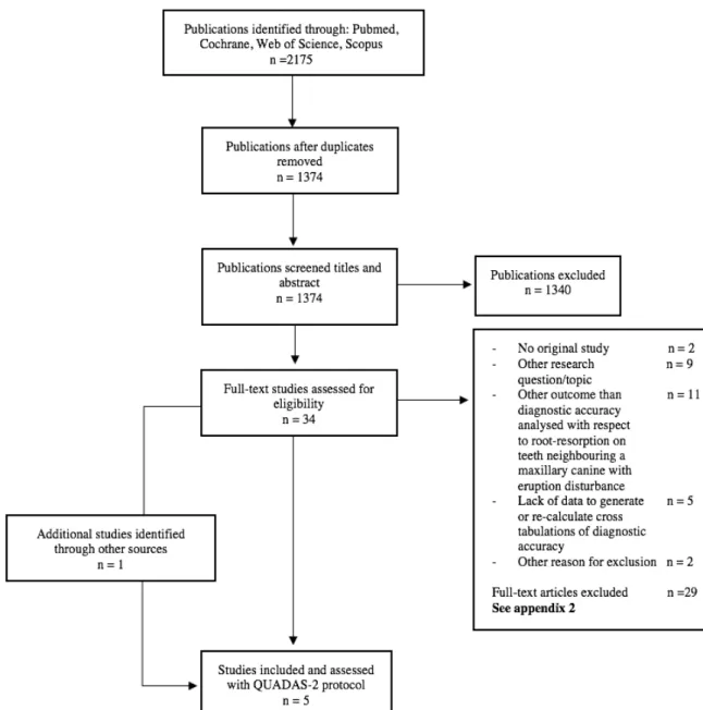
The two authors independently reviewed all full-text publications and included or excluded them using a pre-established protocol (Appendix 1). Excluded studies/publications and reasons for exclusion are listed in figure 1.
2.5 Data extraction and quality assessment:
Study characteristics of included publications were tabulated (Table 2). The index test, reference standard, results on diagnostic accuracy; and other relevant information was plotted into the table. Quality assessment was performed by the authors and their mentors (LG, JS, AS, HC) using the QUADAS-2 tool (Appendix 3) (6). Included studies in this systematic review were assessed regarding the risk of bias in patient selections, index test, reference standard and flow and timing. The first three domains also assessed the applicability. Any uncertainties were discussed until consensus decision was made.
3. Results
3.1 Literature identification
The literature identification and reasons for exclusion are presented in a flow diagram (Fig.1). A total of 2175 studies were retrieved with 1374 remaining after removal of duplicates. 34 publications were assessed in full-text. The most frequent reasons for excluding a study were if it presented “other outcome than diagnostic accuracy with respect to root-resorption of teeth neighbouring a maxillary canine with eruption disturbance” or concerned “other research question/main topic”. All studies excluded during full-text assessment are presented in Appendix 2. Reference lists for the four included studies were reviewed and resulted in the inclusion of one additional study. In January 2019 the PubMed search was updated (table 1). It resulted in 122 new titles and abstracts, none of which met the criteria for inclusion.
3.2 Study characteristics
Key characteristics from the five included studies are summarized in Table 2 (48-52). The sample size varied between one to 156 maxillary canines, both unilateral and bilateral. Root resorption was diagnosed with different methods as index test -intraoral radiography, panoramic radiography, CT or CBCT. Two studies used eight maxillary laterals with simulated cavities as reference test and two intact laterals incisors as control group (48,49) while Ericson et al. used extracted teeth with root-resorption (50). Two studies used CT for the reference standard (51,52).
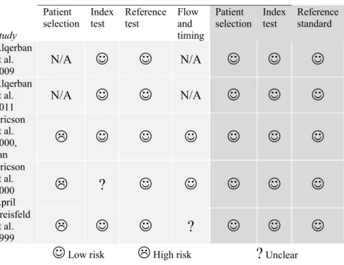
3.3 Assessment of methodological quality
The quality assessment using the QUADAS-2 tool (6) is presented in Figure 2. Low risk of bias was shown in approximately half of the domains for risk of bias and for all domains regarding applicability. No study presented low risk of bias throughout. For domain patient selection risk of bias varied, no study presented low risk. Alqerban et al. 2009 and 2011 presented as N/A (not applicable) in domains patient selection and flow and timing due to in vitro studies.
3.4 Diagnostic accuracy
Two-by-two (2x2) tables of diagnostic accuracy estimates were presented and re-calculated or generated using data derived from the studies. The outcome was presented as values for sensitivity and specificity (Table 2). Thresholds for root-resorption were similarly defined in all five studies. Ericson et al. developed and used a four category resorption scale, from “No resorption” to “Severe resorption” (50) and three other studies also used this scale (48,49,51). Freisfeld et al. defined root resorption as dentin layer that was “distinctly narrowed” (52).
Alqerban et al. 2009 showed a significant difference (P < 0.05) between panoramic examination and CBCT system regarding detection of root resorptions for all resorption sizes (including teeth with no resorption) (49). Though, there was no significant difference between the two CBCT systems, Accuitomo and Scanora, that was compared. Alqerban et al. 2011 compared six different CBCT systems and no significant difference was found between the systems in grading the root resorptions (48). Ericson et al., found a high level of agreement between CT-examination and the clinical in-vitro evaluation of the resorbed lateral incisor roots although CT scans revealed significantly more resorption damages on both lateral and central incisors compared with intraoral films (50,51). Freisfeld et al. did not find any significant differences between CT and panoramic radiography in diagnosis of resorptions (52).
Study
Risk of bias Applicability
Patient
selection Index test Reference test Flow and timing
Patient
selection Index test Reference standard Alqerban et al. 2009 N/A
J
J
N/AJ
J
J
Alqerban et al. 2011 N/AJ
J
N/AJ
J
J
Ericson et al. 2000, JanL
J
J
J
J
J
J
Ericson et al. 2000 AprilL
?
J
J
J
J
J
Freisfeld et al. 1999L
J
J
?
J
J
J
J
Low riskL
High risk?
UnclearTable 2– Characteristics of included studies on diagnostic accuracy regarding root resorption on neighbouring teeth caused by maxillary canines First author, year, country, reference Population, setting, number of teeth included, number of resorbed teeth Impacted maxillary canine (N)
Index test Reference test Diagnostic
accuracy Sensitivity and Specificity % Calculated: tic accuracy Sensitivity Specificity Predictive value%, CI Comments Alqerban, A et al., 2009, Belgium (49) Child cadaver skull in early mixed dentition with impacted maxillary canine 8 maxillary laterals with simulated resorption. Control group: 2 maxillary laterals with intact root N 1 Panorama Accuitomo CBCT Scanora CBCT Teeth with simulated resorption Control group: 2 maxillary laterals with intact root Panorama Sensitivity 78 Specificity 38 Accuitomo CBCT Sensitivity 95 Specificity 75 Scanora CBCT Sensitivity 94 Specificity 75 Panorama False-positive errors 63 False-negative errors 22 Accuitomo CBCT False-positive errors 25 False-negative errors 5 Scanora CBCT False-positive errors 25 False-negative errors 6 Alqerban, A et al., 2011, Belgium (48) Child cadaver skull in early mixed dentition with impacted maxillary canine 8 maxillary laterals with simulated resorption. Control group: 2 maxillary laterals with intact root N 1 Six different CBCT-machines Table 1 Teeth with simulated resorption Control group: 2 maxillary laterals with intact root Accuitomo Sensitivity 93,75(100)* Specificity 87,50(75) Galileos Sensitivity 87,5(87,5) Specificity 70,83(100) Scanora Sensitivity 95,83(100) Specificity 95,83(50) Kodak Sensitivity 86,46(93,75) Specificity 91, 67(100)
There was no significantly difference among the tested
CBCT-systems * = sens. and spec. from the second evaluation, this time with two radiologists
Picasso Sensitivity 85,42(100) Specificity 95,83(100) ProMax Sensitivity 98,96(100) Specificity 58,33(75) Ericson, S and Kurol, P.J, January 2000, Sweden (50) Children with ectopically diagnosed maxillary canines 12 children 16 resorbed neighbouring teeth N 17 CT Extracted teeth Sensitivity 100 Specificity 87,5 Ericson, S and Kurol, P.J, March 2000, Sweden (51) Orthodontics specialist clinic 107 children 72 resorbed incisors N 156 Intraoral periapical radiograph CT Sensitivity 69 Specificity 90
Uses the CT calculations from Ericson et al(50) Sensitivity 1 Specificity 0,875 Freisfeld, M et al., 1999, Germany (52) Patients from dental school 30 patients 16 resorbed incisors N 39 Panorama CT Sensitivity 45,6 Specificity 88,9
4. Discussion
The results show that the sensitivity of CBCT regarding root resorption of teeth neighbouring maxillary canines is higher than for that of conventional radiography.
4.1 Methodological considerations
It is important to search a several databases to avoid missing relevant diagnostic accuracy studies (4). Diagnostic accuracy studies are poorly indexed in databases of scientific literature, such as PubMed and Web of Science. Therefore, it is recommended to perform wide searches to retrieve relevant publications within this field. Since wide search strategies were adopted, using free-text terms, a majority of relevant publications of interest were assessed as being captured. In addition, reference list of included publications were searched and it has been deemed the best way to find relevant studies (1).
Two reviewers, LG & JS, read and included or excluded retrieved publications. Under optimal circumstances, at least one more group of assessors could have been used. However, any unclarities were discussed to consensus was met within the research group (including supervisors AS, HC).
The lack of transparency regarding data on diagnostic accuracy presented a major difficulty in assessing the full-text publications for inclusion or exclusion. Many compared different radiographic systems in relation to prediction of ectopic maxillary canines. Orthodontic treatment planning was also a common research topic, which did not mention diagnostic accuracy regarding root resorption. Prevalence of impacted maxillary canines was also a common subject for excluded publications.
During full-text reading, a protocol (Appendix 1) was used to determine whether the studies contained index test and reference tests and of what kind. It was also recorded if sensitivity and specificity data were presented, if a thresholds was determined and an overall assessment of whether the study was relevant for this review. In full-text reading 34 studies were reviewed, 29 of which were excluded. Many of the excluded items were missing 2x2 tables.
CRD guidance states that there is a loss of transparency and reporting on collection of materials in diagnostic test accuracy studies (4). This was also the case for retrieved studies where the materials and method sections was inadequate regarding patient selection. Therefore, it was problematic to determine if there was a risk of bias in this category. In general, the included studies showed low risk of bias in QUADAS-2 assessment, see Figure 2. Regarding concern of applicability, all studies were at low risk in all categories as the studies that weren’t relevant were excluded during full-text assessment.
Since the majority of the studies were retrospective and the authors used materials from patients who were all diagnosed with impacted canines there was an increased risk of root resorption. This, however, is expected in diagnostic studies (4). In this review, Ericson et al. (50,51) and Freisfeld et al. (52) were assessed as high risk of bias due to their non-consecutive or randomized selection. Eslami et al. discussed the problem regarding patient selection in their review and considered all studies without randomization as high risk of bias in subject selection (53). “These undiagnosed patients through conventional radiographs were basically considered those with a difficult diagnosis; this might have caused underestimation of CBCT efficacy”. They suggest that the high risk of selection bias might be reduced by recruiting consecutive participant from several clinics – prospective study design.
Both studies by Alqerban et al. were in vitro studies and was assessed as not applicable (N/A) in patient selection (48,49). The same authors also received N/A under the category flow and timing. Because of its study design disease progression will not be a factor of influence.
Due to the low number of included studies and lack of homogenises between them, GRADE was excluded in this review.
4.2 Discussion of results
In general, two-dimensional index tests showed a lower sensitivity (0.45-0.78) compared to three-dimensional tests (0.85-1.0), see table 2.
Alqerban et al. 2009 compared panoramic imaging against two different CBCT modalities (49). Showing a significant difference in the detection of root resorptions. In 2011 Alqerban et al instead compare six different CBCT modalities (48). In both of these studies they used simulated root resorptions on laterals in child cadaver cranium with a neighbouring impacted canine in an effort to investigate how small root resorptions the modalities could detect. CBCT showed a high sensitivity for minor (0.2 mm) as well as greater damage as compared to panoramic imaging. The panoramic image was not able to detect damage 0.2 mm or less. This is in line with Ren et al., reporting that two-dimensional images showed a significantly lower detection rate of mild and moderate root resorption compared to CBCT (54). On the other hand, severe root resorption was visible on both modalities. In in vitro studies such as Alqerban et al. and Ren et al. the environment created is well controlled and mimics reality.
Further, methodological factors within studies affect the outcome of diagnostic accuracy measures. For example, in the study by Alqerban et al., 2011, orthodontists obtained CBCT slices that best showed root resorption in axial, coronal, sagittal view and one 3D model. In contrast to the orthodontists, the radiologists received all the data from the OnDemand 3D software and could reconstruct the image. The results differed between the orthodontists and the radiologists, hypothetically as radiologists are more used to 3D imaging assessment and received all data. The observers were informed before they received the images that some do not contain root resorption. This could be an advantage to the authors; giving out this information may reduce the proportion of false positives and thus affect the sensitivity and specificity positively. The reviewers’ judgement regarding root resorption is likely affected by this information.
Ericson et al. compared CT against extracted teeth. The majority of the extracted teeth had distinct and severe root damage (50). The authors considered this to be a problem as it might simplify the CT diagnosis and presumably affect the diagnostic accuracy measures. In addition, sensitivity and specificity often over estimates diagnostic accuracy (55) .
Nowadays CT is not the common diagnostic method regarding the dentomaxillofacial area, instead, it is CBCT. Because of the technology behind CBCT the foci of interest is not sliced but instead it is created by the cone beam that is covering the field of view. Ericson et al., saw misinterpretations in CT images due to the slice thickness in CT and morphology of the resorption(50). This gave the impression of decreased dentin content in the resorption cavity that could give misleading information about the size of the resorption. By using CBCT this misinterpretation can be avoided.
Another finding by Ericson et al. were that resorptions were predominantly buccally and palatally located. This, according to the authors, speaks against intraoral images as diagnostics for root resorption (51). Freisfeld et al., reaches the conclusion that diagnosis of root resorption, by panoramic imaging, in the area of the maxillary incisors is subject to a strong subjective bias, which also point to the fact that 2D may be insufficient regarding diagnosis of root resorption caused by impacted maxillary canines (52).
CBCT provides images with a higher degree of detail with a higher sensitivity regarding resorption. Regarding patients with maxillary canines with eruption disturbances information of any root resorption could be useful if there is a severe space insufficiency that requires tooth extraction. In these cases, CBCT can be used to avoid extraction of a healthy tooth. For most patients however, CBCT examination is probably not beneficial as this additional information in most cases will not change the treatment decision (46). Furthermore, the monetary cost of CBCT is significantly higher than that of conventional radiography and the cost of CBCT in terms of radiation is around 100 times that of intraoral radiography (56).
Due to ethical aspect radiation should always be well indicated. Patients diagnosed with impacted maxillary canines are usually young. The risk for developing cancer later in life when exposed to radiation in childhood than in adulthood is 2-3 times greater. Therefor it is important that the CBCT examination is justified (57).
Freisfeld et al. showed a reversed distribution of sensitivity and specificity for panorama compared to Alqerban et al. (49,52). It is possible that the ten-year timespan between the studies creates this discrepancy, image quality is constantly under development. The reversed accuracy could also depend on different thresholds between the two studies. Freisfeld diagnosed root resorption when “the dentin layer was distinctly narrowed” and Alqerban used a four category resorption scale. The heterogeneity between the studies agree well with CRD and is often pronounced in diagnostic reviews (4). The source of the heterogeneity can be explained due to different thresholds, different patient compositions such as prevalence and disease spectrum and also the variation in interpretation by observers and categories used. Ericson et al. developed a four category resorption scale for evaluating root resorptions (50). They used CT as index test and extracted teeth as reference test. The scale proves to be well-matched between in vivo and in vitro. The scale is used as threshold in four of five included studies (48-51). This point to a well-constructed scale and if incorporated in future studies would ease meta-analyses and the synthesis of evidence.
5. Conclusion
According to this systematic review, three-dimensional imaging showed the highest sensitivity and specificity regarding root resorptions caused by impacted maxillary canine. CBCT showed higher accuracy than two-dimensional imaging. Though, CBCT has higher accuracy it also has a higher radiation dose and might not always be beneficial regarding treatment planning. Therefore, CBCT should be used with respect and not routinely.
6. Reference
1. Whiting P, Harbord R, de Salis I, Egger M, Sterne J. Evidence-based diagnosis. J Health Serv Res Policy 2008 October 01;13 Suppl 3:57-63.
2. Kottner J, Streiner DL. The difference between reliability and agreement. J Clin Epidemiol 2011 June 01;64(6):701-2; author reply 702.
3. SBU. Utvärdering av metoder i hälso- och sjukvården: En handbok. Available at: https://www.sbu.se/globalassets/ebm/metodbok/sbushandbok.pdf.
4. Centre for Reviews and Dissemination. Systematic Reviews: CRD's Guidance for Undertaking Reviews in Health Care. 2009; Available at:
https://www.york.ac.uk/media/crd/Systematic_Reviews.pdf. Accessed 02/04, 2019. 5. Fryback DG, Thornbury JR. The efficacy of diagnostic imaging. Med Decis Making 1991
June 01;11(2):88-94.
6. Whiting PF, Rutjes AW, Westwood ME, Mallett S, Deeks JJ, Reitsma JB, et al.
QUADAS-2: a revised tool for the quality assessment of diagnostic accuracy studies. Ann Intern Med 2011 October 18;155(8):529-536.
7. White S C, Pharoah MJ e. Oral radiology: principles and interpretation. 7th ed. St. Louis, Mo.: Mosby: Elsevier; 2015.
8. Clark CA. A Method of ascertaining the Relative Position of Unerupted Teeth by means of Film Radiographs. Proc R Soc Med 1910;3(Odontol Sect):87-90.
9. Husain J, Burden D, McSherry P, Morris D, Allen M, Clinical Standards Committee of the Faculty of Dental Surgery, Royal College of Surgeons of England. National clinical guidelines for management of the palatally ectopic maxillary canine. Br Dent J 2012 August 01;213(4):171-176.
10. Ericson S, Kurol J. Incisor resorption caused by maxillary cuspids. A radiographic study. Angle Orthod 1987 October 01;57(4):332-346.
11. Agnini M. The panoramic X-ray as a detector for preventing maxillary canine impaction. Int J Orthod Milwaukee 2007 January 01;18(4):15-23.
12. Ericson S, Kurol J. Early treatment of palatally erupting maxillary canines by extraction of the primary canines. Eur J Orthod 1988 November 01;10(4):283-295.
13. Preda L, La Fianza A, Di Maggio EM, Dore R, Schifino MR, Campani R, et al. The use of spiral computed tomography in the localization of impacted maxillary canines.
Dentomaxillofac Radiol 1997 July 01;26(4):236-241.
14. Scarfe WC, Farman AG, Sukovic P. Clinical applications of cone-beam computed tomography in dental practice. J Can Dent Assoc 2006 February 01;72(1):75-80.
15. Liang X, Jacobs R, Hassan B, Li L, Pauwels R, Corpas L, et al. A comparative evaluation of Cone Beam Computed Tomography (CBCT) and Multi-Slice CT (MSCT) Part I. On subjective image quality. Eur J Radiol 2010 August 01;75(2):265-269.
16. Alqerban A, Jacobs R, Lambrechts P, Loozen G, Willems G. Root resorption of the maxillary lateral incisor caused by impacted canine: a literature review. Clin Oral Investig 2009 September 01;13(3):247-255.
17. Strålsäkerhetsmyndigheten. för ett strålsäkert samhälle. Available at: https://www.stralsakerhetsmyndigheten.se. Accessed 02/09, 2019.
18. Al-Okshi A, Lindh C, Sale H, Gunnarsson M, Rohlin M. Effective dose of cone beam CT (CBCT) of the facial skeleton: a systematic review. Br J Radiol 2015 January
01;88(1045):20140658.
19. Kjaer I. Mechanism of human tooth eruption: review article including a new theory for future studies on the eruption process. Scientifica (Cairo) 2014;2014:341905.
20. Nanci A, Ten Cate A. Ten Cate's oral histology : development, structure, and function. : St. Louis : Mosby Elsevier, cop. 2013; 8. ed; 2013.
21. Koch Ge, Poulsen Se. Pediatric dentistry : a clinical approach. : Chichester, UK ; Ames, Iowa : Wiley-Blackwell, 2009; 2. ed; 2009.
22. Welbury Re, Duggal Me, Hosey MTe. Paediatric dentistry. : Oxford : Oxford University Press, 2012; 4. ed; 2012.
23. Thilander B, Jakobsson SO. Local factors in impaction of maxillary canines. Acta Odontol Scand 1968 May 01;26(2):145-168.
24. Litsas G, Acar A. A review of early displaced maxillary canines: etiology, diagnosis and interceptive treatment. Open Dent J 2011 March 16;5:39-47.
25. Ericson S, Kurol J. Longitudinal study and analysis of clinical supervision of maxillary canine eruption. Community Dent Oral Epidemiol 1986 June 01;14(3):172-176.
26. Thilander B, Myrberg N. The prevalence of malocclusion in Swedish schoolchildren. Scand J Dent Res 1973;81(1):12-21.
27. Tsai HH. Eruption process of upper permanent canine. J Clin Pediatr Dent 2001 January 01;25(3):175-179.
28. Yan B, Sun Z, Fields H, Wang L. Maxillary canine impaction increases root resorption risk of adjacent teeth: A problem of physical proximity. Orthod Fr 2015 June
01;86(2):169-179.
29. Walker L, Enciso R, Mah J. Three-dimensional localization of maxillary canines with cone-beam computed tomography. Am J Orthod Dentofacial Orthop 2005 October 01;128(4):418-423.
30. Ericson S, Bjerklin K, Falahat B. Does the canine dental follicle cause resorption of permanent incisor roots? A computed tomographic study of erupting maxillary canines. Angle Orthod 2002 April 01;72(2):95-104.
31. Liu DG, Zhang WL, Zhang ZY, Wu YT, Ma XC. Localization of impacted maxillary canines and observation of adjacent incisor resorption with cone-beam computed tomography. Oral Surg Oral Med Oral Pathol Oral Radiol Endod 2008 January 01;105(1):91-98.
32. Ericson S, Kurol J. Resorption of maxillary lateral incisors caused by ectopic eruption of the canines. A clinical and radiographic analysis of predisposing factors. Am J Orthod Dentofacial Orthop 1988 December 01;94(6):503-513.
33. Cernochova P, Krupa P, Izakovicova-Holla L. Root resorption associated with ectopically erupting maxillary permanent canines: a computed tomography study. Eur J Orthod 2011 October 01;33(5):483-491.
34. Strbac GD, Foltin A, Gahleitner A, Bantleon HP, Watzek G, Bernhart T. The prevalence of root resorption of maxillary incisors caused by impacted maxillary canines. Clin Oral Investig 2013 March 01;17(2):553-564.
35. Chaushu S, Kaczor-Urbanowicz K, Zadurska M, Becker A. Predisposing factors for severe incisor root resorption associated with impacted maxillary canines. Am J Orthod Dentofacial Orthop 2015 January 01;147(1):52-60.
36. Schindel RH, Sheinis MR. Prediction of maxillary lateral-incisor root resorption using sector analysis of potentially impacted canines. J Clin Orthod 2013 August 01;47(8):490-493.
37. Lindauer SJ, Rubenstein LK, Hang WM, Andersen WC, Isaacson RJ. Canine impaction identified early with panoramic radiographs. J Am Dent Assoc 1992 March 01;123(3):91-2, 95.
38. Sajnani AK, King NM. Early prediction of maxillary canine impaction from panoramic radiographs. Am J Orthod Dentofacial Orthop 2012 July 01;142(1):45-51.
39. Yu JN, Gu YG, Zhao CY, Liu K, Mo SC, Li H, et al. Three-dimensional localization and assessment of maxillary palatal impacted canines with cone-beam computed tomography. Shanghai Kou Qiang Yi Xue 2015 February 01;24(1):65-70.
40. Ericson S, Kurol J. CT diagnosis of ectopically erupting maxillary canines--a case report. Eur J Orthod 1988 May 01;10(2):115-121.
41. Naoumova J, Kurol J, Kjellberg H. Extraction of the deciduous canine as an interceptive treatment in children with palatally displaced canines - part II: possible predictors of success and cut-off points for a spontaneous eruption. Eur J Orthod 2015 April 01;37(2):219-229.
42. Naoumova J, Kurol J, Kjellberg H. Extraction of the deciduous canine as an interceptive treatment in children with palatal displaced canines - part I: shall we extract the deciduous
canine or not? Eur J Orthod 2015 April 01;37(2):209-218.
43. Becker A, Chaushu S. Etiology of maxillary canine impaction: a review. Am J Orthod Dentofacial Orthop 2015 October 01;148(4):557-567.
44. Power SM, Short MB. An investigation into the response of palatally displaced canines to the removal of deciduous canines and an assessment of factors contributing to favourable eruption. Br J Orthod 1993 August 01;20(3):215-223.
45. Stivaros N, Mandall NA. Radiographic factors affecting the management of impacted upper permanent canines. J Orthod 2000 June 01;27(2):169-173.
46. Christell H, Birch S, Bondemark L, Horner K, Lindh C, SEDENTEXCT consortium. The impact of Cone Beam CT on financial costs and orthodontists' treatment decisions in the management of maxillary canines with eruption disturbance. Eur J Orthod 2018 January 23;40(1):65-73.
47. Moher D, Liberati A, Tetzlaff J, Altman DG, PRISMA Group. Preferred reporting items for systematic reviews and meta-analyses: the PRISMA statement. Int J Surg
2010;8(5):336-341.
48. Alqerban A, Jacobs R, Fieuws S, Nackaerts O, SEDENTEXCT Project Consortium, Willems G. Comparison of 6 cone-beam computed tomography systems for image quality and detection of simulated canine impaction-induced external root resorption in maxillary lateral incisors. Am J Orthod Dentofacial Orthop 2011 September 01;140(3):e129-39. 49. Alqerban A, Jacobs R, Souza PC, Willems G. In-vitro comparison of 2 cone-beam
computed tomography systems and panoramic imaging for detecting simulated canine impaction-induced external root resorption in maxillary lateral incisors. Am J Orthod Dentofacial Orthop 2009 December 01;136(6):764.e1-11; discussion 764.
50. Ericson S, Kurol J. Incisor root resorptions due to ectopic maxillary canines imaged by computerized tomography: a comparative study in extracted teeth. Angle Orthod 2000 August 01;70(4):276-283.
51. Ericson S, Kurol PJ. Resorption of incisors after ectopic eruption of maxillary canines: a CT study. Angle Orthod 2000 December 01;70(6):415-423.
52. Freisfeld M, Dahl IA, Jager A, Drescher D, Schuller H. X-ray diagnosis of impacted upper canines in panoramic radiographs and computed tomographs. J Orofac Orthop 1999;60(3):177-184.
53. Eslami E, Barkhordar H, Abramovitch K, Kim J, Masoud MI. Cone-beam computed tomography vs conventional radiography in visualization of maxillary impacted-canine localization: A systematic review of comparative studies. Am J Orthod Dentofacial Orthop 2017 February 01;151(2):248-258.
54. Ren H, Chen J, Deng F, Zheng L, Liu X, Dong Y. Comparison of cone-beam computed tomography and periapical radiography for detecting simulated apical root resorption.
Angle Orthod 2013 March 01;83(2):189-195.
55. Grimes DA, Schulz KF. Refining clinical diagnosis with likelihood ratios. Lancet 2029 April 23;365(9469):1500-1505.
56. Christell H, Birch S, Horner K, Rohlin M, Lindh C, SEDENTEXCT consortium. A framework for costing diagnostic methods in oral health care: an application comparing a new imaging technology with the conventional approach for maxillary canines with eruption disturbances. Community Dent Oral Epidemiol 2012 August 01;40(4):351-361. 57. UNSCEAR. Sources, Effects and Risks of Ionizing Radiation. SCIENTIFIC Annex B.
Effect of radiation exposure of children.
http://www.unscear.org/docs/reports/2013/UNSCEAR2013Report_AnnexB_Children_13-87320_Ebook_web.pdf ed.
Appendix 1:
Protocol for inclusion and exclusion of full-text publications.STUDY TYPE:
AIM OF PUBLICATION Main topic on diagnostic accuracy of root resorption regarding… Yes □ No □ Other □
SAMPLE AND METHODS
RELEVANCE FOR THIS REVIEW YES □ NO □
(If NO see “Reasons for exclusion”) REASONS FOR EXCLUSION
First author, year:
Diagnostics test analysed with respect to root-resorption on
teeth neighbouring a maxillary canine with eruption disturbance Yes □ No □ Sample size (teeth):
INDEX TEST
Index test described: Yes □ No □
Type(s) of x-ray method: REFERENCE STANDARD
Reference standard described: Yes □ No □
Type of method:
Thresholds defined for index test and reference standard Yes □ No □ OUTCOME MEASURES
Data presented as diagnostic accuracy (i.e.
sensitivity/specificity/predictive values positive and negative, ROC/AUC curves, likelihood ratios, relative risk) possible to generate or re-calculate
Yes □ No □
- No original study (see study type) □
- Other research question/ topic □
- Other outcome than diagnostic accuracy analysed with respect to root-resorption on teeth neighbouring a maxillary canine with eruption disturbance
□ - Extensive lack of description of index test/reference standard or its thresholds
□ - Lack of data to generate or re-calculate cross tabulations of
diagnostic accuracy
□
Other reasons for exclusion, specify: ________________ ________________ ________________
Appendix 2
: excluded studies from full-text assesmentAuthor Reason for decline
Alqerban, A et al., 2009 Lack of data to generate or re-calculate cross tabulations of diagnostic…
Alqerban, A et al., 2011 Lack of data to generate or re-calculate cross tabulations of diagnostic…
Alqerban, A et al., 2013 Other outcome than diagnostic accuracy analysed with respect to…
Alqerban, A et al., 2016 Lack of data to generate or re-calculate cross tabulations of diagnostic…
Amintavakoli,N et al., 2018 Other outcome than diagnostic accuracy analysed with respect...
Broer, N et al., 2005 Other research question/topic
Cernochova, P et al., 2011 Other research question/topic
Cuminetti, F et al., 2017 Other research question/topic
Ericson, S and Kurol, J., 1987 Other research question/topic
Flint, D.J et al., 1998 Other research question/topic
Guarnieri, R et al., 2016 Other research question/topic
Haney, E et al., 2010 Other outcome than diagnostic accuracy analysed with respect to…
Jawad, Z et al., 2016 Lack of data to generate or re-calculate cross tabulations of diagnostic…
Jung, Y.H et al., 2012 Other outcome than diagnostic accuracy analysed with respect to...
Katheria, B.C et al., 2010 Other outcome than diagnostic accuracy analysed with respect to...
Kim, H et al., 2008 Written in foreign language
Krennmalr, G et al., 1995 Other research question/topic
Li, Z et al., 2013 Written in foreign language
Pico, C.L et al 2017 Other outcome than diagnostic accuracy analysed with respect...
Preda, L et al., 1997 Other outcome than diagnostic accuracy analysed with respect…
Rossini, G et al., 2012, Italy No original study
Sawamura, T et a., 2003 Other research question/topic
Schmuth, G.P et al., 1992 Other outcome than diagnostic accuracy analysed with respect to…
Traxler, M et al., 1989 Other outcome than diagnostic accuracy analysed with respect to…
Tsolakis et al. 2017 Lack of data to generate or re-calculate cross tabulations of diagnostic…
Ucar, F.I et al., 2017 Other outcome than diagnostic accuracy analysed with respect to…
van Vlijmen, Oliver J.C et al., 2012 No original study
Witcher, T.P et al., 2010 Other research question/topic
Appendix 3
QUADAS-2Phase 1: State the review question:
Patients (setting, intended use of index test, presentation, prior testing): Index test(s):
Reference standard and target condition:
DOMAIN 2: INDEX TEST(S)
If more than one index test was used, please complete for each test.
Phase 3: Risk of bias and applicability judgments
QUADAS-2 is structured so that 4 key domains are each rated in terms of the risk of bias and the concern regarding applicability to the research question (as defined above). Each key domain has a set of signalling questions to help reach the judgments regarding bias and applicability.
DOMAIN 1: PATIENT SELECTION
A. Risk of Bias
Describe methods of patient selection:
v Was a consecutive or random sample of patients enrolled? Yes/No/Unclear
v Was a case-control design avoided? Yes/No/Unclear
v Did the study avoid inappropriate exclusions? Yes/No/Unclear Could the selection of patients have introduced bias? RISK: LOW/HIGH/UNCLEAR
B. Concerns regarding applicability
Describe included patients (prior testing, presentation, intended use of index test and setting):
Is there concern that the included patients do not match the review question?
CONCERN: LOW/HIGH/UNCLEAR
A. Risk of Bias
Describe the index test and how it was conducted and interpreted:
v Were the index test results interpreted without knowledge of the results of the reference standard?
Yes/No/Unclear v If a threshold was used, was it pre-specified? Yes/No/Unclear Could the conduct or interpretation of the index test
have introduced bias?
RISK: LOW /HIGH/UNCLEAR
B. Concerns regarding applicability
Is there concern that the index test, its conduct, or
DOMAIN 3: REFERENCE STANDARD
A. Risk of Bias
Describe the reference standard and how it was conducted and interpreted:
v Is the reference standard likely to correctly classify the target condition?
Yes/No/Unclear v Were the reference standard results interpreted without
knowledge of the results of the index test?
Yes/No/Unclear Could the reference standard, its conduct, or its
interpretation have introduced bias?
RISK: LOW /HIGH/UNCLEAR
B. Concerns regarding applicability
Is there concern that the target condition as defined by the reference standard does not match the review question?
CONCERN: LOW /HIGH/UNCLEAR
DOMAIN 4: FLOW AND TIMING
A. Risk of Bias
Describe any patients who did not receive the index test(s) and/or reference standard or who were excluded from the 2x2 table (refer to flow diagram):
Describe the time interval and any interventions between index test(s) and reference standard:
v Was there an appropriate interval between index test(s) and reference standard?
Yes/No/Unclear v Did all patients receive a reference standard? Yes/No/Unclear v Did patients receive the same reference standard? Yes/No/Unclear v Were all patients included in the analysis? Yes/No/Unclear