Role of YopE and LcrH in effector translocation, HeLa
cell cytotoxicity and virulence
Margareta Aili
Department of Molecular Biology Umeå University
Cover: “Birch trunks”, original artwork Copyright © 2005 by Helena Hörberg
Copyright © 2005 by Margareta Aili
ISBN 91-7305-977-3
TABLE OF CONTENTS
ABSTRACT ... 5
PAPERS IN THIS THESIS... 6
INTRODUCTION ... 7
Yersinia spp. and their diseases...7
Protective antigens and treatment of Yersinia infections ...8
Virulence properties of Yersinia ...8
Motility...8
Adhesion proteins...9
Capsule and LPS ...9
Target cells and cellular model systems ...10
Type III Secretion System ...11
The Secretion Apparatus ...11
T3SS chaperones ...12
The secretion signal...13
Substrate specificity switch...13
Translocation of effector proteins into eukaryotic cells...14
Regulation of the T3SS...15
Positive regulation of T3S...15
The Low calcium response...15
De-repression of Yop synthesis by secretion of negative regulatory elements...16
LcrH is a negative regulator in Yersinia...17
Effector proteins of Yersinia ...18
The family of RhoGTPases ...20
Regulation of RhoGTPases ...20
RhoGTPases and the actin cytoskeleton ...21
Control of endocytosis by RhoGTPases...22
RhoGTPases as targets for bacterial toxins...23
RESULTS AND DISCUSSION ... 25
In vitro GAP activity towards RhoA, Rac1 and Cdc42 can be uncoupled from cytotoxicity ...25
In vivo substrate specificity of YopE in infected cells ...26
The role of YopE in prevention of pore formation...28
Regulation of effector translocation into target cells – a role for YopE ...29
Functional differences between YopE and ExoS ...31
Another potential mechanism of translocation control in Yersinia...33
Delineation of YopD ...34
Low levels of the translocators is sufficient for effector translocation ...35
ACKNOWLEDGEMENTS ... 37
REFERENCES ... 39
ABSTRACT
In order to establish an extra-cellular infection the gram-negative bacteria Yersinia
pseudotuberculosis uses a type III secretion system (T3SS) to translocate a set of
anti-host effectors into eukaryotic cells. The toxins disrupt signalling pathways important for phagocytosis, cytokine production and cell survival. Secretion and translocation via this T3SS is strictly regulated on several levels. In this context, the function of YopE and LcrH during Yersinia infections has been analysed.
YopE is an essential translocated effector that disrupts the actin cytoskeleton of infected eukaryotic cells, by inactivating small GTPases through its GTPase activating protein (GAP) activity. However, cytotoxicity can be uncoupled from in
vitro GAP activity towards the RhoA, Rac1 and Cdc42 GTPases. Furthermore, in vivo studies of the YopE GAP activity revealed that only RhoA and Rac1 are
targeted, but this is not a pre-requisite for Yersinia virulence. Hence, YopE must target one or more additional GTPases to cause disease in mice.
YopE was the only Yersinia effector that blocked LDH release from infected cells. Moreover, translocated YopE could regulate the level of subsequent effector translocation by a mechanism that involved the YopE GAP function and another T3S component, YopK. Loss of translocation control elevated total T3S gene expression in the presence of eukaryotic cells. This indicated the existence of a regulatory loop for feedback control of T3S gene expression in the bacteria that originates from the interior of the eukaryotic cell after effector translocation is completed. This might represent the true virulence function of YopE.
Exoenzyme S (ExoS) of Pseudomonas aeruginosa has a YopE-like GAP domain with similar activity towards RhoA, Rac1 and Cdc42. However, ExoS is unable to complement hyper-translocation resulting from loss of YopE. This indicates a unique function for YopE in translocation control in Yersinia that might be dependent on correct intracellular localisation. It follows that the Membrane Localisation Domain in YopE was important for translocation control, but dispensable for cytotoxicity and blockage of LDH release.
YopD and its cognate chaperone LcrH are negative regulatory elements of the T3S regulon and together with YopB, are involved in the effector translocation process. Randomly generated point mutants in LcrH specifically effected stability and secretion of both the YopB and YopD substrates in vitro and prevented their apparent insertion as translocon pores in the membranes of infected cells. Yet, these mutants still produced stable substrates in the presence of eukaryotic cells and most could mediate at least partial effector translocation. Thus, only minimal amounts of the YopB and YopD translocator proteins are needed for translocation and the LcrH chaperone may regulate this process from inside the bacteria.
PAPERS IN THIS THESIS
This thesis is based on the following publications referred to in the text by their roman numerals (I-V).
I. Aili, M., Telepnev, M., Hallberg, B., Wolf-Watz, H. and Rosqvist, R. 2003.
In vitro GAP activity towards RhoA, Rac1 and Cdc42 is not a prerequisite for
YopE induced HeLa cell cytotoxicity. Microb. Pathog. 34(6): 297-308. Reprinted with permission from Elsevier.
II. Aili, M., Isaksson, E.L., Hallberg, B., Wolf-Watz, H. and Rosqvist, R. 2005.
Functional analysis of the YopE GAP activity of Yersinia pseudotuberculosis. Cell. Microbiol. Submitted manuscript.
III. Aili, M., Isaksson, E.L., Carlsson, S., Rosqvist, R., and Francis, M.S. 2005.
Regulation of Yersinia Yop-effector delivery by translocated YopE. Manuscript.
IV. Francis, M.S., Aili, M., Wiklund, M-L., and Wolf-Watz, H. 2000. A study of
the YopD-LcrH interaction from Yersinia pseudotuberculosis reveals a role for hydrophobic residues within the amphipathic domain of YopD. Mol. Microbiol. 38(1): 85-102. Reprinted with permission from Blackwell Publishing.
V. Edqvist, P.J., Aili, M., and Francis, M.S. 2005. Minimal secretion of the
YopB and YopD translocators is sufficient for Yop-effector translocation by
Yersinia. Manuscript.
Contribution by the author to the studies listed below will not be extensively discussed in this thesis.
Aili, M., Hallberg, B., Wolf-Watz, H., and Rosqvist, R. 2002. GAP activity of
Yersinia YopE. Methods Enzymol. 358:359-370.
Henriksson, M.L., Francis, M.S., Peden, A., Aili, M., Stefansson, K., Palmer, R, Aitken, A. and Hallberg, B. 2002. A nonphosphorylated 14-3-3 binding motif on exoenzyme S that is functional in vivo. Eur. J. Biochem. 269(20):4921-4929.
INTRODUCTION
As human beings living on the planet Earth, we are constantly in contact with organisms too small for us to see: microbes. They are everywhere and in many ways dictate the life we live. Some are beneficial to us, some do not care about us, and some are harmful to us, yet many generally see us as perfect food and housing sources. One group of micro-organisms we encounter causes diseases, ranging from mild to life-threatening. Disease-causing bacteria come in many variants and have developed a multitude of strategies to inflict harm on humans, and other host organisms. Two broad avenues of bacterial life-styles can be identified within a human host; extra-cellular and intra-cellular. Each bacterial species has developed specific mechanisms for survival, in addition to general ones shared by all bacteria, which arise from their choice of habitat e.g. the human body. The work described in this thesis has dealt with the use of an ingenious toxin distribution apparatus used by a wide array of bacterial pathogens, to infect an equally impressive number of hosts within the plant and animal kingdoms. This thesis primarily focuses on the human gastro-intestinal pathogen Yersinia pseudotuberculosis.
Yersinia spp. and their diseases
Gram-negative Yersinia spp. belong to the family Enterobacteriaceae and include 11 species, of which three are human pathogens [211, 274]. Y. pestis, the causative agent of plague, is transmitted by flea bites or aerosols, and can produce three clinical manifestations; bubonic plague with a mortality rate of between 40-60% (if left untreated), septicemic plague without bubo manifestation and pneumonic plague, both with a mortality rate close to 100% [211].
Y. pestis is closely related to the enteropathogenic Y. pseudotuberculosis, a food-
and water-borne pathogen which causes self-limiting gastrointestinal infections in healthy humans [186]. The divergence of Y. pestis from Y. pseudotuberculosis may be as recent as 1500-20000 years ago [1]. Compared to Y. pseudotuberculosis, Y.
pestis has down-sized its genome, thereby restricting itself to a life-style as a strict
pathogen dependent upon the infected host for a number of essential nutrients, including amino acids and nucleotide precursors [56, 203].
Y. enterocolitica is a more distant relative to Y. pestis and Y. pseudotuberculosis,
but causes a similar disease in humans as does Y. pseudotuberculosis [41, 42]. Enteric Yersinia causes terminal septicemia in mice, termed yersiniosis [55], which
valuable tool in identifying virulence determinants in Yersinia [39, 102, 185, 289]. Non-pathogenic Yersinia have been isolated from food and water as well as sick and healthy humans [274].
Protective antigens and treatment of Yersinia infections
Survivors of plague and humans exposed to enteric Yersinia, develop protective antibodies towards primarily the F1 capsule of Y. pestis and the V antigen (LcrV) of all three species [42, 211]. Historically, either live attenuated strains or killed whole cell vaccines have been used to protect against primarily plague although with limited success, especially against pneumonic plague [286]. Several of the
Yersinia outer proteins (discussed below) are antigenic, but do not provide
protection against plague in mice [159]. Today Y. pestis is considered a potential biological weapon, and this emphasizes the need for an effective vaccine.
Identified Yersinia infections can usually be successfully treated in the early stages by administration of classical antibiotics such as streptomycin and tetracycline [41, 211]. However, new anti-bacterial agents should be considered [146], as multi-resistant isolates of Y. pestis have been identified [101].
Virulence properties of Yersinia
The three human pathogenic species of Yersinia share several common virulence determinants despite the very different diseases they cause in humans. Motility enhances infection by enteric Yersinia [313, 314]. Attachment to the extra-cellular matrix and eukaryotic cells is facilitated by several different bacterial cell surface adhesins. A Type III Secretion System (T3SS) is used by the bacteria to disarm immune cells [66]. The T3SS is used to introduce toxins, anti-host effector proteins commonly named Yersinia outer proteins (Yops), into target cells, thereby preventing activation of the immune system [183, 198, 249, 298, 309]. All pathogenic Yersinia harbour a common, inter-changeable virulence plasmid of roughly 70 kb [23, 83, 220, 303], which encodes the components of the T3SS as well as the translocated Yops [24, 37, 40, 65, 116, 163, 220, 238, 269]. In addition,
Y. pestis carries two specific plasmids encoding important virulence traits such as
the F1 capsule and Plasminogen activator protease (Pla) [135] and the ymt gene, the product of which facilitates survival in, and spread via the flea [128].
Motility
All Yersinia spp. possess the genes required for synthesis of flagella which confer motility to enteric Yersinia at low temperatures [62]. The expression of flagella
genes is repressed at high temperatures (37°C) [144, 145] and is possibly under the control of a quorum sensing system [18]. Motility of Yersinia plays a role in invasion of host cells and expression of the flagella genes is coordinated with that of the adhesin invasin [20, 313]. Motility is likely to enhance the ability of the bacteria to reach the intestinal epithelium during the early stages of infection. One of the flagella gene clusters in Y. pestis contains multiple mutations and would appear to be non-functional, whereas the other cluster seems functional [203]. However, the conditions during which it is expressed remain elusive, since motility has not been observed for Y. pestis [203].
Adhesion proteins
Yersinia expresses several adhesins that exhibit diverse functions during infection.
Adhesins allow enteric Yersinia to attach to host epithelial cells in the gastro-intestinal tract and initiate uptake through M cells [63]. However, several adhesins are non-functional in Y. pestis [56], perhaps as a result of the radically different infection route of this species compared to the enteropathogenic Yersinia. No adhesin so far studied in enteric Yersinia is absolutely required for virulence in mice, although adhesin deficient mutants are slower in penetrating the intestinal epithelium [210, 291].
Four adhesins have been studied in Yersinia; Invasin [140], YadA [260], pH6 antigen [308] and Ail [179]. Invasin is a chromosomally encoded adhesin which interacts with host cell β1-integrins to promote uptake of Yersinia, presumably during the initial phase of infection when enteric Yersinia penetrates the M cells of the intestinal epithelium [63, 251]. YadA promotes tight binding to eukaryotic cells and efficient uptake of the bacteria [34, 78] at 37°C when invasin is repressed [20]. In addition, YadA confers serum resistance to the bacteria [26], which is presumably important for their dissemination. Ail was identified by its ability to confer invasive properties to non-invasive Escherichia coli [179], however its role in Yersinia virulence is not clear [291]. pH6 antigen promotes adhesion to eukaryotic cells and is involved in hemagglutination [308], but a role of pH6 antigen in preventing phagocytosis is also proposed [136]. pH6 antigen is required for full virulence in Y. pestis [165], likely due to the absence of the other three adhesins in this species [56]. Recent genome projects have revealed a number of potential adhesin molecules in Y. pestis, but their functions are still unknown [203].
Capsule and LPS
The capsule produced by Y. pestis at 37°C is a unique feature not shared with enteric Yersinia [135, 211]. The capsule is implicated in the ability of the bacterium
to resist phagocytosis by monocytes [74, 211] and expression obstructs bacterial binding to eukaryotic cells [74]. Enteric Yersinia produces a protective lipopolysaccharide (LPS) layer which confers resistance to complement-mediated killing as well as protection from antimicrobial peptides [259, 261]. In addition, LPS plays a role in effective colonisation of the intestinal tract [259]. A rough type of LPS is produced by Y. pestis [203, 259], where presumably the capsule is sufficient protection against the innate immune system.
Target cells and cellular model systems
Historically, Yersinia was thought of as an intra-cellular pathogen, residing in phago-lysosomes of macrophages [211, 224]. However, this view changed with the observation that virulent Yersinia prevents phagocytosis by professional phagocytes [51, 123, 238, 239, 278]. All pathogenic Yersinia have tropism for lymphoid tissues in infected hosts [66]. Lymphoid organs are not easily accessible for the bacteria, as they need to cross specialised barriers in order to reach this niche. Enteric Yersinia have the ability to promote uptake by specialised M cells in the intestinal epithelium [63, 112, 172]. By utilizing the unique features of antigen sampling exhibited by M cells, Yersinia is delivered to the Peyer’s patches underneath the epithelial layer in the intestine [63, 172]. In this immune cell dense milieu, the T3SS is presumed to be activated and the translocated Yop effectors disarm the innate immune response to Yersinia, thus enabling extra-cellular growth of the pathogen [122, 187]. In a recent murine infection study, macrophages and dendritic cells were identified as the in vivo target cells for translocated Yop effectors [171], presumably as an indirect effect of the phagocytic nature of these cells. However, all pathogenic species of Yersinia are considered to be facultative intra-cellular bacteria and can replicate within macrophages [224, 271, 272, 320]. This ability might be of importance during initial stages of infection.
Several in vitro cell infection models for Yersinia have been developed over the years. HeLa cells have been extensively used to study both the invasive properties and the ability of the bacteria to prevent uptake [73, 213, 290]. Upon infection with wild type Yersinia, HeLa cells undergo a morphology change resulting in disassembly of the actin cytoskeleton [240, 242]. Macrophages have also been a favoured cell type for functional studies of Yersinia to stimulate infection-like conditions in vitro [108, 290]. Both HeLa cells and immune cell lines have been used to identify eukaryotic targets of essential virulence effectors of Yersinia [30, 72, 106, 213]. Yeast has also been successfully used to reveal functions of translocated effector proteins [188, 304].
Type III Secretion System
Type III Secretion Systems (T3SS) can be viewed upon as syringes used by numerous gram-negative bacteria to directly transport toxins from the bacterial cytoplasm across two bacterial membranes and a eukaryotic cell membrane to the interior of a eukaryotic cell [100]. Among the proteins necessary for building up the secretion machinery spanning the bacterial envelope is a core set with high identity in all known T3SS [6, 137, 191, 200]. A second set of proteins with lesser identity between systems most likely reflects a level of built-in specificity needed to sense the environment so that the bacteria knows where and when the injection apparatus should be used to modulate host responses [100, 200]. This would be different for each species, because they normally occupy unique niches defined by a set of distinctive environmental cues [137].
T3SSs can be separated from other bacterial secretion systems [285] by four distinguishable features; 1) the N-terminal secretion signal is not cleaved upon secretion [60, 177] 2) customised intra-bacterial chaperones are needed for many of the secreted proteins [141, 190, 293, 302] 3) high induction of the system requires tight interaction with target cells during infection [214, 241]. 4) one-step introduction of toxins from the bacterial cytoplasm to the interior of eukaryotic cells [161].
The Secretion Apparatus
Analysis of secretion-deficient Yersinia revealed a role for about 20 ysc (Yersinia secretion) genes, located on the common Yersinia virulence plasmid, in secretion of virulence determinants [9, 24, 86, 116, 178, 217, 301]. Ysc products is believed to assemble in the bacterial envelope into a syringe-like structure to enable transport of T3SS secreted substrates over the bacterial double-membrane, as has been visualised for T3SS from other bacteria [36, 152, 252, 281]. The Ysc-T3SS is responsible for secretion and translocation into eukaryotic cells of at least six different Yop effectors (discussed below). The ysc genes were indirectly identified as regulators of the Low calcium response (discussed below) [24, 86, 178, 217], since a non-functional T3SS prevents induction of secreted substrates [24]. In this way, correct regulation of the system requires a secretion competent apparatus, a process referred to as feedback inhibition.
Recently, a second T3SS was identified in Yersinia, the Ysa (Yersinia secretion apparatus) system. It is encoded on the chromosome and contains at least 13 Ysa proteins which build up the Ysa secretion apparatus, independently of the Ysc T3SS [90, 121]. Initial studies showed that at least 8 proteins, called Ysps (Yersinia
these Ysps have been identified as known secretion substrates of the Ysc T3SS; YopN, YopJ/P and YopE [311]. Four novel Ysps are known only to be substrates of the Ysa T3SS, specificity provided in part by a Ysp specific chaperone, all of which are encoded on the chromosome in close proximity to the ysa operon [91]. Nevertheless, to be able to secrete common substrates, the Ysa-T3SS and the Ysc-T3SS must engage in regulatory cross-talk in a way that is not yet known.
T3SS chaperones
Functional Type III Secretion (T3S) depends on two classes of cytosolic chaperones; effector class chaperones and translocator class chaperones [293, 302]. Common to both classes is their ability to confer increased stability and efficient secretion of a cognate substrate(s) (PAPER IV, [68, 69, 89, 96, 247, 293, 294]). T3SS chaperones are small, acidic globular proteins [167, 293] that bind to their substrate generally as homodimers, or as in the case of YscB and SycN, heterodimers [27, 68, 216].
Effector class chaperones bind to translocated effectors of the T3SS and consist of
SycH, YerA (SycE) and SycT in Yersinia. YerA and SycT bind YopE and YopT [96, 138, 294], respectively, while SycH binds not only YopH, but also LcrQ (YscM1 and YcsM2 in Y. enterocolitica) [216, 293]. A regulatory role has been proposed for the SycH/LcrQ complex in establishing a secretion hierarchy through the Ysc T3SS, allowing for rapid secretion of YopH before YopE [305]. Similarly, the YerA and SycT chaperones would secure secretion of YopE and YopT before secretion of the chaperone-less effectors YopM, YopJ and YpkA [167]. Only moderate sequence similarity exist between the known effector chaperones from different T3SSs, which is surprising in light of their structural similarities – a common mix of β-sheet and α-helical folds [27, 169, 216, 268]. In general, the effector class chaperones keep the N-terminus of its substrate in an unfolded state, whereas in contrast the C-terminal enzymatic domain of the substrate often is folded and active [7, 28, 268].
Translocator class chaperones usually bind at least two substrates, the
translocator proteins essential for effector translocation into eukaryotic cells (PAPER IV, [197, 293]). Structurally, this class of chaperones harbour a distinct tetratricopeptide repeat motif involved in protein-protein interactions [201], which are of functional importance to the Yersinia chaperone LcrH [76]. LcrH (SycD) of
Y. pseudotuberculosis binds and stabilises YopB and YopD in the bacterial
cytoplasm prior to secretion (PAPER IV, [190, 197]). A YopD-LcrH complex is also required for proper regulation of secretion and translocation (discussed below) [92].
The secretion signal
Proteins secreted by the T3SS are not processed [177]. The secretion signal of T3S substrates is located in the very N-terminus of the protein [160, 177, 247], but its nature (peptide or mRNA) is still under debate [12, 168, 226-228]. Recently, evidence for promiscuous secretion of substrates by both T3SS and the flagellar system of Yersinia have been reported [292, 311, 312], suggesting a general secretion targeting signal for secreted substrates of both systems. This is not surprising in light of evidence that flagella assembly requires its own T3SS, from which it is believed the pathogenic T3SSs have evolved [110, 200, 244]. Yersinia encodes three structurally independent T3SSs that all secrete a common subset of substrates. Thus, bacteria might well regulate the secretion specificity by only expressing one system at a certain time [86, 91, 155, 295]. This suggests a cross-talk between the regulatory circuits of the three systems in Yersinia. A further illustration of this is the activation of the yop regulon in the absence of the flagellar regulatory genes [33]. However, secretion from the bacteria via a different T3SS is not always followed by translocation into eukaryotic target cells [160]. Yet, studies have shown that in Yersinia the effector protein YopJ is targeted by both the Ysc and the Ysa T3SS into the cytosol of eukaryotic cells [311].
A second secretion signal has been described for T3SS substrates that is dependent on specific chaperones [44, 197, 247, 293]. Chaperone-dependent secretion of pre-made proteins is rapid [168] and has been suggested to confer a hierarchy to secretion, such that proteins essential for the translocation step are secreted prior to the translocated effectors [167]. Recently a study in Salmonella enterica proposed a model for chaperone-dependent secretion of effectors [7]. The effector-chaperone complex docks to the T3SS ATPase in the cytoplasm of the bacteria via a binding domain in the chaperone. ATP hydrolysis by the ATPase unfolds the enzymatic domain of the effector and releases the effector from the chaperone for secretion through the T3SS [7]. The T3SS ATPase is a highly conserved component [137], so that the observed functional relationship between chaperone-ATPase is likely to be a universal feature among all T3SSs. It is also very likely that it is this relationship that enables chaperones to confer secretion specificity, so that substrates are secreted by the appropriate T3SS [160].
Substrate specificity switch
A common theme for T3SS is a change in substrate secretion between components constituting the secretion apparatus, regulatory substrates, the translocators and finally the translocated effector proteins. How this specificity switch is achieved is not completely clear, but a key role is played by YscP.
YscP is a secreted component of the T3SS affecting yop expression and secretion of Yops [205, 265]. Deletion of yscP results in an increase in the amount of YscF (the needle protein of the T3S apparatus) on the surface of the bacteria and longer YscF-filaments [4, 77]. This indicates that an yscP mutant fails to switch from secretion of needle components to translocator/effector substrates. YscP must control secretion specificity together with YscU, an inner-membrane protein with a large cytosolic domain [9], since suppressor mutants in YscU restore controlled secretion of Yops in a yscP mutant [77]. YscU can be cleaved in the C-terminus [157], which is essential for Yop secretion [156]. Consistent with this regulatory effect, is the decrease in yop expression and secretion when YscU is over-expressed [157].
Translocation of effector proteins into eukaryotic cells
The final goal for pathogenic bacteria utilizing a T3SS is the introduction of anti-host effector proteins into the cytosol of eukaryotic target cells [67, 100]. In
Yersinia, the translocon operon lcrGVHyopBD encodes the translocator proteins
YopB, YopD and LcrV [25, 212, 222]. Secretion of these proteins by the T3SS is essential for subsequent translocation of Yop effectors into eukaryotic cells [118, 119, 125, 192, 195, 214, 243, 264, 283]. The translocators are believed to form a pore in the eukaryotic cell membrane through which the effector proteins gain access to the eukaryotic cell [133, 170, 197, 243, 297].
LcrV was first recognized as a protective antigen against Y. pestis produced at 37°C [19, 50], and subsequently identified as part of the Low calcium response (Lcr) [212], meaning that it promotes effector gene expression upon deprivation of Ca2+ (in vitro) [221]. More recently however, LcrV was shown to be involved in the translocation process per se by being the only purified component of the translocation apparatus capable of pore formation [48, 133, 170]. Consistent with this is localisation of LcrV at the bacterial surface, presumably at the zone of contact between bacteria and target cell, including the tip of the YscF needle [85, 184, 215]. LcrV also interacts with YopB and YopD [190, 245], the two other essential secreted components of the translocation machinery that are also considered to be pore forming proteins [117, 119, 197]. LcrG has no direct role in formation of the translocation pore [189], rather LcrG is a negative regulator of T3SS (discussed below) [173, 193, 255].
Finally, LcrH is also encoded by the translocon operon. It was first identified as a negative regulator of translocation and essential for virulence [25, 189, 212, 223]. Later it was demoted to a “mere” chaperone function when it was identified as essential for the stability of the translocator proteins YopB and YopD inside
bacterial cells and for their subsequent efficient secretion [190, 293]. As essential components of the translocation apparatus, YopB and YopD also form pores in plasma membranes of eukaryotic cells [119, 189, 283]. In addition, YopD has a regulatory role inside bacteria, where it acts as a negative regulator of yop expression in partnership with LcrH and another regulatory element, LcrQ [11, 92, 197, 299]. Furthermore, YopD is itself translocated into target cells [93], but the role of translocated YopD is unknown.
Regulation of the T3SS
Regulation of the T3SS is achieved on several levels. In response to physical and chemical cues, such as temperature and other extra-cellular signals, the bacteria can determine a spatial reference during infection and decide if this is where it needs to induce the yop regulon. In addition, functional completion of the secretion apparatus is an important check-point, as is the monitoring of translocation of the effectors in order to recognize when sufficient intoxication is achieved.
Positive regulation of T3S
The T3SS in Yersinia is repressed at 26°C, the optimal growth temperature for
Yersinia, and activated at 37°C, host temperature [88, 129, 130]. Positive
transcriptional regulation of the yop regulon at 37°C is achieved by activation of LcrF (VirF in Y. enterocolitica), an AraC-like transcriptional activator [24, 64, 129, 130, 155, 310]. In simulated in vivo like conditions, production and translocation of Yops by Yersinia requires close contact with the eukaryotic target cell [119, 134, 214, 241].
The Low calcium response
During in vitro growth, pathogenic Yersinia exhibits growth restriction at 37°C in the absence of calcium, termed the Low calcium response (Lcr) [49, 99, 109, 120, 127, 154]. This directly coincides with high level production and T3SS-mediated export of Yops and LcrV to the culture supernatant [38, 39, 86, 175, 176, 215, 220, 270, 273]. For the most part, all components necessary for this temperature and calcium regulation are encoded on the common 70 kb virulence plasmid of Yersinia [23, 83, 104, 105, 218, 219].
Growth restriction is a fascinating phenomenon in Yersinia, presenting an intriguing paradox – no growth when the key virulence determinant, T3S, is maximally induced – which is not yet solved. Nevertheless, growth restriction and the Lcr have allowed the characterisation of several T3S genes. Wild type Yersinia,
which require calcium for growth at 37°C in vitro (termed Calcium Dependent, CD) maintain normal Yop regulatory control under these conditions [39, 127, 154]. On the other hand, strains able to grow at 37°C even without the addition of calcium (termed Calcium Independent, CI), are usually avirulent in virulence models of yersiniosis, since they do not express the T3SS [99, 109, 196]. These strains are likely defective in a positive activator of T3S or fail to assemble a functional secretion system. In contrast, strains unable to grow at 37°C regardless of calcium condition, are termed Temperature Sensitive (TS). These strains are constitutively producing Yops, likely due to loss of negative regulation [87, 92, 223, 255, 299]. Of late, these broad groups appear inadequate, since loss of some regulatory proteins result in a similar growth phenotype, although the proteins act on different levels of T3S regulation. In addition, studies reported here clearly highlight that in vitro regulation phenotypes are not always mirrored by regulatory phenotypes observed in vivo (PAPER III and PAPER V).
De-repression of Yop synthesis by secretion of negative regulatory elements
Negative regulation of the T3SS in the absence of inductive signals at 37°C is believed to be achieved by blocking of the secretion system. The cytosolic protein LcrG controls the yop regulon during non-inducing conditions [255], presumably by forming a complex together with LcrV hindering secretion through the T3SS [71, 158, 173, 193]. Upon T3SS activation, LcrG is secreted and the negative regulation is lifted, allowing positive regulatory elements to induce expression and secretion of the translocation machinery and subsequently translocation of the anti-host effectors [193, 255, 257]. In addition, YopN forms a complex in the cytoplasm with TyeA, SycN and YscB which has the potential to function as a block for secretion through the T3SS from the inside [61, 84, 87, 139, 250, 275]. The regulatory relationship between the LcrV-LcrG complex and the YopN-TyeA-SycN-YscB complex is unclear.
Yet another degree of repression of the yop regulon is asserted by the negative element LcrQ [8, 236, 266]. Repression by LcrQ can be lifted by an interaction with the cytosolic chaperone SycH, since this promotes secretion of LcrQ [52, 54, 277, 305]. An emerging idea is that LcrQ acts together with a complex of YopD and LcrH by binding to certain yop mRNA in order to prevent translation during non-inducing conditions [11, 53]. Presumably, during inducing conditions SycH would bind LcrQ and direct it for secretion, as LcrH would do for YopD. This suggests a model in which a cytoplasmic YopD-LcrH complex hinders transcription of yop genes and de-repression would require YopD secretion.
The sequential building up of the T3SS is in itself a regulatory event. The needle protein YscF is the last protein to assemble into the T3S apparatus. This generates a signal for assembly completion and a subsequent switch to secrete Yop substrates [4, 132]. YscF may also function as a regulator of T3S by potentially sensing extra-cellular calcium levels [8]. It also has a crucial role in translocation of secreted proteins into target cells in a cell contact dependent manner [287], perhaps by allowing for correct localisation of LcrV in close contact to the target cell [184].
LcrH is a negative regulator in Yersinia
Further analysis of the LcrH-YopD complex has revealed a regulatory role for LcrH, separate from its chaperone function in stabilising YopD and YopB (PAPER IV, [190, 293]). Regulation in an lcrH null mutant cannot be restored by overproduction of stable YopD [92]. Hence, the LcrH-YopD interaction is required for correct regulation in Yersinia. Significantly, however, some LcrH mutants can still bind and stabilise YopD, but remain deregulated for Yop expression. These mutants fail to bind YscY (T3SS chaperone to the secreted component YscX), indicating that this interaction must also be important for the regulatory effect of LcrH [45, 92].
Collectively, these data illustrate a fundamental role of LcrH and YopD in regulation of T3S genes. In fact, the secretion of YopD may even be a molecular switch that signals gene expression to be turned on [305]. This secretion event, and also that of YopB, is dependent in part on the chaperone LcrH (PAPER IV, [76, 92, 190]). In addition, Yop effector translocation depends on YopB and YopD secretion and even low levels of substrate secretion appears sufficient for successful translocation (PAPER V, [46, 47, 124, 197]). It follows that LcrH may have the capacity to regulate translocation from within the bacterial cytoplasm. All in all, regulatory control of T3S occurs on several levels, with a strict hierarchy in order to ensure proper activation of the components of the system only when they are needed. Why Yersinia T3S displays such a multi-layered regulation is still an enigma. Perhaps the different regulatory proteins blocking the secretion apparatus respond to different extra-cellular signals providing spatial and temporal information to the bacteria. If all regulatory proteins would interpret the same signals, it is surprising that redundancy is not displayed in the system. Our limited understanding of the in vivo signals for T3S is likely to lead to an over-simplified notion of when and where T3S is employed by Yersinia.
Effector proteins of Yersinia
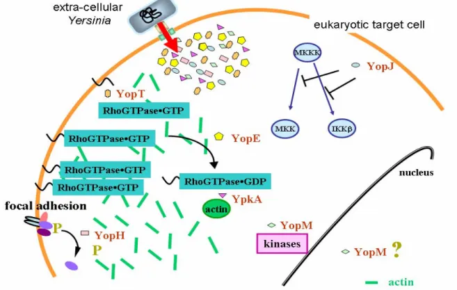
The T3SS of Yersinia is used by the bacteria to translocate a minimum of six anti-host effector proteins into target cells (figure 1). Taken together the joint action of these toxins allows the bacteria to escape the host’s immune system and remain in an extra-cellular milieu to replicate. At least four effector proteins are essential for
Yersinia to cause systemic infections in mice; YopM, YopH, YopE and YpkA [40,
88, 102, 162]. The role of YopJ and YopT in virulence is still unclear [103, 181, 289].
YopJ (YopP in Y. enterocolitica) induces apoptosis in macrophages [180, 182,
317] by suppression of pro-inflammatory cytokines via inhibition of the MAP kinase pathways (figure 1) [199, 202, 248, 317] and is involved in inhibition of the adaptive immunity [288]. YopM is a Leucine Rich Repeat (LRR) protein [80, 163] which upon secretion can inhibit platelet aggregation [162] and also binds α-thrombin in vitro [230, 258]. It is thereby thought to promote dispersion of the bacteria in the blood. The function of YopM in eukaryotic cells is not known, but it
Figure 1. Translocated effectors of Yersinia disrupt cellular signalling. The co-ordinated attack on the actin cytoskeleton and focal adhesions by YopH, YopT, YopE and YpkA is believed to prevent phagocytosis. YopJ inhibits MAP kinase induction of cytokines and induces apoptosis. The function of YopM is unknown, but the protein can be found associated with cellular kinases and in the nucleus.
interacts with two cellular protein kinases [174] and can also be detected in the nucleus (figure 1) [256]. YopM may, however, interfere with cytokine-dependent activation of vital cellular components of the innate immunity [148].
The remaining four effector proteins (YopH, YpkA, YopT and YopE) all target different aspects of actin dynamics in the eukaryotic cell (figure 1). YopH is one of the most potent tyrosine phosphatases described [318] and an essential virulence determinant of Yersinia [40] involved in anti-phagocytosis [238], inhibition of calcium signalling in neutrophils [14] and inhibition of T and B cell activation [10, 309]. YopH de-phosphorylate eukaryotic signalling proteins important for focal adhesion function and T cell activation such as p130Cas, FAK, Fyb, LAT and Slp-76 [13, 32, 35, 72, 106, 213]. YpkA (YopO in Y. enterocolitica) is a serine/threonine protein kinase [102] found at the inner surface of the eukaryotic cell membrane after translocation [118]. YpkA requires actin as an activating co-factor in infected cells (figure 1) [143] and although YpkA interacts with the small GTPases RhoA and Rac1 (important regulators of the actin cytoskeleton – discussed below) [22, 75], there exists no evidence of YpkA-dependent phosphorylation of these proteins. It may be that the actin disruption by YpkA is due to its ability to block activation of RhoA without interfering with GEF-induced GDP/GTP exchange [75, 143, 188].
In contrast to YopH and YpkA, direct effects on the distribution and activity of small GTPases have been identified for YopT and YopE [2]. YopT is a cysteine protease responsible for removing the C-terminus of RhoA [3, 254]. This induces a re-distribution of membrane-bound RhoA to the cytosol (figure 1) and uncoupling of the RhoA-effector interaction necessary for RhoA regulation of the actin cytoskeleton [262]. The end result of YopT activity is cellular cytotoxicity [138].
YopE is a GTPase Activating Protein (GAP) with in vitro activity towards RhoA,
Rac1 and Cdc42 (figure 1) [31, 304]. Upon bacterial infection of eukaryotic cells or micro-injection of purified protein, YopE induces a rapid collapse of the actin cytoskeleton [240], presumably via its ability to down-regulate small RhoGTPases [5, 15, 31, 304]. Together with YopH, YopE was initially recognized as one of the Yop effectors responsible for blocking phagocytosis of Yersinia [238, 239]. Other effects of translocated YopE have also been attributed to the GAP activity, such as prevention of Yersinia-induced pore formation in target cell membranes [296], inhibition of pro-inflammatory signalling [183, 249] and disruption of tight junctions [279]. Taken together, this emphasises YopE as a key effector in Yersinia virulence. By targeting members of the small RhoGTPase family, one effector can have multiple effects on signalling pathways in target cells. This is evident from results reported in this thesis (see Results and Discussion section).
Sub-groups Members
Cdc42 Cdc42, TC10, TCL, Chp, Wrch-1
Rac Rac1, Rac2, Rac3, RhoG
Rho RhoA, RhoB, RhoC
RhoD RhoD, Rif
Rnd Rnd1, Rnd2, Rnd3/RhoE
RhoH/TFF RhoH/TFF
RhoBTB RhoBTB1, RhoBTB2
Miro Miro-1, Miro-2
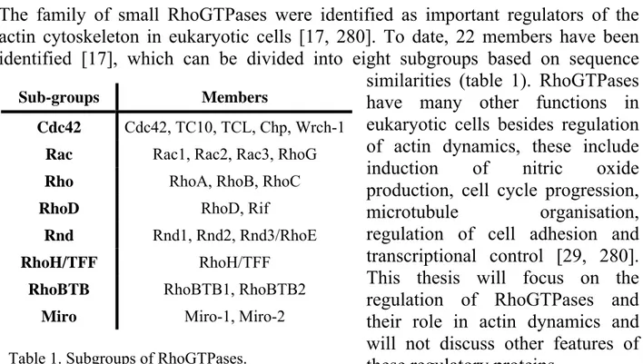
Table 1. Subgroups of RhoGTPases.
The family of RhoGTPases
The family of small RhoGTPases were identified as important regulators of the actin cytoskeleton in eukaryotic cells [17, 280]. To date, 22 members have been identified [17], which can be divided into eight subgroups based on sequence
similarities (table 1). RhoGTPases have many other functions in eukaryotic cells besides regulation of actin dynamics, these include induction of nitric oxide production, cell cycle progression,
microtubule organisation, regulation of cell adhesion and
transcriptional control [29, 280]. This thesis will focus on the regulation of RhoGTPases and their role in actin dynamics and will not discuss other features of these regulatory proteins.
Regulation of RhoGTPases
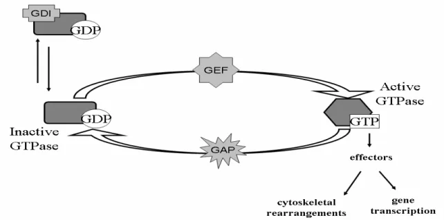
RhoGTPases function as molecular switches, alternating between a GTP-bound active conformation and a GDP-bound inactive conformation (figure 2) [280]. A post-translational modification of the C-terminal CAAX-domain confers membrane-localisation to the GTPase [280]. Important regulators of RhoGTPase activity are Guanine Exchange Factors (GEF), GTPase Activating Proteins (GAP) and Guanine nucleotide Dissociation Inhibitors (GDI) (figure 2) [280]. The proteins regulating RhoGTPases out-number the GTPases by at least 3 to 1 for the GEFs and the GAPs [206], showing the importance of tight regulation of RhoGTPases for correct function in eukaryotic cells. The GEF and GAP functions are often found as discrete domains in multi-functional proteins [206]. GEFs activate RhoGTPases by exchanging the GDP for a GTP upon an activation signal (figure 2) [280]. RhoGDIs bind to GDP-bound GTPases and block spontaneous release of the nucleotide, thereby preventing activation (figure 2) [246]. Extra-cellular signalling results in release of GDI from the GTPase thereby allowing for subsequent activation by GEFs [246, 280].
RhoGTPases have an intrinsic GTP hydrolysis activity, which can be increased by GAPs (figure 2) [206]. Hydrolysis of GTP to GDP inactivates the RhoGTPase by inducing a conformational change resulting in loss of interaction with downstream effector proteins [29, 206]. Common to all eukaryotic RhoGAPs is an essential arginine residue, positioned to stabilize the transition state during hydrolysis of the GTP nucleotide [315].
RhoGTPases and the actin cytoskeleton
RhoGTPases were first discovered as regulators of the actin cytoskeleton, with distinct morphological changes dependent upon activation of Rac1 (lamellipodia), RhoA (stress fibre formation) or Cdc42 (filopodia) [17, 194, 233, 234]. Most studies have focused on these three members, but successive identification of numerous other RhoGTPase family members have painted a picture of sophisticated cross-talk within the eukaryotic cell for control of the actin cytoskeleton and cellular processes dependent upon functional actin dynamics. Such processes include motility, endocytosis, cell cycle progression, adhesion and phagocytosis [225, 231, 232, 280].
RhoGTPases do not directly interact with the actin cytoskeleton. Rather regulation occurs via down-stream effector proteins (figure 2) [29]. Most members of the RhoGTPase family have been implicated in direct regulation of the actin
Figure 2. Regulation of small RhoGTPases. Guanine Exchange Factors (GEFs) activate the GTPase upon stimulation. GTPase Activating Proteins (GAPs) increases the intrinsic GTP-hydrolysis, thereby inactivating the GTPase. Guanine nucleotide Dissociation Inhibitors (GDIs) sequester inactive GTPases and prevent spontaneous exchange of GDP to GTP. Only active GTPases interact with down-stream effector proteins.
cytoskeleton [17]. However, RhoH and the Rnd proteins are GTPase deficient [58, 164]. RhoH rather seems to function as an inhibitor of the NFκB and p38 pathways, which are activated by other members of the Rho family [164]. Rnd1-3 are thought to antagonize the action of RhoA on stress fibre formation [17, 58]. In addition, phosphorylation of Rnd3 by a RhoA-effector increases its stability and activity, thereby suggesting a way of down-regulating RhoA induced pathways in the cells [235].
Most members of the Cdc42-group affect the formation of filopodia in cells, albeit to different extent [17]. In addition, Chp and Wrch-1 both act as activators of the JNK MAP kinase pathway [16, 282]. Rif induces the formation of long filopodia in a Cdc42-independent manner [209], and both RhoD and Rif reorganise the actin cytoskeleton extensively [17]. The RhoA-family induce stress fibres when over-expressed in cells [17]. All members of the Rac1-family induce lamellipodia in cells and Rac1 and Rac2 can also induce thick bundling of actin filaments [17]. RhoBTB1 and 2 have a modular organisation with a RhoGTPase domain followed by other domains important for protein-protein interactions [229]. No role for the RhoBTB proteins in actin regulation has been identified [17]. Miro proteins are atypical RhoGTPases and function within mitochondrial homeostasis, rather than actin regulation [17, 94].
Control of endocytosis by RhoGTPases
Several GTPases regulate endocytic pathways, including phagocytosis and receptor recycling [79, 225, 232]. RhoB closely associates with late endosomes and retards receptor trafficking through the endosomal pathway [232]. RhoD localises to endosomes and is proposed to modulate endosome distribution and motility [232]. TCL is implicated in regulation of receptor endocytosis and recycling [70]. TC10 also plays a role in recycling of receptors via the endocytic pathway, perhaps by remodelling of cortical action [57, 59, 113]. RhoGTPases may also function in exocytosis in a variety of cell types and are implicated in the release of neuro-transmitters [232].
Phagocytosis is a specialised form of endocytosis and the first line of defence against invading micro-organisms by the innate immune system. Two major types of phagocytosis by professional phagocytes have been identified and both processes are regulated by small GTPases of the Rho family. Rac1 and Cdc42 are involved in Fc-receptor phagocytosis (type I) whereas RhoA has been identified as an important regulator of complement receptor phagocytosis (type II) [225].
signalling pathways down-stream of the Fc- and complement receptors [13, 82, 300].
RhoGTPases as targets for bacterial toxins
Many pathogens exploit the actin cytoskeleton of eukaryotic cells either to promote attachment to cells and uptake into intra-cellular compartments for replication [111, 319], or as is the case with Yersinia, the actin cytoskeleton is targeted in order to prevent phagocytosis and ensure an extra-cellular milieu for bacterial replication [67, 111]. A common way used by many bacterial species to interfere with the actin cytoskeleton is by targeting members of the RhoGTPase family [111]. Significantly, these are not necessarily substrates of T3SS. Cytotoxic Necrotising Factor (CNT) of Escherichia coli and Dermonecrotic toxin (DNT) of Bordetella species activate members of the Rho family by constitutive modifications preventing nucleotide hydrolysis [131]. Exoenzyme S (ExoS) of Pseudomonas
aeruginosa has a C-terminal ADP-ribosyltransferase activity [153, 166], which
modifies members of several different GTPase families, either resulting in activation (in the case of Rac1 [237]) or deactivation (in the case of Ras and RhoA [126]). Clostridium spp. localise toxins into target cells to constitutively inactive RhoGTPases [131].
Another group of bacterial toxins mimic the eukaryotic regulatory proteins of RhoGTPases (figure 2). By interfering with the proper control of the GTPase activity, the bacteria can modulate the host response to suit its purposes [111]. YopE of Yersinia has been shown to act as a GAP towards RhoA, Rac1 and Cdc42
in vitro [5, 31, 304] and disrupt Rac1 dependent signalling pathways in eukaryotic
cells [15], resulting presumably in prevention of phagocytosis [15, 31, 238].
Salmonella spp. promotes its own uptake into epithelial cells by actin remodelling
and then subsequent host cell recovery [319]. Uptake is mediated by translocation of the T3SS effectors SopE and SopE2, GEFs for Cdc42 and Rac1 [95]. Cell recovery requires translocation of SptP that possesses a YopE-like GAP domain that targets Rac1 and Cdc42 in vitro [97, 98, 267]. Another GAP domain is found in ExoS, which targets Rac1, RhoA and Cdc42 both in vitro and during infection of eukaryotic cells [126, 150, 208, 306]. P. aeruginosa also encodes a second GAP-containing toxin, Exoenzyme T (ExoT) with 76% identity to ExoS [21]. The GAP activity of ExoT targets the same substrates as ExoS [147], making it hard to understand why the bacteria carries two proteins with seemingly the same function. Several other identified T3SS effector proteins can modulate the activity of small RhoGTPases in infected eukaryotic cells, but their exact mechanisms of action are unknown [111].
GENERAL AND SPECIFIC AIMS
One general aim of this study was to investigate the function of the translocated protein YopE, an essential virulence determinant of Yersinia pseudotuberculosis. YopE belongs to a small group of bacterial GTPase Activating Proteins (GAPs). My work focused on trying to understand the biological functions of YopE in relation to its GAP activity.
Specific Aims:
I. Define regions in YopE essential for its known properties; in vitro GAP activity towards small GTPases and disruption of the actin cytoskeleton? II. Identify the in vivo targets of YopE in infected eukaryotic cells.
III. Identify amino acids essential for the in vivo activity of YopE, focusing on virulence in mice and inactivation of small GTPases in infected cells. A second general aim was to study the control of translocation during Yersinia infection of eukaryotic cells. Not only did this examine potential regulatory roles of pore-forming components, since this feature is a pre-requisite for translocation, but also the idea that translocated effectors could exhibit regulatory effects.
Specific Aims:
IV. Investigate the molecular mechanisms underlining the LcrH-YopD interaction.
V. Examine whether LcrH-dependent secretion of YopB and YopD can
influence efficiency of effector translocation.
RESULTS AND DISCUSSION
YopE is an essential virulence determinant of Yersinia pseudotuberculosis with an actin disrupting activity and the ability to prevent bacterial phagocytosis by macrophages [88, 239, 240].
YopE is a 219 amino acids long protein (figure 3) [88], with a T3SS secretion signal in the first 11 amino acids [168, 247]. Stability, secretion and translocation of YopE is facilitated by binding to the cytosolic chaperone YerA to a domain in the N-terminus of YopE (figure 3) [247, 263, 264, 293, 294, 302]. In recent years YopE has been shown to function as a GTPase Activating Protein (GAP) towards three members of the RhoGTPase family in vitro and to prevent Rho dependent signalling in yeast [304]. Moreover, mutation of the essential arginine at position 144, renders the YopE protein non-functional towards RhoA, Rac1 and Cdc42 in
vitro and the corresponding mutant bacteria is avirulent in mice hence the GAP
activity of YopE is essential for virulence [5, 31, 304]. A Membrane Localisation Domain (MLD) for targeting to eukaryotic membranes have been identified in the N-terminus of the protein (figure 3), overlapping in part with the domain necessary for translocation and YerA binding [44, 151].
In vitro GAP activity towards RhoA, Rac1 and Cdc42 can be
uncoupled from cytotoxicity
We wanted to characterise regions in YopE important for cytotoxicity towards HeLa cells and determine if this correlated to in vitro GAP activity towards RhoA, Rac1 and Cdc42. By deletion analysis of the N- and C- terminal ends of YopE we defined an essential minimal region of amino acids 90-215 for YopE dependent Figure 3. Organisation of the YopE protein. Amino acids 1 to 11 are essential for secretion. The YerA chaperone binds to amino acids 11 to 74 as a homodimer. The C-terminal (amino acids 89 to 219) is a GTPase Activating Protein (GAP) towards small RhoGTPases. Amino acids 54 to 75 constitute a Membrane Localisation Domain (MLD) in YopE for membrane targeting inside eukaryotic cells.
90-215) of YopE was carried out by smaller in frame deletions. Using this approach, all except four mutants lost the cytotoxicity phenotype. The four regions dispensable for cytotoxicity encompassed the residues 131 to 135, 162 to 165, 189 to 192 and 211 to 215 (figure 2 and 3, PAPER I). Despite this wild-type phenotype, none of these four deletion mutants could inactivate RhoA, Rac1 or Cdc42 in vitro (figure 5, PAPER I). These results show for the first time that it is possible to genetically separate the cytotoxic function of YopE from its in vitro GAP activity towards RhoA, Rac1 and Cdc42. This brings into question the essential nature of RhoA, Rac1 and Cdc42 as targets for YopE during cell infection.
Even small deletions can cause changes to a protein’s overall tertiary structure. With the help of structural modelling [114, 115, 149] and by using the combined knowledge from the solved structures of YopE, ExoS and SptP [81, 267, 306], we showed that changes in the poly-peptide structure could occur in at least four of our deletion mutants. Therefore, we generated more subtle mutations using alanine substitution to further analyse the GAP domain of YopE. Remarkably, alanine mutagenesis revealed only one amino acid triplet essential for cytotoxicity (aa 181-183), supporting the idea that our original deletions caused disturbances in the overall structure (table 1, PAPER I). In contrast, only one triplet substitution mutant (aa 210-212) approached the level of the GAP activity of the wild type protein (figure 5 and table 1, PAPER I). This further supports the notion that cytotoxicity is not dependent on functional in vitro GAP activity towards RhoA, Rac1 and Cdc42.
The finding that in vitro YopE GAP activity towards RhoA, Rac1 and Cdc42 could be separated from its cytotoxic effect on target cells is important. This offers a way to dissect the molecular mechanisms of one function in isolation from the other. However, as a note of interest, the data could also imply that the main in vivo target of YopE does not include RhoA, Rac1 and/or Cdc42. Thus, the question really is what the critical target of YopE is during infection of the host?
In vivo substrate specificity of YopE in infected cells
To better understand the function of YopE during infection, we employed pulldown assays to look at the activation state of RhoA, Rac1 and Cdc42 in infected HeLa cells. This approach has previously identified RhoA and Cdc42 as targets for the GAP activity of wild type ExoS [126]. We showed that YopE does not target Cdc42 in cells, although it is an efficient GAP in vitro towards this GTPase (PAPER II, [5, 304]). However, inactivation of Rac1 and RhoA by wild type
Yersinia occurs as early as 5 and 30 minutes, respectively, after initiation of
infection (figure 7, PAPER II).
Extensive mutagenesis of the GAP domain of YopE revealed a function of amino acids 181-183 in separating in vitro GAP activity towards RhoA, Rac1 and Cdc42 from cytotoxicity (PAPER I). This amino acid triplet lies in a domain (aa 178 to aa 187) with a high degree of similarity in the three bacterial GAPs and this region will be referred to as the “homology domain” (figure 1, PAPER II). The homology domain could be involved in target binding by SptP [267]. We created six single alanine substitutions of amino acids 178 to 183 to further dissect the functions of YopE during Yersinia infections. A range of unique phenotypes was revealed when these mutants were analysed.
First, we showed that the cytotoxicity of YopE is dose-dependent. In other words, a cytotoxic phenotype was observed when the mutant protein (W181A) was expressed in trans from a multi-copy plasmid, but not when this same mutant protein was produced under native conditions in cis on the Yersinia virulence plasmid (table 1, PAPER II). This was not seen with the R144A GAP mutant, devoid of the catalytically essential arginine residue (table 1, PAPER II). Perhaps the W181A mutant can sequester an essential regulator of the actin cytoskeleton when hyper-translocated. Yet, if this was true, the GAP mutant would be expected to behave similarly, since mutation of the arginine 144 is not believed to alter the tertiary structure of YopE. We also considered the possibility that W181A still possessed some residual GAP activity, but at least in vitro, it displayed no activity towards RhoA, Rac1 and Cdc42 at concentrations at least 100 times higher than that required by wild type YopE to completely hydrolyse bound GTP to GDP (figure 6, PAPER II). The other five mutants in the homology domain were all cytotoxic towards HeLa cells irrespective of their level of expression (table 1, PAPER II). All further studies were conducted with mutants where the mutation was introduced on the virulence plasmid.
Only two mutants created in the “homology domain” (F178A and S179A) were virulent in mice (table 1, PAPER II), while all the mutant proteins tested were defective for in vitro GAP activity towards RhoA, Rac1 and Cdc42 (figure 6, PAPER II). This result further substantiates our conclusions from Paper I that there is little correlation between cytotoxicity, in vitro GAP activity and virulence.
It is prudent to consider that in vitro GAP activity profiles of YopE are only a guide to what might be happening in vivo. After all, studies on eukaryotic GAP proteins show differences between in vitro and in vivo GAP activity [253, 316]. Thus, to
clarify the effect of the single alanine mutants on endogenous RhoA, Rac1 and Cdc42 during cell infections, we performed pulldown assays of active GTPases at 30 minutes post-infection (figure 8, PAPER II). Surprisingly, one virulent mutant (F178A) was severely reduced in its ability to inactivate RhoA and Rac1 in infected cells at 30 minutes post-infection. In addition, the attenuated mutant G182A was indistinguishable from wild type at this time point (figure 8, PAPER II). Furthermore, the avirulent mutant T183A, which showed wild type activity towards Rac1, was unable to inactivate RhoA (figure 8, PAPER II). Taken together these mutants suggest that additional target(s) for YopE inside eukaryotic cells are necessary for virulence, and although YopE targets RhoA and Rac1 during cell infection, these GTPases are not the essential targets.
This is the first evidence that the “homology domain” of YopE is involved in target recognition and that individual amino acids in a bacterial GAP domain can influence binding to distinct GTPases. If the proposed anti-phagocytic function of YopE [31, 239] is critical for Yersinia virulence, a defect in GAP activity as seen with the F178A mutant, should have had severe consequences on bacterial virulence. However, this is not the case, suggesting that alternative targets of YopE are essential to cause disease or that YopE possesses an alternative, as yet undescribed, function during infection.
The role of YopE in prevention of pore formation
We also analysed the single alanine mutants for their ability to prevent pore formation in the eukaryotic cell membrane, analysed as release of lactate dehydrogenase (LDH) from infected cells (figure 5, PAPER II), an activity dependent on the GAP function of YopE (PAPER III, [296]). Whether this phenotype has relevance in the context of in vivo YopE function is clouded. However, it correlates well with the ability of YopE to cause cytotoxicity and LDH release is dependent on functional actin cytoskeleton regulation [296], which suggests an additional role of YopE that could be investigated with our new assortment of phenotypically distinctive mutants. Indeed, only non-cytotoxic mutants of YopE (R144A and W181A) have lost the ability to block LDH release (figure 5, PAPER II), presumably by an inability to prevent actin dependent pore formation in the eukaryotic plasma membrane [296]. All cytotoxic mutants of YopE blocked LDH release (figure 5, PAPER II). This establishes a clear link between the YopE-dependent functions of cytotoxicity and prevention of pore formation. This is consistent with the role for host cell actin in both phenotypes. Interestingly, the relationship between pore prevention (blockage of LDH release)
and virulence is less clear, because the avirulent T183A mutant is still cytotoxic and efficiently blocks LDH release (table 1 and figure 5, PAPER II).
We also analysed the contribution of all known translocated effector Yops with respect to LDH release from infected cells. No other Yop effector was found to be involved (figure 2, PAPER III). Control of LDH release is a phenomenon specific to YopE. Thus, the YopH- and YpkA-induced cytotoxicity must be distinct from that induced by YopE and generates different cell fates [2, 43, 143]. In addition, although a yopK null mutant induces bigger pores in sheep erythrocytes and hyper-translocates effector proteins (figure 3, PAPER III, [134]), it still prevents LDH release to the same extent as wild type Yersinia (figure 2, PAPER III). This data elevates YopE towards a specific role in preventing Yersinia-induced pore formation, an activity dependent on the polymerization/de-polymerization cycle of the actin cytoskeleton [296]. On the basis of our avirulent T183A mutant, which is still cytotoxic, prevents pore formation and functions as a GAP in vivo towards Rac1 (PAPER II), we propose a role for Rac1 in YopE-mediated control of LDH release and cytotoxicity. However, the fully cytotoxic F178A mutant is defective for Rac1 inactivation in infected cells (PAPER II). This suggests that an additional GTPase is targeted for the actin disruption phenotype associated with YopE or that very low GAP activity is sufficient for cytotoxicity to occur, since F178A has a low
in vitro GAP activity towards Rac1, RhoA and Cdc42 (PAPER II).
So are these phenotypes a true reflection of the role of YopE inside eukaryotic cells? The answer might be both yes and no – yes, because YopE targets Rac1 and RhoA in vivo, and no, because one virulent mutant (F178A) could not inactivate Rac1 and RhoA in infected cells. Perhaps YopE targets a number of GTPases in eukaryotic cells during infection. Although YopE-targeting of Rac1 leads to actin cytoskeleton disruption, we do not believe that Rac1 is the target ultimately responsible for the contribution of YopE in Yersinia virulence. The family members of RhoGTPases have a high degree of identity and eukaryotic GAPs often target more than one member [206, 316]. Both Rac1 and RhoA possess close homologous counterparts that function within vesicle transport and cellular immune responses (e.g. RhoB, RhoD and Rac2) [142, 225, 307]. Thus, the effect of YopE on Rac1 and RhoA could be a secondary effect of its ability to target another closely related GTPase.
Regulation of effector translocation into target cells – a role for YopE
The lack of correlation between control of pore formation and virulence is intriguing. As a result we initiated studies to try and uncover the consequences of
Figure 4. Model of YopE activities inside eukaryotic cells. Inactivation of Rac1 (and to some extent RhoA) by YopE accounts for the actin disruption activity of YopE. YopE regulates the level of effector translocation together with YopK by inactivation of an unknown GTPase. The latter function of YopE is essential for virulence in mice.
the inability of YopE to prevent pore formation. Interestingly, the YopE GAP mutant, which does not prevent LDH release (figure 5, PAPER II), had also lost control of translocation into eukaryotic cells – translocating elevated levels of itself and YopH (figure 3, PAPER III). Moreover, the GAP mutant maintained constitutive Yop synthesis in the presence of eukaryotic cells (figure 4, PAPER III). In contrast, wild type Yersinia demonstrates a tight control of effector translocation levels and also produced less Yops in the presence of cells than the GAP mutant (figure 3 and 4, PAPER III). This suggested a role for the GAP activity of translocated YopE in sending a signal back to the bacterium to turn off further synthesis, secretion and translocation of Yops. This highlights one mechanism by which Yersinia can strike a balance between paralysing target cells and avoiding activation of the innate immune response through excessive toxin-induced cellular damage. This notion of YopE-dependent translocation control was further strengthened by the observation that hyper-translocation also occurred in the avirulent mutants W181A and T183A (figure 3, PAPER II). This is the first time a translocated T3SS effector has been shown to be involved in regulation of translocation of itself and other effectors. Importantly, this regulatory effect is specific to the presence of eukaryotic cells because during in vitro growth conditions, all yopE mutants regulate Yop synthesis normally (figure 4, PAPER II).
If regulation of translocation initiation is understudied in the T3SS, almost nothing is known about the signals that control termination of translocation. One study has observed hyper-translocation of effector proteins by a
yopK mutant that renders the
bacteria avirulent in the mouse model of yersiniosis [134]. This finding has significant similarities to our observation with YopE. It appears that Yersinia utilises YopE and YopK to determine when enough translocated effectors have been introduced into target cells (figure 4). The absence
of this mechanism spells doom for the infecting bacteria. Thus, bacteria must weigh the energetic cost of infinite Yop production against the programmed functions of the translocated effectors through a function of translocated YopE. The net result is translocation control during infection and effective colonisation of the host. This unique function for YopE in translocation control requires GAP activity. However, what might be the target of this activity is still unclear, for avirulent mutants in other parts of YopE, having lost regulatory control, still maintain some functional GAP activity towards established targets in vivo (PAPER II).
We therefore propose a model whereby YopE exerts a role on two functional pathways inside eukaryotic cells, targeting at least two different GTPases (figure 4). One pathway influences cytotoxicity and control of pore formation, and at least in part is mediated by Rac1 targeting. The other pathway involves effector translocation control, which is an essential virulence trait of YopE (PAPER II), and is mediated by targeting of an as yet unknown target (figure 4). The second pathway controlling translocation also requires functional YopK [134].
Functional differences between YopE and ExoS
Exoenzyme S (ExoS) of Pseudomonas aeruginosa is a bi-functional T3SS translocated effector with an N-terminal YopE-like GAP domain. ExoS has been shown to act as a GAP towards Rac1, RhoA and Cdc42 [107, 126]. We wanted to investigate if ExoS has the same function as YopE in effector translocation control (PAPER III). Wild type ExoS has two enzymatic activities, so we analysed mutants devoid of either GAP activity or the ADP-ribosyltransferase (ADPRT) activity that also targets small GTPases [126]. The GAP domains of YopE and ExoS share 40% identity. Surprisingly, the ExoS GAP activity was not sufficient to complement a
yopE null mutant phenotype with respect to elevated LDH release or
hyper-translocation of YopH during infection of HeLa cells (figure 6 & 7, PAPER III). ExoS was translocated efficiently because all ExoS variants induced cytotoxicity on HeLa cells after 1 hour (table 1, PAPER III), fully complementing the yopE null mutant for this activity.
The reason behind an inability of ExoS to complement a yopE mutant defective in pore formation control or translocation control is very fascinating, especially since ExoS targets Rac1 both in vitro and in vivo [107, 126, 150] and Rac1 targeting appears to be important for at least YopE-mediated pore formation control (PAPER II and PAPER III). The ADP-ribosyltransferase activity of ExoS has been suggested to mask the GAP activity [126, 237]. However, it would then be expected that ADPRT negative ExoS should be able to complement the yopE null
mutant – this was not the case (figure 6 and 7, PAPER III). Interestingly, a recent study claimed that the effect of ExoS on Rac1 is cell type specific, perhaps due to the subcellular localisation of Rac1 in different cell lines [237]. In this context, it is worth noting that both ExoS and YopE contain a MLD, rich in hydrophobic amino acids (figure 5A), but with low amino acid homology, in the N-terminal part of the protein [151, 207]. Moreover, they appear functionally inter-changeable, in that domain swapping experiments between ExoS and YopE still enable hybrid proteins to be translocated and cause cytotoxicity [151]. We have also identified a similar sequence in the N-terminus of ExoT and SptP (figure 5A). This indicates a conserved localisation targeting mechanism for the bacterial GAP proteins.
Intriguingly, the MLD of YopE renders YopE dependent on YerA binding for protein secretion and translocation [44], although sufficient information for secretion and translocation of YopE is encoded in the first 50 amino acids of the protein [44, 247, 302]. Perhaps conservation of the MLD reflects the essential nature of this domain in targeting of YopE inside eukaryotic cells. The significance of the MLD in translocation control has so far not been investigated. We are in the Figure 5. The Membrane Localisation Domain (MLD) of YopE is involved in regulatory control of yop expression. (A) Alignment of MLDs from bacterial GAPs. Red (small, hydrophobic and aromatic side groups), blue (acidic side groups), green (hydroxyl, amine and basic (Q) side groups) and magenta (basic side groups). (B) Control of Yops production during bacteria-cell contact by YopE. Wild type (YPIII/pIB102), yopER144A (YPIII/pIB562) GAP defective mutant, yopK (YPIII/pIB155) or yopE∆MLD (YPIII/pIB529) were added to wells with (+) or without (-) HeLa cell monolayers for 90 min. Analysis performed as described in PAPER II and PAPER III.