ANALYSIS AND KINASE-MEDIATED DECAY OF RNAS WITH 5´-HYDROXYL TERMINI
by
SALLY E. PEACH
B.S., Massachusetts Institute of Technology, 2009
A thesis submitted to the Faculty of the Graduate School of the University of Colorado in partial fulfillment
of the requirements for the degree of Doctor of Philosophy
Molecular Biology Program 2016
© 2016 SALLY PEACH ALL RIGHTS RESERVED
This thesis for the Doctor of Philosophy degree by Sally E. Peach
has been approved for the Molecular Biology Program
by
Sandra L. Martin, Chair David L. Bentley Richard E. Davis Katerina J. Kechris
Jeffrey S. Kieft Jay R. Hesselberth, Advisor
Peach, Sally E. (Ph.D., Molecular Biology Program)
Analysis and Kinase-mediated Decay of RNAs with 5´-hydroxyl Termini Thesis directed by Assistant Professor Jay R. Hesselberth
ABSTRACT
RNA cleavage by metal ion-independent endoribonucleases and self-cleaving ribozymes produces RNA fragments with 5´-hydroxyl (5´-OH) and 2´,3´-cyclic phosphate termini. However, RNAs with 5´-OH termini (5´-OH RNAs) are not detected by conventional sequencing methods, and 5´-OH RNAs cannot be directly degraded by canonical 5´-phosphate-dependent 5´→3´ exoribonucleases.
To identify new 5´-OH RNA fragments, I exploited the unique ligation mechanism of E. coli RtcB RNA ligase to develop 5OH-seq, a novel method to capture and sequence 5´-OH RNAs. I applied 5OH-seq to budding yeast and captured known 5´-OH fragments produced by tRNA Splicing Endonuclease (SEN) during processing of intron-containing pre-tRNAs and by ribonuclease Rny1 during stress-induced cleavage of tRNA anti-codon loops. I identified numerous novel 5´-OH fragments derived from mRNAs: some 5´-OH mRNA fragments were derived from single, localized cleavages, while others were likely produced by multiple, distributed cleavages. Many 5´-OH mRNA fragments were produced upstream of codons for highly electrostatic peptides, suggesting that the fragments may be generated by co-translational mRNA decay, such as no-go decay (NGD). 5´-OH RNAs must be phosphorylated to become substrates for 5´-phosphate-dependent 5´→3´ exoribonucleases, a process I term “kinase-mediated decay” (KMD). During the unfolded protein response (UPR), the ER-localized Ire1 endoribonuclease cleaves HAC1 mRNA, yielding 2´,3´-cyclic phosphate and 5´-OH RNA fragments that are
ligated by Trl1 tRNA ligase to produce mature HAC1 mRNA and activate downstream target genes. The HAC1 fragments cleaved by Ire1 have previously been identified with 5´-phosphate (5-PO4) termini in vivo in xrn1∆ yeast, suggesting the HAC1 fragments are KMD substrates. I found that HAC1 3´-exons accumulate with 5´-PO4 termini in xrn1∆ yeast when Trl1 is present, but not kinase-dead Trl1-D425N or RtcB ligase, suggesting that Trl1 phosphorylates the HAC1 3´-exon for decay by Xrn1. Failure to degrade the HAC1 3´-exon promotes aberrant splicing and UPR activation without stimulus.
In summary, this thesis describes a method to directly capture and sequence 5´-OH RNAs (5OH-seq), a comprehensive analysis of 5´-OH RNAs in budding yeast, and evidence for a unified theory of 5´-OH RNA degradation by kinase-mediated decay (KMD). I also present preliminary data on the abundance of 5´-OH RNAs in mammalian tissues that lack the RNA 5´-kinase CLP1.
The form and content of this thesis are approved. I recommend its publication. Approved: Jay Hesselberth
DEDICATION
To Mom, for her boundless love and support throughout all of my crazy journeys, and for her timeless feminist wisdom, which helped shape my path:
ACKNOWLEDGEMENTS
First and foremost, I would like to acknowledge and thank my advisor, Jay Hesselberth. He approaches science with an excitement and curiosity that is contagious, and he has been beyond essential in helping to push my research forward. As I’ve said on numerous occasions, I definitely won the PI lottery.
The rest of the Hesselberth lab has been equally important for my success. Monica Ransom is an incredibly thoughtful person, and keeps up the spirits with scientific advice, presentation feedback, and cupcakes. Patrick Cherry provided important data for the kinase-mediated decay model (Figure 3.4), and is faster than Googling a biochemistry question. Erich Chapman has been a great role model and science brother, and he has made things far more interesting for us all. Big thanks to Austin Gillen(s) for secret -80ºC communications, and Kerri York for teaching me so much during that first year in the lab. Finally, to *Dr. Suzi*: it just wasn’t the same without you around. It has been a pleasure completing my graduate studies in this lab, and I will miss everyone greatly.
I am extremely thankful to my thesis committee. My chair Sandy Martin has been a fantastic role model. Dick Davis has been the most thorough and thoughtful critic of manuscripts. Jeff Kieft and David Bentley and their labs have been exceptionally generous with ideas, reagents, and machinery. Katerina Kechris has patiently answered my naïve statistics questions and helped me develop more rigorous data analysis approaches.
I have been fortunate to be a part of two fantastic graduate programs and one amazing department. Thanks to Arthur Gutierrez-Hartmann, director of the Medical Scientist Training Program (MSTP), for years of solid leadership and advice, and to Angie Ribera, former associate director of MSTP, for incredible support during one of the hardest
moments of my life. Bob Sclafani, director of the Molecular Biology Program, and Mark Johnston, chair of the Biochemistry and Medical Genetics Department, have been extremely encouraging and have taken an active role in my development as a scientist. The administrative staff behind the scenes made my life ultra easy, including: Annie Vazquez, Sabrena Heilman, Sue Brozowski, Lindsay Quandt, Karen Vockrodt, Michele Parsons, Emily Dailey, and Jodi Cropper. I have also appreciated the financial support of the Molecular Biology NIH training grant T32 GM008730.
Science has been mailed to me from all over the country and the world. Many thanks to Marc Hammarlund from Yale University; Stewart Shuman and Beate Schwer from Sloan Kettering; Roy Parker and Yuriko Harigaya from University of Colorado Boulder; Jingyan Wu and Anita Hopper from the Ohio State University; Ender Karaca and James Lupski from the Baylor College of Medicine; and Javier Martinez and Stefan Weitzer from the Institute of Molecular Biotechnology in Vienna. All of these scientists have shared samples and data with me during my efforts to find novel 5´-OH RNAs.
Several previous advisors have helped me on my way here. Ten years ago, Roger Summons accepted me into his lab at MIT with no previous experience, and D’Arcy Meyer-Dombard put up with teenaged-Sally and taught me how to wield a pipette. Dianne Newman supervised my first significant research project as a senior at MIT, which certainly changed my life’s direction. Jake Jaffe was a truly phenomenal mentor at the Broad Institute without whom I would have never applied to MD/PhD programs.
My friends and colleagues in the medical and graduate school have been critical to keeping me afloat. The list is large and incomplete, but here goes… From MSTP: Eric Nguyen for Battlestar Galactica, hosting all of my parties, and explaining happiness; Chris
Knoeckel for being my free Udi’s psychiatrist and teaching me the secrets to graduate school success; Mira Estin, Sarah Nelson, Hannah Scarborough, Veronica Searles Quick, and Genevieve Park for forming a core group of MD/PhD women that I could both look up to and befriend; Jing Zhang for that, plus an ever-growing collection of strange banana experiences; and the rest of my surviving MSTP “2011” cohort, especially Tamara Burleson Garcia for bringing some small-town Southern sense to this crazy world and just enough pink. From graduate school: my MolBio cohort, especially Ryan Sheridan, Alexis Zukowski, and Maggie Balas; Becky Fusby for keeping it real in the truest sense of that phrase; Seena Shah for one all-too-short year and “a proper English garden”; and Charlotte Siska for cows, tutus, and rolling in the dust. From medical school: Garland Castañeda for just enough sarcasm, spreadsheets, and summits to help me survive the first two years; and, of course, Stevie Kohler for continuously teaching me how to be human.
I’m also extremely thankful for those outside my Anschutz academic bubble, especially: Christie Ciarlo, who has been an essential adventuring partner, scientific comrade, and dear friend; MinWah Leung for setting an extremely high bar for both exploration and cheerfulness; and Camilo Guáqueta, for being my rock during this last grad school crunch time, BBB.
Finally, I would like to thank my family, who have been there for me since the beginnings of Little Woo: my sister Katie, who has taught me so much about overcoming adversity; my brother James, for his unfailing kindness and strength; my dad, for introducing me to the world of a physician; and, most importantly, to my amazing mom for her endless love, which will follow me always.
TABLE OF CONTENTS CHAPTER
I. INTRODUCTION ... 1
Overview of RNA Modifications ... 2
Generation of RNAs with 5´-hydroxyl Termini ... 4
5´-OH RNAs in Biological Systems ... 10
RNA Decay Pathways ... 30
Kinase-Mediated Decay ... 40
Methods for Global Mapping of RNA Modifications ... 48
II. GLOBAL ANALYSIS OF RNA CLEAVAGE BY 5´-HYDROXYL RNA SEQUENCING ... 53
Introduction ... 53
Materials and Methods ... 55
Results ... 60
Discussion ... 80
Summary ... 89
III. KINASE-MEDIATED DECAY OF EUKARYOTIC MRNA CLEAVAGE PRODUCTS ... 91
Introduction ... 91
Materials and Methods ... 93
Results ... 99
Discussion ... 110
IV. ANALYSIS OF 5´-HYDROXYL RNAS IN MAMMALS ... 117
Introduction ... 117
Materials and Methods ... 118
Results ... 122
Discussion ... 128
Summary ... 134
V. SUMMARY AND FUTURE DIRECTIONS ... 135
Applications and Improvements of 5OH-Seq ... 136
Future Studies and Considerations of Kinase-Mediated Decay ... 142
LIST OF TABLES TABLE
1.1 Functions of several eukaryotic metal ion-independent endoribonucleases. ... 11
2.1 Oligonucleotides ... 57 2.2 Yeast strains ... 57 3.1 Plasmids. ... 94 3.2 Yeast strains. ... 95 3.3 Oligonucleotides ... 98 4.1 Oligonucleotides ... 119
LIST OF FIGURES FIGURE
1.1 Two distinct mechanisms of endoribonuclease cleavage. ... 9
1.2 Yeast and human pathways for processing of intron-containing tRNAs. ... 15
1.3 Rny1 (yeast) and Angiogenin (humans) cleave the anticodon loops of tRNAs under stress conditions. ... 19
1.4 The unfolded protein response (UPR) in yeast is primarily regulated by the non-canonical cytosolic splicing of HAC1 mRNA. ... 25
1.5 Human IRE1α cleaves XBP1 and ER-proximal mRNAs. ... 28
1.6 Canonical cytosolic mRNA decay pathways: Xrn1 and the exosome. ... 34
1.7 Decay pathways for defective mRNAs: nonsense-mediated decay (NMD), non-stop decay (NSD), and no-go decay (NGD). ... 37
1.8 Examples of kinase-mediated decay (KMD). ... 46
2.1 Purified E. coli RtcB ligates RNA substrates with 3´-PO4 and 5´-OH termini .... 62
2.2. Schematic of method to clone and sequence RNAs with 5´-OH termini (5OH-seq). ... 63
2.3 5OH-seq captures RNAs with 5´-PO4 termini after in vitro dephosphorylation. ... 67
2.4 Cleavages in tRNA in introns, anticodon loops, and D arm and loop ... 68
2.5 Analysis of mRNAs captured by 5OH-seq. ... 71
2.8 5´-OH RNAs accumulate upstream of regions encoding
consecutive polycharged amino acids. ... 77 2.9 HAC1 mRNA cleavage fragments accumulate with
5´-OH and 5´-PO4 termini. ... 81
2.10 Some 5´-OH RNAs increase with tunicamycin treatment and
occur in a common motif. ... 83 3.1 Xrn1 degrades the phosphorylated HAC1 3´-exon cleavage product. ... 102 3.2 The HAC1 3´-exon mRNA cleavage product requires
the kinase activity of Trl1 to promote degradation by Xrn1. ... 105 3.3 Xrn1 suppresses HAC1 mRNA splicing and UPR activation. ... 108 3.4 Trl1 phosphorylates NGD 3´ fragments prior to
5´→3´ degradation by Xrn1. ... 111 3.5 Models of kinase-mediated decay of mRNA cleavage fragments. ... 113 4.1 CLP1 shRNA knockdowns validated by qRT-PCR and Western blot. ... 123 4.2 CLP1-deficient cell lines have increased 5OH-signal
surrounding PolyA sites. ... 124 4.3 CLP1 kinase-dead mutants differentially accumulate 5OH-RNAs
in mouse spinal cords. ... 126 4.4 SeC 3´-tiRNAs accumulate in the spinal cords of mice. ... 129 4.5 SeC tRNA Northern blot requires optimization. ... 132
CHAPTER I INTRODUCTION
RNA is a diverse molecule comprising the majority of nucleic acid content in a given cell. It can act as the messenger encoding protein sequence (mRNA), the molecule decoding the sequence and carrying amino acids during protein translation (tRNA), and a structural and catalytic component of the ribosome (rRNA). The constant production of this essential molecule necessitates robust decay mechanisms to break down RNA and recycle nucleotides. For instance, the canonical Xrn1 and exosome decay pathways in yeast maintain cytosolic homeostatsis by processive mRNA degradation. However, little is known about RNAs which exist outside of the known decay pathways. What happens when an RNA is cleaved and the resulting RNA terminus is immune to degradation by known exoribonucleases?
This dissertation seeks to address this question by employing both broad, transcriptome-wide analyses of RNAs with 5´-hydroxyl termini (5´-OH RNAs) as well as a focused study of the molecular mechanism of kinase-mediated decay (KMD) for specific 5´-OH RNAs within the unfolded protein response (UPR) and the no-go decay (NGD) pathway. This chapter provides an overview of RNA modifications, the generation and regulation of 5´-OH RNAs, the known RNA decay mechanisms, and the current methods of terminal RNA modification detection by next-generation sequencing. In Chapter II, I discuss the development and validation of the novel 5OH-seq method, which combines the unique ligation specificity of E. coli RtcB with massively parallel next-generation sequencing to directly capture and sequence 5´-OH RNAs. I describe the findings of the first transcriptome-wide analysis of 5´-OH RNAs in Saccharomyces cerevisiae, including
the enrichment of 5´-OH RNAs upstream of regions encoding polycharged amino acids and 5´-OH RNAs cleaved during UPR induction. In Chapter III, I further explore the 5OH-seq results in yeast. I demonstrate the molecular mechanism of the decay of two 5´-OH RNAs: the exon fragment of HAC1 mRNA generated by Ire1 cleavage and the 3´-exon fragment of a no-go decay (NGD) reporter construct. I term these and other related processes “kinase-mediated decay” (KMD). In Chapter IV, I apply the 5OH-seq method to mammalian RNA samples and describe results of a preliminary analysis of 5´-OH RNAs in mouse spinal cords, human cell lines, and human patient-derived fibroblasts with compromised 5´-OH kinase activity. In Chapter V, I summarize the collective findings and describe future directions and applications to RNA decay, including extensions to human disease. Together, these chapters provide a comprehensive overview of 5´-OH RNAs in eukaryotes and define the molecular mechanism of their degradation by KMD.
Overview of RNA Modifications
Modification of nucleobases and ribose sugars drastically increases the information coding potential of RNA, creating a “combinatorial chemistry playground” (Jackman and Alfonzo 2013). The first glimpse of RNA modification arose when the hydrolyses of yeast RNA with a phosphodiesterase and 5´ nucleotidase yielded inorganic phosphate, suggesting that some phosphoryl groups were attached to C5 of the ribose (Gulland and Jackson 1938). Since this early discovery of what is now known as the 5´-phosphate terminal modification (5´-PO4), over 100 RNA modifications, both terminal and internal, have been identified on all major RNA types (tRNA, rRNA, and mRNA) and several minor RNA subspecies (snRNA and miRNA) in all domains of life (Machnicka et al. 2012).
tRNA provides a stunning example of internal RNA modifications. In 1957, Davis and Allen described an abundant “fifth nucleotide” that comprised 4% of yeast tRNA nucleotides (Davis and Allen 1957). The chemical structure and properties of this pseudouridine (Ψ) nucleotide, the C5-glycoside isomer of uridine, were revealed shortly thereafter (Cohn 1960). Over the next half century, dozens of tRNA modifications were discovered (Reviews: Hopper 2013; Jackman and Alfonzo 2013; Helm and Alfonzo 2014). An analysis of 561 sequenced tRNA molecules from a variety of species found 11.9% of the all nucleotides differed from the canonical bases and 16.4% of nucleotides in 34 sequenced yeast cytoplasmic tRNAs are modified (Sprinzl and Vassilenko 2005; Phizicky and Alfonzo 2010). Though the role of most tRNA modifications is not well-understood, it is abundantly clear that many RNA modifications are required for normal cellular functioning. Over 200 diseases have been linked to tRNA hypomodification, including cancers, asthma, diabetes, and neurological diseases (Torres et al. 2014; Abbott et al. 2014). In addition to modifications of the internal nucleotides, the 5´ and 3´ termini of RNA can be covalently modified. The nature of these termini can inhibit or promote recognition by enzymes that modify the end chemistry by phosphorylation, cyclic phosphate diesterase activity, adenylation, guanylation, ligation, and so on. Perhaps the most well-studied example of a regulated 5´-terminal RNA modification is the 7-methylguanosine (m7G) cap on the 5´ terminus of mRNA. In the mid 1970s, a series of publications demonstrated the presence of the m7G cap on the 5´ terminus of mRNAs from viruses, slime mold, yeast, and mammals (Shatkin 1976). During transcription, nascent pre-mRNA is dephosphorylated by RNA triphosphatase Cet1 (Tsukamoto et al. 1997). A guanine nucleotide is transferred to the resulting 5´-bisphosphate mRNA by mRNA
guanylyltransferase Ceg1 creating a 5´-to-5´ triphosphate linkage. Methylation of the guanine by mRNA (guanine-N7-)-methyltransferase Abd1 yields a m7G-mRNA (Mao et al. 1995). The m7G cap recruits eukaryotic initiation factor 4E (eIF4E) to promote translation (Sonenberg et al. 1979), and it also promotes 5´ proximal intron excision (Ohno et al. 1987). Importantly, the m7G cap prevents recognition by 5´→3´ exoribonuclease Xrn1, and must first be removed by Dcp1/Dcp2 to promote decay of the mRNA (discussed further in the “RNA Decay” section).
In this dissertation, I seek to characterize another terminal RNA modification: the 5´-hydroxyl terminus. I consider the 5´-OH terminus a “hidden” terminus for two reasons. First, 5´-OH RNAs have evaded extensive characterization due to their incompatibility with sequencing methods that rely on 5´-phosphate-dependent chemistries. Therefore, in order to comprehensively identify 5´-OH RNAs with precise resolution, I needed to develop a new method to specifically capture and map this previously “hidden” RNA species (Chapter II and Chapter IV). The second reason is that, similar to the m7G capped mRNAs, 5´-OH RNAs cannot be recognized or degraded by 5´-phosphate-dependent 5´→3´ exoribonucleases. In Chapter III, I describe how the “hidden” 5´-OH terminus can be phosphorylated, creating a competent substrate for 5´→3´ RNA decay.
Generation of RNAs with 5´-hydroxyl Termini
Within this section, I introduce three key processes generating 5´-OH RNAs: intrinsic cleavage, self-cleaving ribozymes, and metal-ion independent endoribonucleases. Cleavages by several metal-ion independent endoribonucleases are of particular interest in
regulated pathways, which are further described in the subsequent section, “5´-OH RNAs in biological systems”.
Intrinsic RNA cleavage
RNA can assume a conformation promoting sequence-independent cleavage. This intrinsic RNA cleavage likely proceeds via an in-line SN2-like reaction that requires an initial deprotonation of the 2´-OH of the upstream ribose. Following deprotonation, the 2´-O functions as the nucleophile and attacks the neighboring 3´-5´ bridging phosphate prompting the 5´-O of the downstream ribose to act as a leaving group. Intrinsic RNA cleavage yields a 5´ RNA fragment with a 2´,3´-cyclic phosphate terminus and a 3´ RNA fragment with a 5´-OH terminus. The cleavage is relatively non-specific and likely accounts for a significant portion of in vitro RNA fragmentation.
Certain metal ions can enhance intrinsic RNA cleavage. Metal ions promote RNA cleavage by acting as Brönsted bases and abstracting a proton from the 2´-OH (Brown et al. 1985). Indeed, acceleration of RNA cleavage by divalent lead cations was first shown over 65 years ago (Dimroth et al. 1950). Other divalent cations Mg2+ and and Zn2+ also increase the rate of cleavage compared to uncatalyzed reactions, and lanthanide ions Eu3+, Tb3+, and Yb3+ are even more efficient catalysts, accelerating the rate of dinucleotide cleavage 3-4 orders of magnitude (Breslow and Huang 1991).
Intrinsic RNA cleavage is important for many RNA sequencing methods. Single-stranded RNA cleaves at a rate ~100 times higher than double-Single-stranded RNA (Soukoup and Beaker, 1999), enabling partial mapping of RNA structure by differential cleavage in the presence of metal cations (Forconi and Herschlag 2009). Intentional RNA
fragmentation is a critical step in the optimization of various sequencing library preparations, including the 5OH-seq method described within this thesis.
Self-cleaving ribozymes
Though the potential for enzymatic activity of RNA had been suggested by Carl Woese, Francis Crick, and Leslie Orgel in 1967, the experimental evidence for the necessity of RNA in catalysis came a decade later. While investigating the mechanism of ribonuclease P (RNase P) in the sequence-independent generation of 5´ termini of E. coli tRNAs, Sidney Altman’s group showed that purified RNase P contained a single protein and a single nucleic acid component. Treatment of purified RNase P with micrococcal nuclease (MN) or RNase A inhibited cleavage of a tRNATyr precursor, and addition of total cellular RNA after MN treatment partially restored the enzymatic activity, suggesting that the RNA component was required for enzymatic function (Stark et al. 1978). Separately, Tom Cech’s group was investigating the removal mechanism of a 413 bp intervening sequence (IVS) from the 26S pre-rRNA in Tetrahymena thermophila. They initially purified an active form of 26S pre-rRNA that completed IVS excision and circularization even following extensive measures to inactivate potential protein contaminants including SDS-phenol extraction, boiling in SDS, and treatment with proteases (Cech et al. 1981). These results suggested, but did not definitely prove, that excision was not performed by a protein. In a follow up study, in vitro transcription of purified plasmid rDNA with a minimal E. coli RNA polymerase system yielded a pre-rRNA independently capable of autoexcision and autocyclization of the IVS (Kruger et al. 1982). This seminal publication also coined the term “ribozyme” for RNA with enzymatic properties. Shortly thereafter,
Altman’s group definitively showed that RNA was the catalyitic subunit of RNase P (Guerrier-Takada et al. 1983). For these discoveries, Thomas Cech and Sidney Altman were awarded the Nobel Prize in Chemistry in 1989.
Self-cleaving ribozymes are a subset of short ribozymes that achieve RNA cleavage and ligation at specific sites. Examples of such ribozymes include: hammerhead (Prody et al. 1986), hairpin (Buzayan et al. 1986), hepatitis delta virus (HDV) (Sharmeen et al. 1988), Varkud satellite (VS) (Saville and Collins 1990), glmS (Winkler et al. 2004), and twister (Roth et al. 2014) ribozymes. Though it was initially thought that all ribozymes were metalloenzymes that required coordination of divalent metal ions to achieve catalysis, analysis of the hammerhead, hairpin, and VS ribozymes in the absence of metal ions revealed that RNA cleavage rates were unaffected (Murray et al. 1998). Instead, self-cleaving ribozymes employ a site-specific internal transesterfication reaction (Das and Piccirilli 2005), which is similar to the mechanism of intrinsic RNA cleavage described above and yields final products with 2´,3´-cyclic phosphate and 5´-OH termini.
Metal ion-independent endoribonucleases
Endoribonucleases can be divided into two distinct categories based on their reliance on divalent cations, their mechanism of action, and the terminal chemistry of the resulting RNA cleavage fragments (Figure 1.1) (Yang 2011). In general, metal ion-dependent endoribonucleases use a conserved aspartic acid residue to coordinate two divalent metal cations, usually Mg2+, and the non-bridging oxygens of the scissile phosphate. One of these divalent metal cations facilitates the deprotonation of a water molecule, creating a hydroxide that acts as nucleophile to attack the phosphate. The 3´-O of the 5´-ribose acts
as the leaving group and is deprotonated by a general acid. The cleavage occurs 5´ of the scissile phosphate and yields a 5´ RNA fragment with a 3´-OH terminus and a 3´ RNA fragment with a 5´-PO4 terminus (Figure 1.1A) (Steitz and Steitz 1993). Because generation and attack of the nucleophile is relatively independent of nucleic acid conformation, metal ion-dependent endoribonucleases are able to cleave ssRNA, dsRNA, and DNA:RNA hybrids. Endoribonucleases in this category include RNases H, P, and Z, as well as Dicer and Drosha, and are relegated to a minor role in this document (Yang 2011).
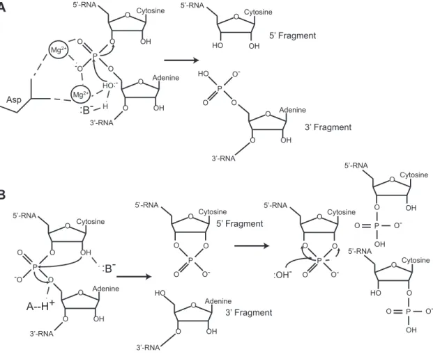
In contrast, metal ion-independent endoribonucleases coordinate amino acids to promote acid-base catalysis. For instance, residue His12 of RNase A deprotonates the 2´-O creating the attacking nucleophile. His119 serves as a general acid catalyst and protonates the 5´-O leaving group. The chemistry of metal ion-independent endoribonucleases ensures a break 3´ of the scissile phosphate and generates a 5´ RNA fragment with a 2´,3´-cyclic phosphate terminus and a 3´ RNA fragment with a 5´-OH terminus (Figure 1.1B) (Raines 1998; Fedor and Williamson 2005). Metal ion-independent endoribonucleases require accessibility of the 2´-OH, and therefore almost exclusively cleave ssRNA.
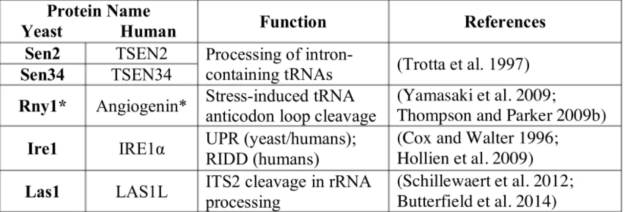
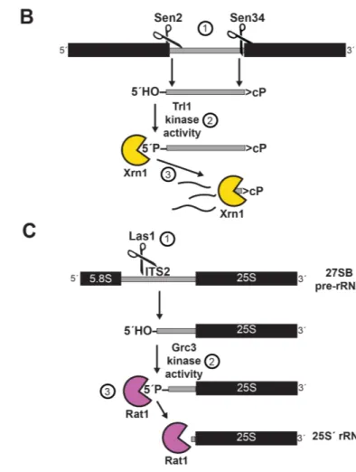
Though mammals have evolved over a dozen metal ion-dependent endoribonucleases, I focus on several well-characterized endoribonucleases that are conserved from yeast through humans (Table 1.1). In the next section, I describe regulatory pathways involving four metal ion-independent endonucleases (Sen2, Sen34, Rny1/Angiogenin, and Ire1). The role of Las1 in rRNA biogenesis is explored in the “Kinase-Mediated Decay” section.
Figure 1.1. Two distinct mechanisms of endoribonuclease cleavage.
A. Metal ion-dependent endoribonucleases (e.g., RNase H, RNase P, RNase Z) coordinate a metal ion to initiate cleavage of the scissile phosphate in ssRNA, dsRNA, and DNA:RNA hybrids. Cleavage generates a 5´-fragment with a 3´-OH terminus and a 3´-fragment with a 5´-PO4 terminus.
B. Metal ion-independent endoribonucleases (e.g., Sen2, Sen34, Ire1, Rny1) use amino acid residues to promote catalysis in the absence of a metal ion. Cleavage generates a 5´-fragment with a 2´,3´-cyclic phosphate terminus and a 3´-5´-fragment with a 5´-OH terminus. Figure used with permission from (Cooper, 2015b).
O OH O O O OH O Mg2+ Mg2+ :B- H HO:-3’-RNA 5’-RNA Cytosine Adenine O O P O HO O OH O Cytosine Adenine 3’-RNA 5’-RNA O O-P -O O O OH O O O OH O 3’-RNA 5’-RNA Cytosine Adenine P -O O A--H+ :B-5’ Fragment 3’ Fragment Asp O OH HO Cytosine 5’-RNA 5’ Fragment O OH O Adenine 3’-RNA 3’ Fragment O-P HO O O :OH-O O P O Cytosine 5’-RNA O O-O Cytosine O Cytosine OH O HO O 5’-RNA 5’-RNA P O OH O-P O OH O-A B
5´-OH RNAs in Biological Systems
Although a transcriptome-wide identification of 5´-OH RNAs via a direct sequencing method had never been performed prior to my work, there are numerous well-studied examples of 5´-OH RNAs in regulated pathways of RNA cleavage. In this section, I discuss the generation of 5´-OH RNAs in three pertinent examples: processing of intron-containing tRNAs by the tRNA splicing endonuclease (TSEN), cleavage of tRNAs by Rny1/Angiogenin to yield stress-induced tRNA fragments (tiRNAs), and the cleavage of HAC1/XBP1 mRNA by inositol-requiring enzyme 1 (IRE1). The regulated decay of these fragments are further described in the “Kinase-Mediated Decay” section.
TSEN: Processing of intron-containing tRNAs
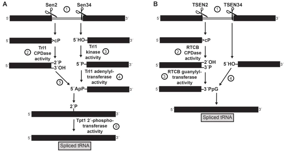
Of the predicted 295 tRNAs encoded in the yeast genome, 59 tRNAs representing 10 codons contain an intron that must be removed following transcription (Lowe and Eddy 1997). The first intron-containing tRNA in yeast was discovered in 1977 when DNA sequencing of SUP4 revealed a 14 bp intervening sequence adjacent to the 3´ end of the anticodon triplet in tRNATyr (Goodman et al. 1977). An intron of ~18 bp was found 3´ of the anticodon loop in tRNAPhe the following year (Valenzuela et al. 1978). Though the functional purpose of tRNA introns remains mysterious, the processing of intron-containing tRNAs is now well-understood (Figure 1.2A).
The first step in maturation of intron-containing tRNAs is processing by the tRNA Splicing Endonuclease (TSEN). Intron-containing tRNAs are transcribed in the nucleus, then exported to the cytoplasm where they are cleaved by the TSEN complex to remove the intron. Sen2 cleaves on the 5´ end of the intron and Sen34 cleaves on the 3´ end of the
Table 1.1 Functions of several eukaryotic metal ion-independent endoribonucleases.
Protein Name
Yeast Human Function References Sen2 TSEN2 Processing of
intron-containing tRNAs (Trotta et al. 1997) Sen34 TSEN34
Rny1* Angiogenin* Stress-induced tRNA anticodon loop cleavage
(Yamasaki et al. 2009;
Thompson and Parker 2009b) Ire1 IRE1α UPR (yeast/humans);
RIDD (humans)
(Cox and Walter 1996; Hollien et al. 2009) Las1 LAS1L ITS2 cleavage in rRNA
processing
(Schillewaert et al. 2012; Butterfield et al. 2014)
UPR: Unfolded Protein Response; RIDD: Regulated IRE1-Dependent Decay of mRNA; ITS2: Internal Transcribed Spacer 2
* Rny1 and Angiogenin are not homologous but perform an analogous function. See text in the “5´-OH RNAs in Biological Systems” section for additional explanation.
intron (Ho et al. 1990). The enzymes generate a 5´ fragment with a 2´,3´-cyclic phosphate terminus and a 3´ fragment with 5´-OH terminus (Peebles et al. 1983; Knapp et al. 1979).
T-RNA Ligase 1 (Trl1) – also known as RNA Ligase 1 (Rlg1) – resolves the termini and ligates the two tRNA exons. Trl1 was first purified and shown to perform tRNA splicing in 1986 (Phizicky et al. 1986). The specific molecular mechanism was worked out in the late 1980s and early 1990s via a series of experiments primarily from John Abelson’s and Craig Greer’s groups. In brief, Trl1 ligates the 5´ and 3´ tRNA halves via key three enzymatic activities (Greer et al. 1983; Phizicky et al. 1986), each performed by a separate modular domain: (i) hydrolysis of the 2´,3´-cyclic phosphate terminus of the 5´ tRNA half to a 2´-phosphate terminus by the 2´,3´-cyclic phosphodiesterase (CPDase) domain; (ii) phosphorylation of the 5´-OH terminus of the 3´ tRNA half by the NTP-dependent polyribonucleotide kinase domain; and (iii) adenylation of the 5´-phosphate terminus to form an RNA-adenylate intermediate which is attacked by the 3´-OH terminus to ligate the ends via a phosphodiester bond (Xu et al. 1990; Apostol et al. 1991). The three domains (CPDase, kinase, and adenylyltransferase/ligase) are all essential for tRNA splicing and yeast viability, but can function independently and perhaps arose from gene fusion events (Sawaya 2003). The ligation produces a 2´-phosphate at the splice junction. For the tRNA to be functional, this phosphate must be removed by the essential 2´-phosphotransferase Tpt1, which transfers the 2´-phosphate to NAD+, forming ADP-ribose 1´,2´-cyclic phosphate and nicotinamide (Culver et al. 1993; 1997; Spinelli et al. 1997) (Figure 1.2A). Following splicing, some tRNAs also undergo retrograde transport to the nucleus where they are further modified (Shaheen and Hopper 2005; Takano 2005). Decay of the tRNA
intron, which also carries 5´-OH and 2´,3´-cyclic phosphate termini is further described in the “Kinase-Mediated Decay” section (Wu and Hopper 2014).
In humans, intron-containing tRNAs are cleaved by conserved TSEN complex members TSEN2 and TSEN34, but the resulting 3´ and 5´ tRNA exons are ligated via a different mechanism than yeast. It had been speculated since the 1970s that the tRNA exons were joined by a direct ligation pathway that differed from the yeast system described above; however, the responsible enzyme was not identified until 2011. Popow et. al followed the ligation of a 3´-PO4 dsRNA over three chromatographic purification steps and identified 91 potential proteins by MS/MS (Popow et al. 2011). One of the proteins, HSPC117, is the human homolog of E. coli RtcB. Due to its genetic proximity to RNA 3´-terminal cyclase RtcA, the E. coli RtcB was thought to be an RNA repair enzyme (Genschik et al. 1998; Galperin and Koonin 2004). Depletion of human RTCB by RNAi reduced ligation of tRNA halves in vivo, and mutation of a conserved cytosine residue for metal ion coordination (Cys122→Ala122) abolished tRNA ligation in vitro, thus demonstrating that RTCB is the mammalian tRNA ligase (Popow et al. 2011). (Additional functions of RTCB and consequences of mutation are discussed in the “IRE1α: non-canonical splicing of XBP1 mRNA and RIDD” section.)
Almost simultaneously, two groups showed that archaeal (Pyrobaculum aerophilum) RtcB and bacterial (E. coli) RtcB also perform ligation of 2´,3´-cyclic phosphate RNAs and 5´-OH RNAs (Englert et al. 2011; Tanaka and Shuman 2011). Further biochemical studies by Shuman’s group demonstrated RtcB joins the ends by a multi-step mechanism: i) resolution of the 2´,3´-cyclic phosphate using cyclic phosphate diesterase (CPDase) activity, ii) formation of a covalent RtcB-GMP intermediate on His337, iii)
guanylation of the 3´-PO4 RNA terminus to form a (3′)pp(5′)G intermediate, and iv) attack of the 5´-OH terminus on the (3′)pp(5′)G to form the ligation junction (Chakravarty et al. 2012; Chakravarty and Shuman 2012) (Figure 1.2B). The mechanism of RtcB represents a novel method of RNA ligation. I take advantage of the unique enzyme specificity of E. coli RtcB to ligate 5´-OH RNAs to a 3´-PO4 RNA/DNA oligonucleotide in the initial step of the 5OH-seq method (Chapter II).
Rny1/Angiogenin: Stress-induced tRNA fragments
A growing body of literature is dedicated to understanding the generation and consequences of tRNA cleavage (Review: Saikia and Hatzoglou 2015). Presently, two major categories of tRNA fragments have been demonstrated in eukaryotic cells: stress-induced tRNA fragments (tiRNAs) and tRNA-derived fragments (tRFs). This discussion will focus on tiRNAs, as they are the most well-studied tRNA fragments and have 5´-OH termini.
In both prokaryotes and eukaryotes, tRNAs are cleaved in the anticodon loop during specific cellular stress conditions to yield 3´ and 5´ halves termed tiRNAs. The 3´-tiRNAs are produced with 5´-OH termini, and the 5´-tiRNAs have 2´,3´-cyclic phosphate termini. Studies in E. coli were the first to identify a mechanism for in vivo tiRNA generation. In response to bacteriophage T4 infection, the PrrC nuclease cleaves tRNALys in the anticodon loop (Levitz et al. 1990). Another study showed that colicin E5 cleaved 3´ of a queuine wobble base in tRNAs for Tyr, His, Asn, and Asp (Ogawa et al. 1999). In contrast to eukaryotic tRNA anticodon loop cleavages, which affect a minority of the tRNA pool, the
15
Figure 1.2. Yeast and human pathways for processing of intron-containing tRNAs. A. Yeast pathway for cleavage and ligation. (1) The TSEN complex cleaves intron-containing tRNAs, yielding a 5´-half with a 2´,3´-cyclic phosphate terminus and a 3´-half with a 5´-OH terminus. (2) Trl1 resolves the 2´,3´-cyclic phosphate terminus to a 2´-PO4, (3) phosphorylates the 5´-OH terminus, and (4) adenylates the new 5´-PO4 terminus. (5) The 3´-OH attacks the adenylated intermediate. (6) Tpt1 removes the 2´-PO4 at the splicing junction.
B. Human pathway for cleavage and ligation. (1) The TSEN complex cleaves intron-containing tRNAs as in A. (2) RTCB resolves the 2´,cyclic phosphate terminus to a 3´-PO4, and (3) guanylates the new 3´-PO4 terminus. (4) The 5´-OH terminus attacks the guanylated intermediate.
prokaryotic stress response shuts down translation by cleaving a large percentage of the pool (Thompson and Parker 2009a).
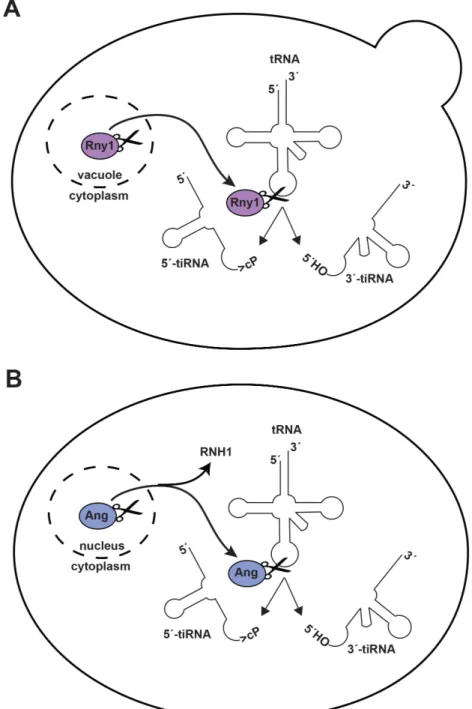
Although tRNA fragments had been identified in the the urine and sera of cancer patients in the late 1970s, evidence of in vivo eukaryotic tRNA cleavage came much later. The first study demonstrating in vivo eukaryotic tRNA cleavage showed that starvation by amino acid deprivation of Tetrahymena induced anticodon loop cleavage in asparagine, glycine, and threonine tRNAs (Lee and Collins 2005). In 2009, RNase T2 family member Rny1 was identified as the endoribonuclease responsible for tiRNA generation in yeast under oxidative stress conditions (Thompson and Parker 2009b). Yeast tiRNAs are also produced in numerous other stress conditions, including: nitrogen starvation, methionine starvation, heat shock, and entrance into stationary phase (Thompson et al. 2008). Rny1 is normally sequestered in the vacuole or secreted, and is only released into the cytoplasm in the presence of cellular stressors (Thompson and Parker 2009b) (Figure 1.3A).
In mammals, Angiogenin is an endoribonuclease that cleaves tRNA anticodon loops under stress conditions. Though Angiogenin shares this common function with Rny1, it is not a homolog, and the mammalian Rny1 homolog RNASET2 does not cleave tRNAs (Yamasaki et al. 2009). Angiogenin is a secreted ribonuclease of the RNase A superfamily that was initially discovered as an angiogenic factor in conditioned medium of HT-29 colon adenocarcinoma cells (Fett et al. 1985). While it was known that Angiogenin hydrolyzes tRNAs when injected into Xenopus oocytes (Saxena et al. 1992), it was not until 2009 that Angiogenin was shown to generate tiRNAs in human fetus hepatic tissue and human osteosarcoma cells (U2OS) (Fu et al. 2009; Yamasaki et al. 2009). To become activated, Angiogenenin must translocate from the nucleus to the cytoplasm and dissociate from
Ribonuclease/Angiogenin Inhibitor 1 (RNH1) (Thompson and Parker 2009a). Once active in the cytoplasm, Angiogenin cleaves tRNAs near the anti-codon loop to yield 3´ and 5´ tiRNAs (Figure 1.3B). Angiogenin is downregulated in several neurodegenerative diseases, and loss of function mutations have been identified in patients with amyotrophic lateral sclerosis (ALS) (Greenway et al. 2004), and Parkinson’s disease (PD) (van Es et al. 2011). However, Angiogenin can also promote cellular growth and is upregulated in many solid tumors including the colorectal, prostate, and breast cancers (Shimoyama et al. 1999; Katona et al. 2005; Montero et al. 1998).
Several studies have implicated tiRNAs in inhibition of both translation and apoptosis. Transfection of 5´-tiRNAs, but not 3´-tiRNAs, promotes stress granule formation and moderately inhibits protein synthesis, independent of eIF2α phosphorylation state (Emara et al. 2010). Synthetic 5´-tiRNAs can also displace eIF4G/A from mRNAs to inhibit translation in rabbit reticulocyte lysate (Ivanov et al. 2011). tiRNAs can saturate the Dicer binding pocket, decreasing cleavage of dsRNA substrates (Durdevic et al. 2013). Finally, both 5´-tiRNAs and 3´-tiRNAs can bind cytochrome c, competitively inhibiting the binding of apoptotic protease-activating factor 1 (APAF1), thus suppressing apoptosis (Saikia et al. 2014).
Ire1: non-canonical splicing of HAC1 mRNA
The unfolded protein response (UPR) is a conserved eukaryotic intracellular signaling pathway activated in response to endoplasmic reticulum (ER) stress (Walter and Ron 2011; Moore and Hollien 2012). When the protein folding load exceeds ER capacity,
Figure 1.3. Rny1 (yeast) and Angiogenin (humans) cleave the anticodon loops of tRNAs under stress conditions.
A. In yeast, Rny1 is normally sequestered in the vacuole or secreted. Following certain stress conditions, Rny1 is released to the cytoplasm. Rny1 cleaves tRNAs in the anticodon loop, generating a 5´-tiRNA with a 2´,3´-cyclic phosphate terminus and 3´-tiRNA with a 5´-OH terminus. Does Rny1 target other RNAs?
B. In humans, Angiogenin (Ang) is usually localized to the nucleus or secreted. During cellular stress, Ang enters the cytoplasm and dissociates from Ribonuclease/Angiogenin Inhibitor 1 (RNH1). Ang generates the same tiRNAs as Rny1 in A. These tiRNAs may
the UPR is triggered, resulting in upregulation of protein-folding machinery (Kozutsumi et al. 1988). The net result is an increase in the size and folding-capacity of the ER and a resolution of the unfolded protein burden (Schuck et al. 2009). The UPR is naturally triggered by a multitude of environmental stimuli, diseases, and developmental processes including: oxidative stress; viral infection (Bechill et al. 2006; 2008); inherited diseases of protein misfolding, such as cystic fibrosis and retinitis pigmentosa; and the development of antibody-secreting plasma cells (Gass et al. 2002; Iwakoshi et al. 2003) or pancreatic ß-cells (Harding et al. 2001; Zhang et al. 2002). The UPR is commonly chemically induced by tunicamycin, an inhibitor of N-linked glycosylation of proteins, which decreases protein stability. Dithiothreitol (DTT), which reduces disulfide bonds and thus results in decreased protein stability, can also be used to induce the UPR.
In budding yeast, the UPR is mediated by a single pathway controlling the non-canonical cytosolic splicing of HAC1 mRNA, which encodes a transcription factor that resolves ER stress by upregulating key protein folding machinery and chaperones. This unexpected and unprecedented means of RNA regulation was revealed by a series of elegant experiments conducted independently by Peter Walter at UCSF and Kazutoshi Mori at UT Southwestern and Kyoto University in the early 1990s, for which they were jointly awarded the Lasker Award in 2012.
Several early studies suggested the existence of a regulated system for increasing protein expression under specific cellular stress conditions. The first of these studies showed that the mRNA levels of two glucose regulated proteins (GRPs) p4A3 and p3C5 increased under conditions of glucose starvation in the hamster K12 cell line (Lee et al. 1983). While it was initially thought that induction of GRPs was due to defective
glycosylation caused by glucose depletion, induction of GRPs by amino acid analogues argued against this theory (Kelley and Schlesinger 1978). Impaired protein folding was also shown to regulate the expression of GRPs. In simian CV-1 cells, expression of a misfolded influenza virus haemagglutinin (HA) but not wildtype HA induced GRP78 and GRP94/HSP90 (Kozutsumi et al. 1988). GRP78 was later renamed BiP and is encoded by the KAR2 gene in yeast (Normington et al. 1989).
The discovery of Ire1 and Hac1 in the molecular regulation of the UPR was spurred by the identification of a 22 nt conserved sequence in the KAR2 promoter. This conserved UPR element (UPRE) is required for induction of yeast BiP in response to tunicamycin (Mori et al. 1992; Kohno et al. 1993). Independently, Walter and Mori expressed the UPRE upstream of the E. coli ß-galactosidase gene (lacZ) creating a colorimetric system for screening yeast genomic libraries and ultimately identifying Ire1/Ern1, a putative protein kinase (Nikawa and Yamashita 1992), as a required gene for upregulation of KAR2 expression following treatment with tunicamycin (Cox et al. 1993; Mori et al. 1993). Further studies revealed Ire1 spans the ER membrane due to an N-terminal ER signal adjacent to a hydrophobic region and contains a C-terminal cytosolic kinase domain whose enzymatic activity is essential for complementation of temperature sensitive ern1-1 mutants (Mori et al. 1993). The initial presumption was that Ire1 would phosphorylate and activate a transcription factor that would subsequently recognize the UPREs upstream of target genes. Indeed, using an adapted yeast in vivo one-hybrid screening system (Mori et al. 1996) or an overexpression system (Cox and Walter 1996), Mori and Walter each identified Hac1/Ern4 as the transcription factor responsible for binding the UPRE and upregulating expression of target genes.
Peter Walter’s group then proceeded to make the revolutionary discovery that Hac1 signaling was not regulated by phosphorylation, but by non-canonical cytosolic mRNA splicing, publishing two essential studies in the same issue of Cell. In one publication, they showed that HA-Hac1 was undetectable by immunofluorescence with an α–HA antibody under normal growth conditions, but was detected in the nucleus when cells were induced with tunicamycin. Northern analysis demonstrated that the HAC1 mRNA shortened in an Ire1-dependent manner following treatment with tunicamycin, and sequencing revealed the excision of a 252 nt intervening sequence (IVS) and splice junctions of the mRNA that differed from consensus spliceosome recognition sequences (Cox and Walter 1996). The second study used a sectoring screen in a kar2-∆HDEL yeast stain to identify Trl1/Rlg1 as the HAC1 mRNA ligase (Sidrauski et al. 1996). The following year, Walter’s group demonstrated that Ire1 cleaves at a conserved 5´-CN|CNNG-3´ motif in stem loops at the 3´ and 5´ splice junctions (Sidrauski and Walter 1997).
The series of experiments described above unveiled the first and only known example of a signaling pathway that is regulated by the cytosolic splicing of an mRNA. Ire1 oligomerizes upon sensing unfolded proteins via its luminal domain (Gardner and Walter 2011). This oligomerization promotes the activation of the Ire1 cytosolic endoribonuclease domain. When the Ire1 endoribonuclease domain is activated, it cleaves HAC1 mRNA at two locations on either side of the excised intron at sequence-specific recognition motifs (Kawahara et al. 1998), generating fragments with 2´,3´-cyclic phosphate and 5´-OH termini (Gonzalez et al. 1999). Non-canonical cytosolic splicing of the exons is then carried out by tRNA ligase Trl1 via the three distinct enzymatic activities previously described in “TSEN: Processing of intron-containing tRNAs.” Following
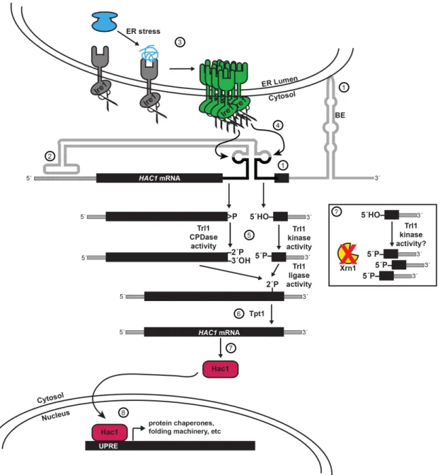
removal of this phosphate by Tpt1 (Spinelli et al. 1997), spliced HAC1 mRNA is translated, yielding a functional transcription factor that upregulates approximately 7% of yeast genes to modulate the ER stress response (Travers et al. 2000), including key UPR markers KAR2, EUG1 (Tachibana and Stevens 1992), and PDI1 (Cox et al. 1993). While the UPR is non-essential for growth in the absence of cellular stress, HAC1 and IRE1 null mutants grow slowly compared to wild type yeast and both are required under UPR-inducing conditions (Figure 1.4).
Key publications in the 2000s further extended this complex model of HAC1 mRNA splicing and resolved two unanswered questions by attributing regulatory significance to both the intron and the 5´- and 3´-UTRs. Previous experiments had revealed curious findings of unspliced HAC1 mRNA (HAC1U): In situ hybridization experiments localized HAC1U to the cytoplasm (Chapman and Walter 1997), and large portions of HAC1U comigrated with polyribosomes on sucrose gradients (Cox and Walter 1996). This suggested that Hac1U is partially translated by ribosomes that subsequently stall on the HAC1U mRNA. These findings were resolved by demonstrating that translational attenuation is mediated by a long-range 16 nucleotide base-pairing interaction between the intron and the 5’ UTR (Rüegsegger et al. 2001). There still remained the question of what might promote the colocalization of HAC1 mRNA and Ire1 prior to cleavage. Mutational probing of the HAC1 3´-UTR revealed a prominent, extended stem-loop with a conserved bipartite element (3´ BE) which targets HAC1 mRNA to Ire1 foci to promote cleavage during ER stress (Aragón et al. 2008) (Figure 1.4).
Given these latest examples of UPR regulation by the mRNA transcript itself, I was particularly curious about the findings of a recent study that captured HAC1 3´-exons with
5´-PO4 termini in dcp2∆ xrn1∆ yeast (these genes are discussed in the “RNA Decay Pathways” section) (Harigaya and Parker 2012). The existence of HAC1 3´-exons with 5´-PO4 termini is perplexing because HAC1 mRNA is cleaved by metal ion-independent endoribonuclease Ire1, and the 3´-exon should therefore be generated with a 5´-OH terminus (Figure 1.4). If the HAC1 3´-exon can exist with a 5´-PO4 terminus, it must be phosphorylated after Ire1 cleavage. Furthermore, because the HAC1 3´-exon with a 5´-PO4 terminus accumulates when Xrn1 is deleted, Xrn1 must be required for the degradation of this exon. In Chapter II, I use 5OH-seq to replicate the capture of HAC1 3´-exons with 5´-PO4 termini in xrn1∆ yeast. This finding sets up the experimentation in Chapter III, where I investigate the phosphorylation and decay of the HAC1 3´-exon intermediate.
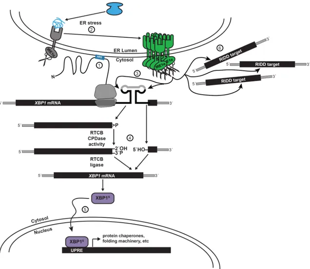
IREα: non-canonical splicing of XBP1 mRNA and RIDD
In contrast to the single UPR pathway found in yeast, the human UPR is mediated via three branches: PERK, ATF6, and IREα (Walter and Ron 2011). (This discussion will be limited to the IRE1α pathway, as it is the only pathway conserved with yeast and the only pathway regulated by mRNA splicing.) Similar to yeast, the IREα branch signals via the non-canonical cytosolic splicing of XBP1 mRNA, the human ortholog of yeast HAC1. In contrast to yeast HAC1 mRNA, XBP1 mRNA is targeted to the ER membrane by the nascent peptide of the partially translated Xbp1 protein (Yanagitani et al. 2009; 2011), and the two conserved recognition motifs that are cleaved by IRE1α excise a 26 nt intron following UPR activation (Figure 1.5).
Until 2014, there was debate as to the primary mammalian pathways for XBP1 mRNA ligation following IRE1α-mediated cleavage: a yeast-like pathway and a direct-
Figure 1.4. The unfolded protein response (UPR) in yeast is primarily regulated by the non-canonical cytosolic splicing of HAC1 mRNA.
(1) HAC1 mRNA is targeted to the ER membrane by a bipartite element (BE) in the 3´-UTR. (2) Translation of HAC1 is attenuated by long-range base-pairing of the intron and the 5´-UTR. (3) During ER stress, unfolded proteins are sensed by Ire1, which promotes oligomerization, autophosphorylation, and activation of the endoribonuclease domain. (4) Active Ire1 cleaves HAC1 mRNA in two conserved stem loops, generating a 5´-exon with a 2´,3´-cyclic phosphate terminus and 3´-exon with a 5´-OH terminus. (5) Trl1 ligates the exons, and (6) Tpt1 removes a 2´-PO4 at the splicing junction. (7) Spliced HAC1 mRNA is translated to Hac1 protein, (8) which translocates to the nucleus, binds UPR elements (UPREs), and upregulates expression of ~7% of the yeast genome. Inset: (?) Ire1 cleavage produces HAC1 3´-exon mRNA intermediates with 5´-OH termini. In dcp2∆ xrn1∆ yeast strains, these intermediates accumulate with 5´-PO termini. Does phosphorylation of the
ligation pathway. In the hypothetical yeast-like pathway, the three enzymatic roles of yeast Trl1 would be carried out by analogous but non-homologous human counterparts: CNP, a cyclic phosphodiesterase; CLP1, a polynucleotide kinase (discussed in further detail in the “Kinase-Mediated Decay” section); and an unknown ligase. TRPT1, a phosphodiesterase homologous to the yeast Tpt1, would be a likely candidate for 2´-PO4 removal. However, CNP and TRPT1 are non-essential in mice, and TRPT1-null mice have intact XBP1 splicing (Harding et al. 2007). Rather than resolve the 2´,3´-cyclic phosphate and 5´-OH termini, ligate, and remove the 2´-PO4 via four enzymes, the direct ligation pathway would join these ends with a single enzyme (Schwer et al. 2008). As previously discussed in the “5´-OH RNAs in Biological Systems” section, RTCB performs such a direct ligation during processing of intron-containing tRNAs in humans (Popow et al. 2011). RTCB can also ligate Ire1-cleaved HAC1 mRNA in a yeast trl1∆ strain (Tanaka et al. 2011). However, a report demonstrating that XBP1 mRNA ligation was not impaired when RTCB was depleted by RNAi in HeLa cells (Iwawaki and Tokuda 2011) argued against RTCB as the ligase for XBP1 mRNA.
In late 2014, a series of three publications revealed that RTCB is the mammalian UPR ligase (Lu et al. 2014; Kosmaczewski et al. 2014; Jurkin et al. 2014) (Figure 1.5). The first created a synthetic circuit wherein an XBP1 splicing-dependent 4-OHT-regulated Cre sensor activates Bim expression to generate a robust apoptotic response in SNL cells (Lu et al. 2014). This clever biological engineering approach enabled an unbiased screen that identified RTCB as the requisite XBP1 mRNA ligase. Kosmaczewski et al. bypassed the essential requirement of RtcB in tRNA splicing in C. elegans by expressing pre-spliced tRNAs for Leu(CAA) and Tyr(GUA) in vivo. They were able to demonstrate impaired XBP1
mRNA splicing of endogenous intron-containing tRNAs in RtcB null worms (Kosmaczewski et al. 2014). (We employ a similar strategy in Chapter III to evaluate the requirement of Trl1 in the phosphorylation of the 3´ cleavage product of HAC1 mRNA in the yeast UPR.) The final publication showed that simultaneous depletion of RTCB and its Archease cofactor by Dox-inducible shRNAs abolishes XBP1 mRNA splicing in HeLa cells (Jurkin et al. 2014).
Recent studies have linked RTCB ligase activity to several essential processes. The RTCB-mediated splicing of XBP1 is required for secretion of antibodies in human plasma cells (Jurkin et al. 2014). In the orthologous C. elegans system, RTCB-1 splicing of xbp-1 protects neurons from degeneration induced by human α–synuclein, which is a hallmark of Parkinson’s disease (Ray et al. 2014). RtcB also localizes to the site of neuronal injuries in C. elegans and inhibits axon regeneration independent of xbp-1 ligation, tRNA ligation, and cofactor Archease (Kosmaczewski et al. 2015).
In many eukaryotes, activated IRE1 broadly cleaves mRNA in a process termed Regulated Ire1-Dependent Decay (RIDD) (Hollien and Weissman 2006) (Figure 1.5). During RIDD, Ire1 cleaves mRNAs that are colocalized to the ER, generating RNA fragments with 2´,3´-cyclic phosphate and 5´-OH termini. RIDD was first identified by microarray studies of Drosophila S2 cells depleted of either IRE1 or XBP-1 by RNAi and induced with DTT. A population of mRNAs were differentially repressed in IRE1-depleted cells but not XBP-1-depleted cells, and this was shown to be the result of mRNA cleavage performed by IRE1 that was dependent on co-translational ER-localization of the mRNA (Hollien and Weissman 2006). Subsequent studies have extended RIDD to plants (Hayashi et al. 2012), fission yeast (Kimmig et al. 2012), and mammals (Hollien et al. 2009). While
Figure 1.5. Human IRE1α cleaves XBP1 and ER-proximal mRNAs.
(1) The nascent peptide of partially translated XBP1 targets the mRNA to the ER. (2) During ER stress, IRE1α oligomerizes, promoting autophosphorylation and activation of the endonuclease domain. (3) Active IRE1α cleaves XBP1, generating a 5´-exon with a 2´,3´-cyclic phosphate terminus and a 3´-exon with a 5´-OH terminus. (4) RTCB ligates the exons. (5) Spliced XBP1 mRNA is translated to XBP1S protein and upregulates expression of protein chaperones and folding machinery. (6) Active IRE1α also cleaves ER-proximal mRNAs in a process termed Regulated IRE1-Dependent Decay (RIDD).
RIDD has not been demonstrated in budding yeast, the conservation of Ire1 and the HAC1/XBP1 pathway in eukaryotes suggests a similar pathway may be present, and I investigate this further in Chapter II. Importantly, although the 3´ mRNA fragments have 5´-OH termini following cleavage by IRE1, the initial RIDD paper demonstrated that these species accumulate in XRN1-depleted cells (Hollien and Weissman 2006). This discrepancy is not addressed and RIDD-substrates are prime candidates for “kinase-mediated decay”, which is explained later in this chapter and discussed in Chapter III.
At the time of this dissertation, hundreds of publications have purported a UPR phenotype in human disease, comprising an extensive breadth of syndromes. In 1999, a group in Osaka, Japan was the first to implicate the UPR in a specific disease, early-onset familial Alzheimer’s disease (FAD). Their study demonstrated that missense-mutations in the human presenilin-1 (PS1) gene – the most common autosomal dominant cause of early-onset FAD – increased sensitivity of neuroblastoma cells to tunicamycin and inhibited induction of GRP78/BIP mRNA (Katayama et al. 1999). Since this initial study, aberrant regulation of the UPR has been linked to multiple disease processes, including: i) protein misfolding (e.g., proinsulin 2 in ß-cell death and procollagen in osteogenesis imperfecta); ii) polyglutamate expansions, such as Machado-Joseph disease; iii) cancers, including myeloma, breast cancer, many others; and iv) neurodegenerative diseases, specifically Parkinson’s disease, Alzheimer’s disease, and amyotrophic lateral sclerosis (Reviews: (Kaufman 2002; Lee and Ozcan 2014)). While description of the causes and implications of UPR misregulation in each of these diseases is beyond the scope of this thesis, such extensive connections between UPR and disease demonstrates the paramount importance of understanding the molecular mechanisms modulating this ubiquitous stress response.
RNA Decay Pathways
Within this section, I provide an overview of RNA decay. I outline the known primary cytosolic mRNA degradation pathways of Xrn1 and the exosome. I also describe several degradation pathways for defective mRNAs: nonsense-mediated decay (NMD), non-stop decay (NSD), and no-go decay (NGD). Finally, I introduce previously unaddressed questions of 5´-OH RNA decay.
5´→3´ degradation by 5´-phosphate-dependent exoribonucleases
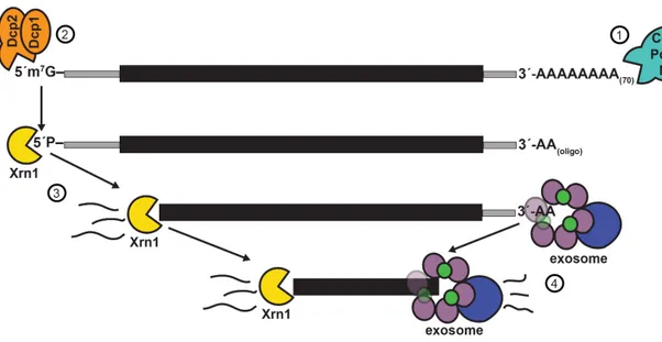
The constant production of mRNAs necessitates robust mechanisms of regulated mRNA turnover. In the late 1970s, studies of the 5´ mRNA cap structure in Xenopus, wheat germ, and mouse L cells suggested that a 5´→3´ decay process might hydrolyze uncapped mRNAs (Furuichi et al. 1977; Shimotohno et al. 1977). Shortly thereafter, Audrey Stevens purified an exoribonuclease from yeast and demonstrated that it performed 5´ hydrolysis of radiolabelled RNA with a 5´-PO4 termini (Stevens 1980). This protein was named eXoRiboNuclease 1 (Xrn1) (Larimer and Stevens 1990), and subsequently shown to degrade mRNA by processive 5´-phosphate–dependent 5´→3´ hydrolysis (Hsu and Stevens 1993; Muhlrad et al. 1994; Stevens 2001).
Most 5´-capped mRNAs are deadenylated and decapped prior to degradation. The first step of decay is deadenylation by the Ccr4/Pop2/Not complex (Brown and Sachs 1998; Tucker et al. 2001) or a secondary Pan2/Pan3 complex (Boeck et al. 1996). Deadenylation is regulated by several RNA binding proteins (RBPs), particularly the poly-A binding protein (Pab1), which binds the polyadenylated mRNA and inhibits the Ccr4 deadenylase (Caponigro and Parker 1995). Decapping proteins 1 and 2 (Dcp1/Dcp2) subsequently
remove the 7-methylguanylate caps of the deadenylated transcripts (van Dijk et al. 2002); however, decapping proceeds independently of deadenylation for a subset of mRNA transcripts (Muhlrad and Parker 2005). As with deadenylation, decapping is a highly regulated process, and likely predominantly regulated by Pat1/Lsm1-7 mediated recruitment of Dcp1/Dcp2 (Review: Parker 2012).
The final product of most mRNA decapping events is a deadenylated mRNA with a 5´-PO4 terminus that Xrn1 recognizes and degrades (Figure 1.6). Xrn1 is non-essential in yeast, presumably due to secondary pathways of degradation; however, xrn1∆ strains have a decreased rate of mRNA decay, which varies between a 1.5 to 6-fold reduction depending on the mRNA transcript (Cao and Parker 2001). In contrast, Xrn1/Pacman knockdowns in higher-order eukaryotes C. elegans and Drosophila confer either developmental failures or lethality, confirming that 3´→5´ decay pathways cannot compensate for defective 5´→3´ decay (Newbury and Woollard 2004; Jones et al. 2012).
In addition to Xrn1, there are two other known 5´-phosphate-dependent 5´→3´ exoribonucleases in yeast. The most well-studied of these is Rat1, known as Xrn2 in higher-order eukaryotes. The essential RAT1 gene was initially identified in a screen for defective poly(A) mRNA transport from the nucleus (thus its name: Ribonucleic Acid Trafficking 1), and encodes an exoribonuclease similar to Xrn1 (Amberg et al. 1992; Kenna et al. 1993). Rat1 is predominantly expressed in the nucleus, and overexpression of wild-type RAT1 can only partially restore growth defects in xrn1∆ yeast (Poole and Stevens 1995). However, expression of RAT1 with mutations that increase cytosolic localization can fully complement xrn1∆ yeast. Similarly, Xrn1 is primarily located in the cytoplasm, but XRN1 tagged with a nuclear localization signal (NLS) can rescue temperature-sensitive
rat1-1 yeast (Johnson 1997). Thus, Xrn1 and Rat1 are functionally interchangeable exoribonucleases, but are restricted to the cytoplasm and nucleus, respectively. Rat1 is required for pre-rRNA trimming (Henry et al. 1994) (described in the “Kinase-Mediated Decay” section), snoRNA processing (Petfalski et al. 1998), and mRNA transcription termination (Kim et al. 2004; Ghazal et al. 2009).
Functional studies of a Rai1 homolog Ydr3706 recently revealed a third 5´-phosphate-dependent 5´→3´ exoribonuclease in yeast. Though initially evaluating the effects of Ydr3706 on decapping, a group at Columbia discovered the protein had distributed 5´→3´ exoribonuclease activity and proposed the new name Decapping eXOnuclease 1 (Dxo1). The study showed that Dxo1 can degrade 5´-PO4 RNA, but cannot proceed through a RNA-DNA heteroduplex, suggesting that Dxo1 is weaker than Xrn1 and more prone to stalling (Chang et al. 2012). The mammalian homolog DXO is restricted to the nucleus and acts as a 5´ end quality control mechanism to detect and degrade incompletely 5´ end-capped pre-mRNAs (Jiao et al. 2013); however, it is not known what role cytosolic yeast Dxo1 might play in cytosolic mRNA decay.
3´→5´ degradation by the exosome
A secondary pathway for RNA degradation following deadenylation is 3´→5´ degradation by the exosome (Figure 1.6). The exosome was first discovered when immunoprecipitation of Ribosomal RNA Processing 4 (Rrp4), a protein required for 3´→5´ exonuclease activity on 5.8S rRNA, revealed that it was part of a much larger complex (Mitchell et al. 1997). Mass spectrometry analysis enabled identification of four associated proteins – thereafter named Rrp41, Rrp42, Rrp43, and Rrp44 – each of which is
required for 5.8S rRNA 3´ end formation (Mitchell et al. 1997). Studies over the subsequent decade demonstrated that the exosome is a complex comprised of 10 key proteins: the exonuclease and endonuclease domain-containing Rrp44/Dis3 protein and a nine-member ring structure formed by six RNase PH proteins (Rrp41, Rrp42, Mtr3, Rrp43, Rrp46, Rrp45) and three small RNA-binding proteins (Rrp4, Rrp40, and Csl4) analogous to bacterial PNPase (Allmang et al. 1999; Liu et al. 2006). While the core exosome complex is present in both the nucleus and the cytoplasm, the exosome requires four Ski proteins (Ski2, Ski3, Ski7, and Ski8) for cytoplasmic mRNA decay (Anderson and Parker 1998; van Hoof et al. 2000b). The Ski2 protein is an ATPase that may act as an RNA helicase to unwind structured RNAs and remove bound proteins; a related protein, Mtr4, may perform the same activity when associated with the nuclear exosome (Lykke-Andersen et al. 2009). Deadenylation occurs prior to 3´→5´ exosomal decay of mRNAs, possibly due to removal of factors bound to the poly(A) tail that would otherwise inhibit the exosome (Parker 2012). In contrast to the 5´-PO4 terminus requirement of Xrn1, the effect of RNA 3´ terminal modifications on recognition and degradation by the exosome is unknown.
The components of the exosome are well-conserved in humans, and mutations can greatly compromise neurodevelopment. For example, a recent study that sequenced the exomes of patients with pontocerebellar hypoplasia type 1 (PCH1) revealed recessive mutations in EXOSC3, the human homolog of yeast RRP40 (Wan et al. 2012). A morpholino knockdown of exosc3 mRNA in zebrafish conferred maldevelopment consistent with the human phenotype, and expression of wildtype exosc3 mRNA restored normal development.
Figure 1.6. Canonical cytosolic mRNA decay pathways: Xrn1 and the exosome. (1) The first step in cytosolic mRNA decay is deadenylation by the Ccr4/Pop2/Not complex or the Pan2/Pan3 complex. (2) Dcp1 and Dcp2 are recruited to the 5´ terminus and Dcp2 removes the m7G cap, leaving a 5´-PO4 terminus. (3) Xrn1-mediated degradation of decapped 5´-PO4 mRNA proceeds 5´→3´. The cytosolic exosome is comprised of a 9-member ring structure and the Rrp44 nuclease. (4) Degradation of deadenylated mRNA by the exosome proceeds 3´→5´. Adapted from (Parker 2012).
Decay pathways for defective mRNAs
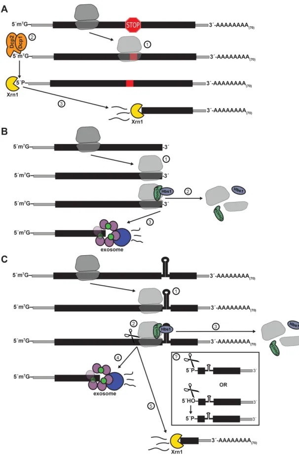
In addition to the major Xrn1 and exosome decay pathways, three other minor pathways for mRNA quality control have been described in yeast: nonsense-mediated decay (NMD), non-stop decay (NSD), and no-go decay (NGD) (Figure 1.7). The most well-studied of these mRNA quality control mechanism, NMD, was first suggested in 1979 when amber nonsense mutations introduced into the yeast URA3 gene decreased mRNA stability (Losson and Lacroute 1979). Other than mRNAs with the nominal nonsense mutations, NMD acts on a broad array of substrates with anomalous translation termination, as well as a subset of “normal” mRNAs (Parker 2012). These defective mRNAs are marked by Upf1, Upf2, and Upf3 interaction and are subject to rapid deadenylation-independent decapping followed by 5´-phosphate-dependent degradation by Xrn1 (Baker and Parker 2004) (Figure 1.7A). NMD is conserved in Drosophila (Gatfield et al. 2003) and mammals (Lejeune et al. 2003); however, metazoans have additional mechanisms for mediating NMD, including SMG6, an endonucleolytic component that cleaves mRNA at premature translation termination codons (PTCs) (Huntzinger et al. 2008). SMG6-mediated cleavage allows cytosolic decay by XRN1 and the exosome to proceed from the site of PTC in the 5´→3´ and 3´→5´ directions, respectively (Stevens et al. 2002).
Nonstop decay (NSD) occurs when a translating ribosome reaches the 3´ end of an mRNA without reaching a stop codon (Figure 1.7B). The stalled ribosome is recycled by Dom34/Hbs1 (discussed in more detail below) and triggers rapid 3´→5´ Ski-dependent degradation of the mRNA by the exosome (Frischmeyer 2002; van Hoof et al. 2002; Tsuboi et al. 2012). NSD of the mRNA occurs concomitantly with rapid proteasome-mediated