swedish dent al journ al, supplement 22 1 , 20 1 2. d oct or al dissert a tion in odont ol og y d aniel n ebel malmö universit y malmö university
daniel nebel
functional importance
of estrogen receptors
in the periodontium
isbn/issn 978-91-7104-388-7/0348-6672 fun ction al import an ce of es tr ogen recept ors in the periodontiumF U N C T I O N A L I M P O R T A N C E O F E S T R O G E N R E C E P T O R S I N T H E P E R I O D O N T I U M
Swedish Dental Journal, Supplement 221, 2012
Cover image:
Gene expression in estrogen treated PDL cells. The microarray chip is displaying the complete human genome on 28,869 genes. Light intensity in each pixel corresponds to level of gene expression in one gene.
© Copyright Daniel Nebel 2012 Illustrations: Daniel Nebel ISBN 978-91-7104-388-7 ISSN 0348-6672 Holmbergs, Malmö 2012
DANIEL NEBEL
FUNCTIONAL IMPORTANCE
OF ESTROGEN RECEPTORS
IN THE PERIODONTIUM
Department of Periodontology
Faculty of Odontology
Malmö University, Sweden 2012
This publication is also available online: www.mah.se/muep
CONTENTS
LIST OF PAPERS ... 9 ABSTRACT ... 11 POPULÄRVETENSKAPLIG SAMMANFATTNING ... 14 ABBREVIATIONS USED ... 17 INTRODUCTION ... 19Anatomy of the periodontium ...19
Cell types ...20
Fibroblasts in the periodontium ...20
Periodontal ligament cells (PDL cells) ...21
Gingival epithelial cells (HGEP) ...22
Human umbilical vein endothelial cells (HUVEC) ...22
Periodontitis ...23
LPS and TLR signaling ...25
Estrogen ...28
Estrogen signaling and functions ...29
Estrogen and periodontitis ...30
Nitric oxide and periodontitis ...33
Animal models of periodontitis ...34
OBJECTIVES ... 36
METHODOLOGY ... 37
Cells ...37
mRNA expression ...40
Affymetrix gene array ...40
Determination of collagen and DNA synthesis
and cell viability ... 41
Protein expression ... 41
Morphometry ... 41
Immunocytochemistry and immunohistochemistry ... 42
Ovariectomy ... 43
Statistics ... 43
Ethical approval ... 43
RESULTS AND DISCUSSION... 44
Effects of LPS on PDL cells and HUVEC ... 44
Effects of estrogen and E.coli LPS on the functional properties of PDL cells ... 46
Estrogen and NOS blocker inhibit cytokine and chemokine production in PDL cells ... 47
Effects of ovariectomy and aging on mouse periodontium... 48
Distribution of estrogen receptors in the gingiva ... 49
CONCLUSIONS ... 53 ACKNOWLEDGEMENTS ... 55 REFERENCES ... 57 PAPER I ... 67 PAPER II ... 77 PAPER III ... 85 PAPER IV ... 95 PAPER V ... 105 APPENDIX ... 123
9
LIST OF PAPERS
This thesis is based on the following five papers, which will be re-ferred to in the text by their Roman numerals. The papers are ap-pended at the end of the thesis together with a review paper, not
included in the thesis
.
I. Daniel Jönsson, Daniel Nebel, Gunilla Bratthall, Bengt-Olof Nilsson. LPS-induced MCP-1 and IL-6 production is not reversed by oestrogen in human periodontal ligament cells.
Archives of Oral Biology 2008;53:896-902.
II. Daniel Nebel, Gunilla Bratthall, Gunnar Warfvinge, Bengt-Olof Nilsson. Effects of ovariectomy and aging on tooth attachment in female mice assessed by morphometric analysis.
Acta Odontologica Scandinavica 2009;67:8-12.
III. Daniel Nebel, Daniel Jönsson, Ola Norderyd, Gunilla
Bratt-halland Bengt-Olof Nilsson. Differential regulation of chemokine
expression by estrogen in human periodontal ligament cells.
Journal of Periodontal Research 2010;45:796-802.
IV. Daniel Nebel, Gunilla Bratthall, Eva Ekblad, Ola Norderyd and Bengt-Olof Nilsson. Estrogen regulates DNA synthesis in human gingival epithelial cells displaying strong estrogen receptor β immunoreactivity.
10
V. Daniel Nebel, Joel Arvidsson, Johan Lillqvist and Bengt-Olof
Nilsson. Differential effects of LPS from Escherichia coli and
Por-phyromonas gingivalis on IL-6 production in human periodontal
ligament cells. Submitted.
Published paper appended, but not included in the thesis:
Daniel Jönsson, Daniel Nebel, Gunilla Bratthall and Bengt-Olof Nilsson. The human periodontal ligament cell: a fibroblast-like cell acting as an immune cell.
Journal of Periodontal Research 2011;46:153-7.
Published papers are reproduced with the permission of the respective copyright holders. Paper I: ©
2008 Elsevier Ltd; Paper II: © 2009 Informa UK Ltd; Papers III and IV and appendix: ©
11
ABSTRACT
The main functions of estrogen are associated with reproduction. However, estrogen has been shown to be of functional importance
also in non-classic target organs. Previous studies, especially
epi-demiologic and clinical ones, have addressed estrogen’s influence on periodontitis, suggesting that estrogen has a beneficial effect, but the biological mechanisms have not been identified. Estrogen exerts genomic effects in the target cells by binding to the nuclear receptors, estrogen receptor (ERs), ERα and ERβ. The expression of the two subtypes of ERs varies depending on the tissue. The overall objectives of this thesis were to study the functional im-portance of estrogen receptors in the periodontium with special fo-cus on inflammation, and stimulators of inflammation and their signaling pathways. The thesis is based on the following five papers.
In Paper I, effects of estrogen on E. coli LPS-induced PDL cell
pro-duction of interleukin-6 (IL-6), monocyte chemoattractant protein-1 (MCP-protein-1) and C-reactive protein (CRP) are assessed, by using ELISA. Furthermore, effects of LPS and estrogen on the normal characteristics of the PDL cell such as collagen synthesis and cell proliferation is determined by using L-[3H]proline incorporation
and measurement of DNA synthesis, respectively. Key findings: E.
coli LPS stimulates PDL cell IL-6 and MCP-1 production but has no effect on the normal physiological properties of PDL cells. LPS-induced IL-6 and MCP-1 is not reversed by estrogen suggesting that estrogen has no anti-inflammatory effect in these experiments.
12
In Paper II, we investigate the effects of ovariectomy and aging on tooth attachment in female mice by using morphometric analysis.
Key findings: Withdrawal of female sex hormone production by
ovariectomy has no effect on alveolar bone height and apical ter-mination of the junctional epithelium. In a second series of experi-ments these parameters are similar in mice sacrificed at 8-26 weeks of age, suggesting that tooth attachment is preserved with age in mice within a period of six months.
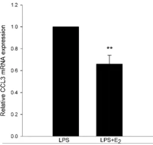
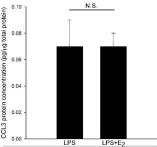
In Paper III, the objective is to investigate the regulation of CCL2/MCP-1, CCL3/MIP-1α, and CCL5/RANTES chemokines by estrogen in human PDL cells by determining mRNA transcript lev-els (using quantitative real-time PCR) and protein levlev-els (using
ELISA). Key findings: A physiological concentration of estrogen
reduces the expression of CCL3 mRNA by about 40% compared to PDL cells treated with LPS alone. In contrast, inter-individual differences in the effects of estrogen on CCL5 mRNA expression are observed. These findings indicate that estrogen affects chemo-kine expression in PDL cells showing a complex pattern involving down-regulation as well as up-regulation of chemokines. Estrogen exerts both anti-inflammatory and pro-inflammatory effects through these mechanisms.
In Paper IV, ER expression in human gingival biopsies, and effects of estrogen on cultured gingival epithelial cell (HGEP) prolifera-tion, are investigated. Expression of ERα and ERβ is determined by immunohistochemistry and effects of estrogen on HGEP
prolifera-tion monitored by measuring DNA synthesis. Key findings:HGEP
cells show strong ERβ immunoreactivity but low ERα
immunoreactivity both in vivo and in culture, suggesting that ERβ
is the predominant ER subtype in HGEP. High, but not low, con-centrations of estrogen attenuates proliferation of gingival epitheli-al cells, indicating a concentration-dependent mechanism.
13 In Paper V, the objective is to investigate the effects of LPS from
Escherichia coli and Porphyromonas gingivalis on IL-6 production in human PDL cells and endothelial cells, and the signaling mecha-nisms involved. Quantitative real-time PCR is used to determine IL-6 mRNA levels and ELISA to determine IL-6 protein. Key
find-ings: E. coli LPS (but not P. gingivalis LPS) stimulates IL-6
produc-tion in PDL cells. Treatment with the non-selective nitric oxide synthase inhibitor L-NAME reduces IL-6 by 30%, while aminoguanidine, an inhibitor of inducible nitric oxide synthase, does not affect IL-6 levels, showing a mechanism probably involv-ing nitric oxide formation via endothelial nitric oxide synthase. Treatment with the glucocorticoid steroid dexamethasone totally
14
POPULÄRVETENSKAPLIG
SAMMANFATTNING
Ordet östrogen brukar associeras till det faktum att det är ett kvinnligt könshormon. Östrogen utövar förvisso viktiga effekter i samband med utveckling och utmognad av kvinnans könsorgan samt vid reproduktionen, men har också visat sig ha många andra funktioner i kroppen, hos både kvinnor och män. Östrogen verkar i cellkärnan genom att binda till en östrogenreceptor (ER), som finns i två olika typer, ERα och ERβ.
Parodontit (tandlossning) är en inflammationssjukdom som drab-bar tandens fäste som svar på bakterier som normalt finns i mun-hålan. I cellmembranet hos vissa bakterier finns en molekyl, LPS, som fungerar retande och startar inflammationen genom att binda till TLRs (en mottagarmolekyl) som sitter på vita blodkroppar, men också till vävnadsceller som till exempel PDL celler. Effekten blir att cellerna börjar producera ämnen, bland annat cytokiner och kemokiner, som får fler vita blodkroppar att komma till plat-sen och inflammationen är då ett faktum. Även om grundorsaken till parodontit är känd vet vi inte fullt ut varför vissa personer drabbas och andra inte. Det är därför viktigt att undersöka om kroppsegna faktorer såsom hormonet östrogen kan påverka mottagligheten för parodontit. Sedan tidigare finns ett flertal studi-er som visar ett samband mellan förändringar i östrogennivåstudi-er och tandlossning. Det övergripande syftet med studierna i denna av-handling var att undersöka ERs betydelse i parodontiet och hur
15 östrogen via ER påverkar cytokin- och kemokinfrisättning i PDL-celler som aktiveras med bakterietoxinet lipopolysackarid (LPS). Studie I: LPS får PDL-celler att drastiskt öka produktionen av in-flammationsproteinerna, IL-6 och MCP-1. Effekten dämpas inte av östrogen. LPS påverkar inte PDL-cellers normala funktioner, vilket betyder att LPS specifikt stimulerar produktionen av inflamma-tionsproteiner i dessa celler.
Studie II: För att studera effekten av östrogen i parodontiet opere-rades de östrogenproducerande äggstockarna bort på en grupp av honmöss. Tändernas fästenivå hos denna grupp jämfördes med en kontrollgrupp med normal östrogenproduktion. Efter sex veckor visade det sig inte vara någon skillnad på tandfästet mellan de två grupperna. Då ingen av grupperna utvecklade tandlossning kan ingen säker slutsats dras om östrogens eventuella effekter på tand-fästet.
Studie III: Östrogen påverkar PDL-cellers produktion av flera oli-ka inflammationsproteiner, men mönstret är komplext. Ett protein som stimulerar rekrytering av vita blodkroppar (kemokin) hämmas av östrogen medan en annan kemokin förblir oförändrad . För ett tredje inflammationsprotein skilde produktionen sig åt beroende på vilket genetiskt ursprung cellerna hade. Detta tyder på att östrogen verkar både pro- och anti-inflammatoriskt och att det genetiska ur-sprunget kan påverka östrogenets funktion.
Studie IV: I tandköttet (gingivan) är ERβ den dominerande östro-genreceptorn. Mönstret går igen både på odlade celler och på väv-nadsprover från patienter. Tätheten av östrogenreceptorerna skiljer sig inte åt när man jämför vävnadsprov från inflammerade ställen med vävnadsprov från friska ställen. Vid höga doser av östrogen minskar förmågan att dela sig hos gingivala epitelceller. Studien vi-sar att östrogen verkar genom ERβ i tandköttet och att höga östro-genhalter minskar gingivala epitelcellers förmåga att dela sig.
16
Studie V: PDL-celler producerar olika mängder av inflammations-proteinet IL-6, beroende på vilket LPS de behandlas med. LPS från
den parodontitassocierade bakterien, P. gingivalis orsakar ingen
IL-6 produktion i PDL-celler medan LPS från tarmbakterien E. coli
ökar IL-6 produktionen med cirka 30 gånger. Effekten verkar in-volvera kväveoxid (NO). När enzymen, som behövs vid bildandet
av NO, blockerades minskade IL-6 produktionen som svar på E.
coli LPS med 30% vilket indikerar att NO är inblandat i IL-6
pro-duktionen.
Sammanfattningsvis visar studierna att östrogen, sannolikt via ERβ, påverkar parodontiets celler på flera olika sätt. Östrogen ut-övar både effekter som kan tolkas som skyddande (minskning av produktionen av inflammationsproteiner) men också effekter som kan innebära reducerat skydd (t ex hämning av gingivala epitelcel-lers celldelning). Studierna bidrar med ny kunskap om den biolo-giska betydelsen av östrogen i parodontiet. Denna kunskap utgör en viktig grundbult för att förstå hur förändringar i produktion av östrogen kan påverka utveckling av tandlossningssjukdomen.
17
ABBREVIATIONS USED
CCL Chemokine ligand
CRP C-reactive protein
E2 17β-estradiol
E. coli Escherichia coli
ELISA Enzyme-linked immunosorbent assay
ER Estrogen receptor
GCF Gingival crevicular fluid
HGEP Gingival epithelial cells
GPR30 G-protein coupled receptor 30
GRO-α Growth-regulated oncogene-α
HUVEC Human umbilical vein endothelial cell
IL Interleukin LPS Lipopolysaccharide
MCP-1 Monocyte chemotactic protein-1 (also CCL2)
MIP-1α Macrophage inflammatory protein-1α (also CCL3)
MyD88 Myeloid differentiation primary-response gene
NO Nitric oxide
NOS Nitric oxide synthase
OvX Ovariectomy
PCR Polymerase chain reaction
PDL Periodontal ligament
P. gingivalis Porphyromonas gingivalis
PMNs Polymorphonuclear leukocytes
18
RANTES Regulated upon activation, normal T-cell expressed
and secreted (also CCL5)
SAA Serum amyloid A
TLR Toll-like receptor
19
INTRODUCTION
Anatomy of the periodontium
The periodontium comprises different parts: the gingiva, the perio-dontal ligament (PDL), the root cementum, and the alveolar bone. These are all different types of connective tissues, two of which are mineralized (cementum and bone) and two fibrous (gingiva and PDL). This complex forms an effective support for the teeth. The junction where the gingiva attaches to the tooth is unique. It is the only place where hard tissue penetrates the epithelium, represent-ing a gateway between the inside of the body and the oral cavity that is full of microorganisms. This barrier is of course a vulnerable spot, but works surprisingly well most of the time. Under certain conditions, the barrier weakens and there is a risk of disease in the periodontium.
The gingiva is the soft tissue covering the alveolar bone. It also at-taches to the surface of the tooth, with hemidesmosomes forming a relatively tight barrier towards the oral cavity. The gingival tissue consists of an epithelial layer with high turnover of cells and an underlying connective tissue called the lamina propria.
The root surface is covered with hard tissue called cementum, which is formed by the cementoblasts. The cementoblasts first lay down the organic collagen matrix, which later becomes mineral-ized. The cementum enables the collagen fibers of the PDL to
at-20
tach to the tooth and is an avascular tissue, which makes it less important in immunological terms.
The periodontal ligament is the connective tissue that attaches to the root cementum on one side and the socket wall of the alveolar bone on the other. The ligament plays an important role in tooth maintenance, tooth mobility, and cementum formation. It also has sensory, nutritive, and homeostatic functions.
The alveolar bone is a specialized mineralized tissue built from an organic matrix of type-I collagen permeated by the mineral hy-droxyapatite. In addition, bone consists of noncollagenous proteins e.g. osteocalcin, alkaline phosphatase, and osteonectin.
Cell types
The periodontium harbors a great variety of different cell types that interact with bacteria of the biofilm and the immune system. There are cells derived from all three germ layers: endoderm, meso-derm, and ectoderm. These cells are in direct contact with the bac-teria and their products, and form the first line of defense against this threat. Traditionally, the host tissues have only been seen as a target for inflammation, but in recent years more attention has been given to the tissues’ role in being a part of the immune re-sponse. This thesis focuses on two different types of primary cells found in the periodontium: the periodontal ligament cell (PDL cell) and the gingival epithelial cell (HGEP). Furthermore, primary en-dothelial cells (HUVEC) and monocytes (THP-1) were used to study endothelial function and immune responses.
Fibroblasts in the periodontium
The fibroblast is a key cell in the periodontal tissues, with its great capacity for synthesis and secretion of fibrous proteins forming the extracellular matrix. It shows a remarkable ability to differentiate and mature into more specialized cell types. Gingival fibroblasts form collagen types I and III, which synthesize the connective tis-sues of the gingiva. In bone, the matrix is secreted by osteoblasts, a differentiated fibroblast. Embryologically, the fibroblast originates
21 from the mesodermal germ layer, in contrast to the gingival epithe-lial cell which is derived from the epidermal germ layer. In addition to collagen formation, fibroblasts may serve other important func-tions in the periodontium.
Periodontal ligament cells (PDL cells)
PDL cells are fibroblasts, and the most common type of cell in the periodontal ligament. Furthermore, the PDL contains endogenous stem and progenitor cells. These can differentiate into mesenchymal lineages such as osteoblasts, adipocytes, and
chon-drocytes in vitro (Shi et al. 2005; Fujii et al. 2008). These features
make the PDL especially interesting in terms of regeneration after loss of attachment resulting from periodontitis. The collagen in periodontal ligament is formed by the periodontal ligament cells. PDL cells account for about 5–6% of the total volume of the peri-odontal ligament. PDL cells have a high production of collagen
(Overall et al. 1987; Somerman et al. 1988). Periodontal ligament
cells have also been suggested to be osteogenic. In contrast to gin-gival fibroblasts, PDL cells produce high levels of alkaline phos-phatase. Furthermore, PDL cells are capable of producing
osteocalcin and form mineral-like nodules (Arceo et al. 1991;
Nohutcu et al. 1997).
PDL cells express both receptor activator of nuclear factor kappa B ligand (RANKL) and osteoprotegerin (OPG), both of which are important signaling proteins that function in bone and immune cell
communication (Kanzaki et al. 2001; Hasegawa et al. 2002).
RANKL is normally expressed in osteoblasts but it serves to differ-entiate and activate osteoclasts.
Apart from the normal functions of PDL cells, e.g. synthesizing col-lagen, they have the capacity to initiate inflammation by expressing different cytokines. PDL cells have functional characteristics simi-lar to those of leukocytes and leukocyte-derived cells involved in innate immunity. Interleukins IL-1β, IL-6, and IL-8, and also MCP-1 and TNF-α are examples of cytokines that are expressed by PDL
22
1995; Ozaki et al. 1996; Okada et al. 1997; Agarwal et al. 1998;
Engels-Deutsch et al. 2003; Shu et al. 2008; Sun et al. 2010;) In
addition, PDL cells have also been suggested to participate in im-mune reactions by expressing surface proteins characteristic of
an-tigen-presenting cells (APCs) (Konermann et al. 2011).
Gingival epithelial cells (HGEP)
The gingiva is composed of an outer epithelium and an underlying connective tissue called the lamina propria. These two layers are separated by the basal membrane, which anchors the epithelium to the loose lamina propria. A thin layer of gingival epithelial cells (HGEP or GEC), the basal layer, is situated on the basal mem-brane. The cells show stem cell properties and are in a constant state of renewal. The proliferation rate is high and the HGEP show high mitotic activity. As new cells proliferate in the basal layer, older cells migrate towards the surface. Shedding of the outermost layers of cells occurs when they reach the surface facing the oral cavity. The desquamation acts as a protective mechanism since bacteria have difficulty in adhering and colonization. The main function of HGEP is to synthesize keratin, and they are therefore often defined as keratinocytes. Depending on the location, the ker-atin production can be low (e.g. in the cheeks and soft palate) or high (e.g. in the alveolar gingiva and hard palate). When maturing, the HGEP loses many of its functions but the keratin production continues until the cell is shed. Cultured HGEPs are obtained by isolating cells from the basal cell layer. The cells are kept in a high-ly proliferative and low keratin-producing state.
Human umbilical vein endothelial cells (HUVEC)
Endothelial cells form a thin layer at the interior surface of blood vessels. These cells are responsible for a number of biological func-tions, e.g. a barrier function by regulating the passage of leucocytes into and out of the bloodstream. Furthermore, endothelial cells have a role in inflammation, angiogenesis, thrombosis, recruitment of white blood cell and fibrinolysis; all important factors in modu-lating the inflammation response. The function of endothelial cells is therefore of importance regarding periodontitis. Cardiovascular
23 disease has been shown to be associated with endothelial dysfunc-tion. High levels of dimethylarginine (ADMA) lead to reduction of nitric oxide (NO) concentrations, which in turn leads to
hyperten-sion (Deanfield et al. 2005). Endothelium of different origin can
differ in phenotype. The cells used in paper V are venous endothe-lial cells derived from the umbilical cord HUVEC.
Periodontitis
There has been little consensus regarding diagnosis and classifica-tion of diseases in the periodontium. Historically, different systems have been used, some of them focusing more on the extent of breakdown of the periodontal attachment and some focusing on susceptibility to development of periodontal disease. The fact that periodontitis often has very slow progression, makes it difficult to determine whether there is an ongoing loss of attachment.
In short, a healthy periodontal condition is characterized by a firm non-bleeding gingiva with a shallow (1–3-mm) periodontal pocket. Gingivitis is defined as inflammation of the gingiva without any loss of attachment. Typical signs of inflammation are redness, bleeding on probing, and an increased pocket depth due to swell-ing. Periodontitis is usually defined as inflammation of the gingiva that extends into the adjacent attachment apparatus. The disease is characterized by loss of clinical attachment due to destruction of periodontal ligament and loss of the adjacent supporting bone. The loss of attachment is usually seen as marginal bone loss in X-rays. In many classification systems, a number of subgroups of gingivitis and periodontitis are defined.
Periodontitis is an inflammatory disease caused by mainly Gram-negative, anaerobic bacteria in a subgingival biofilm, the plaque. Already in the 1960s, Loe and coworkers showed that bacterial
plaque causes gingivitis (Loe et al. 1965). The mechanism behind
gingivitis developing into periodontitis is still not fully understood. Not all patients with gingivitis will go on to develop periodontitis. Periodontitis patients have a wide range of progression rates. While patients with faster-progressing periodontitis will suffer tooth loss,
24
most patients can, despite the loss of attachment, keep their teeth for the rest of their life. Still, gingival inflammation is seen as a risk
factor for the development of periodontitis (Hamp et al. 1972).
Teeth with consistently inflamed gingiva had a 46-times higher risk of being lost than teeth without gingivitis, within a 26-year period. Gingival inflammation is thus a risk factor for tooth loss (Schatzle
et al. 2004).
It is now generally accepted that periodontitis is caused by bacteria
in the dental plaque (Jenkinson et al. 1999). In early periodontal
research, much effort was made to identify specific bacterial species that were believed to cause the inflammation. In a periodontal pocket, there are about 500 different bacterial species. Even though several species of bacteria have been isolated and identified as be-ing more likely to be found in a site of periodontitis, the pathogen-esis appears to be even more complex. The ecology of the bacterial culture appears to be a more important factor than the specific bacterial species. A study carried out by Loe and coworkers on Sri Lankan laborers who did not get any dental treatment showed that different individuals can respond completely differently to the same bacterial load. It is clear that the patient’s susceptibility to perio-dontitis is crucial to whether attachment loss will occur or not. 8% of the population got periodontitis with rapid progression and sub-sequent tooth loss, 81% suffered moderate progressive periodonti-tis, and a group of 11% showed no progression of periodontal
dis-ease beyond gingivitis (Loe et al. 1986). This study clearly shows
that there is a correlation between severity of periodontitis and the susceptibility of the patient. Several other epidemiological studies have shown the same ratio between individuals who develop severe periodontitis, those who develop moderate periodontitis, and those who do not experience any loss of periodontal attachment
T co 2 h es F p is n su fl ti T b ti
L
L ce Tobacco smo ose levels ar 2002; Emrich host response strogen can Figure I: Perio plaque, but pa s clear that th not he/she will usceptibility a fluence of estr ion. The studies c basic research icular periodPS and TLR
Lipopolysacc ell membran oking and di re risk facto h et al. 199 e to the bac modulate th odontitis is a atients do not he patient’s su ll develop per are not fully u rogen and on conducted an h and are th dontal diagnR signaling
charides (LP ne of Gram abetes melli ors for perio 91) (Figure I cterial biofilm he host respo an inflammato t respond equ susceptibility i riodontitis. Th understood. T n the role of h nd presented herefore not osis.S) are large m-negative ba
tus with fluc odontal dise I). These fac m. This thes onse.
tory disease in ually to the sa
is a crucial fa The factors tha
This thesis co host tissue in d in this thesi directly app molecules f acteria. LPS ctuating blo ease (Jansson ctors modul sis focuses o initiated by b ame bacterial factor for whe
at affect the p oncentrates on n initiating inf is are experi plicable to an found in the consist of 25 od glu-n et al. ate the on how bacterial l load. It hether or patient’s n the in- flamma-imental ny par-e outpar-er a lipid
26
and a polysaccharide bound together with a strong, covalent bond. The main function of LPS is to protect the bacteria from chemical substances and to stabilize the structure of the bacteria. LPS also work as an endotoxin by promoting the secretion of pro-inflammatory cytokines e.g. lymphokines, interleukins, and chemokines. Accordingly, LPS trigger a strong immune response. This takes place especially in immune cells, e.g. macrophages and B cells, but also in many other cell types as presented in this thesis. In short, LPS bind to a complex with LPS-binding protein (LBP), which then binds to the cell-surface receptor CD14. The complex
subsequently activates Toll-like receptor 4 (TLR4) (Poltorak et al.
1998), which activates multiple intracellular signaling pathways. The intracellular TIR domain (Toll/interleukin-1 receptor-like do-main) of TLR4 interacts with other TIR domain-containing intra-cellular adaptor molecules such as TIRAP (TIR domain-containing adaptor molecule) and MyD88 (myeloid differentiation primary-response protein 88). A signaling cascade continues downstream with activation of several factors, including TRAF6 (TNF receptor-associated factor 6) and IRAK4 (interleukin-1 receptor-receptor-associated kinase 4).
Two major TLR4 pathways have been identified: the MyD88-dependent pathway and the MyD88-inMyD88-dependent pathway. The early-phase MyD88-dependent pathway involves nuclear factor-кB (NF-кB). Activation of NF-кB leads to production of pro-inflammatory cytokines. The MyD88-independent pathway volves late-phase NF-кB activation and leads to production of
in-terferon beta (IFN-β) (not shown in figure) (Akira et al. 2004;
Krishnan et al. 2007). The signaling via MyD88 leads to
stimula-tion of genes encoding inflammatory cytokines, thus triggering in-flammation (Figure II).
F o in up up in 2 T T ef so P Figure II: LPS or TLR4. The nvolving a sig up in the activ upregulation o ndependent p 2004; Krishnan The major t TLR4, but LP ffects of E. c ome period P. gingivalis S from Gram-e figurGram-e show gnaling cascad vation of nucl of genes encod pathway of TL an 2007) ransducer o PS from som coli LPS are dontal-associ LPS-induced negative bact ws the early-p ade including clear factor-кB ding pro-infla LR4 is not sh of the cellul me bacterial s mediated by ated bacter d IL-6 produ
teria exert eff phase MyD88 a number of B (NF-кB). Th ammatory cyto hown. (Figure lar effects o species act th y TLR4. In c ria are reco uction is obs ffects on eithe 8-dependent p f factors, whi he activation l tokines. The M e adapted from of bacterial hrough TLR contrast, LP ognized by served in U8 27 er TLR2 pathway ich ends leads to MyD88-m Akira LPS is R2. The PS from TLR2. 87 cells
28
transfected with human TLR2 but not with TLR4, indicating that
LPS from P. gingivalis signals via TLR2 but not via TLR4
(Hirschfeld et al. 2001). LPS from P. gingivalis and C. ochracea
bind to TLR2 but can also work as an antagonist on human TLR4
(Yoshimura et al. 2002).
In HUVEC, E. coli LPS was found to be a potent inducer of
E-selectin, yielding significant expression (Darveau et al. 1995). In
contrast, LPS obtained from P. gingivalis did not induce E-selectin
expression, indicating that E. coli-derived LPS but not P.
gingivalis-derived LPS regulates E-selectin expression in this cell type.
P. gingivalis has also been shown to promote endothelial cell
inva-sion, suggesting that P. gingivalis may regulate proliferation of
en-dothelial cells (Deshpande et al. 1998). Furthermore, animal
exper-iments have shown that oral injection of P. gingivalis accelerates
atherosclerotic lesion formation in hyperlipidemic mice (Koizumi et
al. 2008).
Estrogen
Estrogens are a group of steroid hormones responsible for
numer-ous functions throughout the body. 17β-estradiol (E2) is the most
potent endogenous estrogen, and was used in our studies. In this thesis the term estrogen will be used synonymous with 17β-estradiol.
In 1929, Adolf Butenandt and Edward Adelbert Doisy inde-pendently discovered estrogen. They isolated it and determined the structure, and also characterized other steroid hormones including testosterone and progesterone. Butenandt and Doisy were later awarded the Nobel Prize, which illustrates the growing importance of the field of hormone research (Tata 2005).
In 1957, Jensen and Jacobsen came to the conclusion, based on
the specific binding of E2 in the uterus, that the biological effects
of estrogen must be mediated by a receptor protein. About thir-ty years later, two groups reported the cloning of the estrogen
29 in 1995 a second estrogen receptor, ERβ, was cloned from rat
prostate and ovary by a Swedish research group (Kuiper et al.
1996).
The expression of ER varies depending on the tissue and organ. Some tissues express one estrogen receptor type exclusively while others express both ERα and ERβ. ERα is widely expressed throughout the reproductive organs such as breast, uterus, and vagina. ERβ has higher expression than ERα in many other organs such as the bladder, intestines, lungs, prostate, salivary glands, and
blood vessels (Andersson et al. 2001; Matthews et al. 2003;
Valimaa et al. 2004). There are also examples where there can be
different expression of ERα and ERβ in the same tissue. Bone has such a pattern. There is higher expression of ERα throughout bone but bone marrow has much higher expression of ERβ (Gustafsson 2003). Importantly, estrogen shows similar affinity for both ER subtypes.
Both men and women produce estrogen. However, estrogen is pre-sent at significantly higher levels in women of reproductive age. In men, estrogen is synthesized in the testes while it is the ovaries that are responsible for estrogen production in women. The maturing follicle in the ovary is responsible for the monthly variation in es-trogen production during the menstrual cycle. No more follicles mature after menopause, thus causing a dramatic fall in estrogen levels.
Estrogen signaling and functions
Estrogen is synthesized from cholesterol in the gonads. Like other steroid hormones, estrogen works by entering the cell across the cell membrane. In the nucleus, it binds to one or both of the ER’s specific domains in the DNA where it can regulate target genes. In this way, estrogen can both stimulate and reduce gene activity, which leads to an increase or decrease in protein synthesis.
30
Estrogen receptors normally reside in the nucleus but they may also occur in mitochondria. These receptors are inactive in the absence of ligand, but when estrogen enters the nucleus a complex between ligand and receptor is formed, which then regulates transcription. This estrogen-ER complex then connects to specific DNA sites, called estrogen response elements. In turn, this complex binds to coactivator proteins and genes become active or inactive. mRNA is produced by the genes, leading to the synthesis of specific proteins. These proteins influence cell behavior differently, depending on the
cell type involved (Dahlman-Wright et al. 2006; Nilsson et al.
2001).
The main functions of estrogen are associated with reproduction, with effects during the different phases of the menstrual cycle and during pregnancy. Promotion of proliferation of breast cells and cells that form the endometrium of the uterus are just two exam-ples of these functions. However, estrogen has also been shown to have a number of other functions in non-classic target organs. Es-trogen receptors are distributed throughout the body, and it has been shown that estrogen affects many processes beside those asso-ciated with reproduction.
In the work included in this thesis, we looked more closely at the effects of estrogen on modulation of inflammation in periodontitis.
Estrogen and periodontitis
There have been several epidemiological, clinical, and experimental studies suggesting that estrogen has an impact on gingivitis and periodontitis. Estrogen has both direct and indirect effects on dif-ferent parameters in periodontitis. Some examples are given below:
Inhibits release of IL-6 by human marrow cells (Gordon et al.
2001)
Reduces T-cell-mediated inflammation (Josefsson et al. 1992)
Suppresses leukocyte production by the bone marrow
(Josefsson et al. 1992; Cheleuitte et al. 1998)
31 Reduces gingival inflammation and frequency of clinical
at-tachment loss in osteopenic/osteoporotic women in early
menopause (Reinhardt et al. 1999)
Low levels of estrogen allow increased local production of the
bone-active cytokine IL-1β in GCF (Reinhardt et al. 1994)
Estrogen levels fluctuate throughout the different phases of a woman’s life. In childhood estrogen levels are low; during puberty, they rise and then become cyclic with the highest peak when
ovula-tion occurs. During pregnancy, the plasma levels of E2 rise
dramat-ically during the second and third trimesters, to as much as 10
ng/ml. The serum levels of E2 during the third trimester of
pregnan-cy are 30–40 times higher than their peak levels during the
men-strual cycle (Tulchinsky et al. 1972). Figure III shows the estrogen
levels during the different stages of a woman’s life. Puberty, preg-nancy, and menopause—phases associated with dramatic altera-tions in estrogen levels—may affect periodontal tissues by altering the host response.
32 Figure through puberty breast and rea trogen tum. In lowered pause. After causes occurs al. 19 (witho velopin 2009). III: Schemat ghout life. In c ty, estrogen l development. each their pea rises dramat n the pre-men ed, and then d These levels a menopause, s permanent s on average 998). It has out hormone ng periodon . tic overview o childhood, es levels rise, le . When the m ak just before tically during nopausal stag decrease and are significant , E2 levels d cessation o at 51 years s been repo e replacemen ntitis than p of fluctuating strogen levels eading to fem menstrual cycle e ovulation. In all trimesters ge, estrogen le remain at a v tly lower than decrease to of the functio s of age in th orted that p nt therapy) h pre-menopau g estrogen le s are low and male characte le starts, estrog
n pregnancy, rs and then re
evels can be b very low leve n in men. a minimum oning of the he western w post-menopa have a great usal women evels in wom d stable. Durin eristics such ogen levels cyc
the level of e everts post pa both raised an el during men m. Menopau e ovaries, an world (Kato ausal wome ter risk of d n (Haas et a men ing as cle es- ar-nd no-use nd et en de-al.
33 17β-estradiol was found to promote human cementoblast cell
pro-liferation in vitro and periodontal regeneration in an experimental
periodontitis model in beagle dogs (Nunez et al. 2010).
Even though estrogen has been considered a female hormone, lev-els of serum estradiol in elderly men are higher than those in post-menopausal women. There have only been a few studies addressing the effect of estrogens on periodontitis in men. Estrogen concentra-tions are not related to periodontal status or number of teeth in
el-derly men (Orwoll et al. 2009).
Importantly, the role of estrogen in bone homeostatis should also be taken into account. It is well known that estrogen stimulates os-teoblast function but inhibits osteoclast function, thereby promot-ing bone formation. At menopause, the low levels of estrogen re-sult in osteoporosis, which has severe effects on the health of wom-en (Lerner 2006).
The general hypothesis is that estrogen has a protective effect via anti-inflammatory mechanisms. However, the picture is complex: some studies have supported the hypothesis while others have not. It can be stated that estrogen exerts effects on the periodontium under both physiological and pathological conditions, but the mechanisms are not fully known. In addition, it has not yet been proven that estrogen has a protective effect against periodontitis as claimed.
Nitric oxide and periodontitis
Nitric oxide (NO) is synthesized endogenously by a group of en-zymes called nitric oxide synthase (NOS). The amino acid arginine
(L-arginine) functions as a substrate for the reaction (Moncada et
al. 1991). There are three isoforms of NOS: endothelial NOS
(eNOS), neuronal NOS (nNOS), and inducible NOS (iNOS). iNOS is present in several cell types such as macrophages and polymorphonuclear cells, and is expressed in response to
inflam-matory stimuli e.g. IL-1 and LPS (Kendall et al. 2001).
inhibi-34
tors of nitric oxide synthase (NOS) in an experimental periodonti-tis model, suggesting that nitric NO modulates inflammation and
bone resorption in periodontitis (Leitao et al. 2005; Herrera et al.
2011). Inflammatory cells have the capacity to stimulate NO pro-duction in the periodontal lesion. There are higher levels of L-arginine in inflamed gingival tissue than in healthy tissue (Matejka
et al. 1998), and increased iNOS expression is seen in macrophages
and endothelial cells in periodontitis (Lappin et al. 2000). Thus,
NO appears to have an important function in periodontal disease.
Animal models of periodontitis
For investigation of the etiology and pathogenesis of periodontitis and for the pursuit of new types of treatment, having a good model is crucial. It is not always possible to do research on humans. Peri-odontal disease often progresses slowly, which complicates the evaluation of treatment results.
Various animal species have been used for the study of periodontal disease, e.g. mice, rats, dogs, ferrets, and pigs. Anatomically, all these animals have a periodontium similar to that of humans with alveolar bone, PDL, root cementum, and gingiva. The number of teeth varies, and of course the size and shape is a not unimportant factor. In rodents, the teeth migrate throughout life. Although dogs have more similarities regarding the human periodontium than mice, murine models are used most, for several reasons. The possi-bility of using transgenic or gene knockout mice in experimental models creates new opportunities. The number of transgenic and gene knockout mouse models is increasing successively, and will undoubtedly be of great importance for future periodontal re-search. In knockout mice lacking the lysosomal-associated mem-brane protein-2 (LAMP-2) gene, periodontitis develops
spontane-ously (Beertsen et al. 2008). This is an example of how the
func-tions of a single gene can be studied in detail.
In order to induce periodontitis in experimental animals, a ligature can be placed in the periodontal pocket. The ligature accumulates bacteria and the loss of attachment is rapid. This method is not
35 suitable for mice because of the small size of their teeth. It has therefore been important to find other models to study periodonti-tis in mice. A different method to induce periodontiperiodonti-tis in mice is to administer components that initiate inflammation. Injection of LPS
adjacent to the teeth has been used for this purpose (Ukai et al.
36
OBJECTIVES
The specific objectives were:
To investigate whether LPS from Escherichia coli affects
pro-duction of IL-6, MCP-1, and CRP by PDL cells and/or the normal functional characteristics of PDL cells, and whether es-trogen modulates the effects of LPS (paper I).
To investigate the effects of ovariectomy, i.e. loss of estrogen production, and aging on tooth attachment in female mice (paper II).
To investigate the regulation of CCL2/MCP-1, CCL3/MIP-1α, and CCL5/RANTES chemokines by estrogen in human PDL cells (paper III).
To investigate gingival ERα and ERβ expression in health and disease, and the effects of estrogen on gingival epithelial cell proliferation (paper IV).
To investigate the effects of LPS from Escherichia coli and
Porphyromonas gingivalis on IL-6 production in human PDL cells and endothelial cells, and the signaling mechanisms in-volved (paper V).
37
METHODOLOGY
Cells
In four out of five papers included in this thesis, human primary cells were used—i.e. PDL cells, endothelial cells, gingival epithelial cells, and monocytes. There are many positive aspects of using primary cells. The fact that the cells are directly derived from the location you want to study is of course crucial. Culture itself may change the phenotype of the cells, but the cells are probably very similar to the cells in tissue, as opposed to animal cell lines or immortalized cell lines which may differ a great deal from native human cells.
The disadvantage of using human primary cells is mainly that they are more challenging to culture than immortalized cell lines. After a number of population doublings, cells undergo senescence and stop dividing. Consequently, donors are required who consent to donate tissue for this purpose. The process by which cells are ob-tained is time-consuming and complicated. In recent years, primary cell lines have become commercially available which facilitates la-boratory work.
In our laboratory, PDL cells were obtained using an explant cul-ture technique. Both female and male patients aged between 12 and 16 years were PDL cell donors. The patients and their parents were informed and the parents gave written consent. The periodon-tal ligament is scraped from teeth extracted for orthodontic indica-tions. Only the middle third is used, to avoid contamination from gingival or apical tissues and to avoid possible pathology. The small pieces of connective periodontal tissue are placed under a
co-38 verslip in the grate f ter jus of PDL a long ed tog PDL c cedure (Some Figure PDL ce buffere ile cure The exp fetal ca Cells w two we p and then c presence of from the exp st a few pass L cells and g, branched c gether, PDL cells were us e is widely rman et al. 1 IV: Teeth ex cells. Directly ed saline and ette in order t xplants are see alf serum and were allowed t
eeks the numb
cultured und f 10% fetal plant, and la sages (Figure fibroblast m cytoplasm an cells align t sed at passag used, and 1988). xtracted for or y after extract the middle th to avoid conta eded in a Petr d then culture to migrate fro ber of cells wa der growth-st l calf serum. arge number e IV). The c morphology: nd one or tw themselves lo ges 3–5 in a was first orthodontic in tion, the toot hird of the PD tamination fro ri dish with ce ed at 37o C in om the explan as high enoug timulating c . The cells d rs of cells are cells show ty spindle-shap wo nucleoli. ocally in par all experimen described b ndications are oth is washed DL is scraped om gingival an ell culture me an atmosphe nts, and after gh for passage conditions, i. divide and m e obtained a ypical featur ped cells wi When crow rallel cluster nts. This pr by Somerma e used to obta d in phosphat d off with a ste and apical part edium and 10 ere of 5% CO r approximate e. .e. mi- af-res th d-rs. o-an ain te- er-rts. 0% O2. ely
In T h F L ch te R w T co ce it th m b ce se so g o ta b a n papers IV These were harvested in a Figure V: Thre Left: PDL cell characteristic s ers. Middle: g Right: human which are cobb The endothel
ord and wer ells from dif ty (Garlanda hat HUVE microvascula been develop ells (DeCarl e cells. Con ources, low ingival endo origin primar al research, been found in link betwee and V, HG bought com accordance w ree different ty l fibroblasts st spindle-shape gingival epith n endothelial v bble-stone shap lial cells that re vein-deriv fferent organ a et al. 1997 ECs have ar endothelia ped for isola
o et al. 2008 ntamination yields, and othelial cells ry human en since period n coronary a en periodont GEP and HU mmercially a with the dire
types of prima stained with h ed morphology
helial cells w vein cells fro ped and grow t we used w ved endothel ns have show 7). Thus, it c a different al cells. In tion of ging 8). Even so, with other slow prolif difficult to i ndothelial ce dontal path arteries (Ma titis and athe
UVEC prima and were se ections from
ary cells were hematoxylin-e gy and are alig with cuboidal
om the umbil w in clusters. were derived
ial cells (HU wn a wide ra cannot be co t phenotyp recent years gival microva it is challen cell types feration are isolate. Even ells are of in ogens (e.g. rcelino et al erosclerosis. ry cells wer eeded, grow m the compan e used in the eosin. The cel igned in paral l or columnar ilical cord, HU from the um UVEC). Endo ange of heter ompletely ru pe from g s, a techniq ascular endo nging to isola in culture, factors that n so, irrespec nterest in per P. gingivalis l. 2010), sug 39 e used. wn, and ny. studies. lls show llel clus-r shape. HUVECs, mbilical othelial rogene-led out gingival que has othelial ate the-limited t make ctive of riodon-is) have ggesting
40
All of the cell experiments were performed in cell culture medium without phenol red, to avoid the estrogen-like activity of phenol red, using dextran-coated charcoal-stripped fetal calf serum in or-der to remove the estrogens in serum.
mRNA expression
Affymetrix gene array
Knowing that periodontitis is an inflammatory disease caused by a complex immune response involving the release of hundreds of dif-ferent proteins, it is difficult to choose any particular one to study. Previous studies have identified a number of proteins that are asso-ciated with periodontitis, but it is likely that a number of im-portant pathways have not yet been identified. The human genome is completely mapped. Even though we do not know the functions of all genes, it is known which gene/genes code(s) for a specific protein. Prior to the study resulting in paper III, we carried out an Affymetrix whole-gene array on PDL cells. One group was treated
with E. coli LPS and the other group was treated with LPS in
com-bination with 500 nM E2. The objective of the array procedure was
to identify genes regulated by E2 that might be interesting for
fur-ther investigation. Total RNA was extracted and purified. A com-plete genome microarray on 28,869 genes was performed, compar-ing gene expression in the two groups. The cut-off limit was set to a twofold change. Estrogen caused an upregulation of 38 genes, while 28 genes were downregulated. Estrogen-regulated genes were associated with cell metabolism and cell signaling, but so were genes associated with an early inflammatory response. Some of the genes, like CCL3, were targeted with this technique prior to qRT-PCR and ELISA assays.
qRT-PCR
Quantitative real-time polymerase chain reaction (qRT-PCR) was the technique used for all gene data presented in the papers. One-step quantitative real-time PCR measurements were performed us-ing QuantiFast SYBR Green RT-PCR. Each sample was analyzed in duplicate. Gene expression was calculated using
glyceraldehyde-41 3-phosphate dehydrogenase (GAPDH) as reference gene, as de-scribed by Pfaffl (Pfaffl 2001).
Determination of collagen and DNA synthesis and cell
via-bility
Synthesis of collagen and DNA was assessed by measuring incor-poration of radiolabeled proline and thymidine into newly synthe-sized collagen and DNA, respectively. Cell viability was determined by trypan-blue exclusion test.
Protein expression
Enzyme-linked immunosorbent assay (ELISA) (Engvall et al. 1971)
was the method used to obtain data on cytokine/chemokine protein production and data on inflammation markers in mice. Levels of cytokines/chemokines were determined in cell supernatants, and these data were normalized to the amount of total protein in each
sample using the method of Lowry (Lowry et al. 1951). Each
sam-ple was analyzed in duplicate.
Morphometry
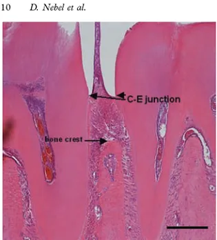
In paper II, sagital tissue sections from mice mandible were used to assess tooth attachment. After decalcification, the tissue was em-bedded and cut into thin, 4-μm sections. These sections included teeth with surrounding periodontal tissues: gingiva, root cementum, PDL, and alveolar bone. Tooth attachment was as-sessed by measuring the alveolar bone height and the apical termi-nation of the junctional epithelium, using image analysis software (Figure VI).
42 Figure measur distal s lar, resp gitudin represe
Immu
In pap were u val bio the ma tive ti (probi inflam probin subjec VI: Alveolar ring the distan surface of the spectively, and nal sections o ents 100 μm.unocytoche
per IV, im used to detec opsies. Ging arginal gingi issue. The b ng pocket d med sites (pr ng) from six ts). Prior to r bone height ance between t e first molar a d the highest of mandibles smistry and
munocytoch ct the estrog gival punch ival epitheliu biopsies wer depth < 4 m robing pock individuals experiment t in mouse m the cemento-e and the mesia point on the stained withimmunohis
hemistry an en receptors biopsies we um, includin re obtained mm and no b ket depth > (three male ts, different mandible was enamel (C-E) al surface of t e alveolar cres hematoxylinstochemistr
d immunoh s in HGEP ce ere used from ng epithelium from both bleeding on 5 mm and subjects and dilutions of determined b ) junction of t the second m st (A.C.) in lo and eosin. Bry
histochemist ells and ging m tissue fro m and conne healthy sit probing) an d bleeding o d three fema f the ERα an by the mo- on-Bar ry gi-om ec-tes nd on ale nd43 ERβ primary antibodies were tested. The sections were stained with either a polyclonal ERα antibody or an ERβ antibody. Mouse (C57BL/6 strain) uterus was included as a positive control for both antibodies. In the negative controls, the primary antibodies were omitted. All scoring was done blind. For each biopsy and staining, at least three sections were analyzed.
Ovariectomy
In the second paper, ovariectomy in female mice was used as a way of studying the effect of estrogen. The method is a well-established experimental technique in rodents, inhibiting endogenous ovarian production of estrogen in order to obtain an experimental model with a hormonal status similar to that observed in post-menopausal women. During the surgical procedure, both ovaries were removed. This led to termination of all estrogen production and the mice therefore become estrogen-deficient.
Statistics
All values are presented as mean ± S.E.M. Statistical significance was calculated using ANOVA and Student’s two-tailed t-test for unpaired comparisons with Bonferroni correction for post hoc analysis as appropriate. P-values of less than 0.05 were assumed to indicate statistical significance.
Ethical approval
The studies were approved by the Human Ethical Committee at Lund University, Lund, Sweden (Paper I, III, IV, V) and the Animal Ethics Committee at Lund University, Lund, Sweden (Paper II).
44
RESULTS AND DISCUSSION
Effects of LPS on PDL cells and HUVEC
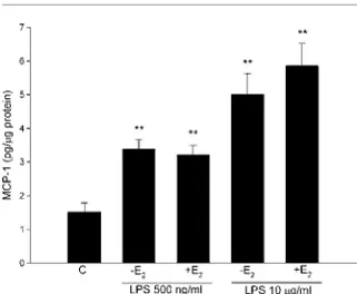
We observe at both the mRNA level and the protein level that PDL
cells are capable of producing IL-6 when exposed to E. coli LPS.
IL-6 concentration increased by about 30 times (Figure VII) within 24–72 h (Papers I and V). Also, an increase in the cellular MCP-1 concentration of 2–3 times was observed when PDL cells were
ex-posed to E. coli LPS (Paper I). PDL cells express factors that are
important in the TLR4 signaling pathway e.g. TLR4, MD-2, and
MyD88 (Hatakeyama et al. 2003). E. coli LPS is a known TRL4
agonist, suggesting that the production of IL-6 and MCP-1 ob-served in response to this stimulation was mediated though TLR4. By contrast, no stimulation of IL-6 production was seen using
P. gingivalis LPS as an inducer (Paper V). Unlike E. coli LPS,
P. gingivalis LPS acts as a TLR2 agonist and has also been shown
to be a TLR4 antagonist (Yoshimura et al. 2002). Although TLR2
is expressed in PDL cells (Hatakeyama et al. 2003), it is clear that
P. gingivalis is a weak inducer of IL-6 production. Several other studies have shown that PDL cells have the capacity to express other cytokines also, e.g. GRO-α, IL-1β, RANKL and TNF-α
(Jönsson et al. 2009; Shu et al. 2008; Wada et al. 2004). Thus,
45 Figure VII: Human PDL cells were stimulated for 24 h with E. coli LPS or P. gingivalis (PG) LPS (1 µg/ml) in the absence or presence of L-NAME
(100 µM) or 17β-estradiol (E2, 100 nM). IL-6 concentration increased by
about 30 times after stimulation with E. coli LPS. The NO synthase blocker, L-NAME, reduced E. coli LPS-induced IL-6 protein production. Values are mean ± S.E.M. *p < 0.05; ***p < 0.001. N.S., not significant.
The standardized culture conditions ensure reproducible experi-ments, and it follows that all the cells have similar phenotype and characteristics. On the other hand, it is likely that periodontal tis-sue from different patients responds differently in terms of cytokine production. We have shown that estrogen regulates cytokine ex-pression differently in PDL cells depending on the genetic origin of the cells (Paper III). It is possible that inter-individual response in cytokine production may be an important factor determining sus-ceptibility regarding development of periodontitis.
The expression of IL-6 in LPS-stimulated PDL cells is considerably lower than that observed in inflammatory cells, e.g. monocytes (Paper V). One can ask whether the secretion of pro-inflammatory cytokines by the periodontal fibroblast cells is of clinical relevance.
46
It is clear that an inflammatory cell has a greater capacity for initi-ating and driving inflammation, but also that the host tissue cells play a role in inflammation and that they should not be neglected. The number of periodontal host cells greatly exceeds that of in-flammatory cells. Furthermore, PDL cells are always present at the location where the inflammation starts, and may have an im-portant role in the initial recruitment of white blood cells to the site of inflammation. When the inflammatory cascade has started, it is likely that the cells of the periodontal tissue have a subordinate role in inflammation.
In contrast to PDL cells, LPS-stimulated HUVEC do not express significantly higher levels of IL-6 than control cells. TLR4 is nor-mally localized in the plasma membrane. In contrast, TLR4 in
HUVEC is mainly located in the Golgi apparatus (Makó et al.
2010). The cellular localization of TLR4 probably explains the poor stimulation of IL-6 in HUVECs. In summary, LPS-stimulated IL-6 production appears to depend on the cell type.
Effects of estrogen and
E.coli
LPS on the functional
proper-ties of PDL cells
E. coli LPS with or without estrogen has no effect on the primary function of PDL cells, i.e synthesis and secretion of collagen. Cell viability and DNA synthesis were also found to be unaffected by LPS and estrogen (Paper I), suggesting that estrogen works mainly as a promoter of PDL cell differentiation to a more specialized
os-teoblast-like cell type, as suggested by Liang. (Liang et al. 2008).
This indicates that there are no off-target effects of LPS in PDL cells.
High levels of estrogen show an anti-proliferative effect on HGEP cells (Paper V). Estrogen causes a reduction in DNA synthesis, both at 500 nM and at 10 μM. This effect is not seen in PDL cells with a lower and more physiological dose of estrogen. It is not clear how the anti-proliferative effect is mediated. Any of the two classical ERs, ERα or ERβ, or the new membrane-bound putative ER, GPR30, may be involved. These high concentrations of
estro-47 gen are uncommon under physiological conditions. The highest peaks of estradiol are seen during the second and third trimester of pregnancy. There have been several studies showing an increase in gingivitis during pregnancy compared to non-pregnant women
(Rakchanok et al. 2010). An anti-proliferative effect due to high
levels of estrogen (e.g. during pregnancy) could reduce the capacity of gingival epithelial cells to resist the bacterial load in a gingivitis situation. Usually, gingivitis in pregnancy is explained by an in-crease in angiogenesis and vascular permeability, but based on the data presented in the present thesis an effect of estrogen on gingival epithelial cell proliferation cannot be ruled out.
Estrogen and NOS blocker inhibit cytokine and chemokine
production in PDL cells
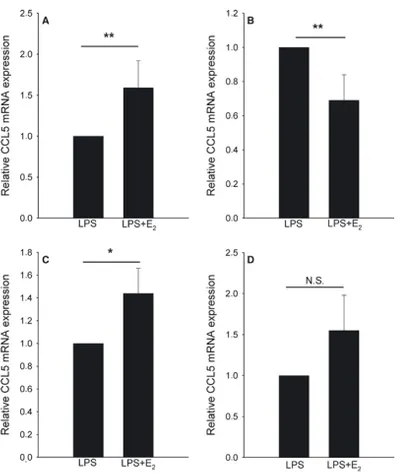
In Paper III, we showed that estrogen regulates chemokine expres-sion in PDL cells. The pattern is complex and involves both a downregulation and an upregulation of chemokines. We showed estrogen-induced downregulation of CCL3 mRNA, while the ex-pression of CCL2 mRNA was unaffected by estrogen. The effect of estrogen on CCL5 expression shows inter-individual variation, suggesting that the genetic origin of PDL cells might influence CCL5 expression (Figure VIII). As will be discussed later, PDL cells express predominantly ERβ. The effect of estrogen is therefore probably mediated through this receptor.
E. coli LPS-induced IL-6 production is inhibited by the NOS block-er L-NAME (100 µM), by about 30% (Papblock-er V, Figure VII). The selective iNOS blocker aminoguanidine had no effect on the LPS-induced IL-6. Our results suggest that eNOS but not iNOS may be involved, and they are supported by the results of Kikuiri et al. who showed that human PDL cells express eNOS but not iNOS at
48
Figure VIII: The effects of estrogen on PDL cell CCL5 mRNA levels de-pend on inter-individual variations. PDL cells were treated for 24 h with lipopolysaccharide (LPS) (0.5 lg/mL) in the absence or in the presence of
100 nM E2. Panels A and B show data from cells derived from the two
boys and panels C and D show data from the two girls. Values are means ± S.E.M. *p < 0.05; **p < 0.01. NS, not significant.
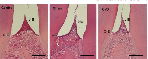
Effects of ovariectomy and aging on mouse periodontium
In Paper II, we did not observe any attachment loss in female mice within the observation period of 26 weeks using a morphometric technique. The distance between the cemento-enamel junction and the alveolar crest was similar at all ages. The apical termination of the junctional epithelium was at the cemento-enamel junction at all ages. These findings show clearly that mice do not develop perio-dontitis spontaneously in the model. The hypothesis was that es-trogen would exert a protective, anti-inflammatory effect. In this case, the ovariectomized (OvX) mice would develop more perio-dontitis than the animals with intact ovaries. However, no differ-ence in tooth attachment was observed between OvX mice and
49 control mice (Paper II). Our results can not exclude a possible ef-fect of estrogen in spite of the fact that we could not see any at-tachment loss in any of the groups.
There have been a few studies suggesting that OvX rats develop
less periodontitis than sham-operated control animals (Duarte et
al. 2004a; Duarte et al. 2004b). In a cotton ligature model,
Anbinder and coworkers showed that there was no effect of OvX
in rats, in contrast to Duarte (Anbinder et al. 2006). Estrogen
re-placement therapy has been shown to have a positive effect on
per-iodontitis in OvX rats (Duarte et al. 2004b). There have been no
previous studies showing effects of OvX on tooth attachment in mice. In conclusion, the effects of OvX on periodontium of rodents are still not clear.
Our study showed that young mice do not develop measurable per-iodontal bone loss. Recently, Liang and coworkers have shown that old mice (>18 months of age) show periodontal bone loss and
elevated expression of pro-inflammatory cytokines (Liang et al.
2010). These studies thus indicate that older mice could be used to serve as controls in future studies on periodontitis.
There are positive aspects to using a periodontitis model in mice without applying ligatures or other methods for induction of in-flammation. The aim when actively inducing periodontitis is to speed up the progression. Even though there is attachment loss, it is not desirable to provoke a fast progression that does not corre-spond to the typical, slowly developing periodontitis often ob-served in the clinical situation. Different effects of OvX in mice as compared to rats may also be due to a different bacterial microflora. Another study design with either induction of perio-dontitis or a longer observation period would better answer the question of whether OvX has any effect on tooth attachment.
Distribution of estrogen receptors in the gingiva
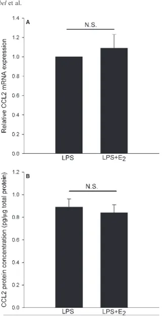
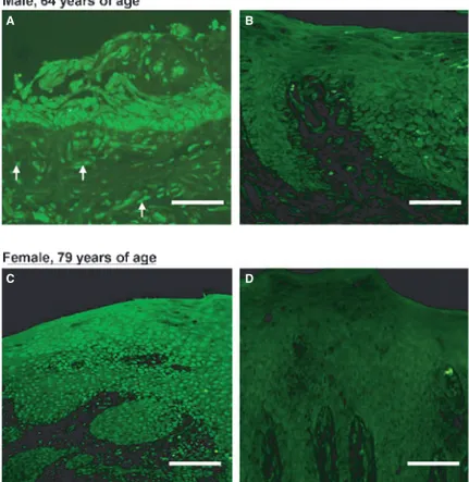
In biopsies from human gingiva, we found strong ERβ immuno-reactivity but no or very weak ERα immunoimmuno-reactivity in all six
sub-50
jects (Figure IX). The signal was found in the nuclei in cells of all ep-ithelial cell layers and also in cells of the lamina propria (Paper IV). The number of subjects is limited, and it cannot totally be ruled out that there are subjects with another ER subtype distribution. These
findings are consistent with those of Valimaa et al. (2004), who
re-ported that ERβ is the predominant ER in the gingival epithelium. In cultured HGEP a weak signal for ERα was also observed beside a very strong ERβ signal. In tissues, the immunoreactivity is more dif-ficult to interpret due to the complexity of the tissue structure. It is possible that there was also a weak ERα signal in the biopsies that was difficult to identify. Another possibility is that degradation of ERα occurs in the tissue but not in the cultured cells.
No differences in ERα/ERβ expression patterns were observed be-tween male and female subjects. High ERβ expression, but no ERα expression, was observed in both healthy and diseased sites, sug-gesting that the effects of estrogen on gingival epithelial cells are mediated through ERβ. In a study carried out by Karthik and coworkers, ER expression was determined in four different groups. In the groups of post-menopausal women, there was a significant reduction in ER expression in the gingiva of women with chronic periodontitis compared to those with healthy periodontium
(Karthik et al. 2009). Comparing Karthik’s experiment with ours,
there are obvious similarities in the study design. However, we used a “split-mouth-like design” where both the healthy biopsy and the diseased biopsy are obtained from the same donor. This has one big advantage: other factors that are not possible to con-trol will exercise the same influence on both groups. On the other hand, one can only see differences between a healthy and a dis-eased site in the same patient. The patient’s ER expression pattern may influence the susceptibility regarding development of perio-dontitis, and it was not possible to study this potential relationship using our study design.
F (H A sa n si se sh T fo P n k b Figure IX: Gi HGEP, lower A stong ERβ s amples. Not o nective tissue, ignal for ERα een in biopsie hown). Bars r The same pa or ERβ has PDL cells (Jo no studies de known to be blasts and os Gingival biops r row) stained signal was se only epithelia i.e. the lamin was seen in t ies from healt
represent 50 μ attern of ER been seen in nsson et al. emonstrating difficult to steoclasts ho sies (upper ro d for ERβ (lef een throughou al cells but als
na propria, ex the HGEP. N thy sites or si μm. subtype dis n other cell t 2004). To m g ER in hum culture since owever, both
ow) and gin ft column) or ut all tissue p lso the cells of
xpressed ERβ No difference i ites of gingiva stribution w types in the my knowledg man cemento e few cells a h ERα and ngival epitheli r ERα (right c preparations a f the underlyi β. No or a ver in ER express al inflammati with a strong periodontiu ge, there hav oblasts, a ce are viable. In ERβ are exp
51 lial cells column). and cell ing con-ry weak sion was ion (not g signal um, e.g. ve been ell type n osteo-pressed
52
(Vanderschueren et al. 2004). It is clear that ER expression is
cell-type dependent, suggesting a differential effect of estrogen depend-ing on ER subtype expression. If most periodontal cells show the same ER expression pattern, the periodontium could be a target for pharmacological treatment if the effects of estrogen prove to be beneficial. A selective ERβ drug affecting only periodontal cells would not have unwanted side effects in breast tissue, causing pro-liferation and increased risk of breast cancer, since the proliferative effects of estrogen in breast tissue are mediated by ERα.
53
CONCLUSIONS
E. coli LPS enhances MCP-1 and IL-6 production in PDL cells, suggesting that PDL cells are capable of recruiting leukocytes to the inflammation site (Paper I).
E. coli LPS with or without estrogen does not affect other properties of PDL cells such as proliferation and collagen syn-thesis. Estrogen does not reverse the LPS-mediated cytokine synthesis (Paper I).
Removal of ovarian production of female sex hormones by ovariectomy for 6 weeks has no influence on tooth attach-ment, suggesting that estrogen does not have any influence on tooth attachment in mice, within 6 weeks post-ovariectomy (Paper II).
Tooth attachment is preserved with age in mice within a od of six months. This indicates that mice hardly develop peri-odontitis without active induction within this time frame (Paper II).
Estrogen regulates chemokine expression in PDL cells in a complex manner involving both downregulation and upregulation of gene activity. Estrogen exerts both anti-inflammatory and pro-anti-inflammatory effects through these mechanisms (Paper III).