ACTA UNIVERSITATIS
UPSALIENSIS
Digital Comprehensive Summaries of Uppsala Dissertations
from the Faculty of Science and Technology
1312
Discovery and evaluation of direct
acting antivirals against hepatitis C
virus
ELDAR ABDURAKHMANOV
Dissertation presented at Uppsala University to be publicly examined in B21, BMC, Husargatan 3, Uppsala, Friday, 18 December 2015 at 09:00 for the degree of Doctor of Philosophy. The examination will be conducted in English. Faculty examiner: Professor David Frick (University of Wisconsin–Milwaukee).
Abstract
Abdurakhmanov, E. 2015. Discovery and evaluation of direct acting antivirals against hepatitis C virus. Digital Comprehensive Summaries of Uppsala Dissertations from the Faculty of Science and Technology 1312. 49 pp. Uppsala: Acta Universitatis Upsaliensis.
ISBN 978-91-554-9398-1.
Until recently, the standard therapy for hepatitis C treatment has been interferon and ribavirin. Such treatment has only 50% efficacy and is not well tolerated. The emergence of new drugs has increased the treatment efficacy to 90%. Despite such an achievement, the success is limited since the virus mutates rapidly, causing the emergence of drug resistant forms. In addition, most new drugs were developed to treat genotype 1 infections. Thus, development of new potent antivirals is needed and drug discovery against hepatitis C is continued.
In this thesis, a FRET-based protease assay was used to evaluate new pyrazinone based NS3 protease inhibitors that are structurally different to the newly approved and currently developing drugs. Several compounds in this series showed good potencies in the nanomolar range against NS3 proteases from genotype 1, 3, and the drug resistance variant R155K. We assume that these compounds can be further developed into drug candidates that possess activity against above mentioned enzyme variants.
By using SPR technology, we analyzed interaction mechanisms and characteristics of allosteric inhibitors targeting NS5B polymerases from genotypes 1 and 3. The compounds exhibited different binding mechanisms and displayed a low affinity against NS5B from genotype 3.
In order to evaluate the activity and inhibitors of the NS5B polymerase, we established an SPR based assay, which enables the monitoring of polymerization and its inhibition in real time. This assay can readily be implemented for the discovery of inhibitors targeting HCV.
An SPR based fragment screening approach has also been established. A screen of a fragment library has been performed in order to identify novel scaffolds that can be used as a starting point for development of new allosteric inhibitors against NS5B polymerase. Selected fragments will be further elaborated to generate a new potent allosteric drug candidate.
Alternative approaches have successfully been developed and implemented to the discovery of potential lead compounds targeting two important HCV drug targets.
Keywords: Direct acting antivirals, Hepatitis C, NS3-4A protease, NS5B polymerase,
structure-based drug discovery, fragment-based drug discovery, surface plasmon resonance
Eldar Abdurakhmanov, Department of Chemistry - BMC, Biochemistry, Box 576, Uppsala University, SE-75123 Uppsala, Sweden.
© Eldar Abdurakhmanov 2015 ISSN 1651-6214
ISBN 978-91-554-9398-1
List of Papers
This thesis is based on the following papers, which are referred to in the text by their Roman numerals.
I Belfrage, A. K., Abdurakhmanov, E., Åkerblom, E., Oshalim,
A., Gising, J., Skogh, A., Danielson, U.H., Sandström, A. Dis-covery of pyrazinone based compounds that potently inhibit the drug resistant enzyme variant R155K of the hepatitis C virus NS3 protease. Submitted.
II Belfrage, A. K., Abdurakhmanov, E., Alogheli, H., Åkerblom,
E., Danielson, U.H., Sandström, A. Pyrazinone based hepatitis C virus NS3 protease inhibitors targeting genotype 1a, 3a and the drug-resistant enzyme variant R155K. Manuscript.
III Winquist, J., Abdurakhmanov, E., Baraznenok, V., Hender-son, I., Vrang, L., Danielson U.H. (2013) Resolution of the teraction mechanisms and characteristics of non-nucleoside in-hibitors of hepatitis C virus polymerase. Antiviral Research, 97:356–368.
IV Abdurakhmanov, E., Solbak, S., Danielson, U.H. Characteri-zation of allosteric inhibitors of hepatitis C virus polymerase – a genotype comparative study. Manuscript.
V Abdurakhmanov, E., Danielson, U.H. A time-resolved surface
plasmon resonance based hepatitis C virus NS5B polymerase assay and its application for drug discovery. Manuscript.
VI Vagrys, D., Abdurakhmanov, E., Fridh, V., Karlsson, O., Danielson, U.H. Fragment library screening addressing hepatitis C protein NS5B from genotypes 1 and 3 using an SPR-based approach. Manuscript.
Contents
Introduction ... 7
Hepatitis C virus ... 7
HCV genetic variants ... 8
Non-structural HCV proteins as drug targets ... 9
NS3-4A ... 9
NS5B ... 12
Drug discovery ... 15
Structure-based drug design ... 15
Fragment-based drug design ... 16
Analysis of biomolecular interactions using SPR biosensor technology ... 17
Interaction mechanisms ... 17
Present investigation ... 20
Evaluation of pyrazinone based NS3 protease inhibitors targeting genotypes 1 and 3 as well as the drug-resistant variant R155K (Papers I and II) ... 21
Mechanisms of action and interaction properties of non-nucleoside NS5B polymerase inhibitors (Paper III) ... 28
Characterization of allosteric inhibitors of HCV polymerase – a genotypes comparative study (Paper IV) ... 31
Establishment of a label free continuous NS5B activity assay (Paper V) ... 35
Identification of novel NS5B polymerase inhibitor scaffolds (Paper VI) ... 36
Conclusions and future perspectives ... 39
Populär sammanfattning på svenska ... 41
Acknowledgments... 43
Abbreviations
DAA Direct acting antiviral
DSF Differential scanning fluorimetry
FBDD Fragment-based drug design
FQ Fit quality
FRET Förster resonance energy transfer
HCV Hepatitis C virus
HTS High throughput screening
IC50 Half maximal inhibitory concentration
LE Ligand efficiency
NI Nucleoside inhibitors
NMR Nuclear magnetic resonance
NNI Non-nucleoside inhibitors
NS3 Non-structural protein 3
NS5B Non-structural protein 5B
NTP Nucleotide triphosphate
PDB ID Protein database identification code
RdRp RNA-dependent RNA-polymerase
RU Response unit
SAR Structure-activity relationship
SBDD Structure-based drug design
SPR Surface plasmon resonance
Introduction
According to the World Health Organization (WHO), about 3% of the hu-man population worldwide is infected with hepatitis C virus (HCV). Around 70% of these individuals will develop a chronic HCV infection, one of the main causes of liver cirrhosis and hepatic cancers. The virus is transmitted by a blood-to-blood route, mainly by intravenous drug use, unsafe medical intervention, or blood transmission. The infection is usually asymptomatic, which makes it difficult to diagnose at an early stage. No vaccine is available against HCV, however research in this field is ongoing. Until recently, the standard therapy for hepatitis C treatment has been interferon and ribavirin. Such treatment has 50% of efficacy and is not well tolerated. Currently, with the emergence of new drugs, treatment success is estimated to be 90% (1). Despite of such an achievement, the long term efficacy is limited since the virus is rapidly mutated. This causes the emergence of drug resistant forms of the virus. In addition, most of the new drugs were developed to treat genotype 1 infections. Thus, the development of new potent direct acting antivirals that can be used for all relevant genotypes and that do not lose efficacy due to resistance is needed. The process of drug discovery against hepatitis C therefore needs to be continued.
Hepatitis C virus
Hepatitis C is a positive-stranded RNA virus, which belongs to the
Flaviri-dae family. It was discovered in 1989 and was previously known as “non-A,
non-B hepatitis” (2). The virus is an enveloped spherical particle, around 40-70 nm in diameter. The virus infects humans and chimpanzees (3). Hepato-cytes are the main target, although it can infect other cell types, for example B lymphocytes (4). The HCV genome is about 9.6 kb long and contains a single large open reading frame that encodes a polyprotein, flanked by 5’ and 3’ non-coding regions (NCR). The viral RNA at the 5’NCR contains an internal ribosomal entry site (IRES) (5). After the virus has entered into the host cells, its RNA is released and translated by host ribosomes into a poly-protein. Subsequently, the polyprotein is processed by host and viral prote-ases into 10 structural and nonstructural proteins. These proteins are associ-ated with the endoplasmatic reticulum (ER), forming an effective replication machinery. The structural proteins, i.e. the core protein (C), envelope
pro-teins 1 (E1) and 2 (E2), and p7, are used for assembly of new virus particles, whereas most of the non-structural (NS) proteins, i.e. NS2, NS3, NS4A, NS4B, NS5A and NS5B participate in viral genome replication. After for-mation of the replication complex, viral RNA synthesis is initiated. The packaging and assembly of new viruses occur in a so called membranous web. The mature viruses are then released from the host cell by exocytosis (6).
HCV genetic variants
There are six major genotypes and several subtypes of HCV identified now-adays (Fig. 1). The distribution of the genotypes varies in different geo-graphic regions. Genotype 1 is predominant in North America and genotypes 1, 2 and 3 in Europe. In Scandinavia, for example, 50% of HCV infected individuals have genotype 3, which is also prevalent in Asia along with gen-otype 6. Gengen-otype 4 is found in the Middle East, gengen-otype 5 in South Africa, and genotype 6 mainly in Hong Kong. Up to 65% sequence similarity is observed among the genotypes (7).
Figure 1. Global distribution of HCV genotypes. Adapted by permission from
Non-structural HCV proteins as drug targets
NS3-4A
The non-structural protein NS3-4A is a multifunctional protein that consists of two domains: a helicase domain and a protease domain (Fig. 2). NS4A is 54-residue protein, which is essential for NS3-4A functionality. Disruption of the NS3-NS4A interaction leads to reduction, or complete loss of protease activity (8, 9). The NS3-4A protease is responsible for proteolytic cleavage of the viral polyprotein into several non-structural proteins, including NS4A, NS4B, NS5A, and NS5B (10). NS3-4A is a classical chymotrypsin-like
ser-ine protease. The catalytic triad includes His57, Asp81, and Ser139 and is
locat-ed at the interface between protease and helicase domains. To be
catalytical-ly active, NS3-4A requires two co-factors: Zn2+ and NS4A. Zn2+ is located
far away from active site of NS3-4A thus it is believed to play a structural role (11). NS4A also has a structural role, influencing the formation of a catalytically functional active site of NS3-4A, while also anchoring it to the membrane (12).
The nomenclature of Schechter and Berger is widely used to describe the alignment of substrate/inhibitor in the active site of a protease (13). For ex-ample, if the side chains of the native substrate are used to define the peptide
sequence as NH2-P6-P5-P4-P3-P2-P1-P1’-P2’-P3’-P4’-OH, the
correspond-ing pockets in the enzyme’s active site are assigned as S6-S5-S4-S3-S2-S1-S1’-S2’-S3’-S4’. The cleavage site is located between the P1 and P1’ posi-tions. A decapeptide substrate is most suitable for cleavage by NS3-4A pro-tease. The consensus substrate sequence is (Asp/Glu)–X–X–X–X– (Cys/Thr)↓(Ser/Ala)–X–X–X, where X corresponds to any amino acid and the arrow shows the scissile bond (10).
The main role of the helicase domain is to unwind RNA, however, the ex-act mechanism is still not clear. The helicase domain also functions as an ATPase to hydrolyze adenosine tri-phosphate (ATP) as a source of energy. Such activity is stimulated upon nucleic acid binding (14). Both the helicase and protease domains can modulate each other’s activities (15, 16).
Although, the helicase domain is also an attractive drug target, the focus of this thesis will be on the protease domain and the inhibition of its activity.
Figure 2. Crystal structure of full-length NS3-4A protease (PDB ID: 1CU1). The
catalytic residues (CPK colored sticks) are located between the helicase (green) and the protease domains (red). The NS4A cofactor is incorporated into the protease domain (blue). The Zn2+ ion is depicted in the protease domain (gray sphere)
Since its identification in 1993, the HCV NS3-4A serine protease has be-come an important drug target for the discovery of potent, selective inhibi-tors against HCV. Although NS3-4A is an attractive target for structure based drug design, the development of the first potent DAAs against NS3-4A has been partially hampered by a misconception about a shallow sub-strate binding pocket. Nevertheless, significant progress has been made in this field and several DAAs targeting the protease have already reached the market.
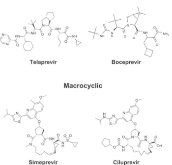
NS3-4A protease inhibitors can be divided into different classes. The first classification is based on their mode of action, such as non-covalent product-based inhibitors and reversible covalent inhibitors, known as serine-trap inhibitors. The second is based on their structure and can be classified as linear and macrocyclic inhibitors (17-20) (Fig. 3). Recently, an allosteric class of NS3-4A inhibitors has been identified. The location of the allosteric site is very close to the catalytic triad, but the mode of action and structure of the inhibitors are different to the active site binding inhibitors (21).
Ciluprevir was the first non-covalent, product-based NS3-4A protease DAA that entered the clinical trial phase, but was discontinued due to serious side effects (22, 23). However, it provided the basis for further development
of inhibitors targeting the NS3-4A. The first generation protease inhibitors launched on the market in 2011 were boceprevir and telaprevir. Both are reversible covalent inhibitors and were approved for combination therapy along with pegylated interferon and ribavirin. Recently, telaprevir was with-drawn from the market due to competition with alternative treatments (24). In 2013, a second generation NS3-4A product-based protease inhibitor, simeprevir, with improved characteristics, was permitted for HCV treatment (25). There are several second and third generation of protease inhibitors currently in development.
Figure 3. Examples of NS3-4A protease inhibitors. Telaprevir and boceprevir are
examples of covalent inhibitors whereas simeprevir and ciluprevir are non-covalent inhibitors.
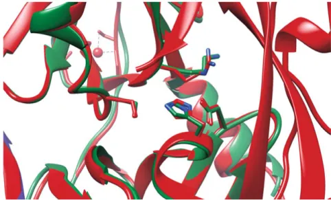
Despite the great treatment efficacy that has been achieved using protease inhibitors, the long term effect may be limited due to a rapid emergence of drug resistance. In most cases, resistance substitutions develop in close prox-imity to the NS3-4A catalytic triad, slightly outside the natural substrate binding site (Fig. 4) (26). Such changes in the protease domain reduce the affinity for the drugs, but still permit the recognition and cleavage of sub-strates, although, usually with a lower catalytic rate (27). The most common drug resistance mutations identified upon treatment with protease inhibitors are: V36A, R155K, A156S, and D168A. In addition, reduced efficacy of DAAs targeting the protease is associated with the natural existence of re-sistance variants in the different HCV genotypes that occur widely. For ex-ample, a substitution in the D168 position of genotype 1 variants causes
resistance to simeprevir. This is consequently observed in all HCV genotype 3 infected patients (28).
Figure 4. Structure alignment of wild-type NS3 (red) (PDB ID: 1CU1) with
re-sistance mutant NS3 R155K (green) (PDB ID: 4I33).
NS5B
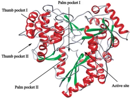
NS5B is an RNA-dependent RNA-polymerase (RdRp) associated with the endoplasmatic reticulum membrane. The membrane spanning helix of the 66 kDa protein consists of 21 C-terminal amino acid residues, which are usually not included in the constructs for most in vitro studies, due to poor solubility of the full-length variant. NS5B shares a common right hand structure with other RdRps and has three main domains: fingers, palm and thumb (Fig. 5). However, it has a distinct feature, that is, close interaction between fingers and thumb domains, resulting in a closed active site structure (6). NS5B catalyzes the synthesis of both positive and negative RNAs during virus replication, and can function in an oligomeric form (29). NS5B is able to initiate RNA synthesis by two different mechanisms: 1) a primer dependent mechanism, exogenous or self-priming (30) and 2) de novo synthesis on the template by joining free nucleotide triphosphates (NTPs) and extending them to a long RNA chain (31). NS5B polymerase is believed to be very dynamic and undergoes large structural re-arrangements in order to perform its func-tion (32). In addifunc-tion, NS5B is an error-prone RdRp, lacking proof reading activity, which explains the large number of genomic variants of the virus (33).
NS5B plays a fundamental role in HCV replication and is therefore con-sidered an important drug target.
Figure 5. Crystal structure of RNA-dependent RNA polymerase with the inhibitor
binding sites indicated (PDB ID: 1QUV).
Five inhibitor binding sites are identified in the NS5B polymerase (Fig. 5). Four are allosteric and bind non-nucleoside inhibitors (NNI). They are locat-ed in the palm and thumb domains. The active site binds nucleoside inhibi-tors (NI). The allosteric inhibiinhibi-tors induce a conformational change, thereby reducing the activity of the enzyme. Active site inhibitors function as chain terminators, blocking the incorporation of incoming nucleotides. Currently, there are plenty of NNI and NI NS5B DAAs in various phases of develop-ment. In 2013, sofosbuvir, a nucleoside analog inhibitor was approved for treatment of HCV infection in combination with pegylated interferon and ribavirin. More recently, in 2015, a non-nucleoside NS5B palm site inhibitor dasabuvir was approved by Food and Drug Administration (FDA) for com-bination therapy of HCV (Fig. 6).
Figure 6. Structures of NS5B polymerase inhibitors. Dasabuvir and sofosbuvir were
approved for clinical use while nesbuvir, lomibuvir and filibuvir were tested in the clinic but discontinued for various reasons.
As in the case of NS3-4A inhibitors, viral resistance against NS5B inhibitors is also a problem. Generally, non-nucleoside inhibitors have a low genetic barrier to resistance. Resistance can easier arise for allosteric inhibitors since the binding site can often be modified without influencing the catalytic activ-ity. However, the advantage of this class of inhibitors is uniqueness of the binding site and therefore low level of side effects. The most common sub-stitution that confer thumb II inhibitors resistance, such as filibuvir and lomibuvir are at positions L419, M423, and I482 (34). The developments of filibuvir was discontinued by Pfizer for strategic reasons, while lomibuvir was not pursued due to lack of efficacy (35). As for palm inhibitors, such as nesbuvir and dasabuvir, the substitutions at the positions C316, M414, Y448, and S556 have been reported. Nesbuvir was discontinued due to liver toxici-ty, however, it possesses quite broad cross-genotypic activity (36).
In contrast to NNIs, the NIs have a higher genetic barrier to resistance. The most frequently occurring substitution is position S282 and is reported to be associated with all NIs, including sofosbuvir (37). This substitution is located in close vicinity of the active site, which in turn causes a reduction of viral replication (38). Thus, resistance to NIs occurs more rarely and slowly.
Due to the conservation of NS5B active site residues, nucleoside inhibi-tors usually have a cross-genotypic activity. It was shown that, for example, sosfosbuvir monotherapy was efficient for treatment of patients infected with HCV genotypes 1, 2, and 3, while dasabuvir has been reported to be potent against genotype 1 (36, 39, 40).
Drug discovery
Since ancient times, human beings have been using medicines prepared from natural resources, mostly from plants. Throughout the history of medical science, new drugs have been discovered by a trial-and-error process, or in other words – by luck. As the demand for new and more effective drugs has increased, a new method of drug discovery was needed. This led to the emergence of several alternative approaches, such rational structure-based drug design (SBDD), fragment-based drug design (FBDD) and high throughput screening (HTS). However, the entire drug discovery process from target identification and validation to the final approval of a drug is a long journey, irrespective of the approach taken.
Structure-based drug design
Advances in genomics, proteomics, bioinformatics, and structural biology have provided a solid basis for SBDD. It is an iterative process that goes through several cycles before optimized lead enters clinical trials. The pro-cess begins with the determination of the protein structure by X-ray crystal-lography or nuclear magnetic resonance (NMR). This is followed by identi-fication and examination of potential drug binding sites, for example, size, hydrophobicity, identification of potential hydrogen bond donors and accep-tors, etc. Binding sites can be active sites, allosteric sites or the interface between two targets. For many protein targets, substrates, cofactors and oth-er modulators can soth-erve excellent starting points for potential inhibitor gen-eration, as for example described above for NS3-4A protease. Next, a set of compounds can be virtually docked into the binding site and ranked accord-ing their most favorable orientation. Then, the selected compounds are syn-thesized and tested experimentally using biochemical assays. The subsequent cycle includes structure determination of the target in the presence of a lead compound. The lead compound can then be further optimized in order to increase its potency. The cycles are repeated several times until a compound
with desired properties is obtained (41). The bioavailability of lead com-pounds is often predicted using Lipinski’s rule of 5. That is, in general, oral-ly available drug are expected to have no more than 5 hydrogen bond donors and no more than 10 hydrogen bond acceptors, molecular weight less than 500 Da, and a calculated partition coefficient (logP) less than 5 (42). SBDD has successfully been applied for the development of antivirals, such as the HCV protease inhibitors boceprevir and telaprevir, HIV protease inhibitors ritonavir and indinavir, influenza virus neuraminidase zanavir (43-47). There are many other examples demonstrating that SBDD is a powerful strategy for drug discovery. This approach provides new designed drugs with high potency, minimum side effects, improved bioavailability, a potential to overcome resistance and many other challenges.
Fragment-based drug design
In recent years, FBDD has become a very popular complementary approach to SBDD in the drug discovery community. Traditionally, many
pharmaceu-tical companies and academic groups use HTS to screen libraries up to 106
compounds. They are quite often not the best candidates for further devel-opment, since such libraries cover a limited fraction of chemical space, which in turn also hampers the finding a good compound as a starting point. FBDD is fundamentally different from HTS. It is based on the screening of smaller compounds libraries, covering a larger fraction of chemical space. Hits are low affinity binders, typically in the micro- to millimolar range. Fragments usually comply with Lipinski’s rule of 3: molecular weights are not more than 300 Da, they include no more than 3 hydrogen bond donors and acceptors, and calculated logP values are not greater than 3 (48).
Since fragments are small, they must make good interactions with the tar-get to bind with sufficient affinity in order to be detected. The important metrics that characterize such low affinity interactions are ligand efficiency (LE) and fit quality (FQ). LE is suitable for ranking fragments according to their average binding energy per atom, defined as kilocalories per mole di-vided by the number of heavy atoms (49). The concept can be further ex-tended to include other physicochemical properties (50). Generally, frag-ments that possesses a LE value (>0.4) are considered suitable to be selected for further development. In order to increase the affinity of a fragment dur-ing its elaboration, addition of more than 15 heavy atoms is required. This corresponds to an increase of the molecular weight up to 200 Da. Therefore, the control of the physicochemical properties of the fragment is required. The development of a fragment into a lead compound, maintaining its LE value, is challenging but may eventually lead to improved drug-like proper-ties (50). The LE depends on the size of the molecule, which makes the di-rect comparison among fragments difficult. To address this problem, the FQ
metric can be applied. The FQ scales the fragment’s LE to obtain a normal-ized score around 1.0, independent of the size.
FBDD generally includes 3 stages: 1) fragment library design, 2) in vitro fragment library screen, and 3) fragment elaboration. Usually, the libraries are designed in such way that fragments contain functional groups that facili-tate their further elaboration, at the same time excluding unstable, reactive and toxic moieties. The screening of the library can be performed virtually or by using biophysical methods such as NMR, X-ray crystallography, mass spectrometry, fluorescence thermal shift, or surface plasmon resonance bio-sensors (SPR) (51). Subsequently, to generate a lead compound, the identi-fied hits are further elaborated by fragment merging, linking or growing strategies. At this stage, FBDD continues according to the iterative process used also for SBDD.
Currently, there are many potential drugs in clinical development phases that were generated using FBDD. An important landmark of this approach was an approval of anti-cancer drug zelboraf in 2011 (52).
Analysis of biomolecular interactions using SPR
biosensor technology
Surface plasmon resonance is an optical detection technique that enables the label-free monitoring interactions between biomolecules in real-time. The SPR phenomenon was first described in 1968 (53), and some years later it was demonstrated that this phenomenon can be used for interaction detection (54). In 1991, the first commercially available SPR-based biosensor instru-ment was launched by Biacore (55). The technology principle is based on attachment of one interaction partner to the chip surface, for example, a pro-tein, whereas another interaction partner, for example, an inhibitor is deliv-ered into the flow cell with the solution. The binding of molecules generates a response, measured in resonance units (RU), and is proportional to the bound mass on the biochip surface. Several useful interaction parameters such as interaction mechanism, specificity, kinetics, affinity, thermodynam-ics, and stoichiometry can be resolved by using this technique.
Interaction mechanisms
The interaction between biomolecules may occur by various mechanisms. With the help of SPR biosensor analysis it is possible to resolve interaction mechanisms and kinetics using predefined models. Quite often, the interac-tion between biomolecules cannot be described by any predefined models due to its complexity. Below is a short overview of the binding models that were used to characterize the interactions presented in this thesis.
1:1 (Langmuir) binding model
This is the simplest model that describes a reversible 1:1 interaction between molecules. It is similar to the Langmuir isotherm for adsorption of gas to a surface (56). The model can be illustrated as following scheme:
where L is an immobilized ligand, A is an injected analyte, and k1 and k-1 are
the rate constants.
The differential equation that describes the model is: 𝑑𝑑[𝐿𝐿𝐿𝐿]
𝑑𝑑𝑑𝑑 = (𝑘𝑘1∙[𝐿𝐿]∙[𝐿𝐿]) − (𝑘𝑘−1∙[𝐿𝐿𝐿𝐿]) and the equilibrium dissociation constant is:
𝐾𝐾𝐷𝐷 =𝑘𝑘𝑘𝑘−1 1
Two-state reaction model
This model describes 1:1 interaction followed by a second step that can be formation of a reversible covalent bond or conformational change, both sta-bilizing a complex:
The following equation is used to describe two-state model:
𝑑𝑑[𝐿𝐿𝐿𝐿]
𝑑𝑑𝑑𝑑 = (𝑘𝑘1∙ [𝐿𝐿] ∙ [𝐿𝐿] − 𝑘𝑘−1∙ [𝐿𝐿𝐿𝐿]) − (𝑘𝑘2∙ [𝐿𝐿𝐿𝐿] − 𝑘𝑘−2∙ [𝐿𝐿∗𝐿𝐿])
The overall equilibrium dissociation constant is defined as:
𝐾𝐾𝐷𝐷 = 𝑘𝑘𝑘𝑘−1 1 ∙ 𝑘𝑘−2 𝑘𝑘−2+ 𝑘𝑘2 L+A k1 LA k-1 L+A k1 LA k-1 L *A k2 k-2
Heterogeneous ligand model
This model describes two independent binding reactions, assuming that the analyte can interact with two forms of the ligand. It is important to realize that such heterogeneity may occur due to inappropriate experimental set-up, for example, arising when the ligand is heterogeneously coupled to the sur-face resulting in various ligand conformations. Thus, the binding model is described as two independent 1:1 interaction models and the kinetic parame-ters, as well as the equilibrium dissociation constants, are obtained separate-ly for each binding reaction:
Steady-state affinity
It is not always possible to resolve the binding kinetics parameters, for ex-ample if the interaction is very fast, e.g. an interaction of a fragment with a target protein. In this case a steady-state analysis can be performed using report points taken at equilibrium. This model is used to calculate the
equi-librium dissociation constant (KD) for 1:1 binding from the response level
(R) plotted against the analyte concentration [A] using following equation:
𝑅𝑅 =[𝐿𝐿] ∙ 𝑅𝑅𝐾𝐾 𝑚𝑚𝑚𝑚𝑚𝑚
𝐷𝐷+ [𝐿𝐿] + 𝑅𝑅𝑅𝑅
where Rmax is a theoretical response level at the steady-state and RI refractive
index contribution caused by bulk effect of the sample.
L1+A k1 L1A
k-1
L2+A k2 L2A
Present investigation
Due to the error prone nature of HCV NS5B polymerase there is a large di-versity of genomic variants of the virus. Six main genotypes and several subtypes are defined nowadays. The most prevalent HCV genotypes in the world are genotypes 1 and 3. Genotype 3 is considered more dangerous as it has a high propensity to cause liver cirrhosis and hepatocellular carcinoma (57-59).
Since the standard treatment of hepatitis C infection with pegylated inter-feron and ribavirin has low affectivity and is poorly tolerated, there is a need for the development of new classes of drugs that specifically target viral proteins. Despite the fact that several DAAs were recently developed and approved for the treatment of HCV infections, the emergence of drug re-sistance mutations requires further development. Moreover, all new DAAs are primarily developed for eradication of genotype 1 infections and thus possess no or limited potency, for example, against genotype 3.
The aim of the present investigation was to contribute to the discovery of new HCV drugs, using novel technologies. The focus was on the combina-tion of using different strategies and addressing both standard target HCV variants as well as potentially resistant variants and genotypes (60).
By using a conventional structure-based approach to design inhibitors that target viral NS3 protease we aimed to develop a new class of pyrazinone based inhibitors, which possess inhibitory activities against wild-type, drug-resistant variant R155K, and NS3 protease from genotype 3 (Papers I and II). Since an effective therapy using DAAs will be a combination of inhibi-tors that target different viral proteins, our next aim was to study the possi-bility of developing allosteric inhibitors targeting the HCV NS5B polymer-ase (Papers III and IV). For this purpose we used a clinically relevant genetic variant, not previously studied, and established a new type of real-time activ-ity assay, which can be used for polymerase characterization as well as inhi-bition studies (Paper V). We also initiated a fragment-based lead identifica-tion approach to provide new starting points for the development of drugs targeting NS5B polymerases from genotype 1 and genotype 3 (Paper VI).
Evaluation of pyrazinone based NS3 protease inhibitors
targeting genotypes 1 and 3 as well as the drug-resistant
variant R155K (Papers I and II)
In 2011, the first DAAs, the electrophilic HCV NS3 protease inhibitors bo-ceprevir and telaprevir, were approved for the treatment of HCV genotype 1 infection. The major problem with these highly potent DAAs and an error prone viral polymerase is the apparent risk for evolution of drug-resistant en-zyme variants. Mutations in the viral genome corresponding to amino acid positions R155, A156 and D168 in the NS3 protease are common in genotype 1 infected patients, and for example, R155K/T/Q substitutions that confer resistance to all NS3 protease inhibitors currently approved or in clinical trials. Besides addressing the challenge for the next generation of NS3-4A protease inhibitors, improving the potency against other genotypes of the virus is also of importance. For example, genotype 3, the second most common worldwide genotype and potentially the most difficult to treat is a variant that needs to be addressed. It differs from genotype 1 variants, for example, in position D168, making it a good model system also for genotype 1a resistance.
In this paper the design and synthesis of 2(1H)-pyrazinone based HCV NS3-4A protease inhibitors with variations in the P1P1’ region and elongat-ed P4P5 urea substituents is presentelongat-ed (Fig. 7). Pyrazinone is uselongat-ed as a uni-versal scaffold to design drugs against proteases, including HCV NS3-4A (61-63). An important feature of the pyrazinone scaffold is the capability to adopt a β-strand structure, which is essential for recognition and binding to the protease active site (64). In addition, the structure facilitates the addition of functional groups at various positions (65).
Figure 7. Structure of a 2(1H)-pyrazinone-based HCV NS3 protease lead
com-pound. The pyrazinone scaffold is highlighted by brackets.
A previous study indicated that an aromatic acyl sulfonomide modification at the P1P1’ position resulted in favorable potencies and a less peptide-like character of the inhibitor (66). Thus, we decided to study the different opti-mization possibilities of new aromatic P1P1’ substituents. Biochemical eval-uation of the synthesized compounds was performed using both wild-type and a resistant variant for genotype 1a, and a wild-type variant for genotype 3a. Series of compounds with variations in the P1P1’ position was designed and evaluated (Table 1).
N N O Cl H N O H N O H N O HNS CF3 O O P1 P1' P5-P4 P3 P2 6 4 3
Table 1. Inhibitory potencies of pyrazinone based inhibitors with various aromatic
modifications at the P1P1’ position
Compound P1P1’ Full-length NS3 1a Ki ± SD (nM) Full-length NS3 1a R155K Ki ± SD (nM) 6 O HNS O O CF3 140 ± 20 30 ± 2 47 H N O S CF3 O O 260 ± 60 60 ± 8 53 H N O S O O CF3 N N 120 ± 20 30 ± 4 54 H N O S O O CF3 F 110 ± 20 20 ± 2 57 O O 120 ± 30 15 ± 2 68 OH O 120 ± 30 17 ± 4 58 N H O S CF3 O O 140 ± 30 24 ± 3 69 N N O Cl H N O OH Bn O H N >10000 Nd
Nd: not determined, SD: standard deviation
Compared to the lead compound (6) in this series, altering the P1P1’ regioi-somer did not affect the inhibitory potencies, despite significant changes in conformation (e.g. compound 47). The move of the electron withdrawing
trifluoromethyl group, as well as replacing the CF3 moiety to an electron
N N O Cl H N O H N O H N P 1P1´
donating methyl group indicated that the P1’-moiety did not have any
specif-ic interactions with the enzyme and that the CF3-moiety did not improve the
potency despite increasing the acidity of the acyl sulfonamide hydrogen, which has been shown to be important in other types of NS3-4A protease inhibitors (67).
The steric and electronic effects by various substituents on the P1-phenyl ring were investigated. It was found that although there were big differences in size and electronic properties, the effects on the inhibitory potencies were insignificant (e.g. compounds 53 and 54). Overall, the SAR shown by the P1P1’ modifications indicated that there were no important interactions with this part.
An attempt to increase the conformational flexibility by incorporation of an methylene linker in compound (58) was tried (Table 1), but in comparison to the carboxylic acid (68) and ester (57) compounds, no additional benefi-cial effects could be seen. To evaluate the importance of the P1-part, a com-pound without the P1-aryl group (comcom-pound 69) was evaluated. The signifi-cant decrease in activity verified that the P2P3 substituent together with the P3-capping group is not only insufficient for nanomolar activities, but also relies on the P1-moiety.
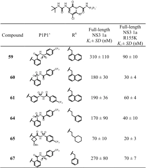
A beneficial property of aryl acyl sulfonamides is the opportunity to easi-ly reverse the acyl sulfonamide functionality which could alter the binding mode and possibly facilitate binding to the oxyanion cavity. Therefore, the compounds presented in Table 2 were evaluated. The varied regioisomers of reversed acyl sulfonamides (e.g. compounds 60 and 61) showed slightly
lower Ki-values compared to the original acyl sulfonamide (59). Next, a R6
group (phenethyl) was combined with the reversed acyl sulfonamide (64) showing similar potency. These results indicated that a distinct binding to the oxyanion hole was still absent and that an optimal P1P1’ substituent needed to be invented for this class of inhibitors.
In order to depart from the flat and highly aromatic structure, which could be advantageous for drug properties, we thought that a spirocyclobutyl-group could be useful, with the inherent opportunity to introduce different functionalities. Such compounds, e.g. compound (65), showed slightly im-proved potency.
The thirteen inhibitors shown Tables 1 and 2, were evaluated against the drug-resistant enzyme variant R155K. Arg155 is positioned in the S2 pocket and is part of a stabilizing salt bridge with Asp168. Thus, the substitution at position R155 disrupts the salt bridge that is important for inhibitors contain-ing extended P2-substituents (68). Generally, this series of compounds were 3- to 8-fold more potent against the mutated NS3-4A variant compared to wild-type. The relatively small P2-substituents in this class of inhibitors probably explain partly the retained potency against the R155K. The most interesting finding was the potency of the P1’-truncated inhibitors (57) and (68) (Table 1) which were shown to be the most active among the evaluated
compounds and more potent against the R155K variant as compared to the wild-type enzyme.
Table 2. Inhibitory potencies of pyrazinone based inhibitors with various aromatic
modifications at P1P1’ and R6 positions.
Compound P1P1’ R6 Full-length NS3 1a Ki ± SD (nM) Full-length NS3 1a R155K Ki ± SD (nM) 59 O HNS O O CF3 310 ± 110 90 ± 10 60 S H N CF3 O OO 180 ± 30 30 ± 4 61 SN H O CF3 O O 190 ± 36 60 ± 4 64 S H N CF3 O OO 170 ± 90 40 ± 10 65 N H OBn O S CF3 O O 70 ± 10 20 ± 3 67 H N O S O O CF3 270 ± 80 70 ± 7
A previous study showed the importance of using a full-length enzyme for the evaluation of inhibitors and for resistance profiling (69). Thus, instead of
further elongating and building out the P2 substituent (R6 of the pyrazinone)
we decided to evaluate an extended urea substituent reaching from the P3-position to P4P5 which would enable additional interactions with the
en-N N O Cl H N H N O O H NP 1P1´ R6
zyme, also found by the natural substrate, and possibly interactions with the helicase domain.
Evaluation of the compounds listed in Table 3 against NS3 protease from genotype 3 showed again that the P1´substituents had no significant effect on the inhibitory potency. However, all inhibitors exhibited a potency in nano-molar range (Table 3).
Table 3. Inhibitory potencies against NS3 protease from genotype 3 of pyrazinone
based inhibitors with various modifications at P1P1’ and R6 positions
Compound P1P1’ R6 Full-length NS3 3a Ki ± SD (nM) 6 H N O S O O CF3 80 ± 40 35 H N O S O O CF3 N N 87 ± 11 36 OH O 70 ± 5 37 N H OBn O S CF3 O O 68 ± 4 38 H N O S O O CF3 565 ± 83 39 S H N CF3 O OO 170 ± 14 N N O Cl H N O H N R6 O H N P 1P1´
The next approach was to alter the P4P5 substituent. Initially, two diverse P3-capping groups were evaluated against genotype 1. Compound (41) showed a six times decreased inhibitory potency compared to (40), which supported the previously suggested positive influence of an internal hydro-gen bond. However, compound (42) which also lacked an internal hydrohydro-gen bond opportunity, showed retained potency (Table 4).
Next, three inhibitors with advanced P4P5 urea substituents were
evaluat-ed. For the inhibitors containing a benzyl in the R6-position, the potency
against genotype 1 was preserved (43, 44, 40). Changing to a R6-cyclohexyl
ethyl group increased the potency about 5 times (45). The most interesting observation was the increased inhibitory potency against the R155K enzyme
variant. Compound (45) showed a Ki value of 2 nM which was 15 times
more active, compared with compounds (44) and (6). Finally, compound
T able 4 . I nhi bi tor y pot enc ie s a ga in st va rio us NS 3 pr ot ea se va ria nt s of py ra zi no ne ba se d inhi bi to rs wi th P4P 5 ur ea subs tit ue nt s C om pound R 1 R 3 R 6 Fu ll-le ng th NS 3 1 a Ki ± S D (n M ) Fu ll-le ng th N S3 1a R 155 K Ki ± S D (n M ) Fu ll-le ng th N S3 3a Ki ± S D (n M ) 40 H H N H N O 270 ± 8 0 70 ± 7 nd 41 H N H O H N 1650 ± 46 nd nd 42 H N N O 380 ± 40 nd nd 43 F N S O N H N O H N O 190 ± 40 40 ± 10 nd 44 F N H N O H N O O 160 ± 60 30 ± 5 135 ± 13 45 F N H N O H N O O 30 ± 2 2 ± 0. 2 5. 4 ± 0. 4 N N O Cl R 3 O H N R 6 O H N S CF 3 O O R 1
In conclusion, we found that compounds without an acidic P1’-acyl sul-fonamide were the most promising inhibitors in this series. They exhibited a beneficial, up to ten fold increase in inhibitory potency against the drug-resistant variant R155K compared to the wild-type enzyme. The compounds in this series retained their inhibitory potency against R155K which is the position that confers resistance to all HCV NS3-4A protease inhibitors ap-proved or in clinical trials. The strategy to extend the P4P5 part showed that the compounds could further improve the interactions to the protease. These results further strengthen the potential of this substance class in the devel-opment of inhibitors less sensitive to mutations.
Mechanisms of action and interaction properties of
non-nucleoside NS5B polymerase inhibitors (Paper III)
The allosteric NS5B inhibitors represent an interesting class of compounds since they interact with a unique binding site of the target protein, which may lead to less side effect development in comparison to nucleoside ana-logs. However, in order to design efficient allosteric drugs, a detailed under-standing of the mechanism of their action is required. In this study we per-formed mechanistic, kinetic chemodynamic and thermodynamic studies of three promising allosteric thumb pocket II inhibitors by using SPR technolo-gy. Their antiviral potencies were assessed using an in vitro cell-based repli-con assay.
The inhibitory effect of the compounds was evaluated by scintillation
count assay. VX-222 was identified as the most potent inhibitor, with an IC50
of 7 nM. Filibuvir displayed a very low inhibition efficiency, showing 80%
inhibition at 100 µM concentration. As a result, no meaningful IC50 value
could be obtained. As for tegobuvir, an even more unusual inhibition was observed. The compound stimulated the NS5B up to 50% at 3.7 µM and then inhibited it up to 40% at 100 µM (Fig. 8). This indicates that the com-pound does not inhibit NS5B by a simple mechanism.
Figure 8. NS5Bd21 activity inhibition by inhibitors: filibuvir (■), VX-222 (♦), and tegobuvir (●).
Next, the inhibitors were tested with respect to their effects on the thermal stability of NS5Bd21 using differential scanning fluorimetry (DSF).
Fili-buvir and VX-222 caused a 4–5 oC increased melting temperature (Tm) of
the enzyme, indicating a stabilization of the protein upon inhibitor binding. Tegobuvir showed no effect on the melting temperature of NS5Bd21.
The mechanisms and kinetics of compound interactions were further as-sessed using SPR technology. The interaction between filibuvir and NS5Bd21 was described by a simple 1:1 Langmuir interaction model, whereas the interaction between VX-222 and NS5Bd21 was better described by a two step interaction model (Fig. 9). Both inhibitors showed similar
af-finities to the enzyme, with the KD-values in the nanomolar range. However,
the residence time was about 15 fold higher for VX-222. Furthermore, the experiment was performed by injecting a single concentration of inhibitor
over native and cross-linked protein surfaces for different time at 5 oC and
25 oC. In this experiment, VX-222 again showed a complex interaction with
NS5Bd21 and remained the same under both temperatures. It was observed that equilibrium was not reached in the interaction with filibuvir at low tem-perature. This can indicate that there is a conformational change, which is rate limiting at low temperatures. Tegobuvir did not show any interaction and was subsequently excluded from further investigation. During the prepa-ration of this paper it is became clear that this compound undergoes an addi-tional modification in the cell in order to be able to act on the NS5B poly-merase. This makes the lack of interaction and strange inhibitory effect logi-cal. -60 -40 -20 0 20 40 60 80 100 -9 -8 -7 -6 -5 -4 % inhibition log([inhibitor])
Figure 9. Sensorgrams representing the interaction between NS5Bd21 and filibuvir
(left), and VX-222 (right). The compounds were injected in two-fold concentration series between 5 and 156 nM.
The thermodynamic and chemodynamic interaction profiles of the inhibitors were examined using SPR technology. Chemodynamic analysis was per-formed by varying pH and ionic strength of the interactions between the inhibitors and the NS5Bd21 polymerase. No significant differences were observed for the two inhibitors. This indicates that that both compounds interact by similar interaction forces.
The thermodynamic profile was analyzed for the interaction with fili-buvir. VX-222 was excluded from this analysis due to the complexity of its interaction. Interaction analysis was performed at various temperatures
rang-ing from 5 oC to 35 oC, and using both native and cross-linked protein. The
results from van’t Hoff analysis demonstrated that the interaction with the native enzyme had favorable entropy and enthalpy, while cross-linked en-zyme had a favorable enthalpy and unfavorable entropy. This shows that the enzyme is dynamic and that conformational changes are involved in the mechanism for thumb pocket II allosteric inhibitors.
In order to determine if conformation changes upon inhibitor binding causes interference with RNA binding to the enzyme, the influence of fili-buvir and VX-222 on the interaction between NS5Bd21 and single stranded RNA was also assessed. In fact, the presence of either filibuvir or VX-222 clearly decreased the amount of RNA-enzyme complex formed, by approx-imately 30% at the highest concentration tested by comparing maximal sig-nal level. The interaction with and without inhibitor was complex since the curves were not well described by any standard interaction models (Fig. 10).
0 2 4 6 8 10 12 -20 0 20 40 60 80 100 120 140 160 Response, [RU] Time, [s]
Figure 10. Interaction between NS5Bd21 and immobilized RNA in the absence and presence of inhibitor. (a) 240 RU of RNA was captured via streptavidin and
NS5Bd21 with 300 nM filibuvir first injected (green), followed by NS5Bd21 alone (red). (b) 200 RU of RNA was captured via streptavidin and NS5Bd21 with 900 nM VX-222 first injected (blue), followed by NS5Bd21 alone (red).
In summary, in this study we demonstrated that the studied allosteric inhibi-tors have different mechanisms of interaction, although they bind to the same site. The induced fit mechanism with prolonged residence time for VX-222 gives it a kinetic advantage over filibuvir. Both compounds were able to interfere with RNA binding to the enzyme, demonstrating that they have a mechanism of action that influences the ability of the enzyme to bind to its substrate. Although today both filibuvir and VX-222 (known as lomibuvir) are discontinued from further development, they can be used as a basis for generation of new allosteric DAAs.
Characterization of allosteric inhibitors of HCV
polymerase – a genotypes comparative study (Paper IV)
Drug discovery efforts against HCV are mainly directed towards eradication of HCV genotype 1, while largely ignoring the development of drugs against other HCV genotypes. Therefore, there is limited information available about the potency of allosteric polymerase inhibitors targeting genotype 3. In addition, despite about 90% sequence identity between NS5B from different HCV isolates, the interaction mechanisms of allosteric inhibitors might still vary, as was previously reported in an earlier study of other non-nucleoside inhibitors (70). In the present study we compared the kinetic parameters and affinities of thumb II allosteric inhibitors filibuvir and lomibuvir (previously known as VX-222) as well as palm inhibitors dasabuvir and nesbuvir (HCV-796) for NS5B polymerase genotype 1b isolates Con1 and BK, as well as for NS5B from genotypes 1b and 3a.First, the activities of NS5B 1b and NS5B 3a were evaluated using a scin-tillation count proximity assay. Both enzymes showed a similar activity, although the sensitivity of the compounds was very different. No dose
de-pendent curve could be obtained for NS5B 3a and thus the IC50 values could
not be determined.
In contrast for NS5B 1b, the thumb II compounds had, as expected, a mi-nor effect on the thermal stability of the NS5B 3a polymerase. Filibuvir
in-creased the melting point by 1 oC while lomibuvir only by 0.5 oC. As for
palm inhibitors, dasabuvir shifted the melting point (Tm) by 0.5 oC and
nes-buvir by 4.5 oC. The presence of dasabuvir had almost similar effect on
NS5B 1b isolate BK and shifted Tm by 6oC whereas nesbuvir shifted only by
2oC.
The interaction kinetics for both filibuvir and lomibuvir with NS5B 3a were very rapid in comparison to those with NS5B 1b. Both compounds appeared to have both faster association and dissociation rates. The interac-tion between NS5B and the inhibitors reached a steady state, except in the case of NS5B 1b Con1. To quantify the differences in kinetics and to esti-mate the kinetic constants, the data were fitted to a 1:1 Langmuir interaction model and a two state reaction model (induced fit). As in a previous study, the simple model was adequate for filibuvir, but the interactions with lomibuvir were best described by a two state reaction model for both geno-types (Fig. 11). The affinity of filibuvir for NS5B from genotype 3a was 10 times lower than for NS5B from genotype 1b. Lomibuvir also displayed a significantly lower affinity to the NS5B 3a.
Figure 11. Interaction of the inhibitors with NS5B from genotypes 1b and 3a (A)
There were no major differences in the interaction profile of the studied compounds between isolates Con1 and BK of genotype 1b. Both exhibited similar interaction kinetics and affinities (Fig. 11).
In agreement with the thermal shift data, dasabuvir exhibited a strong binding with a slow dissociation rate when using an SPR assay. Thus, a sgle cycle kinetic experiment was performed in order to characterize the in-teraction (Fig. 12). The inin-teraction between dasabuvir and NS5B 1b was best described by heterogeneous model, probably due to immobilization of NS5B polymerase. NS5B 3a did not interact with dasabuvir. No interaction was detected between nesbuvir and either of the NS5B variants, again probably due to palm domain rearrangement upon enzyme immobilization.
Figure 12. A single-cycle kinetic sensorgram representing the interaction between
NS5B 1b and dasabuvir. The curves were fitted to a heterogeneous interaction model (black line).
Next, we investigated the interaction between NS5B 3a and RNA in order to compare it to the previously studied interaction between NS5B 1b and RNA. In contrast to that observed for NS5B 1b, a saturable binding of NS5B 3a to RNA was observed. The interaction was well described by a 1:1 interaction model and the kinetic parameters and the affinity could thus be extracted.
The association rate of 8.4 ·105 M-1 s-1 and dissociation rate of 0.225 s-1
re-sulted in an affinity of 26 nM. Interestingly, the binding of NS5B 3a to the RNA was still affected when the protein was injected together with filibuvir or lomibuvir, despite their low affinity to NS5B 3a. The effect was both on the kinetic rate constants and on the affinities. Filibuvir decreased the affini-ty and increased the association rate of the interaction about 8-fold and the dissociation rate about 4-fold. Lomibuvir exhibited less effect, although it showed the same trend on the interaction parameters.
A sequence comparison between NS5B from genotypes 1b and 3a re-vealed substitutions in the filibuvir interacting residues, including L419I and I482L. The substitutions in these positions in genotype 1b are already known resistance mutation for various thumb pocket II non-nucleoside compounds. Examination of palm site sequences revealed resistance associated substitu-tions at S556G in NS5B 3a and C316N in NS5B 1b BK sequence. The
S556G substitution confers resistance to dasabuvir, whereas C316N to nes-buvir. Overall, NS5B from genotype 1b and genotype 3a had about 77 % sequence identity, while it was about 96% between 1b isolates Con1 and BK.
Since a crystal structure of NS5B 3a could not be obtained, despite many attempts, we performed a virtual structure prediction using the RaptorX web server (71). The structures of the apo enzyme and the enzyme in complex with filibuvir were aligned and analyzed. Examination of the thumb II bind-ing site suggested that a substitution at position L419 may be a major cause of decreased filibuvir binding (Fig. 13).
Figure 13. Close up view of the thumb II binding site of NS5B 1b with filibuvir
(PDB ID: 3FRZ) superimposed with predicted NS5B 3a. Filibuvir and NS5B 1b structures are represented in green sticks, while predicted NS5B 3a is in red sticks. The main cause of the reduced affinity of filibuvir is the L419I substitution, which partially protrudes into the binding site of the cyclopentyl group of the inhibitor (white arrow).
In conclusion, neither of the thumb II compounds had an effect on the cata-lytic activity of NS5B 3a polymerase. They interacted with a low micromo-lar affinity, hypothesed to be due to several substitutions in compound ing site that impairs the interaction. These residues form a hydrophobic bind-ing pocket in which the inhibitors can be anchored by their hydrophobic groups. However, the thumb II site inhibitors interfered with NS5B-RNA interaction. A substitution at the S556G position found in the NS5B 3a se-quence most likely has a major contribution for dasabuvir resistance. Nes-buvir exhibited potency against NS5B 1b Con1 and NS5B 3a, but not against NS5B 1b BK, shown by DSF analysis. This information might be useful for the discovery and development of new potent DAAs against HCV NS5B polymerase from genotype 3a.
Establishment of a label free continuous NS5B activity
assay (Paper V)
In order to assess the catalytic activity of in house produced HCV NS5B polymerase and to study the mechanisms of its inhibition, a continuous label free activity assay needed to be established. We decided to use SPR technol-ogy for this purpose. Since the signal increase is proportional to the bound mass on the biosensor chip, it was speculated that it would be possible to observe the incorporation of nucleotides into a growing RNA chain, cata-lyzed by NS5B polymerase in real time.
We show that the incorporation rate of the nucleotides as a result of NS5B polymerase activity can be obtained within 10 minutes, exhibited as an increased signal as a consequence of an elongation process. The proces-sivity of the enzymes derived from genotype 1a and 3a were estimated, re-sulting 290 bases/min and 350 bases/min, respectively (Fig. 14).
Figure 14. NS5B polymerase activity on a biosensor chip.
The allosteric inhibitors filibuvir and lomibuvir decrease the signal in a
con-centration dependent manner, thus permitting IC50 values to be calculated
from maximal responses. Applying an exponential decay equation, the IC50
values were calculated to be 32 nM for filibuvir, and 24 nM for lomibuvir, which are in accordance with the previously reported data obtained by a scintillation count assay (34).
NS5B has no template discrimination property. It can even catalyze the synthesis of RNA on a DNA template, although with a two fold lower pro-cessivity than on a DNA/RNA hybrid. The activities of NS5B 1b and NS5B 3a were comparable. The polymerase could not synthesize DNA on a DNA template since no activity could be observed when using a mixture dNTPs as a substrate. Moreover, the interaction between single stranded DNA and NS5B was completely abolished at high dNTPs concentration. Possible ter-minal nucleotidyl transferase (TNTase) activity of NS5B was also observed.
The established biosensor based continuous NS5B polymerase assay can readily be used to monitor the polymerase activity and inhibition in real time. The assay can be also used for evaluation of compound inhibition and their mode of action.
Identification of novel NS5B polymerase inhibitor
scaffolds (Paper VI)
We initiated the FBDD project to identify fragments binding to the thumb site II NS5B 1b as well as fragments that interact with NS5B 3a. Fragment screening can be performed by various techniques. However, an SPR-based approach possesses several advantages, including being fast, having low resource consumption, and high-throughput.
The Maybridge Ro3 fragment library of 500 compounds that covers a large chemical space was screened by SPR. All target NS5B proteins, used from genotypes 1b and 3a, were membrane helix truncated variants. Fili-buvir was used as a positive control and as a thumb site II competitor. The screening was performed in several steps. First, the library was subjected to a Clean Screen experiment in order to remove fragments that can disturb subsequent screening steps. In total, 16 fragments that caused a baseline increase by 10 or more RUs were removed from the library. Next, in a Bind-ing Level Screen, the fragments that exhibited non-specific bindBind-ing, slow
dissociation and superstoichometric binding (R>Rmax) were removed (Fig.
15).
Figure 15. Graphical representation of different undesired binding behaviors of
fragments
Applying a threshold limit of 10% of the library, 62 fragments were selected as well-behaving for both enzyme variants. From them, 31 bound to both tar-gets, 13 interacting uniquely with NS5B 1b and 18 uniquely with NS5B 3a.
Subsequently, the 62 fragments remaining after the Binding Level Screen were further analyzed in an Affinity Screen. This screen is based on the steady-state analysis of fragment concentration series to verify a 1:1 binding
stoichiometry and to rank them according to their binding affinities (KD).
Out of the 62 fragments analyzed in the Affinity Screen 21 were found to have affinities in the 0.2-5 mM range and were selected for competition ex-periments. All 21 fragments exhibited a 1:1 binding behavior for NS5B 1b, whereas 13 fragments did so for NS5B 3a (Table 5).
Table 5. Affinities, Ligand Efficiencies (LE), Fit Qualities (FQ), and structural
similarities of fragments that interacted with NS5B 3a polymerase Compound KD (µM) nHA LE (kcal
mol-1) FQ Tversky max
similarity 1 446 15 0.30 0.38 0.21 2 316 11 0.43 0.45 0.24 3 244 8 0.61 0.63 0.22 5 264 16 0.30 0.40 0.34 7 308 13 0.37 0.42 0.34 8 365 11 0.42 0.44 0.40 9 376 16 0.29 0.38 0.15 10 365 14 0.33 0.40 0.35 11 340 12 0.39 0.43 0.52 12 311 11 0.43 0.46 0.20 13 971 15 0.27 0.34 0.12 14 662 14 0.31 0.37 0.32 15 262 13 0.37 0.43 0.16
The evaluation of the quality of the hits (Table 5) demonstrated that the in-teraction between fragments and NS5B 3a was relatively ligand inefficient. Fragments 2, 3, 8, and 12 showed relatively high ligand efficiencies, with compound 3 exhibiting the highest ligand efficiency as well as the highest fit quality.
To identify fragments that bind to the thumb II domain of NS5B 1b, a Competition Assay of the 21 selected compounds from the Affinity Screen was performed. An allosteric thumb II compound, filibuvir, was used as a site specific competitor (Fig. 16).
Figure 16. Competition assay examples of fragments competing with filibuvir and
potentially binding to the thumb II site of NS5B (A) and a fragment binding to a different site of the polymerase and demonstrating a baseline increase (B).
From 21 fragments subjected to Competition Assay, 6 were identified as competitive hits. Again, compound 3 showed the highest LE and FQ. The fragment affinities differed from those obtained in the Affinity Screen for NS5B 3a by 10 fold (Table 6).
Table 6. Affinities, Ligand Efficiencies (LE), Fit Qualities (FQ), and structural
similarities of fragments that compete with filibuvir and potentially bind to thumb pocket II of NS5B 1b polymerase
Compound KD (µM) nHA LE (kcal
mol-1) FQ Tversky max
similarity 1 3260 15 0.22 0.28 0.21 2 4150 11 0.29 0.31 0.24 3 2290 8 0.45 0.46 0.22 4 471 14 0.32 0.39 0.32 5 1060 16 0.25 0.33 0.34 6 1060 18 0.22 0.32 0.47 Filibuvir 0.04 37 0.27 0.70 1.00
To conclude, 21 fragments that exhibit a 1:1 interaction with NS5B 1b were identified. From these 21 fragments, 6 were found to potentially interact with thumb pocket II of NS5B, with compound 3 identified as the most promising candidate for further development. The structures of the 6 identified hits
show some structural similarities to filibuvir, lomibuvir and GS-9669. In
addition, 13 fragments were determined to be binders of NS5B 3a, however further studies are needed in order to locate their binding sites. All com-pounds revealed affinities in the typical fragment range with LE ranging
from 0.20 – 0.63 kcal mole-1. The calculated Tversky similarity scores show
that the fragments have novel substructure scaffolds (<0.5 Tversky score), except compounds 6 and 11.
Conclusions and future perspectives
The race in the development of new generations of anti-HCV DAAs will continue and new drugs with improved properties will most likely enter the market in the near future. The pyrazinone based inhibitors presented and evaluated in papers I and II showed good potencies, not only against geno-type 1a NS3-4A protease, but also against genogeno-type 3 as well as the re-sistance variant R155K. To confirm their inhibitory properties, further inves-tigations using a cell-based replicon assay is required. Next, to understand the mechanism of binding and inhibition of this class of inhibitors in detail, determination of a crystal structure in complex with an inhibitor would be beneficial. In addition, the in silico ADMET properties could be examined.
Development of allosteric inhibitors into efficient drugs is hampered by their indirect mode-of-action and complex structure–kinetic relationships. To enable the design of efficient allosteric drugs targeting the NS5B polymer-ase, the interaction characteristics of three thumb site II NNIs as well as two palm NNIs have been analyzed using SPR technology in papers III and IV. We demonstrated that inhibitors have different mechanisms of interaction. Both thumb II and palm compounds exhibited low affinities towards NS5B polymerase from genotype 3. Further interaction studies may include full length variants of the polymerase, associated into a membrane and/or in a complex with other non-structural proteins, in order to create a more closely related physiological environment. Resolving a crystal structure of NS5B from genotype 3 will greatly facilitate the discovery of drugs against this second most prevalent genotype in the world. Despite several attempts to determine the structure of NS5B 3a, we could not obtain any diffraction data.
A commonly used biochemical polymerase assay to evaluate the potential inhibitors is a scintillation count assay. Although such an assay proved to be useful for compound evaluation, an alternative method that enables fast data acquisition, and that is label-free and continuous would be advantageous. We employed SPR technology to establish a time-resolved continuous NS5B polymerase assay and demonstrated its application for compound characteri-zation. This assay needs to be further optimized for characterization of active site inhibitors, which in turn will provide more detailed information about the assay functionality.
Finally, we demonstrated how an SPR-based FBDD approach can be effi-ciently used to screen a fragment library and for identifying potential binders
against NS5B from genotypes 1 and 3. We identified hits that can be used as starting points for the development into lead compounds. The future perspec-tive of this project is to co-crystallize the selected hits with the targets in order to determine their binding mode, as well as to understand how they can be further elaborated to generate leads.
Populär sammanfattning på svenska
Hepatit C är en leversjukdom som orsakas av hepatit C viruset (HCV). Sjuk-domen kan ha akuta och kroniska former och sjukdomsförloppet kan variera från några veckor till att vara livslångt. Ofta är infektionen asymtomatisk, vilket gör det svårt att upptäcka sjukdomen tidigt. Detta leder till en allvarlig sjukdomsprogression med cirros och levercancer som resultat. HCV- infekt-ioner är idag den huvudsakliga orsaken till levertransplantatinfekt-ioner. Viruset överförs via blod, främst via osteriliserad medicinsk utrustning. Det beräknas att 130 till 150 miljoner människor i världen lider av en kronisk HCV infekt-ion. Idag finns det inget vaccin mot viruset, men forskning på detta område pågår. HCV finns över hela världen. Det finns sex genotyper och olika sub-typer av viruset. De vanligaste genosub-typerna i världen är genotyp 1 och 3.
Tills nyligen var standardbehandlingen av en HCV infektion interferon och ribavirin. Tyvärr ger behandlingen önskad effekt hos endast 50% av patienterna och ger ofta svåra bieffekter. Med hjälp av nya läkemedel har andelen lyckade behandlingar stigit till 90%. Trots detta behövs nya läkeme-del då viruset muterar snabbt, vilket orsakar uppkomsten av läkemeläkeme-delsresi- läkemedelsresi-stenta virusformer. Dessutom har de flesta nya läkemedlen utvecklats för att behandla genotyp 1 infektioner. Behovet av en fortsatt läkemedelsutveckling mot HCV är alltså stort.
Vid infektion av leverceller bildar viruset ett effektivt replikationskom-plex som består av flera virala proteiner. Proteinerna kodas av virusets en-kelsträngade RNA-genom. Vissa HCV proteiner är enzymer som är vitala för virusets förökning och överlevnad. Våra studier fokuserades på NS3-4A proteaset och NS5B polymeraset som båda är viktiga läkemedelsmål vid behandling av HCV infektioner.
I denna avhandling beskrivs hur vi utvärderade substanser som skiljer sig strukturellt från redan godkända och utvecklande läkemedel mot NS3 pro-teaset. Vi identifierade nya pyrazinonbaserade föreningar som uppvisar en inhiberande effekt mot genotyp 1 (både ursprungliga och läkemedelsresi-stenta varianter av viruset) och genotyp 3. Dessa föreningar har potential att utvecklas till läkemedelskandidater med aktivitet mot ovan nämnda enzym-varianter.
Eliminering av HCV hos en patient kan lättast uppnås genom att använda en kombination av olika antivirala läkemedel med skilda läkemedelsmål. Därför har vi även studerat ett annat av HCVs essentiella enzymer: NS5B – ett RNA-beroende RNA-polymeras.
![Figure 1. Global distribution of HCV genotypes. Adapted by permission from Macmillan Publishers Ltd: [Nat Rev Gastroenterol Hepatol] (5), copyright (2013)](https://thumb-eu.123doks.com/thumbv2/5dokorg/4270482.94722/8.727.114.612.473.734/distribution-genotypes-adapted-permission-macmillan-publishers-gastroenterol-copyright.webp)