No. 1405
Immunological interactions between mother and child during pregnancy
in relation to the development of allergic diseases in the offspring
Martina Abelius
Division of Pediatrics
Department of Clinical and Experimental Medicine Faculty of Health Sciences, Linköping University, Sweden
© Martina Abelius, 2014 ISBN: 978-91-7519-330-4 ISSN: 0345-0082
Cover illustration by Björn Böke.
Paper I was published by Journal of Reproductive Immunology and has been reprinted with permission from Elsevier.
Paper II was published by Pediatric Research and has been reprinted with permission from Nature publishing group.
Paper III is accepted for publication in Pediatric Allergy and Immunology and has been reprinted with permission from John Wiley & Sons Ltd.
ORIGINAL PUBLICATIONS 3
SUPPLEMENTARY RELEVANT PUBLICATIONS 5
ABSTRACT 7
SAMMANFATTNING 9
ABBREVIATIONS 11
REVIEW OF THE LITERATURE 13
General aspects of allergic disease 13
Genetic and environmental factors 14
Allergen exposure 15
Immunological mechanisms 16
Brief overview of the innate and adaptive immune system 16
T helper cells 18
Activation of CD4+ T cells 18
Differentiation of CD4+ T cells 19
B cells and antibodies 22
The allergic immune response 22
Cytokines in allergic disease 24
Chemokines in allergic disease 25
Th1-associated chemokines 26
Th2-associated chemokines 27
Pregnancy 28
Pregnancy immunology 28
Allergy and pregnancy 29
Immunological interactions between mother and child during pregnancy 30
Development of the immune system during childhood 33
Prenatal development of the immune system 33
Postnatal development of the immune system 34
AIMS OF THE THESIS 37
MATERIAL AND METHODS 39
Design and study population 39
Study subjects, paper I-IV 42
Clinical methodology 43
Clinical definitions 43
Sensitisation 44
Laboratory methodology 44
Collection of blood and placenta samples 44
Isolation of CBMCs/PBMCs 44
Cell cultures 45
Total and allergen-specific IgE levels 46
ELISA 46
Luminex 47
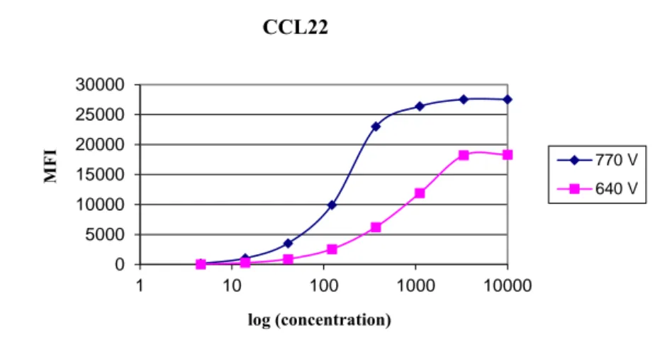
Development of an in-house Luminex assay 48 Coupling of antibodies to microspheres 48 Luminex assay for detection of chemokines 49
Real-time PCR 51
Methodological aspects 55 Optimisation and evaluation of the in-house Luminex assay 55 Th1/Th2 immunity during pregnancy in allergic and non-allergic women 60 Immunological interactions between mother and child during pregnancy 72 Th1- and Th2-like immunity at birth and during childhood in allergic and non-allergic 74 children
SUMMARY AND CONCLUDING REMARKS 79
ACKNOWLEDGEMENT / TACK TILL 83
ORIGINAL PUBLICATIONS
I. Total and allergen-specific IgE levels during and after pregnancy in relation to maternal
allergy.
Sandberg* M, Frykman A, Jonsson Y, Persson M, Ernerudh J, Berg G, Matthiesen L,
Ekerfelt C, Jenmalm M.C.
Journal of Reproductive Immunology 2009, 81:82-8
II. High cord blood levels of the Th2-associated chemokines CCL17 and CCL22 precede
allergy development during the first 6 years of life.
Abelius M.S., Ernerudh J, Berg G, Matthiesen L, Nilsson L.J., Jenmalm M.C.
Pediatric Research 2011, 70:495-500
III. Th2-like chemokine levels are increased in allergic children and influenced by maternal
immunity during pregnancy.
Abelius M.S., Lempinen E, Lindblad K, Ernerudh J, Berg G, Matthiesen L, Nilsson L.J.,
Jenmalm M.C.
Pediatric Allergy and Immunology 2014, in press, DOI:10.1111/pai.12235
IV. Gene expression in placenta, peripheral and cord blood mononuclear cells from allergic
and non-allergic women.
Abelius M.S., Janefjord C, Ernerudh J, Berg G, Matthiesen L, Duchén K, Nilsson L.J.,
Jenmalm M.C. Manuscript
SUPPLEMENTARY RELEVANT PUBLICATIONS
SI. Cord blood cytokines and chemokines and development of allergic disease. Sandberg* M, Frykman A, Ernerudh J, Berg G, Matthiesen L, Nilsson L, Ekerfelt C,
Jenmalm M.C. Pediatric Allergy and Immunology 2009, 20:519-527
SII. A Th1/Th2-associated chemokine imbalance during infancy in children developing
eczema, wheeze and sensitization.
Abrahamsson T, Abelius M.S., Forsberg A, Björkstén B, Jenmalm M.C. Clinical and Experimental Allergy 2011, 41:1729-39
SIII. Reduced IFN-γ and IL-10 responses to paternal antigens during and after pregnancy in
allergic women.
Persson M, Ekerfelt C, Ernerudh J, Matthiesen L, Abelius M.S., Jonsson Y, Berg G, Jenmalm M.C.
Journal of Reproductive Immunology 2012, 95:50-8
SIV. Increased circulating paternal antigen-specific IFN-γ and IL-4 secreting cells in allergic
and non-allergic pregnant women.
PerssonM, EkerfeltC, Ernerudh J, Matthiesen L, Jenmalm M.C., Jonsson Y, Sandberg* M and Berg G.
Journal of Reproductive Immunology 2008, 79:70-8
SV. Placental immune response to apple allergen in allergic mothers.
Abelius M.S. ψ, Enke Uψ, Varosi F, Hoyer H, Schleussner E, Jenmalm M.C., Markert U.R.
Submitted to Journal of Reproductive Immunology
Ψ = shared first authorship
ABSTRACT
Background: Pregnancy and allergic disease have both been postulated as T-helper 2 (Th2)
phenomena. Thus, the increased propensity of allergic mothers to mount Th2-responses might generate favourable effects on the maintenance of pregnancy, but might also be unfavorable, as fetal exposure to a strong Th2 environment could influence the immune development in the offspring to a Th2-like phenotype, favouring IgE production and possibly allergy development later in life. The influence of the intrauterine environment on the immunity and allergy development in the offspring needs to be further investigated.
Aim: The aim of this thesis was to explore the Th1/Th2 balance in allergic and non-allergic
women during pregnancy and its influence on the shaping of the Th1/Th2 profile in the neonate and the development of allergic diseases in the offspring.
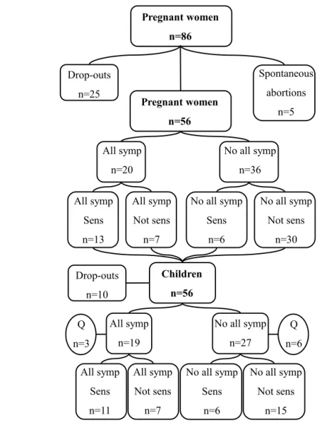
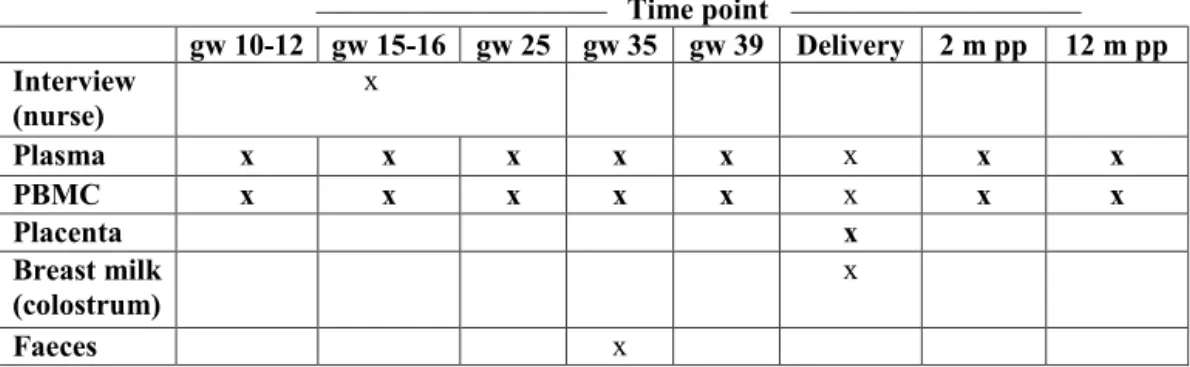
Material and methods: The study group included 20 women with and 36 women without
allergic symptoms followed during pregnancy (gestational week 10-12, 15-16, 25, 35, 39) and 2 and 12 months postpartum, and their children followed from birth to 6 years of age. The circulating Th1-like chemokines CXCL9, CXCL10, CXCL11, Th2-like chemokines CCL17, CCL18 and CCL22, and the allergen-induced secretion of interleukin-4 (IL-4), IL-5, IL-10, IL-13, Interferon-γ (IFN-γ), CXCL10 and CCL17 were measured by Luminex and ELISA. The allergen-specific and total IgE levels were quantified using ImmunoCAP Technology. mRNA expression of Th1-, Th2-, Treg- and Th17-associated genes were measured by PCR arrays and real-time PCR.
Results: We found that sensitised women with allergic symptoms had increased total IgE
levels and birch- and cat-induced IL-5, IL-13 and CCL17 responses during pregnancy as compared with postpartum. The non-sensitised women without allergic symptoms had elevated cat-induced IL-5 and IL-13 responses and lower birch- and cat-induced IFN-γ during pregnancy, but similar IgE levels as compared with postpartum.
Maternal total IgE levels during and after pregnancy correlated with cord blood (CB) IgE and CCL22 levels (regardless of maternal allergy status). Circulating CXCL11, CCL18 and CCL22 levels during pregnancy and postpartum correlated with the corresponding chemokine levels in the offspring at various time points during childhood. Maternal IL-5 expression in peripheral blood mononuclear cells (PBMC) was associated with neonatal Galectin-1, and placental p35 expression was negatively associated with neonatal Tbx21 expression. Increased mRNA expression of CCL22 in cord blood mononuclear cells (CBMC), and increased CCL17 and CCL22 levels in CB were observed in children later developing allergic symptoms and sensitisation as compared with children who did not. Development of allergic symptoms and sensitisation were associated with increased total IgE, CCL17, CCL18 and CCL22 levels during childhood.
Conclusions: Maternal allergy was associated with a pronounced Th2 deviation during
pregnancy, shown as increased total IgE levels and birch- and cat-induced IL-5, IL-13 and CCL17 responses during pregnancy, possibly exposing their fetuses to a particular strong Th2 environment during gestation.
Correlations were shown between the maternal immunity during pregnancy and the
offspring’s immunity at birth and later during childhood, indicating an interplay between the maternal and fetal immunity.
Allergy development during the first 6 years of life was associated with a marked Th2 deviation at birth and a delayed down-regulation of this Th2-skewed immunity during childhood.
SAMMANFATTNING
Bakgrund: Det har länge varit känt att en atopisk hereditet är en riskfaktor för utveckling av
allergiska sjukdomar hos barnet. Denna risk har visats vara betydligt större om modern är allergisk jämfört med om pappan är det. Det är därför troligt att modern bidrar med något mer än bara den genetiska faktorn. Det nyfödda barnet skulle kunna påverkas av moderns
immunitet både under graviditeten och det första levnadsåret via placentan och bröstmjölken. T-hjälpar (Th) celler kan mycket förenklat anta olika profiler; Th1, Th2, Th17 och T
regulatorisk, beroende av vilka signalmolekyler, så kallade cytokiner som de utsöndrar. Både graviditet och allergisk sjukdom karaktäriseras av en Th2-dominant immunitet. Den ökade benägenhet som allergiker har att rikta Th2-svar mot allergener skulle således kunna utgöra en evolutionär fördel, med gynnsamma effekter med avseende på att bli gravid och att upprätthålla graviditeten. Å andra sidan skulle en mycket stark maternell Th2-immunitet under graviditeten skulle kunna påverka barnets immunitet och leda till utveckling av allergiska sjukdomar.
Mål: Att studera det immunologiska samspelet mellan moder och barn under graviditeten och
dess betydelse för utveckling av immunitet och allergisk sjukdom hos barnet.
Material och metod: Tjugo allergiska och 36 friska gravida kvinnor och deras barn har följts
prospektivt, under och efter graviditeten (graviditetsvecka 10-12, 15-16, 25, 35, 39 samt 2 och 12 månader efter förlossningen) samt barnen upp till 6 års ålder. De Th1-associerade kemokinerna CXCL9, CXCL10, CXCL11 och de Th2-associerande kemokinerna CCL17, CCL18 och CCL22 i perifert blod, samt den allergen-inducerade produktionen av Th1-associerade IFN-γ och CXCL10 samt Th2-Th1-associerade IL-4, IL-5, IL-10, IL-13 och CCL17 från mononukleära celler i blodet har mätts med Luminex och ELISA. De allergen-specifika och totala nivåerna av IgE-antikroppar har kvantifierats med ImmunoCAP-teknologi. mRNA nivåerna har analyserats med PCR-array och realtids-PCR.
Resultat: De allergiska mödrarna visade högre nivåer av IgE-antikroppar samt högre björk-
och katt-inducerat IL-5, IL-13 och CCL17 produktion under graviditeten jämfört med postpartum. De friska mödrarna visade en ökad katt-inducerad utsöndring av IL-5 och IL-13 och en minskad utsöndring av björk- och katt-inducerat IFN-γ under graviditeten medan nivåerna av IgE-antikroppar var oförändrade under och efter graviditeten.
Vi har observerat positiva korrelationer mellan moderns IgE (oavsett om modern är allergisk eller inte) och nivåerna av IgE-antikroppar och det Th2-associerade kemokinet CCL22 i navelsträngsblod. Moderns CXCL11-, CCL18- och CCL22-nivåer korrelerade dessutom med motsvarande kemokin hos barnet vid flera tidpunkter under de första 6 åren i livet.
De barn som utvecklat allergiska sjukdomar vid 6 års ålder hade signifikant högre mRNA-uttryck av CCL22 och högre nivåer av CCL17 och CCL22 i navelsträng samt högre nivåer av IgE-antikroppar, CCL17, CCL18 and CCL22 i blodet vid flera tidpunkter under barndomen, i jämförelse med de barn som inte utvecklat allergi.
Slutsatser:De allergiska mödrarna visade en ökad Th2-immunitet under graviditeten, vilket skulle kunna leda till att fostret exponeras för en mycket stark Th2-miljö under graviditeten. Flera samband mellan moderns immunitet under graviditeten och barnets immunitet vid födseln och under barndomen hittades, vilket tyder på ett immunologiskt samspel mellan mor och barn. En ökad Th2-immunitet vid födseln i kombination med en misslyckad nedreglering av denna Th2-immunitet under barndomen skulle kunna bidra till utveckling av allergiska sjukdomar.
ABBREVIATIONS
α-IL-4R anti-IL-4 receptorAD atopic dermatitis APC antigen-presenting cell ARC allergic rhino-conjuntivitis BALF bronchoalveolar lavage fluid BSA bovine serum albumin
CB cord blood
CBMC cord blood mononuclear cells
CCL CC ligand
CD cluster of differentiation Ct threshold cycle
CTLA-4 cytotoxic T lymphocyte antigen 4 CV coefficient of variance
CXCL CXC ligand
CX3CL CX3C ligand DC dendritic cell DMSO dimethyl sulphoxide EBI3 epstein-barr virus induced 3 FCS fetal calf serum
FcεRI Fcε receptor type I Foxp3 forkhead box p3
GATA-3 GATA binding protein 3 gw gestational week
hCG human chorionic gonadotropin HLA human leukocyte antigen ICOS inducible co-stimulator IDO1 indoleamine 2,3-dioxygenase 1 IFN-γ interferon-γ
Ig immunoglobulin
IL interleukin
ILC2 type 2 innate lymphoid cells LAG3 lymphocyte-activation gene 3 MHC major histocompatibility complex NK natural killer
PBMC peripheral blood mononuclear cells PBS phosphate buffered saline
PBSB PBS with 1% BSA
PBS-TBN PBS with 0.05% Tween, 0.1% BSA and 0.05% NaN3
PBT PBS with 0.05% Tween PCR polymerase chain reaction PD-1 programmed death 1 PHA phytohemagglutinin PMT photomultiplier tube RNA ribonucleic acid
RORC retinoic acid-related orphan receptor C RT-PCR reverse transcription polymerase chain reaction SA-PE streptavidin R-phycoerythrin conjugate SPT skin prick test
STAT signal transducer and activator of transcription Tbet T-box expressed in T cells
Tbx21 T-box transcription factor 21 TCR T cell receptor
TGFβ transforming growth factor-β
Th T-helper
TLR toll-like receptor Treg regulatory T cells
TREM-2 triggering receptor expressed on myeloid cells 2 TSLP thymic stromal lymphopoietin
YWHAZ tyrosine 3-monooxygenase/tryptophan 5-monooxygenase activation protein, zeta polypeptide
REVIEW OF THE LITERATURE
General aspects of allergic disease
There are four types of hypersensitivity reactions; immediate hypersensitivity (type I), antibody mediated (type II), immune complex mediated (type III) and T cell mediated (type IV). Type I and IV hypersensitivity reactions include allergy. Type I represents IgE antibody mediated allergy with engagement of mast cells and their mediators and type IV represents T-helper (Th) 1/Th17 cell mediated inflammation (non-IgE antibody mediated allergy) such as allergic contact dermatitis. Type II and III hypersensitivity reactions involve Immunoglobulin G (IgG) and IgM antibodies to antigens on the cell surface or extracellular matrix (type II) or to soluble antigens forming immune complexes (type III) and activation of the complement system [1]. In this thesis, the term allergy refers to IgE mediated allergy.
Some common environmental antigens, e.g. allergens, are able to induce immediate hypersensitivity reactions. Allergens are in general small, highly stable, soluble and glycosylated proteins, with enzymatic activity.Everyone is exposed to allergens, which are acknowledged by the immune system, and allergen-specific IgG- and IgM antibodies are produced. The majority of individuals do not synthesise IgE antibodies in response to allergens [1].
Atopic individuals have a genetic predisposition, personal and/or familiar, to become sensitised and produce allergen-specific IgE antibodies. Thus, the clinical definition of atopy is an IgE antibody high-responder and confirmation of sensitisation with positive skin prick test (SPT) or presence of allergen-specific IgE antibodies in the circulation must be done when using the term atopy [2].
Sensitised individuals usually develop symptoms of asthma, food allergy, atopic dermatitis (AD) and allergic rhinoconjunctivitis (ARC), but some sensitised individuals do not develop allergic symptoms despite repeated allergen exposure, for unknown reasons.
The clinical symptoms of allergic disease are common in Sweden, with a prevalence of 7% for ARC symptoms, 10% for asthma symptoms and 22 % for symptoms of eczema at 6-7 years of age, according to the International Study of Asthma and Allergies in Childhood, phase III [3]. Furthermore, the Swedish population based cohort (BAMSE) reported that 58% of the children had had any allergic disease at some time during the first 12 years of life [4].
An increased prevalence of allergic diseases has been reported during the last decades, predominantly in countries with a westernised lifestyle [3, 5, 6]. The occurrence of allergic symptoms tends to vary with age, with a typical pattern called “the atopic march”[7]. AD and food allergy are the most prominent allergic manifestations during infancy. These symptoms decline with age and asthma and ARC usually develop in pre-school age. The early origin of AD and food allergen sensitisation has shed light on their capacity as predictors for development of sensitisation and other allergic symptoms. They are both associated with an increased risk for development of sensitisation to inhalant allergens, asthma and ARC later in life [7-15]. Lowe et al. demonstrated that children with atopic dermatitis were at greater risk for development of asthma and allergic rhinitis if they were sensitised than if they were not [14]. Furthermore, sensitisation in asymptomatic infants independently predicted development of wheeze, asthma and rhinitis [13], underlining the importance of early sensitisation.
Genetic and environmental factors
Allergic diseases are probably caused by interactions between genetic and environmental factors in combination with allergen exposure. The strong hereditary component in allergic disease has resulted in identification of over 100 genes associated with disease susceptibility, with a few overlapping associations for different allergic diseases, reviewed in [16]. Genetic factors are clearly important, but cannot independently explain the increased prevalence of allergic diseases during the last 40-50 years. A time period of a few decades is far too short for human genetics to undergo such substantial changes causing this increasing prevalence. Thus, a lot of research has focused on environmental factors associated with a westernised lifestyle. Several environmental factors have changed along with the increase in allergy prevalence, many of them leading to reduced microbial exposure, such as increased cleanliness, reduced family size and living in urban areas with an improved general living standard. The “hygiene hypothesis” originates in the observation of a lower prevalence of eczema and hay fever in large families [17], which was later shown for asthma prevalence as well [18]. The protective effect of being surrounded by other children was strengthened by data on day-care attendance, in particular attendance to day-care centres early in life [18, 19]. The beneficial effect of day-care has shown to be modified by variations of the Toll-like receptor 2 (TLR2) gene (TLR2/-16934 polymorphism), as children with the T allele, but not
the AA-homozygotes, were less likely to be sensitised if they attended to day-care, indicating complex gene-environment interactions [20].
The association between large families/attendance to day-care and reduced allergy prevalence led to the interpretation that infections and/or microbial exposure during childhood might protect against allergy. Studies on the effect of respiratory infections on allergy development are inconsistent, without any clear protection, while a substantial amount of findings indicate that microbial exposure is important [21-23]. A low diversity of the intestinal microbiota very early in life has been associated with development of sensitisation, AD, asthma and allergic rhinitis [24-26]. The moderate increased risk of allergy development in children delivered by caesarean section [27], might be associated with a lower total microbiota diversity in these children [28]. Many studies have shown that living on a farm is associated with a relatively low prevalence of allergic diseases, with ingestion of unpasteurised milk and exposure to livestock as identified protective factors [29-31]. A recent study found no clear evidence of synergy or saturation of the protective effects of sibling or farm exposure, suggesting that different mechanisms may underlie these protective factors [32].
Use of broad-spectrum antibiotics has been associated with allergy development [33]. An anthroposophic lifestyle, characterised by limited use of antibiotics and vaccines and a diet rich in Lactobacilli, has been associated with a reduced prevalence of atopy [34]. A lot of other factors, such as dietary changes with a decreased intake of omega 3 fatty acids, obesity, reduced breast-feeding, vaccinations, air pollution and smoking, have also been proposed to influence allergy development [35-37].
Allergen exposure
A repeated exposure to allergens is required for development of sensitisation and allergic symptoms. Low doses of allergens favour development of sensitisation and allergic symptoms while high doses of allergens may induce tolerance [38]. The use of
immunotherapy as a treatment of allergic disease indicates that increasing doses of allergen can promote tolerance.
Exposure to farm animals early in life, in particular before birth, has shown a protective effect on allergies [39], but the effect of pet exposure is less clear. One should keep in mind that it is difficult to distinguish between the contribution of allergens and microbes when evaluating the effect of animal contact on allergy development. A meta-analysis from 2012 reported no associations at all between pet keeping early in life and asthma and ARC in
school age [40], while another meta-analysis from 2013 reported a decreased risk for AD in children exposed to pets during infancy [41]. Neonatal exposure to cats increased the risk for AD in children with mutations in the Filaggrin gene (associated with disease susceptibility), indicating that early pet exposure might be unfavourable for subgroups of children [42]. The possibility of allergen exposure and sensitisation in utero, will be discussed later, on page 32.
Immunological mechanisms
The main task of the immune system is to protect us against harmful microbes, such as certain bacteria, viruses, fungi and parasites. Thus, the ability to distinguish between self and non-self is obligatory in order to achieve optimal protection and minimal damage to the host. A plastic immune system has evolved to keep this balance, involving mechanisms for both up- and down-regulation of immune responses [1].
Brief overview of the innate and adaptive immune system
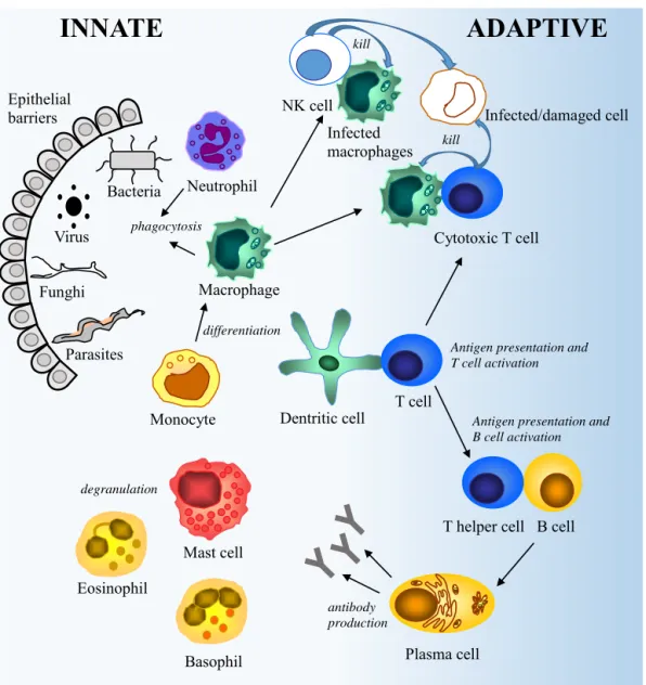
The innate immune system is our first line of defence, comprising epithelial barriers and specialised immune cells, namely granulocytes (neutrophils, eosinophils, basophils) mast cells, dendritic cells (DC), macrophages and Natural killer (NK) cells (Figure 1). Upon infection, the invader is recognised by innate immune cells, predominantly macrophages and neutrophils, bearing receptors for molecular structures on the surface of the microbe, leading to phagocytosis and/or secretion of inflammatory mediators. This is the initiation of the innate immune response. Secreted cytokines and chemokines contribute to the inflammation by increasing the permeability of blood vessels, regulating immune responses and recruiting other immune cells to the site of infection. The phagocytosis is further triggered by microbes coated with molecules acting like opsonins, originally generated by the complement system. Cells infected with viruses or intracellular bacteria are effectively killed by NK cells. Eosinophils and mast cells play an important role in the defence against helminths, by their ability to degranulate and release inflammatory mediators.
Figure 1. A brief overview of the innate and the adaptive immune system.
The DCs migrate to the lymph nodes and display the antigen generating activation, clonal expansion and differentiation of antigen-specific naïve T cells to effector T cells. T cells are not activated by soluble antigens that are not processed by APCs. Activated cytotoxic T cells promote destruction of infected cells and Th cells stimulate macrophages to clear their microbial load. This type of adaptive immune response is called cell-mediated immunity. The other type, humoral immunity, is mediated by antibodies secreted by B cells and plasma cells. B cells can be independently activated by antigen stimulation, but they usually need
INNATE
ADAPTIVE
Epithelial barriers Virus Bacteria Parasites Funghi Macrophage Neutrophil Dentritic cell phagocytosis T cellAntigen presentation and T cell activation
T helper cell B cell Antigen presentation and B cell activation Plasma cell Cytotoxic T cell kill Mast cell Eosinophil Basophil degranulation antibody production NK cell kill Monocyte Infected macrophages differentiation Infected/damaged cell
assistance of Th cells, who interact with the antigen presented on the cell surface of the B cell. Cytokines secreted by Th cells are important for the function of the adaptive immune system e.g. T and B cell proliferation and differentiation, but one should keep in mind that many cytokines influence both innate and adaptive immune responses. Upon activation, B cells differentiate into antibody secreting plasma cells or memory B cells. The main assignment for the antibodies is to facilitate extracellular protection by binding to pathogens causing neutralisation, opsonisation and activation of the complement system.
The innate immune system is particularly important in the early stage of the infection as there is a delay of several days for the adaptive immune system to attain full function. The immune responses are down-regulated after successful clearance of the infection. The majority of the antigen-specific lymphocytes die by apoptosis, while the remaining make up a pool of long-lived memory cells. Memory cells can remain for decades and provide a fast immune response upon reinfection [1].
T helper cells
Activation of CD4+ T cells
T cell activation is initiated by interactions between an APC, predominantly DCs and T cells. Tissue resident immature DCs capture and process the antigen and present an antigen-derived peptide on the major histocompatibility complex (MHC) molecule class II on the cell surface. The DCs mature and migrate to the lymph nodes and activate naïve CD4+ T cells by
interactions between the MHC-peptide complex and the T cell receptor (TCR). The first signal for T cell activation is antigen recognition and the second signal is co-stimulation. The binding of the cluster of differentiation 28 (CD28) receptor on the T cell and the CD80/86 complex expressed on the DC is the best characterised co-stimulatory pathway. Another pathway for co-stimulation is mediated by signaling through the inducible co-stimulator (ICOS) expressed on T cells and ICOS ligand expressed on DCs. The activated T cells also express CD40L, which interacts with CD40 on the DC, generating up-regulation of co-stimulatory molecules and cytokine secretion. Thus, like a positive feed-back loop, engagement of CD40L-CD40 “improve” the DC and promote T cell proliferation and differentiation.
Activation signals are counterbalanced by inhibitory signals. Suppressive effects are mediated by interactions between the inhibitory receptors cytotoxic T lymphocyte antigen 4
(CTLA-4) and programmed death 1 (PD-1) and their ligands; CD80/86 and PDL1/L2 [1]. Immune suppression of activated Th cells is also mediated by Interleukin-10 (IL-10) and transforming growth factor (TGF-β) secreting regulatory T cells (Treg).
Differentiation of CD4+ T cells
The naïve Th cells will differentiate into different subsets of effector cells depending on the immunological micro milieu at the time of activation, i.e. presence or absence of certain stimuli. Cytokines are secreted by the surrounding immune cells generating distinct subsets of Th cells, Th1, Th2 and Th17 for host defence and Treg for immune regulation/suppression. The effector functions of the subsets are markedly different, each one, except Tregs, adapted to combat a certain type of infection.
Th1 immunity is the appropriate defence against intracellular microbes. This defence is predominantly mediated by the signature cytokine for Th1 cells; Interferon-γ (IFN-γ), which promotes cytotoxic activity of cytotoxic T cells and pathogen killing by macrophages [43]. DCs, macrophages and NK cells stimulate Th1 differentiation by providing IL-12 and IFN-γ (Fig 2). IL-12 is considered to be the most potent inducer of Th1 differentiation [44]. T cell mediated IFN-γ secretion is induced by activation of the transcription factors signal transducer and activator of transcription 4 (STAT4), STAT1 and T-box expressed in T cells (Tbet). IFN-γ itself amplifies Th1 differentiation and stimulates production of the Th1-associated chemokines CXCL9, CXCL10 and CXCL11 [45-47]. These chemokines are predominantly secreted by macrophages and they attract CXCR3 expressing Th1 cells, B cells, mast cells and NKT cells to the site of inflammation [48].
Th2 immunity is mounted in response to helminth infections. IL-4 plays a major role in Th2 differentiation and for effector functions. Mast cells and eosinophils are thought to be the initial source of IL-4 in case of helminth infection, even though the Th2 cell itself is the major source of IL-4. It has been speculated that naïve Th cells default to the Th2 pathway in absence of innate stimuli [49]. IL-4 activates STAT6 and the master regulator of Th2 differentiation GATA binding protein 3 (GATA-3), inducing IL-4, IL-5 and IL-13 production in order to amplify the immune response and accomplish effector functions. IL-4 and IL-13 improve humoral immunity by inducing IgE class switch and they also up regulate MHC class II molecules on B cells. IL-5 is important for eosinophil growth, maturation, activation
and survival, and the eosinophils are mainly recruited by CCL11-CCR3 interactions. Mast cell activation is mediated by IL-5 and IL-13 [43]. Furthermore, the IL-4 and IL-13 induced chemokines CCL17 and CCL22 attract Th2 cells, DCs, basophils, mast cells, NK cells and NKT cells by interaction with the CCR4 receptor [48, 50, 51]. CCL18 is induced by 4, IL-13 and IL-10, suggesting that this chemokine has Th2-associated as well as
anti-inflammatory features [52].
Figure 2. The figure shows a simplified overview of the cytokines needed for differentiation of the
Th subsets, the main transcription factors, cytokine secretion and primary function for each Th subset.
Th17 cells are the third subset of Th cells essential for satisfactory host defence, namely for clearance of fungi and extracellular bacteria. Five cytokines are acknowledged for Th17 differentiation, TGF-β, IL-1β, IL-6, IL-21 and IL-23, leading to activation of STAT3 and the lineage specific transcription factor retinoic acid-related orphan receptor C (RORC) [53]. IL17A/F and IL-22 are the main effector cytokines, primarily involved in the maintenance of inflammation, neutrophil activation and epithelial barrier function [53, 54]. The chemokine CCL20 is secreted by Th17 cells and induces migration of Th cells, DCs and B cells via CCR6 [53]. TGF-β IL-2 TGF-β IL-1β IL-6 IL-21 IL-23 IL-12 Naive T cell Th1 STAT1 STAT4 Tbet Th2 STAT6 GATA3 Th17 STAT3 RORC IL-4 Treg STAT5 Foxp3 IL-4, IL-5 IL-9, IL-13 IL-25, IL-31 TNF-β IFN-γ IL-17A/F IL-21 IL-22 TGF-β IL-10 IL-35 Macrophage
activation IgE production Mast cell and eosinophil activation Immune suppression Neutrophil activation Barrier function
Mechanisms for down-regulation of immune responses after successful clearance of the infection are essential in order to minimise tissue damage. Tregs are specialists of immune suppression, targeting T effector cells and other cell types, possessing cell contact dependent as well as independent mechanisms. These include (i) modulation of DC function mediated by CTLA-4 and lymphocyte-activation gene 3 (LAG3), (ii) metabolic disruption by IL-2 starvation, (iii) granzyme A/B and perforin induced cytolysis and (iv) secretion of anti-inflammatory cytokines such as TGF-β, IL-10 and IL-35 [55, 56].
In addition to the thymus-derived natural occurring CD4+CD25+Forkhead box p3 (Foxp3)+ Tregs, CD4+CD25- cells can differentiate into a regulatory subset, i.e. induced Tregs, when stimulated with antigen and an appropriate cytokine environment, e.g. TGF-β [54, 57]. The division of Th cells into subtypes generates a working model to help immunologists to understand the immune system, but it is important to remember that the model is a simplification. Other subpopulations of Th cells have been suggested as well, but their lineage specific transcription factors have not been identified yet, challenging their existence. IL-9 secreting Th9 cells probably involved in the fight against helminths and IL-22 secreting Th22 cells, possibly important for barrier function, are two examples [43, 54]. Another subpopulation is the follicular Th cells, which help the B cells to become activated in the germinal center reaction. The signature cytokine of follicular Th cells is IL-21 [1]. It is generally accepted that the Th1 and Th2 subsets cross-regulate each other at the
transcriptional and cytokine level [54, 58-60]. Th1-associated Tbet and IFN-γ have inhibitory effects on Th2 differentiation and the Th2-associated GATA-3 and IL-4 exert inhibitory effects on Th1 differentiation [58, 59]. TGF-β inhibits development of Th1 and Th2 cells [1]. Th17 differentiation is down regulated by IFN-γ and IL-4, suggesting that suppressive actions of TGF-β are necessary to allow development of Th17 cells (reviewed in [53]).
The immune system allows short-term alterations in the Th1/Th2 balance, for example during infection, a normal pregnancy and very early in life [61, 62]. However, an extensive
activation of Th1 or Th2 immunity can be pathological. Autoimmune diseases such as rheumatoid arthritis, multiple sclerosis and type I diabetes are associated with a Th1 deviation, probably in combination with a Th17 deviation [63] and allergic diseases with a Th2 deviation [64].
B cells and antibodies
Naïve B cells are activated by antigen recognition, i.e. cross-linking of antigen receptors (T cell independent activation) or a cell-cell contact dependent pathway (T cell dependent activation). The naïve B cell antigen receptor comprises membrane bound IgD and IgM antibodies. Upon activation, antigen-specific B cells proliferate and differentiate into memory B cells or antibody secreting plasma cells. T cell independent B cell activation, with e.g. polysaccharides, results in IgM and IgG2 antibody secretion, leading, for example, to
activation of the complement system. Antibody production to protein antigens such as allergens, is dependent on the assistance of Th cells. The antigen-specific B cells take up the encountered antigen, process it and present a protein derived peptide on their MHC class II molecules. Antigen-specific Th cells interact with this MHC-peptide-complex and provide the B cells with the signals needed for antibody isotype switching i.e. CD40L-CD40
stimulation and the appropriate cytokines [1]. IL-4 and IL-13 induce IgE antibody production [65] in response to helminth infections or allergens. IgA is a neutralising antibody, mostly important at mucosal surfaces, and its isotype switching is predominantly stimulated by TGF-β [66]. IgG antibody synthesis seems to be promoted by Th1- and Th2-like immunity, but the regulation of antibody isotype switching is not completely elucidated. A presumed Th1 dominant situation such as Lyme Borreliosis has been associated with high IgG1 and IgG3
subclass antibody levels, suggesting that IFN-γ might stimulate the production of these subclasses [67]. In mice, Th1 immunity has been shown to be associated with production of complement activating and opsonising IgG2a, and IgG2b corresponding to human IgG1 and
IgG3 [68, 69].
Allergic diseases, representing a Th2-biased situation, have been associated with high IgG4
levels to allergens [70]. IL-4 has shown to induce IgG4
switching, supporting the assumption
of IgG4 as a “Th2-associated antibody” [65], in addition to IgE.
The allergic immune response
There are three phases of an allergic reaction; sensitisation, immediate hypersensitivity and the late phase reaction. The sensitisation phase begins with interactions between DCs and the encountered allergen. Allergens are in general distributed transmucosally, at very low doses, by diffusion through the epithelial layer. Specialised DCs capture and process the allergen and present an allergen derived peptide to the naïve T cells. They differentiate into Th2 cells, which help B cells to become activated and induce B cell IgE class switch by secretion of
IL-4 and IL-13. Furthermore, Th2 cells activate eosinophils by secretion of IL-5. The allergen-specific IgE antibodies bind to the high affinity Fcε receptor type I (FcεRI) expressed on the cell surface of mast cells, basophils and to some extent on eosinophils. The cells are “sensitised” and an immediate hypersensitivity reaction is possible at the next encounter of the allergen.
The activation of mast cells is induced by binding of allergens to the IgE antibodies, mediating cross-linking of the FcεRI molecules and an immediate degranulation. A mixture of biogenic amines, e.g. histamine, enzymes such as tryptase and chymase, lipid mediators including prostaglandins, leukotrienes and platelets-activating factor and immune mediators, e.g. cytokines and chemokines, are released into the extra-cellular space, generating allergic symptoms. Histamine induces vasodilation, increased vascular permeability and plasma leakage. The enzymes cause tissue damage, the lipid mediators stimulate for example bronchoconstriction and cytokines such as IL-9 and IL-13 promote mucus secretion. The allergic inflammation is maintained by actions of cytokines and chemokines secreted by APCs, mast cells, Th2 cells and surrounding epithelial cells. The immunological milieu in the inflamed tissues comprise pro-inflammatory cytokines such as TNF, IL-1β, IL-6, eosinophil recruiting/promoting CCL11, IL-3, IL-5, GM-CSF as well as the Th2 cell
recruiting/promoting CCL17, CCL22, IL-4, IL-13 [1, 71]. The inflamed tissue is infiltrated with eosinophils and basophils. This is the late phase reaction and this stage can become chronic in tissues frequently exposed to allergens.
Cytokines in allergic disease
It is generally accepted that established allergic disease is associated with increased Th2-deviated immune responses to allergens, shown as increased GATA-3 expression and IL-4, IL-5, IL-9, IL-13 secretion, both systemically [72, 73] as well as locally in the affected tissues [74, 75]. IL-31 was identified as a Th2-associated cytokine around a decade ago [76] and it has shown to be associated with pruritus and allergic manifestations, such as AD and allergic asthma [77, 78]. The epithelial derived cytokines IL-25, IL-33 and thymic stromal lymphopoietin (TSLP) enhance Th2-like cytokine responses directly by influencing cytokine secreting immune cells and possibly indirectly by polarisation of DCs [77]. Accordingly, increased IL-25, IL-33 and TSLP expression has been reported in lesional AD skin, nasal mucosa and elevated levels in nasal secretions in subjects with allergic rhinitis [77, 79, 80]. A newly discovered cell type, type 2 innate lymphoid cells (ILC2), are activated by IL-25, IL-33 and TSLP and they produce the effector cytokines IL-5 and IL-13 [81]. Thus, they are expected to be involved in allergic diseases. The percentage of ILC2s in peripheral blood was increased after nasal allergen challenge in allergic subjects [82] and ILC2s were detected in the lung tissue and bronchoalveolar lavage fluid (BALF) in a murine ovalbumin-induced asthma model [81]. The role of ILC2s in allergic diseases needs further investigation. The ability of allergic individuals to secrete Th1-associated cytokines such as IFN-γ upon allergen stimulation is usually equal [72, 83] or diminished [84] as compared with controls. The Th2-associated immunity in allergic disease might be explained by an impaired frequency and/or capacity of Tregs to down regulate Th2-like immune responses, allowing a sustained Th2-driven allergic inflammation. Allergen-specific IFN-γ-, IL-4- and IL-10 secreting T cells, presumably representing Th1, Th2 and Tr1 cells, have been detected in allergic and healthy individuals but in different proportions [85]. Th2 cells were the predominant subset in the allergic group and the Tr1 cells in the non-allergic group, indicating an important role for IL-10 secreting Tr1 cells in the development of tolerance to allergens. Moreover, one of the immunological mechanisms behind the successful induction of allergen tolerance during allergen-specific immunotherapy could be induction of Tr1 cells, suppressing allergen-specific Th1- and Th2-responses by secretion of the anti-inflammatory cytokines IL-10 and TGF-β [86].
Th17 cells and their inflammatory mediators attract and promote neutrophil development, and are not known to drive Th2-responses, implying a role for Th17 cells in non-allergic asthma [87]. Lei et al. failed to report any differences in circulating IL-17A levels between allergic
asthmatics and healthy controls [78], while Ciprandi et al. revealed increased serum levels of the same cytokine in a study group with allergic rhinitis as compared with controls [88]. However, all patients in the latter study were previously treated with allergen-specific immunotherapy. In another study, elevated birch-induced mRNA expression of IL-17A after allergen-specific immunotherapy in sensitised children with allergic rhinitis was associated with poor therapeutic outcome [89]. Clearly, the role of Th17 cells in allergy is not settled and needs additional investigation.
Chemokines in allergic disease
Chemokines comprise a large protein family, with more than 50 members. They are divided into four groups depending on the location of two N-terminal cysteine residues. The two major groups are the CC ligands (CCL, cysteine residues are adjacent) and the CXC ligands (CXCL, cysteine residues separated by another amino acid). The two minor groups are represented by the C ligands with only one N-terminal cysteine residue and the CX3C ligands (CX3CL) with cysteine residues that are separated by three amino acids. In concurrence, the chemokine receptors are named CCR, CXCR, XCR and CX3CR, representing receptors for the corresponding chemokine groups.
Microbial recognition and cytokine stimulation induce chemokine production. Chemokines are secreted by leukocytes, predominantly macrophages, but also endothelial cells, epithelial cells and fibroblasts are important chemokine producers [1, 90].
Chemokines are crucial not only for recruitment of leukocytes from the circulation to the tissues but also in the regulation of leukocyte maturation. Leukocyte migration is mediated by a chemical gradient. The chemokine ligand-receptor interaction generates increased cell motility and integrin affinity promoting leukocyte migration. Thus, the composition of infiltrating leukocytes is organised by chemokines present at the site of inflammation.
Table 1. The chemokine ligands and the distribution of chemokine receptors on immune cells
involved in allergic disease
Cell Chemokine receptors Chemokine ligands
Eosinophil CXCR4 CCR1 CCR3 CXCL12 CCL3/5/7/9/10/14/15/16/23 CCL5/7/8/11/13/15/24/26 Basophil CXCR4 CCR1 CCR2 CCR3 CXCL12 CCL3/5/7/9/10/14/15/16/23 CCL2/7/12/13/16 CCL5/7/8/11/13/15/24/26 Mast cell CXCR4 CCR3 CCR4 CXCR3 CXCL12 CCL5/7/8/11/13/15/24/26 CCL17/22 CXCL9/10/11 Dendritic cell CXCR4 CCR1 CCR4 CXCL12 CCL3/5/7/9/10/14/15/16/23 CCL17/22 Monocyte CXCR4 CCR1 CCR8 CXCL12 CCL3/5/7/9/10/14/15/16/23 CCL1 Th1 cell CXCR4 CCR2 CCR5 CXCR3 CXCL12 CCL2/7/12/13/16 CCL3/4/5/8/14 CXCL9/10/11 Th2 cell CXCR4 CCR3 CCR4 CCR8 CXCL12 CCL5/7/8/11/13/15/24/26 CCL17/22 CCL1 NKT cell CXCR4 CCR1 CCR2 CCR4 CCR5 CXCR3 CXCR6 CXCL12 CCL3/5/7/9/10/14/15/16/23 CCL2/7/12/13/16 CCL17/22 CCL3/4/5/8/14 CXCL9/10/11 CXCL16
Chemokines and receptors studied in this thesis are marked with bold. References [1, 48].
Th1-associated chemokines
The IFN-γ induced chemokines CXCL9, CXCL10 and CXCL11 [45, 46] attract CXCR3 expressing cells such as Th1 lymphocytes, NKT and mast cells [48] (Table 1). CXCL9, CXCL10 and CXCL11 are predominantly associated with Th1-like diseases, e.g. sarcoidosis, tuberculosis [91], Sjögren’s syndrome [92], rheumatoid arthritis [93] and Crohn´s disease [94]. A few studies have implicated a role for the CXCR3 ligands in allergic disease. Increased expression of CXCL10 has been reported in the airways of atopic asthmatics, but this increase was less pronounced as compared with CXCL10 expression in patients with sarcoidosis [91]. Moreover, the increased CXCL10 expression was accompanied with high
CCL11 expression in the atopic asthmatics, but not in the sarcoidosis or tuberculosis patients, indicating an up-regulation of both Th1- and Th2-associated chemokines locally in the asthmatic airways [91]. Furthermore, CXCL9 was elevated in the nasal lavage of allergic rhinitis patients and CXCL10 in BALF of asthmatics after allergen challenge [95, 96]. Acute asthma exacerbations were associated with increased CXCL9 and CXCL10 levels in the circulation in contrast to stable asthma [97].
A murine model of asthma implicated an important role for CXCL10 in airway
hyperreactivity and Th2-associated inflammation [98]. Transgenic mice, overexpressing CXCL10 in the lung, experienced increased airway hyperreactivity and augmented recruitment of eosinophils and Th2 cells to the lung [98]. Conversely, CXCL9 inhibited migration and CCR3-mediated functional responses in murine eosinophils, indicating that CXCL9 negatively regulates Th2-associated allergic inflammation [99, 100]. More research is needed to elucidate if the CXCR3 ligands actively promote allergic inflammation, or if they represent a negative feedback mechanism leading to suppression of Th2-associated immune responses.
Th2-associated chemokines
There are plenty of studies suggesting a role for CCL11, CCL17, CCL18 and CCL22 in allergic disease [101-110]. These Th2-associated chemokines are predominantly induced by IL-4 and IL-13, but production of CCL18 is considerably enhanced in synergy with IL-10 [50-52, 111]. In contrast, the production of CCL17 and CCL22 is suppressed by IL-10 [50, 112]. The amplification of the allergic response is partly driven by these chemokines, as they influence the composition of infiltrating leukocytes at the site of the allergic inflammation. CCL11 recruits eosinophils, mast cells, basophils and Th2 cells via CCR3, while CCL17 and CCL22 induce migration of Th2 cells, DC, basophils, mast cells, NK cells and NKT cells by interaction with the CCR4 receptor [48]. CCL18 has shown to attract T cells [113]. The receptor for CCL18 is not yet determined, but CCR8 was recently suggested [114]. Increased levels of CCL11, CCL17, CCL18 and CCL22 in the circulation have been associated with established allergic disease, predominantly AD [101-105], but also with asthma [105, 106] and ARC [105]. Others have reported similar levels of these associated chemokines in ARC patients and controls [104, 115]. An increase in Th2-associated chemokines has also been reported in the affected tissues; CCL17, CCL18 and CCL22 were elevated in BALF of asthmatics [107, 108] and after allergen challenge [109].
Furthermore, epithelial cells in the nasal mucosa of ARC patients expressed more CCL17 than controls [110].
Chemokines have been used as markers for Th1/Th2 immunity in allergic diseases and other immune-mediated disorders, but little is known about the predictive value of circulating chemokines, before disease onset. Although established allergic disease is characterised by a Th2-skewed immunity, the timing of the development of this Th2 skewing is unknown.
Pregnancy
Pregnancy immunology
The maternal immune system tolerates the fetus during pregnancy, despite the fetal
expression of paternal antigens, which are foreign to the mother. The maternal tolerance must be kept without compromising the protection against infections. Today, modulation of the maternal immune system to a Th2-/anti-inflammatory phenotype and appropriate interactions between maternal and fetal immune system are thought to be the key events in the
maintenance of the feto-maternal tolerance [116].
The placenta and the amniotic sac comprise a physical barrier between the mother and the fetus, although there are several possibilities of immunological interactions between the maternal and fetal immune system i.e. a “local interaction” in the decidua and a “systemic interactions” in the intervillous space in the placenta [117].
In the first trimester of pregnancy, fetal extravillous trophoblasts migrate into the decidual tissue and establish contact with decidual stroma cells and maternal NK cells, macrophages and T cells. The extravillous trophoblasts express MHC class I products; human leukocyte antigen (HLA) class I molecules HLA-C, HLA-E and HLA-G but not HLA-A, HLA-B or MHC class II molecules. This specific expression profile is believed to protect the fetus, probably by prevention of T and NK cell activation [118].
The systemic interaction takes place in the intervillous space of the placenta. Villous trophoblasts, with fetal blood vessels, are bathing in maternal blood, allowing exchange of nutrients. Thus, the fetal and maternal blood streams are in very close proximity. A normal pregnancy is traditionally described as a Th2-deviated condition [61]. Thus, an imbalance between Th1 and Th2 immunity in pregnancy, leading to increased Th1-like
immune responses has been associated with spontaneous abortions [119, 120], pre-eclampsia [121] and pre-term labour [122]. Similarly, Th17-like immunity has been associated with spontaneous abortions [123, 124] and might be detrimental for the pregnancy in combination with a Th1 deviated immunity. In addition to a Th2-deviated immunity, the suppressive mechanisms by Tregs and alternative activated macrophages are also considered to be important for the maintenance of the feto-maternal tolerance [125, 126].
Clinical data support the hypothesis of pregnancy as a Th2-deviated situation. Patients with Th1-like diseases such as rheumatoid arthritis and psoriasis experience a temporary reduction of symptoms during pregnancy [127, 128]. Similarly, the rate of relapse declines during pregnancy and increases postpartum in patients with multiple sclerosis [129].
The influence of pregnancy on the course of asthma and allergy are inconclusive. Generally, asthma symptoms decrease in one-third, remains the same in one-third and increase in one-third of the asthmatic women during pregnancy (reviewed in [130]). Kircher et al. reported a concordance between symptoms of asthma and rhinitis during pregnancy, indicating that symptoms of rhinitis may undergo similar alterations as asthma [131]. The atopic status of the women was not assessed in that study, however.
Allergy and pregnancy
The immunological similarities between allergy and pregnancy, i.e. Th2-deviated immune responses to allergens [72, 132, 133] and at the feto-maternal interface [61] have raised the question whether maternal allergy is beneficial in a reproductive perspective. This
assumption embraces an influence of allergic disease on the immune system, beside the allergen-specific immune responses. On one hand, the Th1/Th2 imbalance in allergic disease has shown to be highly antigen-specific, e.g. atopic children produced more Th2-like cytokines in response to the allergen they were sensitised to, but not to other allergens [72]. On the other hand, there are studies on unrelated antigens indicating generally altered immune responses in the allergic group. Allergic women had reduced IFN-γ and IL-10 production in response to fetal and paternal alloantigens during and after pregnancy [134-136]. Furthermore, low IFN-γ and high IL-4 and IL-5 production has been observed in allergic individuals after stimulation with bacterial antigens, i.e. a purified protein derivate from Mycobacterium tuberculosis [137, 138]. In addition, diminished IL-10 responses have been shown in allergic individuals in response to lipopolysaccharide [139] and viral antigen (Influenza A) [140]. Thus, the disturbance in immune regulation associated with allergic
disease might influence the immune system in different ways, beside the allergen-specific immune responses.
A few studies have indicated favorable effects of maternal allergy on becoming pregnant and on the maintenance of the pregnancy. Maternal allergy was associated with a shorter waiting time to pregnancy [141], longer gestational age, higher birth weight and length [142-144], and a lower risk of pre-term birth [145]. Furthermore, allergic mothers have shown to give birth to more children than non-allergic mothers [146] and a possible increased fertility rate in women with allergic rhinitis and eczema has been reported [147]. In contrast, others have shown an inverse relationship between maternal allergy and the number of children [148-150].
Quite a few studies have been conducted to explore the Th1/Th2 balance in allergic and non-allergic women during pregnancy. The idea of exaggerated Th2-responses during pregnancy in allergic women is supported by a study on allergen-induced cytokine secretion by PBMCs in allergic and non-allergic women during the second and third trimesters of pregnancy and 6 weeks postpartum [151]. The non-allergic women showed decreased allergen-induced IL-13 secretion in late gestation while the IL-13 secretion remained high in the allergic women [151]. Furthermore, allergic women had reduced IFN-γ and IL-10 production in response to fetal and paternal alloantigens during and after pregnancy [134-136].
Allergy and pregnancy are characterised by a Th2 deviation, but whether pregnancy magnifies the Th2-skewed immunity of allergic women needs further investigation.
Immunological interactions between mother and child during pregnancy
The developmental origins of allergic disease probably precede disease manifestation. The impact of early life events on the development of diseases later in life, i.e. “fetalprogramming of diseases” was highlighted in 1989 by D.J.P Barker. Inadequate nutrition in utero was found to be linked to development of coronary heart disease in adult life, indicating long-term effects of the intra-uterine environment [152].
Studies on the protective effect of farm exposure on childhood allergy development initially indicated the first year of life as a particular important time period [30], but more attention has been drawn to prenatal influences during the last years. Thus, in support of pregnancy as
an important “time window” for determination of future health and disease, maternal exposure to farms and stables during pregnancy protects against development of asthma symptoms, ARC, AD and allergic sensitisation in the offspring, whereas exposures during infancy had weaker effects [39, 153, 154]. Prenatal farm exposure was also associated with immunological modulation, as shown by increased number and improved suppressive effect of cord blood (CB) Tregs as well as increased expression of receptors of the innate immune system in children of mothers exposed to farm environment during pregnancy [39, 155]. An appropriate maternal (prenatal) exposure to microbes has been suggested to underlie the protective “farm effect”. Data from a mouse model of allergic asthma support this hypothesis, as asthma protection in the offspring was dependent on bacterial exposure and functional maternal TLR signalling [156]. Environmental exposures several years before pregnancy could also be important for the immune development in the offspring. Maternal exposure to cats during her first year of life predicted a positive maternal record for
Toxoplasma gondii, and maternal immunity to Toxoplasma gondii was inversely related with CB allergen-specific IgE in her offspring [157]. Trans-generational effects of prenatal exposures have also been observed. The risk of childhood asthma was increased if the child’s maternal grandmother smoked during pregnancy, even if the child’s mother did not smoke during pregnancy with the index child, suggesting an inherited asthma susceptibility possibly mediated through epigenetic mechanisms [158].
The observation of maternal allergy as a more significant risk factor for allergy development in the offspring as compared with paternal allergy [159-161] has shed more light on the pregnancy as an important time period for fetal immune development [162]. The immunological mechanisms behind this phenomenon are not known, but indicate an influence of the maternal immunity on allergy development, besides the contribution of the genes. Fetuses of allergic mothers might be exposed to a particular strong Th2 environment, due to an exaggerated Th2 immunity of allergic mothers during pregnancy, possibly
influencing the development of immune responses in the offspring, to an IgE favouring, Th2-like phenotype. Accordingly, higher CB IgE levels and higher percentages of IgE-coated CB basophils have been observed in children of allergic mothers as compared to children with paternal or no allergic history [159, 163-165]. Maternal allergic sensitisation was associated with elevated allergen-induced IL-13 in the human neonate [166] and a decreased production of mitogen-induced IFN-γ in newborn mice [167]. A diminished Th1 immunity has also been
observed in human neonates of allergic mothers, shown as lower numbers of IL-12-producing CBMCs as compared to the neonates of non-allergic mothers [168].
An impaired regulatory function in children of atopic mothers has also been suggested. Thus, a reduced number of Lipid A/peptiodoglycan induced Tregs and a reduced suppressive capacity of mitogen induced T effector cells were reported at birth in children of atopic mothers [143].
It is not completely elucidated if allergic sensitisation can occur prenatally or not. The possibility of fetal allergen exposure is supported by the detection of house dust mite allergen in the amniotic fluid and in the fetal circulation, indicating a transamniotic and a
transplacental transfer [169]. A maternal-fetal passage of β-lactoglobulin, ovalbumin and birch pollen was shown in dual placenta perfusion experiments, [170-172] but a lot of allergen was also retained in the placenta, predominantly localised in the syncytiotrophoblast cell layer of the villous trees [173]. Allergen-induced T cell responses have been shown in fetal blood during gestation, already in gestational week 22 and at birth, shown as a capability of PBMCs/CBMCs to proliferate and produce cytokines in response to allergens [62, 174-176]. On the other hand, the neonatal CD4+ T cell population has shown a typical phenotype of recent thymic emigrants, with receptors lacking the specificity of conventional T cells and possibility to interact with a multitude of antigens, e.g. allergens [177].
Allergen-specific IgE antibodies to food and inhalant allergens have been detected in CB indicating that the neonate is capable of producing IgE antibodies before birth [178, 179]. IgE antibodies are not believed to be transported across the placenta [180], but there might be an uncertainty regarding contamination of CB with maternal IgE [181].
The mechanism for uterine programming of the fetus is not known, but factors operating in the intrauterine milieu and/or epigenetic inheritance are possible routes. It is widely accepted that maternal IgG antibodies are transferred to the fetus over the placenta, probably in order to protect the neonate from infections [182]. High CB levels of IgG antibodies to inhalant allergens have been associated with less allergic symptoms during childhood, but the role of these antibodies in allergy development are not completely understood [183].
The bidirectional maternal-fetal trafficking of small numbers of cells during pregnancy, i.e. naturally acquired microchimerism, is another well-recognised immunological exchange between mother and child. A recent study indicated a protective effect of maternal microchimerism on asthma development. The rate of asthma was lower in children who
harboured maternal cells in the circulation as compared to the children who did not [184]. On the other hand, maternal microchimerism has been associated with autoimmune diseases indicating harmful effects of these cells on the offspring’s health [185]. The immunological consequences of maternal cells in her offspring, pre- and postnatally, on the immune maturation and development of allergy need to be further investigated.
Similarly, little is known about the influence of maternally secreted immunological mediators such as cytokines and chemokines during pregnancy on allergy development in the offspring. In fact, there is an uncertainty with respect to which factors in the intrauterine milieu that contribute to programming of the fetal immune system, when, how and where these factors are operating.
In conclusion, the mother may influence the immune development of her offspring, genetically and immunologically. Little is known about possible mechanisms for fetal programming in allergic diseases, but factors operating in the intrauterine milieu, epigenetic inheritance and appropriate prenatal microbial stimulation are thought to be important.
Development of the immune system during childhood
Prenatal development of the immune system
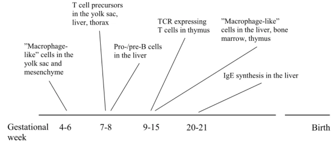
The development of the fetal immune system shows temporal variations between species. In mice, for instance, mature α/β T cells are found in the periphery very late in gestation, while mature α/β T cells appear in peripheral tissues in the first trimester of human pregnancy [186]. Thus, the human fetal immune system seems to be relatively early developed (Fig 3). “Macrophage-like cells” have been detected in the yolk sac and mesenchyme already in gestational week (gw) 4-6, in the liver in gw 9-14, in the bone marrow in gw 12-13, in thymus and the gut in gw 11-22. Moreover, the presence of “macrophage-like cells”, including a MHC class II positive subpopulation, was accompanied by CD3+ T cells and CD20+ B cells in the lymph nodes of 14½-15 weeks old fetuses [187].
T cell precursors, defined by their expression of CD7, have been detected in embryonic tissues i.e. liver, yolk sac and upper thorax regions as early as 7 weeks of fetal gestation. Functional studies on these CD7+ cells, purified from fetal liver, revealed an ability to express markers of mature T cells, such as CD2, CD3, CD4, CD8 and CD25 after culture in T cell conditioned medium with IL-2 [188]. Furthermore, T cells expressing the TCR, CD2 and CD3 have been identified in thymus in gw 13-15 [189].
Figure 3. An overview of the development of the fetal immune system during early pregnancy.
Pro/pre B cells have been observed in the liver at gw 8 [190] and in the lymph nodes at gw 14½-15 [189]. Prenatal IgE production has been detected, by using VDJCε transcripts as a marker for IgE production, in the liver at gw 20 [191], indicating a fetal ability to produce IgE.
Postnatal development of the immune system
The increased susceptibility to infections and decreased immune responses to vaccines in newborns are probably explained by an impaired immune function at birth [192]. Neonates have inadequate cell-mediated immunity, inflammatory responses, antibody production and a poor defense against intra-cellular pathogens, indicating a reduced function of the innate as well as the adaptive immune system [193].
Functional impairments of the innate immune system are reflected by less NK cell cytotoxity and reduced microbicidal activity of neonatal neutrophils as compared with their adult counterparts [194, 195]. Neonatal APC and monocytes have shown a general reduced ability to mount Th1-polarising immune responses. The expression of TLR and the downstream signaling molecules are similar in infants and adults, but TLR-mediated cytokine responses of monocytes and APC at birth indicate less TNF, IFN-α, IFN-γ, IL-1β and IL-12 production as compared with these cells in peripheral blood from adults. In contrast, the TLR-induced production of IL-6, IL-8, IL-10 and IL-23 has shown to be enhanced in newborns, indicating that the neonatal immunity may not always be diminished (summarised in [195, 196]).
Birth Pro-/pre-B cells
in the liver
”Macrophage-like” cells in the yolk sac and mesenchyme
4-6
”Macrophage-like” cells in the liver, bone marrow, thymus
9-15 7-8
IgE synthesis in the liver T cell precursors
in the yolk sac,
liver, thorax TCR expressing
T cells in thymus
Gestational
The ability to produce antibodies is impaired in newborns, probably influenced by the immaturity of neonatal Th cells and B cells [197]. The mother provides her offspring with humoral protection by active transfer of IgG antibodies through the placenta and postnatally by IgA antibodies through breast-feeding [198].
The T cell population at birth consists of a high proportion of naïve T cells and a low proportion of memory T cells, probably as a result of the protective environment during gestation. The proportion of naïve/memory T cells is modified by the postnatal antigen exposure, reaching adult proportions at 12-18 years of age [199]. Neonatal CBMCs respond in a dampened fashion to mitogen and innate stimuli, i.e. with less proliferation, IFN-γ, IL-10 and IL-17 production and less suppressive capacity of isolated Tregs than adult cells [200]. The reduced ability of neonates to mount Th1-associated immune responses is particularly pronounced in children who develop allergic diseases later in life. Lower numbers of IL-12 and IFN-γ-producing CBMCs and lower IFN-γ secretion after allergen stimulation have been associated with development of allergic symptoms and sensitisation later in life [174, 175, 201]. The discrepant immune response at birth, might be associated with exposure to a strong Th2 environment in utero. Accordingly, Th2-associated cytokines are readily produced by CBMCs after mitogen and allergen stimulation [62, 200]. The neonatal Th2-deviated allergen-specific immune responses are down regulated with age and the Th1-like immune responses are up regulated. A delayed down-regulation of the neonatal Th2-like immunity during childhood has been observed in children developing allergic disease [62, 202]. In summary, the development of the immune system starts early in fetal life, implying a possibility for the maternal immune system to interact with the developing fetal immune system from early to late gestation. It is unknown if a certain time period during gestation is particularly important for the shaping of the immune responses in the offspring.
The neonatal immune system is considered to be immature and a delayed maturation of the immune system might be associated with development of allergic disease.
AIMS OF THE THESIS
The overall aim of this thesis was to explore the Th1/Th2 balance in allergic and non-allergic women during pregnancy and its influence on the shaping of the Th1/Th2 profile in the neonate and the development of allergic diseases in the offspring. We hypothesised that the immune profile would be biased towards Th2 during pregnancy in allergic and non-allergic women, with a more pronounced deviation in the allergic group, and that allergy
development in the offspring would be preceded by a pronounced Th2 profile at birth. The specific aims of each individual paper were:
I To study the Th1/Th2 balance in allergic and non-allergic women by measuring specific and total IgE antibody levels during pregnancy and after delivery. II To investigate if CB Th1- and Th2-like chemokine levels are associated with
allergy development during the first 6 years of life.
III To determine (i) if the pregnancy magnifies the Th2 immunity in allergic and non-allergic women, (ii) if the maternal chemokine levels during pregnancy influences the offspring’s chemokine levels during childhood and (iii) to evaluate the relationship between circulating Th1/Th2-associated chemokines and allergy in mothers and children.
IV To explore (i) if maternal allergy influences the gene expression locally in the placenta, systemically in PBMC and fetally in CBMC, (ii) if the gene
expression in the placenta and PBMC influences the gene expression in CBMC and (iii) how the gene expression at birth relates to allergy development during childhood.