This is an author produced version of a paper published in Physical Review A. This paper has been peer-reviewed but does not include the final
publisher proof-corrections or journal pagination.
Citation for the published paper:
Stråhlman, Christian; Kivimäki, Antti; Richter, Robert; Sankari, Rami.
(2017). Negative- and positive-ion fragmentation of core-excited formic-acid molecules studied with three- and four-ion coincidence spectroscopy.
Physical Review A, vol. 96, issue 2, p. null
URL: https://doi.org/10.1103/PhysRevA.96.023409
Publisher: American Physical Society
This document has been downloaded from MUEP (https://muep.mah.se) / DIVA (https://mau.diva-portal.org).
Negative– and positive–ion fragmentation of core–excited formic
acid molecules studied with three– and four–ion coincidence
spectroscopy
Christian Str˚ahlman∗
Malm¨o University, 20506 Malm¨o, Sweden
Institute Methods and Instrumentation for Synchrotron Radiation Research, Helmholtz-Zentrum Berlin f¨ur Materialien und Energie GmbH,
Albert-Einstein-Str. 15, 12489 Berlin and MAX IV Laboratory, Lund University,
P.O. Box 118, 22100 Lund, Sweden Antti Kivim¨aki
Nano and Molecular Systems Research Unit,
University of Oulu, P.O. Box 3000, 90014 Oulu, Finland and MAX IV Laboratory, Lund University,
P.O. Box 118, 22100 Lund, Sweden Robert Richter
Elettra–Sincrotrone Trieste, Area Science Park, 34149 Trieste, Italy Rami Sankari
MAX IV Laboratory, Lund University, P.O. Box 118, 22100 Lund, Sweden
Abstract
The negative ion fragmentation of formic acid (HCOOH) is studied with negative–ion/positive– ion coincidence spectroscopy. We report for the first time four–body ionic fragmentation where up to three positive ions are collected in coincidence with one negative ion. We report yields for 21 three-body channels and five four-body channels. More than 80 % of all negative ion fragmentation involves production of O−, and it is dominated by complete dissociation of all
molecular bonds. Negative ion creation is most abundant at high-Rydberg resonances and just above the molecule’s core ionisation potentials. The existence of four–body fragmentation channels evidences a strong charge redistribution in the molecule.
I. INTRODUCTION
Formic acid (HCOOH), the simplest of the carboxylic acids, attracts continuous attention from researchers due to its importance in many fields of astronomy, biology, chemistry and physics. Formic acid in low concentration is almost omnipresent in Earth’s troposphere, as gas or dissolved in natural sources such as rain water [1]. Emission of formic acid comes from both anthropogenic and biogenic sources. In chemical engineering, synthesis of formic acid from CO2 is extensively studied [2]. The growing need for efficient energy storage
suggests its increased use in fuel cells, combining easy storage in liquid phase with high energy density [3, 4]. Formic acid has been suggested as a suitable product in carbon capture reactions, where CO2 is captured either directly from the atmosphere or from
high-concentration industry sources, and catalysed to useful chemicals [5].
Beyond our planet, the formic acid molecule is present in varying concentrations in stellar atmospheres; it has been discussed whether it is a precursor in the formation of amino acids, such as glycine, in space [6]. The natural abundance of x-ray radiation in stellar environments has been a prominent reason for earth-based investigations of photochemical reactions following core-level excitation.
X-ray excitation of molecules induces electronic processes which lead to fragmentation of the molecule. Core excited and core ionized states (i.e. excitation of a core electron to an unoccupied orbital and to the continuum, respectively) of molecules containing low–Z atoms typically decay by emission of an Auger electron on a fs time–scale. Further ionisation can occur if the molecule holds excess energy after Auger decay. The decay of a core–excited or –ionized molecule by emission of an x-ray photon is less probable. The resulting dissociation of decaying molecules can produce positive ions, neutral fragments, and, with much lower probability, negative ions.
The role of negative–ion production has generally attracted little attention. For formic acid, a study by Guillemin et al. [7] reported negative ion yields (NIY) at the C 1s edge. They used a magnetic mass spectrometer to reveal four negative ionic species, in addition to 21 positive ones. Their study did however not allow for a direct comparison of branching ratios between the fragments. NIYs at the O 1s edge have been measured in photon stimulated ion desorption from condensed formic acid [8], but to our knowledge not in gas-phase. Positive ion creation, in contrast, has been measured at the C 1s [7, 9, 10] and O 1s [11] ionisation
edges. The deuterated versions of formic acid (DCOOH, HCOOD and DCOOD) have also been subject to fragmentation studies with positive ion detection [10, 12]. Fragmentation following VUV excitation was studied with photofragment fluorescence spectroscopy [13].
Our recent study on water in gas-phase showed that negative–ion/positive–ion coinci-dence spectroscopy can be a sensitive probe of weak features in the decay and fragmenta-tion channels of small molecules [14]. Since producfragmenta-tion of negative ions necessarily must be accompanied by positive ions, it is desirable to detect them in coincidence. Most core– excited molecules relax by electronic emission. The net positive charge suggest that neg-ative ion production should be associated with either highly charged positive fragments, or several singly charged fragments. A complete picture of the fragmentation patterns thus requires the ability to detect several positive ions in coincidence with a negative ion. In this paper we will report on such fragmentation patterns involving three and four ionic fragments, namely negative–ion/positive–ion/positive–ion coincidences (NIPIPICO) and negative–ion/positive–ion/positive–ion/positive–ion coincidences (NIPIPIPICO). Four– ion fragmentation following absorption of a single x-ray photon has not been reported pre-viously.
II. EXPERIMENT
Experiments were performed at the high resolution (E/∆E > 104) Gas Phase
Photoe-mission beamline of the Elettra synchrotron radiation laboratory (Trieste, Italy) [15, 16]. An effusive jet of gaseous formic acid was let into a vacuum chamber through a gas needle controlled by a leak valve. The sample was purified from dissolved gases by repeated freeze–pump–thaw cycles. The vapour pressure of formic acid at ambient temperature was sufficient to drive a steady gas flow through the leak valve. The gas jet and the photon beam crossed in the centre of the interaction region of the coincidence setup. The chamber pressure was kept at 6 · 10−7 mbar, but it is estimated that the gas pressure was 10–50 times higher in the interaction region. The intensity of the light was monitored with a photo-diode at the downstream exit of the chamber.
Our negative–ion/positive–ion coincidence setup has been described in detail before [17]. Briefly, it consists of two time–of–flight (TOF) spectrometers operating in tandem. Posi-tive and negaPosi-tive fragments are separated by an electrostatic field, mass–dispersed in flight
tubes and detected with two single–anode multichannel plate (MCP) detectors. Both spec-trometers are installed with their axes at the magic angle (54.7 degrees) with respect to the electric vector of the linearly polarized incident radiation, ensuring that the detected intensity is independent of the angular anisotropy parameter [18]. A weak magnetic field in the negative–ion flight tube deflects electrons, preventing them from reaching the negative– ion detector. We made amendments to the spectrometer compared to our most recent publication [14]. The negative ion detector was reinstalled with better electrical insula-tion, allowing us to put a higher attraction potential on the detector surface (+2000 V, compared to +900 V previously) and a higher gain potential over the double MCP stack (1850 V, compared to 1700 V previously). We could thereby run both TOF spectrometers with approximately twice the extraction potentials, increasing the collection efficiency of, in particular, the positive–ion TOF spectrometer. The cut–off kinetic energy for full detection was increased to 5.5 eV in the positive-ion TOF spectrometer (previously 2 eV) [19]. This, in effect, allows for more efficient detection of fast H+ ions.
Due to the single-anode readout of the positive–ion detector, the instrument cannot record ions separated by less than 10 ns. This reduces the count rate of coincidence channels with two identical positive fragments, i.e. O−/H+/H+ and H−/O+/O+. Their quantitative yield should thus not be directly compared to other channels.
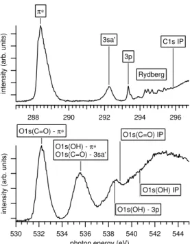
Data was recorded while setting the photon energy at the known resonances and features in the NEXAFS spectrum. At the C 1s and O 1s edge, a negative–ion/positive–ion coinci-dence spectrum at each photon energy was measured for 60 min and 80 min, respectively. The positions of the resonances and features were determined from total positive ion yields (TPIY) measured by the same instrument, as displayed in Figure 1. These resonant features have been studied earlier with near edge x-ray absorption spectroscopy [20] and inner shell electron energy loss spectroscopy [21, 22].
The ground state geometry of HCOOH is planar and has Cs symmetry, and thus all
orbitals have either A’ or A” symmetry [23]. Many previous works have identified the lower unoccupied orbitals as π and σ orbitals [11, 20–22]. Others have considered HCOOH to belong to the C1 symmetry group [7], and thus designated all orbitals as A. We will in this
paper use the notation of Prince et al. [20] which is the most widespread; in particular π* for the lowest unoccupied molecular orbital (LUMO) and 3sa’ for the LUMO+1. These are otherwise designated as 13A and 14A in the C1 symmetry, and should be designated 3A”
intensity (arb. units) 296 294 292 290 288
intensity (arb. units)
544 542 540 538 536 534 532 530
photon energy (eV) O1s(C=O) - π∗ O1s(OH) - π∗ O1s(C=O) - 3sa' O1s(OH) - 3p O1s(C=O) IP O1s(OH) IP π∗ 3sa' 3p C1s IP Rydberg
Figure 1. Total positive ion yields (TPIY) at the C 1s and O 1s absorption edges with designations of resonant features. [20]
and 11A’ in the Cs symmetry. The π∗ orbital is mostly located at the C=O group [22, 24].
The 3sa’ has been identified as an antibonding orbital in OH [23, 25]. While the O 1s TPIY only contains broad resonances, C 1s TPIY shows a prominent set of Rydberg resonances above the π* and 3sa’ peaks, leading up to the IP [20]. A broad σ* resonance is visible in part above IP. The O 1s TPIY contains contributions from both constituent O atoms. The O 1s (OH) → π* resonance is believed to overlap with O 1s (C=O) → 3sa’ [21, 22].
III. DATA ANALYSIS
Time-stamps for detected ions at the two detectors were recorded in separate data li-braries. A positive ion was considered coincident if it was detected within 3000 ns before or 5000 ns after a negative ion. If two or three positive ions arrived within this window, a 3-body or 4-3-body coincidence event was recorded. Only the difference in flight–time between a negative and a positive fragments was established in data analysis. The identity of peaks in the arrival–time difference spectrum, corresponding to negative–ion/positive–ion pairs, were determined from electrostatic simulations and acquisition of photoelectron/positive–ion co-incidence spectra, as described in our previous publications [14, 17]. The NIPIPIPICO map
60 40 20 0 second PI count -2500 -2000 -1500 -1000 -500 0
TOF difference, second PI (ns) 60 0 third PI count -1000 -500 0 500
TOF difference, third PI (ns)
3500 3000 2500 2000 1500 1000 500 0 first PI count -2500 -2000 -1500 -1000 -500 0 TOF difference first PI (ns)
O-/H+/H+/C+ O-/H+/H+/O+ O-/H+/H+/CO+ O-/H+/C+/O+ O-/H+/CH+/O+ C - /H + O - /H + O - /C + O - /O + H - /H +
Figure 2. Intensity map showing NIPIPIPICO events of type O−/H+/PI2/PI3, where PI2 and PI3
are two positive ions, measured at the Rydberg resonances close to the C 1s edge. To increase clarity, we have included all data from the measurements performed in the photon energy range 293.3-295.4 eV (see Figure 3). From the events in the two-ion NIPI1CO spectrum (inset) we select
the O−/H+peak, and plot a contour map of all PI2and PI3 that have been detected in coincidence
with this ion pair. Each island in the map corresponds to a four-body coincidence channel. The TOF differences of the first, second and third PI are calculated relative to the arrival of the negative ion.
in Figure 2 illustrates how four-body channels were identified from arrival–time difference spectra.
All measured data were normalised to the intensity of the light such that one coincidence event equals between 0.9 and 1.4 arb. units, as they are presented in the figures and tables.
IV. RESULTS AND DISCUSSION
The dominant decay channel for core excited HCOOH below threshold is emission of a resonant Auger electron. Production of a singly charged negative fragment from the dissociation process leaves remaining fragments with a net charge of +2. If the doubly
charged fragment is not a single atom, it likely dissociates further into two singly charged fragments:
hν + ABC → ABC∗∗→ ABC+∗+ e−Auger
ABC+∗ → A−+ B++ C+, (1)
where A, B, C are atoms or small molecular fragments, ** denotes a core excited species and * a valence excited species.
We can identify four negative ion species at the C and O edges: H−, C−, O− and OH−. The same species have previously been observed at the C 1s edge [7]. O−channels are dom-inating, followed by H− and C−. The OH− channels are very weak and can only be distin-guished at few strong resonances. Counting the individual yields, we identify 21 three-body channels and 5 four-body channels with statistical significance. No five-body coincidences have been observed.
A. Three–body coincidences at the C 1s edge
Figure 3 shows the total NIPIPICO yield and the branching ratios of all three–body coincidence channels involving O−, H−and C−ions at the C 1s edge. The top panel displays the total NIPIPICO yield as the sum of all individual NIPIPICO channels. In comparison to the TPIY, the total NIPIPICO yield is much enhanced at the Rydberg resonances and grows as the IP is approached. A comparison with non-coincident NIYs measured by Guillemin et al. [7] confirm that negative ion production increases as the photon energy is tuned to high Rydberg resonances, peaking just below the IP. However, in their study, the NIYs at Rydberg resonances are lower than at the π* resonance. Above IP, the NIYs have exponential decays, attributed to post-collision interaction, where a slow photoelectron is overtaken by a fast Auger electron and subsequently recaptured when it becomes exposed to the molecule’s doubly charged core. Similar behaviour has been observed for several small molecules [26– 29].
Figure 3(b) and (c) shows branching ratios for all NIPIPICO channels except for two weak channels involving the OH− ion (OH−/H+/C+ and OH−/H+/O+). Branching ratios are extracted as the fraction of the total NIPIPICO yield, obtained as a sum of all individual
0.06 0.05 0.04 0.03 0.02 0.01 0.00 296 294 292 290 288
Photon energy (eV)
H-/H+/C+ H-/H+/O+ H-/H+/CO+ H-/H+/COO+ H-/C+/O+ H-/O+/O+ H-/O+/CO+ H-/O+/HCO+ C-/H+/O+ (c) 4000 2000 0 Intensity 296 294 292 290 288 0.35 0.30 0.25 0.20 0.15 0.10 0.05 0.00
NIPIPICO branching ratio
O-/H+/H+ O-/H+/C+ O-/H+/CH+ O-/H+/O+ O-/H+/CO+ O-/H+/HCO+ O-/C+/O+ O-/C+/OH+ O-/CH+/O+ O-/CH+/OH+ TPIY Total NIPIPICO IP (a) (b)
Figure 3. Negative-ion/positive-ion/positive-ion coincidences at the C 1s edge. (a) Total NIPIPICO yield, i.e. the sum of all counts in the coincidences channels, and total positive ion yield (TPIY). The two yields are scaled to the same height at the π∗ resonance. (b) The branching ratios of the NIPIPICO channels with NI = O−. (c) The branching ratios of the NIPIPICO channels with NI = H− and C−. The energy point at 286.5 eV has been omitted in panels (b) and (c) due to insufficient statistics.
NIPIPICO channels. No channels involving doubly charged positive ions were observed; neither in two–body NIPICO yields, nor in three–body NIPIPICO yields. The positive charge is thus preferentially distributed over several fragments. Tables with normalised intensities for all channels are provided as supplementary material.
O− production accounts for more than 80% of the total NIPIPICO yield. It is dominated by channels that suggest a complete fragmentation of the HCOOH molecule – O−/H+/C+,
O−/C+/O+ and O−/H+/O+. The branching ratios of these channels change only by few
% across the different C 1s excitations and the C 1s ionization potential. It is, however, interesting to note that the branching ratios of O−/H+/C+ and O−/H+/O+ are in general
mirror images of each other. The same applies to the O−/H+/H+and O−/H+/CO+channels. Both the observations could imply that the O−/H+ pair in these NIPIPICO channels mostly
originates from the hydroxyl group (OH) of the molecule, while the other positive ion results from a further breakage of the CO+ or HCO+ units. Channels where parts of the O=C– O molecular framework remain intact are scarce; the strongest of them is O−/H+/CO+.
The O−/H+/H+ channel is in reality more intense than what appears in the figure because of the experimental difficulty to observe two protons that arrive close in time. Only one C− channel is distinguishable: C−/H+/O+ has its peak branching ratio at the Rydberg
resonances, while it is slightly lower at the π* resonance. The C− channel necessarily means a complete fragmentation of the O=C–O framework.
The branching ratios of the NIPIPICO channels involving the H− ion do not show con-clusive changes in the C 1s core excitation region – they are within statistical uncertainty of ∼ ±0.5% of the data points – but they seem to decrease in general when the C 1s threshold is crossed. Adding up the individual channels, we find that the H− production is largest at the C 1s → 3sa’ resonance with the branching ratio of 19%, an increase of 4% from the C 1s → π∗(CO) resonance. The higher abundance of H− at the C 1s → 3sa’ resonance has previously been proposed to hint at a C–H breaking process with the charge localizing on the H fragment [7]. To assess this hypothesis it is relevant to consider H− in coincidence with CH+. Unfortunately, H− in coincidence with CH+ or OH+ as separate channels are
below the detection limit. However, a summation of all data at the C 1s edge shows evi-dence of weak H−/CH+/O+ and H−/C+/OH+ channels, with the latter stronger than the former. This implies that both H atoms can hold negative charge, but does neither confirm nor disprove the suggestion by Guillemin et al. that the H in the C–H group is preferred for negative ion production.
One of the measurements was performed at the high vibrational levels of the C 1s → π∗(CO) excitation. It shows that the branching ratios of the O−/C+/O+ and O−/C+/OH+
channels increased when compared to the spectrum taken at the maximum of the C 1s → π∗(CO) resonance. The latter channel is likely coupled with the ejection of O− from the C=O unit, whereas the origin of O− can not be deduced in the case of the O−/C+/O+
channel. The branching ratio of the O−/H+/CH+ clearly went down with the increase of
the vibrational energy in this core-excited state, indicating that less O−/H+ ion pairs are
produced (from the hydroxyl group). In summary, the behavior of the NIPIPICO channels at the C 1s → π∗(CO) resonance hint that O− ejection from the C=O site becomes more favored when the vibrational excitation increases.
B. Three–body coincidences at the O 1s edge
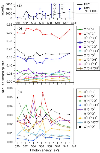
NIPIPICO branching ratios and the total NIPIPICO yield at the O 1s edge are given in Fig. 4. Also at this edge, the total coincidence yield increases as the two IPs are approached. The high yield at IP and decrease at higher photon energies is consistent with the PCI recapture process that has been observed for other molecules. The yield above IP has a significant drop, which resembles how negative ion production drops above IP in many small molecules (see above). NIYs at the O 1s edge of HCOOH have to our knowledge not been published. The same O− NIPIPICO channels are most intense at the O 1s edge as at the C 1s edge and their branching ratios are quite similar. The behavior of the O−/H+/C+
and O−/H+/O+ channels is less mirror-like at the O 1s edge than at the C 1s edge, but some
of it is still visible. The O− channels that are relatively most enhanced at the first O 1s resonance, O 1s(C=O) → π∗, are O−/H+/CH+ and O−/H+/CO+, which again point to the
hydroxyl group as the source of the O−/H+pair. The O−/H+/CH+peaks also at the second O 1s resonance around 535.5 eV. The four strongest O- NIPIPICO channels increase at the high energy side of the O 1s(C=O) → π∗ resonance, where the vibrational excitation of the core-excited state is largest. In contrast the branching ratios of H− NIPIPICO channels decrease, when compared to the ratios determined at the resonance energy.
The only C− channel, C−/H+/O+, has a rather constant branching ratio of about 3% at
O 1s excitations.
The minority channel O−/H+/HCO+ has a peculiar behavior at the O 1s edge. We
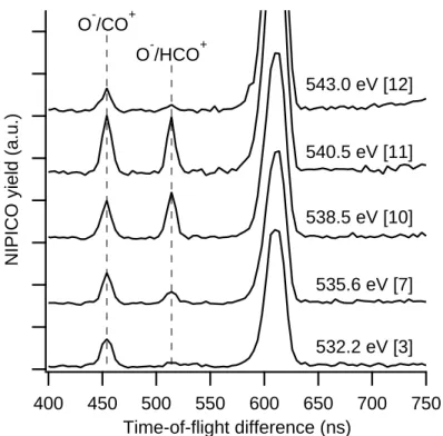
observe that this channel has a larger relative increase than any other channel as the IP is approached (Fig. 4). We demonstrate that the observed behavior is statistically significant by showing the O−/HCO+ two-ion coincidence spectra at certain photon energies in Fig. 5.
O−/HCO+ and O−/CO+ have the same ratio at the 3p resonance (538.5 eV) and just above the first IP, while O−/HCO+ is depressed both above and below these resonances; in
6000 4000 2000 0 Intensity 544 542 540 538 536 534 532 530 0.35 0.30 0.25 0.20 0.15 0.10 0.05 0.00
NIPIPICO branching ratio
O-/H+/H+ O-/H+/C+ O-/H+/CH+ O-/H+/O+ O-/H+/CO+ O-/H+/HCO+ O-/C+/O+ O-/C+/OH+ O-/CH+/O+ O-/CH+/OH+ IP(C=O) IP(OH) TPIY Total NIPIPICO (a) (b) 0.06 0.05 0.04 0.03 0.02 0.01 0.00 544 542 540 538 536 534 532 530
Photon energy (eV)
H-/H+/C+ H-/H+/O+ H-/H+/CO+ H-/H+/COO+ H-/C+/O+ H-/O+/O+ H-/O+/CO+ H-/O+/HCO+ C-/H+/O+ (c)
Figure 4. Negative-ion/positive-ion/positive-ion coincidences at the O 1s edge. See Figure 3 caption for details.
particular at the O 1s (C=O) → π* resonance, the O−/HCO+ yield is barely detectable.
PIY measurements do not explain this behaviour; HCO+ and CO+ are produced at similar rates at O 1s (C=O) → π* while the HCO+ is suppressed to half of the CO+ yield at
the second resonance [11]. The same study shows an increase of non-coincident HCO+ production above IP, remaining firmly below the CO+ yield. PIY–measurements of formic
acid with a deuturated hydroxyl group (HCOOD) showed that HCO+ has a much higher
yield than DCO+at the O 1s edge [11], which suggests that non-coincident HCO+in HCOOH preferably originates from breakage of the C–OH bond.
NIPICO yield (a.u.) 750 700 650 600 550 500 450 400 Time-of-flight difference (ns) O-/CO+ O-/HCO+ 532.2 eV [3] 535.6 eV [7] 538.5 eV [10] 540.5 eV [11] 543.0 eV [12]
Figure 5. Extract of the TOF difference spectrum indicating the O−/CO+ and O−/HCO+ peaks for five selected photon energies at the O 1s edge. (The energy point numbers should go away. I have no access to Igor.) The data for each energy were measured for 80 min and normalized to the intensity of the photon beam. The larger peak at 620 ns is a PEPICO channel arising from non-filtered electrons. The evolution of the O−/HCO+ channel differs clearly from that of O−/CO+.
The only possible route to O−/H+/HCO+ after resonant Auger decay is
hν + HCOOH → HCOOH∗∗→ HCOOH+∗+ e− Auger
HCOOH+∗ → O−+ HCO++ H+. (2)
O−/H+/CO+ has a similar production mechanism
HCOOH+(+)∗ → O−+ CO++ H++ H(+), (3)
with only one additional broken bond. (Creation of an H+2 ion is possible, but not prob-able.) If, as suggested above, the hydroxyl group is the primary source for O−/H+, these
yields suggest that the C–H bond is significantly less likely to break at resonances where O−/H+/HCO+ is observed.
C. Four–body coincidences
Yields for the statistically significant four–body coincidence channels (NIPIPIPICO) are shown in Figures 6 and 7, together with a selection of three-body coincidence spectra for reference purposes. We have only observed channels including the O−/H+ pair. All yields containing the H+/H+ pair are expected to be under-represented with respect to the
O−/H+/C+/O+ and O−/H+/CH+/O+ channels because of the experimental difficulty to observe two protons that arrive close in time.
The channels where all created fragments are recorded, O−/H+/H+/CO+and O−/H+/CH+/O+,
are weak, indicating that a complete fragmentation of the molecule dominates. Due to the small yields and the expected H+/H+ suppression, it is not possible to assess which of the
two complete fragmentation patterns is stronger.
The ions recorded as four-body coincidences in this study carry a net charge of +2, since we have only observed singly charged fragments. Above threshold such channels are expected, following normal Auger decay.
hν + ABCD → ABCD+∗∗+ e−ph
ABCD+∗∗→ ABCD++∗+ e− Auger
ABCD++∗→ A−+ B++ C++ D+, (4)
Below threshold, such fragmentation can occur after resonant double Auger decay, where a core-excited state decays by emitting simultaneously two electrons which share the avail-able energy. For high core-to-Rydberg excitations, doubly ionized final states can also be produced in sequential decay, where final states of resonant Auger decay autoionise. The prominence of NIPIPIPICO close to the C 1s and O 1s ionisation potentials could be ex-plained by such decay. The opening of the normal Auger decay channel above IP explains well the high abundance there.
V. CONCLUSION
We have studied the negative ion fragment formation of formic acid at its core-edges by means of three- and four-body ion coincidence spectroscopy. O− is the most prominent
100 80 60 40 20 0
NIPIPIPICO count (arb. units)
296 294 292 290 288
photon energy (eV) 1200 1000 800 600 400 200 0
NIPIPICO count (arb. units)
O-/H+/H+/C+ O-/H+/H+/O+ O-/H+/H+/CO+ O-/H+/C+/O+ O-/H+/CH+/O+ O-/H+/H+ O-/H+/C+ O-/H+/CH+ O-/H+/O+ O-/H+/CO+ O-/C+/O+
Figure 6. (top panel) TPIY at the C 1s IP. Blue triangles indicate the measured photon energy positions. (middle panel) Yields of the five observed four-body coincidence channels. (bottom panel) Selected three-body coincidence channels for comparisons. All yields are normalised using the same normalisation factors.
negative fragment, followed by H−, C− and OH−. If a negative ion is released, the molecule has a strong preference for complete fragmentation. The three most prominent NIPIPICO channels – O−/H+/C+, O−/C+/O+and O−/H+/O+– bear direct evidence of fragmentation where all bonds in the O=C–O framework are broken, whereas the channels where one or both of the bonds in this framework remain intact are minor. Two pairs of NIPIPICO channels involving the O−/H+ ion pair displayed quite mirror-like trends in their branching ratios especially at the C 1s edge, hinting that the hydroxyl group is the predominant source
160 140 120 100 80 60 40 20 0
NIPIPIPICO count (arb. units)
542 540 538 536 534 532 530
photon energy (eV) 2000
1500
1000
500
NIPIPICO count (arb. units)
O-/H+/H+/C+ O-/H+/H+/O+ O-/H+/H+/CO+ O-/H+/C+/O+ O-/H+/CH+/O+ O-/H+/H+ O-/H+/C+ O-/H+/CH+ O-/H+/O+ O-/H+/CO+ O-/C+/O+
Figure 7. Total positive ion, NIPIPIPICO and selected NIPIPICO yields at the O 1s edge. See Figure 6 caption for details.
of O− ions. Detection of four-body coincidences shows that negative ions can be produced even in processes where the accompanying fragments have a combined positive net charge of +2, evidencing a strong charge redistribution in the molecule. Doubly charged positive ions were not observed in any fragmentation processes that yield negative ions. Branching ratios of individual fragmentation channels typically vary by a couple of % among resonances below a given core IP, with the striking exception of an (almost) complete suppression of the O−/H+/HCO+ coincidence channel at the O 1s (C=O) → π* resonance. A more complete understanding of these elaborate fragmentation paths require a deeper theoretical
understanding of the negative-ion producing dynamics. Further studies in this field should involve theoretical investigation of the electronic processes governing negative ion creation at core edges.
ACKNOWLEDGMENTS
The authors thank El Sayed El Afifi and Lars Christiansson, MAX IV Laboratory, for their help with the design and construction of the instrument upgrade. Mikael Odelius con-tributed with fruitful discussions. We acknowledge Elettra–Sincrotrone Trieste for providing beamtime (proposal number 20150229) and the staff at Elettra for their kind assistance.
[1] A. Chebbi and P. Carlier, Atmos. Environ. 30, 4233 (1996).
[2] W. Leitner, Angewandte Chemie International Edition in English 34, 2207 (1995).
[3] C. Rice, S. Ha, R. Masel, P. Waszczuk, A. Wieckowski, and T. Barnard, J. Power Sources 111, 83 (2002).
[4] X. Yu and P. G. Pickup, J. Power Sources 182, 124 (2008).
[5] S. Moret, P. J. Dyson, and G. Laurenczy, Nat. Commun. 5, 4017 (2014).
[6] S.-Y. Liu, J. M. Girart, A. Remijan, and L. E. Snyder, Astrophys. J. 576, 255 (2002). [7] R. Guillemin, W. C. Stolte, and D. W. Lindle, J. Phys. B: At., Mol. Opt. Phys. 42, 125101
(2009).
[8] D. Andrade, H. Boechat-Roberty, S. Pilling, E. da Silveira, and M. Rocco, Surf. Sci. 603, 3301 (2009).
[9] H. M. Boechat-Roberty, S. Pilling, and A. C. F. Santos, Astron. Astrophys. 438, 915 (2005). [10] A. Guerra, J. Maciel, C. Turci, H. Ikeura-Sekiguchi, and A. Hitchcock, Chem. Phys. 326,
589 (2006).
[11] K. Tabayashi, K. Yamamoto, O. Takahashi, Y. Tamenori, J. R. Harries, T. Gejo, M. Iseda, T. Tamura, K. Honma, I. H. Suzuki, S. Nagaoka, and T. Ibuki, J. Chem. Phys. 125, 194307 (2006).
[12] M. S. Arruda, A. Medina, J. N. Sousa, L. A. V. Mendes, R. R. T. Marinho, and F. V. Prudente, J. Phys. Chem. A 120, 5325 (2016).
[13] M. Schwell, F. Dulieu, H.-W. Jochims, J.-H. Fillion, J.-L. Lemaire, H. Baumg¨artel, and S. Leach, J. Phys. Chem. A 106, 10908 (2002).
[14] C. Str˚ahlman, A. Kivim¨aki, R. Richter, and R. Sankari, J. Phys. Chem. A 120, 6389 (2016). [15] K. C. Prince, R. R. Blyth, R. Delaunay, M. Zitnik, J. Krempasky, J. Slezak, R. Camilloni, L. Avaldi, M. Coreno, G. Stefani, C. Furlani, M. de Simone, and S. Stranges, J. Synchrotron Rad. 5, 565 (1998).
[16] R. Blyth, R. Delaunay, M. Zitnik, J. Krempasky, R. Krempaska, J. Slezak, K. Prince, R. Richter, M. Vondracek, R. Camilloni, L. Avaldi, M. Coreno, G. Stefani, C. Furlani, M. de Si-mone, S. Stranges, and M.-Y. Adam, J. Electron Spectrosc. Relat. Phenom. 101-103, 959 (1999).
[17] C. Str˚ahlman, R. Sankari, A. Kivim¨aki, R. Richter, M. Coreno, and R. Nyholm, Rev. Sci. Instrum. 87, 013109 (2016).
[18] P. Morin, I. Nenner, P. M. Guyion, O. Dutuit, and K. Ito, J. Chim. Phys. 77, 605 (1980). [19] C. Str˚ahlman, Time-of-Flight Ion and Electron Spectroscopy: Applications and Challenges at
Storage Ring Light Sources (Doctoral Thesis, MAX IV Laboratory, Lund University, Sweden, 2016) available at http://lup.lub.lu.se/record/8866027.
[20] K. C. Prince, R. Richter, M. de Simone, M. Alagia, and M. Coreno, J. Phys. Chem. A 107, 1955 (2003).
[21] I. Ishii and A. P. Hitchcock, J. Chem. Phys. 87, 830 (1987).
[22] I. Ishii and A. Hitchcook, J. Electron Spectrosc. Relat. Phenom. 46, 55 (1988).
[23] S. Leach, M. Schwell, D. Talbi, G. Berthier, K. Hottmann, H.-W. Jochims, and H. Baumg¨artel, Chem. Phys. 286, 15 (2003).
[24] U. Hergenhahn, A. R¨udel, K. Maier, A. Bradshaw, R. Fink, and A. Wen, Chem. Phys. 289, 57 (2003).
[25] S. Leach, M. Schwell, F. Dulieu, J.-L. Chotin, H.-W. Jochims, and H. Baumg¨artel, Phys. Chem. Chem. Phys. 4, 5025 (2002).
[26] G. ¨Ohrwall, M. M. Sant’Anna, W. C. Stolte, I. Dominguez-Lopez, L. T. N. Dang, A. S. Schlachter, and D. W. Lindle, J. Phys. B: At., Mol. Opt. Phys. 35, 4543 (2002).
[27] M. N. Piancastelli, R. Guillemin, W. C. Stolte, D. Ceolin, and D. W. Lindle, J. Electron Spectrosc. Relat. Phenom. 155, 86 (2007).
[28] W. C. Stolte, G. ¨Ohrwall, M. M. Sant’Anna, I. D. Lopez, L. T. N. Dang, M. N. Piancastelli, and D. W. Lindle, J. Phys. B: At. Mol. Opt. Phys. 35, L253 (2002).
[29] W. C. Stolte, M. M. Sant’Anna, G. ¨Ohrwall, I. Dominguez-Lopez, M. N. Piancastelli, and D. W. Lindle, Phys. Rev. A 68, 022701 (2003).