No. 1159
Protein Engineering of
Extracellular Superoxide Dismutase
- Characterization of Binding to Heparin
and Cellular Surfaces
Ing-Marie Ahl
Division of Cell Biology
Department of Clinical and Experimental Medicine
Faculty of Health Sciences
Linköping University
581 85 Linköping
Sweden
© Ing-Marie Ahl, 2010
ISBN: 978-91-7393-473-2 ISSN: 0345-0082
Paper I has been reprinted with permission from:
Biochemistry 2009, 48, 9932-9940. © 2009 American Chemical Society
Paper III has been reprinted with permission from:
Protein Expression and Purification 2004, 37, 311-319. © 2004 Elsevier Inc.
Cover: ECSOD binding to HepG2 cells
Gör det du kan, med det du har, där du är.
Lena Tibell, Associate Professor Department of Science and Technology Linköping University
Linköping, Sweden
COSUPERVISOR
Bengt-Harald “Nalle” Jonsson, Professor Department of Physics, Chemistry and Biology Linköping University
Linköping, Sweden
FACULTY OPPONENT
Steen Vang Petersen, Associate Professor Department of Medical Biochemistry Aarhus University
Aarhus, Denmark
MEMBERS OF THE EXAMINATION BOARD Ingemar Rundquist, Professor
Department of Clinical and Experimental Medicine Linköping University
Linköping, Sweden
Sophia Hober, Professor AlbaNova University Center Royal Institute of Technology Stockholm, Sweden
Arne Lundblad, Professor Emeritus Uppsala, Sweden
HOST AT THE PUBLIC DISSERTATION Peter Påhlsson, Professor
Department of Clinical and Experimental Medicine Linköping University
TABLE OF CONTENTS LIST OF PAPERS 1 ABSTRACT 2 POPULÄRVETENSKAPLIG SAMMANFATTNING 4 ABBREVIATIONS 5 BACKGROUND 7
The essentiality and toxicity of oxygen 7
Various kinds of radicals 7
Sources of free radicals 8
Reactive oxygen species 9
Free radical reactions 11
Oxidative damage 11
Diseases and pathological conditions 12
Myocardial ischemia-reperfusion injury 12
Defense systems 13
Antioxidative molecules 14
Antioxidative enzymes 15
Superoxide dismutase 16
Four types of SOD 16
The substrate 17
ECSOD 18
The biological role of ECSOD 21
The potential therapeutic use of ECSOD 21
PseudoECSOD 22
Glycosaminoglycans 22
Heparin and heparan sulfate 22
Heparin-protein interactions 23
MAIN METHODS AND PROTEIN CONSTRUCTS 26
Isothermal titration calorimetry 26
Phage display 27 Ischemia-reperfusion model 28 Protein constructs 29 SUMMARIZING DISCUSSION 31 CONCLUSIONS 37 ACKNOWLEDGMENT 38 REFERENCES 41
LIST OF PAPERS
This thesis is based on the following papers, which will be referred to by their Roman numerals as follows:
Paper I Thermodynamic Characterization of the Interaction between the C-Terminal Domain of Extracellular Superoxide Dismutase and Heparin by Isothermal Titration Calorimetry
Ing-Marie Ahl, Bengt-Harald Jonsson and Lena A. E. Tibell Biochemistry, 2009, 48:9932-9940
Paper II Analysis of Effects of Mutations in the C-Terminal Domain of Extracellular Superoxide Dismutase by Isothermal Titration Calorimetry and Phage Display
Ing-Marie Ahl, Bengt-Harald Jonsson and Lena A. E. Tibell In manuscript, 2010
Paper III Coexpression of yeast copper chaperone (yCCS) and CuZn-superoxide dismutases in Escherichia coli yields protein with high copper contents Ing-Marie Ahl, Mikael J. Lindberg and Lena A. E. Tibell
Protein Expression and Purification, 2004, 37:311-319
Paper IV Cell Association and Protective Effects of PseudoECSOD – a progress report
Ing-Marie Ahl, Sally K. Nelson, Camilla Enström, Ann-Charlotte Ericson and Lena A. E. Tibell
ABSTRACT
Accumulating evidence indicates that oxygen free radicals are involved in many diseases and pathological conditions, such as aging, inflammation, reperfusion damage of ischemic tissue and various cardiovascular diseases. Extracellular superoxide dismutase (ECSOD) thus plays a major role in the maintenance of cells by providing protection against these toxic substances in the extracellular space. Various animal studies have shown that ECSOD has the ability to protect against many of these disorders, and interest has therefore evolved in the potential therapeutic use of the enzyme.
However, despite strenuous efforts, large-scale production of the enzyme has not been achieved. To overcome this problem, a mimic of the enzyme, PseudoECSOD, has previously been constructed. This chimera is easy to produce in large amounts and has all the structural, enzymatic and heparin-binding characteristics of ECSOD, making it a potential substitute for ECSOD in therapeutic situations. However, the copper content of PseudoECSOD has been shown to be rather low, and since the copper ion is very important for the catalytic function of the enzyme, a production system that utilizes a copper chaperone for proper insertion of copper into the active site of the enzyme was constructed. The results show that the copper content of PseudoECSOD produced by this system is close to 100 %.
In order to use PseudoECSOD therapeutically, further investigations of its binding capability and protective properties are needed. Therefore, the binding of ECSOD and PseudoECSOD to heparin was investigated using isothermal titration calorimetry. The results show that although some purely ionic interactions are important for the binding between ECSOD and heparin, there is also a substantial contribution from non-ionic interactions. The investigation also showed that the C-terminal domain is the only part of ECSOD that contributes to productive binding, and that the binding of PseudoECSOD and ECSOD to heparin is similar.
In addition, analysis of mutant proteins strongly indicated that the amino acids R210, K211 and R214 are important for optimal binding of ECSOD to heparin, accounting for about 30 % of the total binding energy. The structural placement of these amino acids in an α-helix also confirms the hypothesis postulated by Margalit et al., that a common structural motif for heparin-binding proteins may be two positively charged amino acids at a distance of approximately 20 Å in the 3D-structure, facing opposite directions of a α-helix. The importance of these residues was also confirmed by analysis of a phage display library of the C-terminal domain of ECSOD.
The binding of PseudoECSOD to heparan sulfate on cell surfaces of two different cell types, HepG2 and endothelial cells, was also investigated. The results clearly show that PseudoECSOD binds to these cells in a very similar manner to ECSOD. To investigate the protective properties of PseudoECSOD against ischemia-reperfusion injuries, an isolated
rabbit heart model was used. The results indicate that the enzyme has a protective effect. However, more experiments using the rabbit heart and other animal models are needed to identify the optimal dose for protective purposes. The protective properties of PseudoECSOD in human tissue should also be thoroughly investigated.
In summary, the findings in these studies, together with earlier results showing the close resemblance of PseudoECSOD to ECSOD in structural, enzymatic and heparin-binding properties, further support the proposition that PseudoECSOD may be a good substitute for ECSOD to use in therapeutic interventions.
POPULÄRVETENSKAPLIG SAMMANFATTNING
Fria syreradikaler produceras konstant i människokroppen, både som biprodukter i cellernas dagliga processer och aktivt t.ex. som försvar mot bakterier. Låga koncentrationer av syreradikaler fungerar som signalmolekyler i cellen, men höga nivåer kan leda till skador på cellens beståndsdelar. För att skydda sig mot dessa skadliga radikaler har cellerna i alla syrelevande organismer utvecklat olika försvarssystem. Ett av de mest effektiva skydden mot fria radikaler är superoxiddismutas, SOD, som är ett enzym som bryter ned superoxidradikaler. Enzymet finns både inuti cellen (intracellulärt) och utanför (extracellulärt). Extracellulärt superoxiddismutas (ECSOD) binder till sockerstrukturer på cellens yta. På så sätt optimeras skyddet av cellerna mot angrepp av syreradikaler utifrån, och denna bindning är därför viktig att studera ur medicinsk synvinkel.
I denna avhandling beskrivs bindningen mellan ECSOD och heparin i detalj genom att använda en mätmetod kallad isoterm kalorimetri. Vi visar bland annat att de positivt laddade aminosyrorna R210, K211 och R214 i enzymet är viktiga för optimal inbindning till negativt laddade strukturer i heparin, och att de intar positioner i enzymets struktur som underlättar inbindning. Resultaten visar vidare att bindningen är beroende av fler samspel mellan oladdade atomer än man tidigare trott samt att produktiv bindning endast sker i enzymets C-terminal.
Eftersom fria radikaler är inblandade i en lång rad sjukdomstillstånd, t.ex. åderförkalkning, cancer, hjärtsvikt, inflammation, lungsjukdomar och åldrande, har ECSOD potential att verka som läkemedel mot dessa. Det har dock visat sig vara svårt att producera ECSOD i stora mängder, och vår forskargrupp har därför tidigare konstruerat ett liknande enzym (PseudoECSOD) som har samma funktion som ECSOD, men som relativt enkelt går att producera i stor skala. Dock har mängden koppar i detta enzym varit låg, och eftersom kopparinnehållet är viktigt för enzymets funktion har vi nu utvecklat ett produktionssystem för att införa koppar i enzymet. Resultatet av detta visar att PseudoECSOD får nära 100 % kopparinnehåll.
Vi har också analyserat PseudoECSODs förmåga att binda till olika celler och vävnader, samt dess skyddande egenskaper mot angrepp från fria superoxidradikaler. Vi har visat att PseudoECSOD binder till båda de undersökta celltyperna, på liknande sätt som ECSOD, och att det har potential att skydda mot radikalangrepp i isolerade kaninhjärtan. Resultaten är mycket lovande, men fler studier, båda i djur och människa, krävs innan enzymet kan verka som en artificiell radikalhämmare.
ABBREVIATIONS
AP-1 Activator protein 1
ARE Antioxidant response element
ATP Adenosine triphosphate
BH4 5,6,7,8-tetrahydrobiopterin
Ca2+ Calcium ion
cDNA Complementary DNA
CS Chondroitin sulfate
Cu2+ Copper ion
CuZnSOD Copper- and zinc-containing superoxide dismutase
CVD Cardiovascular disease
DNA Deoxyribonucleic acid
DS Dermatan sulfate
EC Enzyme Commission
ECM Extracellular matrix
ECSOD Extracellular superoxide dismutase
Fe2+ Iron ion
FeSOD Iron-containing superoxide dismutase
FITC Fluorescein isothiocyanate
GlcA β-d-glucuronic acid
GlcN β-d-glucosamine
GlcNAc N-acetylated β-d-glucosamine
GlcNS N-sulfated β-d-glucosamine GSH Glutathione GSSG Glutathione disulfide H+ Hydrogen ion HA Hyaluronic acid H2O Water H2O2 Hydrogen peroxide
HOCl Hypochlorous acid
•HO2 Perhydroxyl radical
HS Heparan sulfate
IdoA α-l-iduronic acid
IFN-γ Interferon-γ
IgG Immunoglobulin G
IL-4 Interleukin-4
IR Ischemia-reperfusion
ITC Isothermal titration calorimetry
KS Keratan sulfate
LDL Low-density lipoprotein
MAPK Mitogen-activated protein kinase
Mn2+ Manganese ion
MnSOD Manganese-containing superoxide dismutase MPO Myeloperoxidase
NAD+ Oxidized form of nicotinamide adenine dinucleotide
NADP+ Oxidized form of nicotinamide adenine dinucleotide phosphate
NADPH Reduced form of nicotinamide adenine dinucleotide phosphate
NF-κB Nuclear factor kappa beta
Ni2+ Nickel ion
NiSOD Nickel-containing superoxide dismutase
N2O3 Dinitrogen trioxide
•NO Nitric oxide
•NO2 Nitrogen dioxide
NOS Nitric oxide synthase
NOX NADPH oxidase
•OH Hydroxyl radical
ONOO - Peroxynitrite
ONOOH Peroxynitrous acid
•O2- Superoxide anion radical
pH -log [H+]
pKa -log Ka (acid dissociation constant)
R• Unspecified radical
RNS Reactive nitrogen species
ROS Reactive oxygen species
SOD Superoxide dismutase
TRITC Tetramethyl rhodamine isothiocyanate
UV Ultraviolet
XO Xanthine oxidase
XRE Xenobiotic response element
BACKGROUND
The essentiality and toxicity of oxygen
On the early Earth the atmospheric oxygen concentration was very low. However, about 2.45 billion years ago atmospheric oxygen levels suddenly rose in what is now termed the Great Oxidation Event. This caused a separation between anaerobic and aerobic organisms. The anaerobic organisms could not adapt to the new conditions across most of the globe, and hence were restricted to niche environments, while the aerobic organisms evolved adaptive mechanisms that paved the way for the evolution of multicellular plants and animals (1).
Today, oxygen is an essential component for the survival of aerobic organisms, since it is the terminal acceptor of electrons during aerobic respiration, which is the main source of energy in these organisms. However, despite its importance it also has harmful effects. At concentrations greater than those in air, oxygen is toxic to all living creatures, due to the oxygen free radicals that are produced in sequential, univalent reactions (Figure 1) that occur in the metabolism of molecular oxygen (2).
Molecular oxygen 3O
2 → 1O2 Singlet oxygen
↓
Superoxide radical •O2- → •HO2 Perhydroxyl radical
↓
Peroxide ion O22- → H2O2 Hydrogen peroxide
↓
O23- → H2O Water
↓
Oxene ion O- → •OH Hydroxyl radical
↓
Oxide ion O2- → H
2O Water
Figure 1 Sequential, univalent reduction of molecular oxygen.
Various kinds of radicals
A free radical is any molecule, organic or inorganic, that has an odd number of, and thus unpaired, electrons. Free radicals are highly reactive and thus transient. Due to the ubiquity of molecular oxygen in aerobic organisms and its capability to readily accept electrons, oxygen-centered free radicals are frequently formed via diverse mechanisms (3). Radicals and other reactive molecules that are oxygen-centered are called reactive oxygen species (ROS). ROS include superoxide anion radicals (•O2-), perhydroxyl radicals (•HO2), singlet oxygen (1O2),
hydrogen peroxide (H2O2), hydroxyl radicals (•OH) and hypochlorous acid (HOCl) (4). A
since they also contain nitrogen they are known as reactive nitrogen species (RNS). Besides nitric oxide, RNS include peroxynitrite (ONOO-), peroxynitrous acid (ONOOH), nitrogen
dioxide (•NO2) and dinitrogen trioxide (N2O3) (4).
Sources of free radicals
Oxygen free radicals in humans and other aerobic organisms can originate from many sources, both endogenous and exogenous. Examples of exogenous sources, or more strictly, agents that induce their formation, are UV light, ionizing radiation, pesticides, heavy metals, air pollution (5), static magnetic fields (6), redox cycling drugs and other xenobiotics from which free radical metabolites are formed (3), tobacco smoking (7), inorganic particles such as asbestos (8) and silica (9), and gases such as ozone (10, 11).
There are also diverse endogenous sources of reactive oxygen species, partly because oxygen is used in a wide range of processes in normal cellular metabolism, and ROS are by-products of many of the oxidations involved (12). Electron transport in both mitochondria (13-16) and chloroplasts (17, 18), photorespiration in peroxisomes and β-oxidation of fatty acids in peroxisomes and mitochondria (17, 18) are some metabolic processes that are major sources of ROS. Free radicals are also generated in the demethylation, hydroxylation and desaturation reactions catalyzed by cytochromes P450 and b5 in the endoplasmic reticulum and
nuclear membrane (3).
However, ROS are also actively produced in other processes that are not involved in the normal metabolism of healthy cells, such as the respiratory burst during phagocytosis by neutrophils and macrophages. Upon phagocytosis pathogens such as bacteria are engulfed in internalized vesicles, phagosomes, and exposed to a variety of toxic molecules produced via several mechanisms. One of the most important of these mechanisms is the formation of superoxide anions in the reaction catalyzed by phagocytic nicotinamide adenine dinucleotide phosphate (NADPH) oxidase (NOX) (19). Superoxide can further be converted to hydrogen peroxide (19), which subsequently can react with other radicals and transition metals such as Fe2+, yielding highly reactive hydroxyl radicals (20). The very powerful oxidant hypochlorous
acid can also be generated from hydrogen peroxide and chloride ions by myeloperoxidase (MPO) (19, 21). In addition, nitric oxide is produced by the action of inducible nitric oxide synthase (iNOS). Superoxide and nitric oxide can then form the highly reactive peroxynitrite (19). These substances have various, highly toxic effects that kill internalized pathogens (21-23). However, although beneficial in host cell defenses they may have damaging effects on the surrounding cells and extracellular matrix.
There are also non-phagocytic membrane-associated NAD(P)H oxidases, expressed in various cell types, such as endothelial cells (24, 25), that generate and release low amounts of superoxide radicals in the extracellular space (26), through electron transfer from cytosolic NADPH to extracellular oxygen (27, 28).
In addition, numerous enzymes are capable of generating significant amounts of free radicals, through one-electron transfers during their normal catalytic action. For instance, xanthine oxidase (XO) generates superoxide by catalyzing the conversion of hypoxanthine and xanthine to uric acid (20), a process which is further increased during ischemia-reperfusion conditions (29). Lipoxygenase produces ROS by oxidizing polyunsaturated fatty acids to hydroperoxy fatty acid derivatives (29). ROS can also be produced by cellular oxidative metabolic processes involving cyclooxygenase and monoamine oxidase (30). Modulation of enzyme activity, cofactor availability, substrate concentration and oxygen tension can all affect the rates of intracellular radical production. Thus, certain cellular metabolic states, for example hyperoxia, ischemia and antibiotic therapy can favor free radical production (3).
Oxygen may also be transformed to superoxide radicals non-enzymatically, by reacting with redox-active compounds such as semiubiquinone of the mitochondrial electron transport chain (29). ROS are also formed during the autooxidation of diverse molecules such as catecholamines (31, 32), hemoglobin (33, 34), myoglobin (35, 36), hydroquinones (37), flavins (38) and thiols (39). In addition, under normal conditions nitric oxide synthase (NOS) catalyzes the production of nitric oxide from the substrate L-arginine, with help from the cofactor 5,6,7,8-tetrahydrobiopterin (BH4). However, if the availability of the substrate or the
cofactor decreases, NOS activity may switch to an uncoupled state in which the product is superoxide rather than nitric oxide (20).
As mentioned above, transition metals play major roles in the generation of free radicals. Iron and copper can catalyze the Fenton reaction, which generates hydroxyl radicals from hydrogen peroxide (Reaction 1) (40-43). If superoxide is present it may participate in the formation of further hydroxyl radicals by reducing the transition metals to their active states (Reaction 2) (44, 45). The net reaction (Reaction 3) is then called the Haber-Weiss reaction (46). Fe2+/Cu+ + H 2O2 →Fe3+/Cu2+ + OH- + •OH (1) Fe3+/Cu2+ + •O 2- → Fe2+/Cu+ + O2 (2) •O2- + H2O2 → O2 + OH- + •OH (3)
Reactive oxygen species
The most reactive ROS are the hydroxyl radical and singlet oxygen, but as a result of their reactivity, these species have very short half-lives and react in the vicinity of their site of origin. The most abundant ROS are hydrogen peroxide and the superoxide anion radical. These are less reactive and thus have longer half-lives. Hence, there is a higher probability that they will react elsewhere from the site of their production (47). Since superoxide anion radicals, unlike hydrogen peroxide, are charged, they do not have the ability to diffuse
through cell membranes. However, they can cross the outer mitochondrial membrane (48) (49, 50), and the plasma membrane of various cell types (51-53) via anion channels. Another potential mechanism is protonation of superoxide to generate the uncharged perhydroxyl radical, which may cross membranes and then reform superoxide on the other side (54, 55). As mentioned above, superoxide may also be directly produced by NAD(P)H oxidases in the extracellular space (26). Further, since the superoxide radical can induce cascade reactions, it may have effects far from the site of production and action of the original radical, which is the main reason for its high toxicity.
Under physiological conditions, in which the concentration of superoxide is low, the favored reaction is the dismutation reaction yielding hydrogen peroxide (Reaction 4). However, during pathophysiological states, when excess superoxide is produced, it frequently reacts with nitric oxide, yielding the highly reactive oxidant peroxynitrite (Reaction 5) (12). The rate of this reaction is approximately three times faster than the SOD catalysis (56).
2 •O2- + 2 H+ → O2 + H2O2 (4)
•O2- + •NO → ONOO - (5)
Hydrogen peroxide is produced mainly from the dismutation of superoxide anions (47). This reaction (Reaction 4) may be either spontaneous or catalyzed by superoxide dismutase (SOD) (57).
Since hydrogen peroxide is not a radical, it is much more stable than superoxide. Further, it is lipid soluble and can thus cross mitochondrial membranes (58), peroxisomal membranes (16, 59) and plasma membranes (12, 60). Both hydrogen peroxide and superoxide radicals can act either as oxidants or reductants, thus they can affect a very wide range of molecules.
ROS were first considered to have no biological function. However, there is now abundant evidence that these partly reduced oxygen metabolites are critical for the normal operation of a wide spectrum of biological processes (3). They participate as second messengers in the modulation of cell signalling cascades and regulation of important cellular event, such as cell proliferation and differentiation (61-64). Hence, there are several pathways that employ oxidants as effectors in diverse processes.
For example, ROS affect the redox state of cells, which in turn influences the activity of various redox-sensitive proteins, and gene transcription can be induced by ROS (65, 66). Thus, reactive oxygen species are implicated in many intracellular signaling pathways leading to changes in gene transcription and protein synthesis and hence cell function (12).
Free radical reactions
As the name implies, ROS are very reactive and may react with virtually every cellular component, causing serious damage, sometimes sufficiently severe to lead to cell death. Free radicals act through various mechanisms, but the most common is abstraction of univalent atoms, such as hydrogen atoms (Reaction 6) or halides (Reactions 7). Another important mechanism is addition to unsaturated bonds, such as those in fatty acids (3).
R• + XH→ RH + X• (6)
R• + CCl4 → RCl + Cl3C• (7)
The formation of a radical is called initiation. This may occur, for example, through electron transfer from transition metals to oxygen species, or via single electron oxidations or reductions, as in electron transport chains. Propagation of free radical reactions may then proceed indefinitely, causing series of damaging events, or may be terminated by collision of pairs of radicals or by a variety of free radical scavengers (3).
Oxidative damage
Cellular targets at risk of free radical damage depend on the nature of the radical and its site of generation (3). Free radicals are capable of damaging many cell components such as proteins, carbohydrates, lipids and nucleic acids (2, 20). The most severe kinds of damage caused by ROS are lipid peroxidation, oxidation/denaturation of proteins and modifications/mutations of DNA.
Proteins containing the amino acids tryptophan, tyrosine, phenylalanine, histidine, methionine and cysteine can undergo free radical-mediated amino acid modifications. Highly reactive radicals such as hydroxyl radicals may also attack peptide bonds and the amino acids lysine and proline, which are resistant to most other oxidative attacks. Enzymes that depend on attacked amino acids for catalytic activity will be inhibited, and if the amino acids are important for the structure of the protein, denaturation may occur. The susceptibility of proteins to free radical damage thus depends on their amino acid composition, the location of susceptible amino acids and their importance for the activity and/or conformation of the protein, and whether the damaged protein can be repaired. The cellular location of proteins and the nature of the free radical also influence the extent of protein damage. Free radicals can also cross-link proteins into dimers or larger aggregates, by inducing the formation of disulfide bonds between different proteins or irreversible reactions between free radical-damaged amino acid residues (3).
The unsaturated bonds of membrane cholesterol and fatty acids can readily react with free radicals and undergo peroxidation. This process, which yields lipid radicals, can become autocatalytic after initiation (3), leading to severe damage to the membranes. Lipid
peroxidation is mainly caused by hydroxyl and perhydroxyl radicals (2). The presence of shortened fatty acids after lipid peroxidation may seriously affect membrane permeability and microviscosity (67). The increased membrane permeability due to lipid peroxidation and oxidation of structurally important proteins can cause the breakdown of transmembrane ion gradients, loss of secretory functions and inhibition of integrated cellular metabolic processes (3).
Free radical reactions with DNA will result in damaged nucleic acids, and hence mutations if the damage is not repaired. Radical attacks can also result in strand scission. Most nucleic acid destruction occurs through alterations in both the bases and the deoxyribose sugars, which are often cross-linked following radical attack. The components of single-stranded DNA that are most susceptible to free radical action are the thymine and cytosine bases, followed by the guanine and adenine bases and finally the deoxyribose sugar. However, for double-stranded DNA, the deoxyribose moiety is modified more frequently than the bases, due to its exposed location in the helix. The hydroxyl radical is the major oxidant in these reactions (2, 3).
Carbohydrates are also targets for free radicals; monosaccharides may be oxidized and polysaccharides may be depolymerized. Hence, glycosylated proteins are especially sensitive to oxidative damage and various studies have shown that the glycocalyx of endothelial cells can be damaged by free radical attacks (68, 69).
Diseases and pathological conditions
Pathological events often result in overproduction of ROS/RNS. If the antioxidative system cannot cope with this change, an imbalance between the prooxidants and antioxidants will occur, resulting in increased bioavailability of ROS/RNS and a state of oxidative/nitrosative stress (12, 70).
The involvement of oxidative stress has been confirmed in a number of diseases and pathological conditions, including cardiovascular diseases (CVD) (20) such as atherosclerosis, heart failure, diabetes and hypertension (56), myocardial (71, 72) and cerebral (73, 74) ischemia-reperfusion injury, cancer (75-77), various lung diseases (78-80) and neurological disorders (81), inflammation and aging (82).
Myocardial ischemia-reperfusion injury
Ischemia-reperfusion (IR) injuries may occur in the myocardium following blood restoration after a critical period of coronary occlusion (83), since the restoration of blood flow is accompanied by a burst of oxygen free radicals (84). The radical burst is due to activation of membrane oxidases, mitochondrial uncoupling and neutrophil activation (85). Xanthine dehydrogenase, which normally utilizes NAD+ as an electron acceptor, is converted
During the ischemic period, excessive ATP consumption leads to the accumulation of hypoxanthine and xanthine, which upon subsequent reperfusion and influx of oxygen are metabolized by xanthine oxidase to yield massive amounts of superoxide and hydrogen peroxide (86). Since the production of free radicals is proportional to oxygen tension, the rise in ROS production during recirculation is related to over-oxygenation during reperfusion (87). Since IR can cause irreversible tissue damage, early restoration of blood flow is essential to reduce the injury. However, injuries are known to progress even after reperfusion (88).
The pathophysiology of IR-induced injury is associated with various events, such as reperfusion arrhythmias (71, 89), microvascular damage due to inadequate oxygen delivery to the microcirculation during reperfusion (90, 91), myocardial stunning (92, 93), inflammatory reactions, changes in arteriolar resistance caused by alterations in oxygen metabolism (94) and changes in tissue homeostasis that lead to necrosis and/or programmed cell death (85, 89). Ischemia-reperfusion can lead to tissue injuries that are serious complications in organ transplantation, myocardial infarction, and stroke (95-97).
Two main hypotheses have been proposed to explain the pathogenesis of ischemia-reperfusion injury (98-100), namely oxidative stress, which is a well established etiopathogenic factor of reperfusion-induced injury and its consequences (29, 101, 102), and overload of Ca2+. These mechanisms are probably related, and oxygen radicals have been
shown to cause myocardial calcium overload by damaging the sarcoplastic reticulum that normally sequesters and stores intracellular calcium ions (72).
Reperfusion of the ischemic tissue leads to the rapid accumulation of neutrophils at the site of injury (72), where they are the principal effector cells of reperfusion damage (29). These leukocytes adhere to the vascular endothelium, transmigrate to the extravascular space, and can adversely affect viable tissue through the release of proteolytic enzymes and the generation of reactive oxygen species, such as superoxide anion and hypochlorous acid (72). Thus, inhibition of neutrophil adhesion to the endothelium attenuates the process (103), and antioxidant treatment ameliorates both leukocyte adhesion and leukocyte-mediated heart injury in the postischemic period (104).
Ischemia and reperfusion in the rat heart has been found to be associated with activation of redox-responsive transcription factors, such as NF-κB, AP-1 and various MAPKs (105, 106), which may account for inflammatory responses and apoptotic cell death in the affected tissue (107, 108).
Defense systems
To protect themselves from these toxic molecules, aerobic organisms have evolved antioxidative defense systems, which include both enzymes and chemical scavengers that trap the oxygen derivatives and thus regulate the concentration of ROS. The defense systems can be divided into primary defenses that decrease the free radical concentration and prevent
initiation of radical chain reactions, and secondary defenses that trap propagator radicals and diminish the damage they cause by reducing oxidized proteins and repairing DNA.
The primary defenses include antioxidant enzymes, like SOD, catalase and glutathione peroxidases and molecules such as ascorbic acid, glutathione (GSH), uric acid, taurine and hypotaurine. The secondary defenses include enzymes such as glutathione transferases with peroxidase activity that protect against lipid peroxidation, and thioredoxin and other oxidoreductases that act as antioxidants by facilitating the reduction of oxidized protein groups such as thiol. Metal sequestering proteins (109), macroxyproteinase and other proteolytic enzymes that degrade irreparably damaged proteins and DNA repair enzymes can also be included in the secondary defense systems, as can molecules like α-tocopherol, β-carotene and bilirubin (2).
These molecules act in various ways to scavenge various prooxidants and keep them at acceptable levels both inside and outside the cell. Many of them have proven to be able to decrease the damage caused by oxidative stress in cellular and ex vivo animal models (110). However, a molecule demonstrated to have antioxidant properties in vitro might have different or additional properties in a more complex system (111), and no clear benefit of antioxidant therapy has yet been demonstrated in clinical intervention studies (112). The failure to protect cells against oxidative stress in vivo may be due to the bioavailability and metabolism of the agent (113). Hence, the concept of an antioxidant in vitro should not be extended to cells, organs, animals or populations until the evidence has been obtained (111). Antioxidants can have beneficial and/or toxic actions on cells depending on the situation and therefore their use should always be made with a full appreciation of the situation, by clarifying the molecular details of the sources of reactive oxygen species, their nature and their regulation. Increasing evidence indicates that the production of reactive oxygen species is precisely regulated and their downstream targets are specific (112).
Antioxidative molecules
Ascorbic acid is another name for vitamin C. The biologically active substance is the ascorbate ion, which is a cofactor of many proteins and also has antioxidative properties. It acts as a reducing agent that reverses oxidation and keeps iron and copper in their reduced states. Ascorbate is water-soluble and thus is active in the cytoplasm and other aqueous solutions (114, 115).
Glutathione can act as a cosubstrate for glutathione peroxidase, thereby reducing peroxides, or it can react directly with oxygen radicals. GSH also reduces dehydroascorbate, which is formed in the reactions of ascorbic acid, back to its active form.
Uric acid is a very efficient antioxidant that traps free radicals and protects against oxidation in plasma. Taurine and hypotaurine are present in many biological fluids where they have a protective function against free radical damage.
α-tocopherol is a lipid-soluble antioxidant that belongs to the vitamin E family. This molecule protects cell membranes from oxidation damage by reacting with lipid radicals produced in lipid peroxidation chain reactions. Scavenging of the radicals terminates the chain reactions and prevents further damage. The oxidized α-tocopheroxyl radical produced in the process is recycled back to its reduced form via reduction by other antioxidants such as ascorbate, retinol and ubiquinol. Although this is a very important activity of this molecule, the significance of its antioxidant properties at physiological concentrations is unclear. Instead, the main role of α-tocopherol may be in cell signaling (116). A novel, natural form of vitamin E, α-tocopheryl phosphate, can substitute for and is more potent than a-tocopherol in in vitro and animal models (117).
β-carotene belongs to the vitamin A family of fat-soluble vitamins that play roles in diverse functions in the body, for example vision, gene transcription, immune responses and hematopoiesis. It also has antioxidant properties by acting as a very efficient singlet oxygen scavenger and inhibitor of lipid peroxidation.
Bilirubin, which is actually a waste product from the breakdown of hemoglobin, breaks radical chain reactions by reacting with the attacking radical.
Antioxidative enzymes
Catalase (EC 1.11.1.6) scavenges hydrogen peroxide in biological systems by catalyzing its decomposition to water and oxygen (Reaction 8) (2).
2 H2O2 → O2 + 2 H2O (8)
Catalase is a tetramer, each subunit of which contains one porphyrin heme group, which is needed in the reaction catalyzed by the protein. The enzyme is located mainly in the peroxisomes and has one of the highest turnover numbers of all known enzymes (118).
Glutathione peroxidase (EC 1.11.1.9) is the general name of an enzyme family with peroxidase activity, the biochemical function of which is to reduce lipid hydroperoxides to their corresponding alcohols and free hydrogen peroxide to water. There are several isoenzymes with different cellular locations, both intracellular and extracellular, and different substrate specificity. Glutathione peroxidase 1 is the most abundant form. It is a homotetrameric protein that is located in the cytoplasm of nearly all mammalian tissues. Its main substrate is hydrogen peroxide and its catalytic action is dependent on selenium (119). The enzyme requires glutathione as a cosubstrate (Reaction 9), and the oxidized form of glutathione (GSSG) is subsequently reduced back to GSH by glutathione reductase (Reaction 10) (2).
2 GSH + H2O2 → GSSG + 2 H2O (9)
Thioredoxin act as an antioxidant by facilitating the reduction of oxidized proteins by cysteine thiol-disulfide exchange. It is ubiquitous and essential for life in mammals (120).
Superoxide dismutase
Superoxide dismutase (EC 1.15.1.1) was first isolated in 1939 by Mann and Keilin (121), but the catalytic function of the enzyme was discovered by McCord and Fridovich in 1969 (57). The main function of superoxide dismutases is to scavenge the superoxide anion radicals generated in various physiological processes, by catalyzing the disproportionation of the radical to hydrogen peroxide and molecular oxygen (Reaction 4). How scavenging of superoxide radicals by SOD to yield the more potent oxidant hydrogen peroxide can be beneficial to biological system may be confusing. However, one accepted explanation of this paradox is that SOD prevents the formation of peroxynitrite, which is a far more toxic substance than hydrogen peroxide. Further, hydrogen peroxide is readily scavenged by catalase.
These enzymes, which are members of the oxidoreductase family, are metalloenzymes that are present in both eukaryotes and prokaryotes. They are located in a wide variety of intracellular compartments and are also active in the extracellular space. Aerobic organisms contain more SOD than aerotolerant organisms, while strict anaerobes have no detectable SOD activity (122).
Four types of SOD
Depending on the prosthetic metals in the active site, SODs are divided into four types: CuZnSOD (Cu2+ and Zn2+), MnSOD (Mn2+), FeSOD (Fe2+) and NiSOD (Ni2+). In vitro,
one can distinguish between these types of SOD by examining the inhibiting effects of cyanide and hydrogen peroxide, since they all show different inhibition patterns (123-127). However, these reactions do not occur under physiological conditions.
The four types of SOD fall into three different phylogenetic families: the CuZnSODs and the Fe/MnSODs and NiSOD. However, SOD enzymes harboring Ni2+ and Fe2+ or Fe2+
and Mn2+ in their active sites has also been reported in cyanobacteria (128). There is no
homology between the three families, but FeSODs and MnSODs show a great deal of amino acid sequence and structural homologies (129, 130). CuZnSOD, MnSOD and FeSOD are all intracellular enzymes, but there is also an extracellular enzyme called ECSOD, which belongs to the CuZnSOD family. The ECSOD gene is 40-60% homologous to CuZnSOD, but shows minimal homology with MnSOD (131).
Manganese superoxide dismutase (MnSOD), which was first isolated by Keele et al in 1970 (132), is present in the cytosol of prokaryotes and the mitochondrial matrix of eukaryotes (133-135), where it scavenges the superoxide radicals that originate from the
respiratory chain reactions. MnSOD is often induced by oxygen (135). In higher eukaryotes, yeast and some bacterial species the enzyme exists as a homotetramer, while in other organisms it is usually dimeric (136).
Iron superoxide dismutase (FeSOD), which was first isolated by Yost et al. in 1973 (137), is found in prokaryotes (133, 137, 138), simple eukaryotes (135) and in chloroplasts of plants (139), where it protects against the action of superoxide generated by electron leakage to oxygen at the reducing site of Photosystem I (140). The enzyme is either homodimeric or homotetrameric.
Nickel superoxide dismutase (NiSOD) has to date been found in various bacteria and cyanobacteria (125, 135, 141). In most organisms, the active form is homohexameric (135, 141), but it remains monomeric in the absence of nickel (126). The enzyme does not show any sequence homology to the other SODs and have a novel SOD fold (135).
CuZnSOD is mainly found in the cytosol of eukaryotes (142), but the protein is also present in various organelles, such as the intermembrane space of mitochondria (134), peroxisomes (142, 143), nucleus (142) and chloroplasts (144). The enzyme has also been reported in the periplasm of bacteria (145, 146).
CuZnSOD is the major intracellular SOD (109). Most CuZnSODs are homodimers, with each monomer containing an active site, but monomeric isoforms have been reported in bacteria (147). Although the function of cytosolic CuZnSODs is known, there is still uncertainty regarding the biochemical pathways in which they participate (140).
The substrate
The substrate for SOD is the superoxide anion radical, which is formed by a one-electron reduction of molecular oxygen. It is very unstable and undergoes spontaneous dismutation. However, at physiological pH, SODs catalyze the reaction 104 times faster (3). The
protonated form of the superoxide radical is the perhydroxyl radical (•HO2.), which is very
reactive and a stronger oxidant than superoxide but has low abundance at physiological pH (2). However, the protic microenvironment near cell surfaces, which are polyanionic, may favor formation of perhydroxyl radicals, due to the locally lower pH (3). Since perhydroxyl radicals can cross the plasma membrane, this may open the door for superoxide radicals for action in the cytosol.
Superoxide has been shown to affect vascular physiology and pathophysiology, mainly through its interaction with nitric oxide (148), and to activate certain signaling pathways, leading to altered gene expression (61, 149). The radical has also been proposed to serve as a signal molecule in growth regulation and development, partly by altering the cellular responses to growth factors and vasoconstrictor hormones. Superoxide radicals are also implicated in inflammatory diseases, ischemia-reperfusion injury, cancer, and the aging process (148).
ECSOD
Extracellular superoxide dismutase (ECSOD) was discovered by Marklund and coworkers in 1982 (124). It is the predominant SOD in extracellular fluids such as lymph, synovial fluid and plasma (150, 151) and the only known antioxidant enzyme that scavenges superoxide anion radicals specifically in the extracellular space (152). The primary location of ECSOD in tissues is in the extracellular matrix (ECM) and on cell surfaces, where it is found at 20 times the concentration present in plasma (109). 90-99 % of the total amount of ECSOD in mammals is located in the extravascular space of tissues (153, 154). The expression of ECSOD in different tissues varies between different species, but the concentrations are generally highest in blood vessels, heart, lung, kidney, pancreas, placenta, while very little is expressed in brain (155, 156). In the vascular system ECSOD is mainly expressed in the arterial walls and vascular smooth muscle cells (157-159). ECSOD is also synthesized and secreted by a variety of fibroblast and glial cell lines (153, 160).
The enzymatic, structural and heparin-binding characteristics of ECSOD have been thoroughly investigated and also compared to the intracellular CuZnSOD enzyme (161). ECSOD from most species has a molecular weight of approximately 135 kDa (124), although there are some variations (162, 163). In most species ECSOD occurs as a homotetramer (164), which is maintained through interactions between the N-terminal domains (165, 166). However, octameric forms of human ECSOD have also been reported (167).
The ECSOD gene (SOD3) is localized to chromosome 4q21 in the human genome (168). The gene, which is approximately 5900 base pairs (bp) long, consists of three exons and two introns, of which exon 3 contains the 720 bp coding region. The promoter region of the gene contains various regulatory elements, including antioxidant response elements (ARE), AP-1 binding sites, NF-κB motifs and xenobiotic response elements (XRE) (109). The human ECSOD cDNA encodes a 240 aa propeptide, containing an 18 amino acid (aa) long signal peptide that is removed to yield a 222 aa mature protein (169).
The middle part of the enzyme contains the active site, which binds a copper ion that plays a major role in the catalytic action by accepting an electron from one superoxide radical and donating it to another. Along with two protons, the intermediate thus formed produces hydrogen peroxide. A specific arginine in the vicinity of the active site attracts the superoxide radical, facilitating the action of the copper. The zinc ion does not have a catalytic function; instead it stabilizes the structure of the active site (170).
With a kcat/Km of 2 x 10⁹ M-1s-1, CuZnSODs are among the most efficient enzymes
known. This efficiency is partly due to the electrostatic guidance of the substrate to the active site of the enzyme (171) arising from the surface of the protein being negatively charged, while the entrance to the catalytic pocket has a positive charge (172-174).
ECSOD is a glycoprotein (124) and the glycosylation site in the human enzyme is found at Asn 89 (169). Structurally, the carbohydrate chain is of biantennary complex type with an internal fucose residue attached to asparagine-linked N-acetyl-D-glucosamine and with terminal sialic acid linked to N-acetyl-lactosamine (124) (Adachi, 1992). No specific biological function of the carbohydrate moiety has been reported and it does not seem to affect most biochemical properties of the enzyme, such as activity, structure, heparin-binding or plasma half-life. However, the non-glycosylated enzyme showed a marked reduction in solubility in aqueous media (175).
An important characteristic of ECSOD that is not shared by other SOD enzymes is its strong affinity for the highly negatively charged glycosaminoglycans heparin and heparan sulfate. Since the isoelectric point of ECSOD is 4.5, the enzyme carries a net negative charge at physiological pH (176). This implies that its binding to negatively charged glycosaminoglycans is mediated by a cluster of positively charged amino acids in the enzyme. Such a cluster is present in the hydrophilic C-terminal domain (amino acids 194-222), which contains nine positively charged amino acids, and this cluster has been shown to be important for interactions with heparin and heparan sulfate (177-182).
The protection it provides against superoxide radicals is optimized by the binding between the enzyme and heparan sulfate proteoglycans on the surfaces of cells. This binding also prolongs the physiological half-life of the enzyme (183-185), which is reported to be about 20 hours in the vasculature (186) and about 85 hours in tissue (187). The C-terminal may be proteolytically processed during biosynthesis (188, 189), resulting in both intact and cleaved subunits of ECSOD. Three different subtypes of the enzyme, with different combinations of subunits, therefore occur: A, with no affinity; B, with intermediate affinity; and C, with high affinity for heparin/heparan sulfate (124). In the vasculature, ECSOD C equilibrates between the plasma phase and heparan sulfate in the glycocalyx of the vessel endothelium and in tissue ECSOD is probably distributed between heparan sulfate on the surfaces of most cells types in the organs and the interstitial matrix. Since most blood cells do not bind ECSOD and no binding to E. coli has been demonstrated, ECSOD C has the potential to protect most normal cells without protecting microorganisms or interfering with the production of superoxide radicals at the surface of activated neutrophils/macrophages (176).
Various hypotheses regarding the mechanism whereby ECSOD binds to heparin have been proposed. In 1996, Oury et al. reported that two heparin-binding domains of ECSOD are linked by a disulfide bond formed by the Cys219 of the C-terminals, and postulated that this may be of structural importance for the affinity of ECSOD to heparan sulfate (164). However, another study found that when alanine was substituted for Cys219 there was no change in the affinity of the enzyme for heparin (181).
ECSOD binding to glycosaminoglycans other than heparin and heparan sulfate is weak (177), although Gao et al. (2008) showed that it protects hyaluronan from oxidative fragmentation by direct binding (190). ECSOD has also been reported to bind to type I collagen. The heparin-binding region was found to mediate the interaction with collagen and bound ECSOD was shown to protect type I collagen against oxidative fragmentation (191).
A naturally occurring mutation in the ECSOD gene (R213G) results in lowered affinities for both heparin and collagen (192). Because of the reduced amount of bound ECSOD to heparan sulfate on cell surfaces, the protective effect is lowered, which in turn is a risk factor associated with cardiovascular disease (193, 194). The lower affinity for collagen may also increase the risk of oxidative damage of collagen and affect the integrity of the vascular wall (192).
The structure of the middle part of ECSOD was recently determined (171) and the overall subunit fold was shown to be very similar to the corresponding structures in CuZnSOD (195). The active site portions of the two enzymes are sequentially and structurally highly homologous (171, 196), as is the mechanism by which they catalyze the disproportionation of superoxide (171, 197, 198), which may explain their similar catalytic properties, as measured by kinetic parameters (124). The structure of the N-and C-terminal domains of ECSOD remains obscure, since attempts to determine the structure of these parts of the enzyme have failed, possibly because they are highly flexible. However, earlier biophysical investigations and secondary predictions have indicated that they both adopt an helical conformation (166, 178), and suggestions have been made that state that the α-helical conformation of the C-terminal may be induced or stabilized by heparin-binding (180).
The CuZnSOD variants that have been structurally characterized, including ECSOD, are all β-proteins containing eight antiparallel β strands, arranged in a β-barrel (195). The strands are mainly connected through hairpin turns in a flattened Greek key motif, but the enzyme also contains three external loops, which constitute the active site. The construction of the β-barrel makes the protein very stable (199), and all CuZnSOD enzymes display marked physical resistance toward high temperature, pH extremes and high urea and guanidinium chloride concentrations (123, 161).
Due to the high degree of sequence conservation among the CuZnSODs, the amino acid residues that are ligands to the copper and zinc ions constitute a “fingerprint” that can be used to identify members of the family. The residues that maintain the geometry of the active site or are involved in dimer formation are also conserved to a high degree, as are the two cysteines that form an intramolecular disulfide bridge in the protein. In ECSOD there is a further disulfide bridge between C219 residues in neighboring subunits, stabilizing the C-terminal part of the enzyme (164). Petersen and Enghild have also reported the occurrence of two different disulfide bridge patterns in the human ECSOD subunit, creating one active
form of subunit and one inactive form (200, 201). The combination of both active and inactive forms in the assembly of the protein regulates the enzymatic activity (202). The folding of these two variants has been reported to occur inside the cell, which also may be important from a regulatory perspective (203).
It has been shown that heparin and heparan sulfate can induce both ECSOD mRNA and protein expression, and that the sulfation level of these molecules determine the degree of expression. However, the mechanism behind these findings is not known, and may be on receptor level or promoter level (204). Various cytokines, such as interferon-γ (IFN-γ) and interleukin-4 (IL-4) can upregulate the ECSOD expression, possibly via stimulation of transcription by NF-κB (205). ECSOD expression can also be regulated by some hormones and a number of oxidizing agents (109).
The biological function of ECSOD
ECSOD plays an important role in regulating blood pressure and vascular contraction, at least in part through modulating the endothelial function by controlling the levels of extracellular superoxide anion radicals and nitric oxide bioavailability in the vasculature (206, 207). By regulating the bioavailability of nitric oxide, it also affects smooth muscle relaxation, neurotransmission, and inflammation. ECSOD has also been proposed to play an important role in neurologic, pulmonary, and arthritic diseases (78, 152).
ECSOD also modulates oxidant injury, inflammation and fibrosis in a number of lung diseases. In hyperoxic lung injury ECSOD expression and activity is disrupted, potentially contributing to oxidative damage and inflammation (208). ECSOD has also been shown to be involved in protection against oxidation of low-density lipoprotein (LDL) (209, 210), which is a major contributor to atherosclerosis.
The potential therapeutic use of ECSOD
Oxidative stress has been shown to be involved in many diseases and pathological conditions, such as cardiovascular diseases (20), cancer (75-77) and aging (82), which affects a large number of people. Because of the beneficial properties of SOD enzymes against the toxic radicals that underlie oxidative stress, interest has evolved in the therapeutic use of SODs. Most work has focused on CuZnSOD (211) and MnSOD (212), since they are readily produced in large amounts. However, ECSOD may be a better agent to use in therapeutic interventions, since it has been designed for extracellular function and has many beneficial properties, such as affinity for heparan sulfate and a long physiological half-life (186, 187). In fact, various animal models have shown its ability to protect the tissue against oxidative damage (184, 213-215). However, despite strenuous efforts, attempts to produce recombinant human ECSOD in various prokaryotic or simple eukaryotic systems have failed, hindering large-scale production of the protein (216, 217). This complicates genetic
engineering of the enzyme and has also hampered the evaluation of the potential therapeutic value of ECSOD.
PseudoECSOD
To overcome the limitations of production of ECSOD, a chimera of human extracellular and intracellular superoxide dismutase has been constructed. This fusion protein is called PseudoECSOD and comprises the N- and C-terminal domains of human ECSOD fused to human CuZnSOD (Figure 5). PseudoECSOD can be produced in large quantities in Escherichia coli and purified with high yields. The characteristics of PseudoECSOD closely resemble those of human ECSOD (196), which means that this protein may be used as an ECSOD substitute in further investigations.
Glycosaminoglycans
Glycosaminoglycans (GAGs) are linear polysaccharides, with building blocks (disaccharides) consisting of an amino sugar and a uronic acid or galactose. The family of GAGs includes hyaluronan (HA), chondroitin sulfate (CS), dermatan sulfate (DS), keratan sulfate (KS), heparan sulfate (HS) and heparin. All except heparin and HA are attached to a core protein to form a proteoglycan in the active form (218).
Among the interactions between proteins and GAGs that have been examined, complexes of proteins binding to the structurally similar heparan sulfate or heparin molecules are the most intensively studied, mainly because of the commercial availability of heparin. However, the biological ligand in most cases is heparan sulfate.
Heparin and heparan sulfate
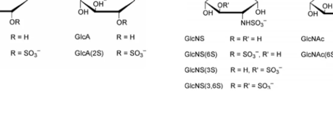
Heparan sulfate and heparin are both linear polysaccharides consisting of repeating uronic acid-(1Æ4)-d-glucosamine disaccharide subunits. The former may be either α-l-iduronic acid (IdoA) or β-d-glucuronic acid (GlcA), both of which may be O-sulfated. The β-d-glucosamine (GlcN) may be either N-sulfated (GlcNS) or N-acetylated (GlcNAc) and both forms may be O-sulfated at various positions (Figure 2) (219). The chemical composition of heparin and heparan sulfate is very similar, the major differences being that heparan sulfate has a lower degree of sulfation, a higher degree of GlcNAc residues and a lower content of epimerized uronic acids (IdoA) (183, 218-220). In addition, heparan sulfate chains are arranged in different domains based on their sulfate content. Variable patterns of substitution of the disaccharide subunits with N-sulfate, O-sulfate and N-acetyl groups give rise to a large number of complex sequences in both heparin and HS, which are structurally the most complex members of the glycosaminoglycan family of polysaccharides (219). This heterogeneity considerably complicates the structural determination of heparin-protein complexes.
a b c d
Figure 2 Monosaccharide building blocks of heparin and heparan sulfate; a) α-L-Iduronic acid, b) β-D-Glucuronic acid, c) N-sulfo-α-D-Glucosamine, d) N-acetyl-α-D-Glucosamine (219).
Heparan sulfate is synthesized by virtually all mammalian cells, while heparin is only produced by mast cells. During biosynthesis heparin chains are attached to a core protein called serglycin, which is only found in mast cells and other hematopoietic cells. This proteoglycan form of heparin, referred to as “macromolecular heparin”, is not secreted by mast cells. Instead, the heparin chains are cleaved from the core protein at random points along the chain and stored in non-covalent complexes with basic proteases within the cytoplasmic secretory granules of the cells. When the contents of the granules are secreted, free heparin is released (183, 219).
In contrast, heparan sulfate usually occurs as a part of a proteoglycan (PG), and rarely in free form. There are several families of proteoglycans, each with a different core protein. The two major subfamilies of cell-surface heparan sulfate proteoglycans (HSPG) are the syndecans and glypicans. The syndecan core proteins are transmembrane proteins while the glypican core proteins are attached to cell membranes by a glycosylphosphatidyl-inositol (GPI) anchor. There are also some subfamilies of HSPG located in the extracellular matrix, e.g. perlecan, agrin and collagen XVIII (219). HSPGs are continuously produced and secreted from a large variety of cells and their composition often shows high degrees of tissue-specificity (183).
Heparin-protein interactions
Hundreds of heparin-binding proteins have been identified, many of which play central roles in important cellular activities and events, e.g. blood coagulation, lipid metabolism and cell growth. Since the functions of these proteins are often dependent on heparin-binding, knowledge of the structural motifs involved in this kind of interaction is essential for a molecular understanding of these processes (183).
Attempts to fully characterize the nature of protein-heparin interactions have been hampered by the heterogeneity of heparin and heparan sulfate, and a lack of structural information regarding the heparin-binding proteins. Many interactions have been described, but the amino acid and oligosaccharide residues that mediate the interactions have not been defined for all of them and X-ray structures have only been obtained for a few heparin-protein complexes (221), e.g. basic fibroblast growth factor (222), antithrombin (223, 224), acidic fibroblast growth factor (225), RANTES (226), eosinophil-granule major basic protein (EMBP) (227) and annexin A2 (228).
The binding of proteins by HS and heparin is primarily electrostatic, involving positively charged groups of the amino acids and negatively charged groups of the polysaccharide. However, there are indications that specific sequences in the GAGs are involved in selective interactions with certain proteins, and that these interactions modulate the protein activities (219).
Heparin and heparan sulfate binding sites have been found within both α-helical and β-sheet frameworks. Several attempts have been made to identify consensus sequences for heparin binding in the primary sequences of the proteins (183), but none of the published sequences are valid for all cases. However, there are often clusters of basic amino acids, e.g. lysine and arginine, in heparin binding regions. In 1993, Margalit et al. proposed that the common structural “motif” amongst heparin-binding proteins may be two basic amino acids, separated by a distance of approximately 20 Å in the 3D structure and facing opposite directions, regardless of the secondary or tertiary protein structure. However, this hypothesis has not yet been verified by structural evidence (229).
AIMS
The general aim of the studies underlying this doctoral thesis was to investigate the requirements for high affinity binding between proteins and heparin/heparan sulfate. The main specific objectives were to characterize the structural requirements and limitations for binding and to identify the amino acids that are of critical importance for the interaction. Since ECSOD has an important biological function that may be utilized therapeutically, I have characterized this enzyme further by using derivatives in the form of fusion proteins and mutant proteins to gain both basic knowledge, and understanding that is interesting from a medical perspective. The investigations included:
• Detailed thermodynamic characterization of the interactions between the C-terminal domain of ECSOD and heparin/defined heparin fragments, using isothermal titration calorimetry (ITC). The measurements included recombinant human ECSOD, fusion proteins and mutant proteins under various experimental conditions (Paper I and II).
• Development of a production system, utilizing a copper chaperone that incorporates copper into the active sites of copper-containing SOD proteins. Since properly incorporated copper is critical for the activity of these SOD enzymes, the production system will be used in the future production of all CuZnSOD variants including the potential therapeutic agent PseudoECSOD (Paper III).
• Analyses of the binding of PseudoECSOD to heparan sulfate on cell surfaces by in vitro immunological techniques, and investigation of the protective properties of PseudoECSOD against oxidative stress in an ex vivo heart model, with the perspective to develop an artificial free radical scavenger that could be used for purposes such as the preservation of transplanted organs (Paper IV).
MAIN METHODS AND PROTEIN CONSTRUCTS
The methods that are central in the studies that this doctoral thesis is based upon are briefly described below.
Isothermal Titration Calorimetry
Isothermal titration calorimetry (ITC) is a technique that is used to measure the thermodynamic parameters of interactions between molecules, based on the fact that every reaction involves the generation or absorption of heat. The method does not require any labeling or immobilization of the analytes (230) and is the only method available for direct measurement of the heat change during complex formation at constant temperature (231). Since a single experiment can provide estimates of all the key thermodynamic variables of reactions, ITC has emerged as the primary tool for characterizing interactions in terms of thermodynamic parameters, elucidating binding mechanisms and, in combination with analyses of relationships between the structure and function of compounds, rational drug design. ITC can be used for numerous applications, e.g. binding studies of antibody-antigen, protein-peptide, DNA-protein, enzyme-inhibitor, enzyme-substrate, carbohydrate-protein, protein-protein interactions as well as enzyme kinetics (231).
In an ITC experiment, a solution of a macromolecule of interest is placed in the sample cell of the calorimeter, while the reference cell is filled with water or blank buffer. Before the experiment starts, the temperature of the two cells is equilibrated and the system tends to maintain this balance. A ligand of interest is then injected into the sample cell, and upon interaction with the macromolecule heat is absorbed or released. Depending on the nature of the association, the system will increase or decrease the power supplied to the sample cell in order to keep it at the same temperature as the reference cell. This change in energy consumption is monitored, yielding a binding isotherm, which provides estimates of the binding constant, the enthalpy and entropy changes associated with the binding and the stoichiometry of the interaction (230, 232). Moreover, ITC experiments performed at different temperatures yield estimates of the constant pressure heat capacity change (ΔCp) (231).
In the ITC experiments performed in the studies underlying this thesis (Paper I and II), a MicroCal VP-ITC microcalorimeter (MicroCal, Northampton, USA) was used. Each protein solution to be tested was placed in the sample cell, and the heparin/oligosaccharide solution was loaded into the syringe injector. The acquired data were then fitted to a theoretical titration curve, with ΔH (enthalpy change, kcal/mol), Kb (binding constant, M-1),
and n (number of binding sites/monomer) as adjustable parameters, by the Origin 5.0 non-linear least squares program implemented in the software supplied by Microcal. A typical binding isotherm is shown in Figure 3.
Figure 3 Binding isotherm for the interaction of ECSOD with heparin. In the top panel, the
major advantage of ITC is that instead of one of the analytes being immobilized, as in oth
Phage Display
lay is a technique in which proteins or peptides of interest are expressed on the surf
peaks indicate the heat released after each addition of heparin into the protein solution. In the bottom panel, the integrated peak area is plotted as a function of molar ratio (heparin/ECSOD). The line represents the best fit of the ITC data, which was used to calculate the thermodynamic parameters.
A
er methods such as SPR, both of the molecules participating in the measured interaction are in solution, hence more reliable data are obtained since this more closely resembles the in vivo situation. The method also has high sensitivity, since only 10-100 nanomols of reactants are needed (231).
Phage disp
ace of a bacteriophage by fusing them to the coat proteins of the phage. By using recombinant DNA technology and random mutagenesis a “library” of phages can be generated, each displaying a unique random peptide. The peptides of the library can then be screened against a ligand of interest by affinity purification. Since phage display provides a direct link between phenotype and genotype, DNA sequencing of the binding phages allows the molecules of interest to be readily identified. The selected phages can then be amplified and purified for further analyses (233).
In the phage display analysis performed in Paper II, a phage display system (T7Select®Phage Display System, Novagen), based on the bacteriophage T7 and E. coli cells (BLT5403), was used. A population of DNA fragments encoding the peptide sequences was cloned into the T7Select vector, packaged into phages, and amplified. To find the most efficient heparin-binding phages, several rounds of biopanning against Heparin Sepharose, with increased salt concentration as the selective factor, were performed. Isolated phages were then amplified and their DNA was analyzed by sequencing across the cloning region.
Since the C-terminal of ECSOD has an α-helical structure, the design of the peptides was based on an optimized helical background, dominated by alanine residues to maintain helical structure (Figure 4). A limited set of mutations at randomized positions, approximately 20 Å away from a fixed arginine residue, was introduced to test a hypothesis regarding structural requirements for heparin-binding proposed by Margalit et al. 1993 (229).
N-AARAAAAQAAAQAXXXXXXAAAQ-C
Figure 4 Design of the peptides of the phage display library. The varied amino acids are indicated by X and the fixed arginine residue in marked.
Ischemia-reperfusion model
To determine the protective properties of PseudoECSOD against ischemia-reperfusion damage, an isolated rabbit heart model was used, which previously has been used to study the effects of various potentially protective agents against ischemia-reperfusion injury (234). The Animal Care and Use Committee in USA gave ethical permission for the animal studies (Animal Protocol # 33901903( )1A, title: “Optimizing the Pharmacological Properties of Superoxide Dismutase.”), and hearts from 14 rabbits were used.
The ischemia-reperfusion experiments were performed essentially as described by Nelson et al. (234). Hearts were mounted through the ascending aorta in a non-recirculating perfusion apparatus and perfused under 80 mmHg with oxygenated, glucose-containing Krebs-Henseleit buffer. After 15 minutes of equilibration the hearts were subjected to 60 minutes of global ischemia followed by 45 minutes of reperfusion. The reperfusion solution contained PseudoECSOD at three different concentrations (200, 300 and 450 U/L), and control experiments were performed in which no exogenous SOD was added to the reperfusion solution. Results were expressed as percent recovery of the developed tension. At the end of the experiments grafts of the hearts were stored at -70°C for further analysis.
Protein Constructs
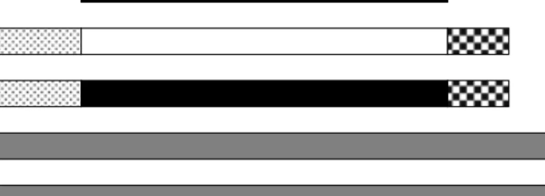
In the studies this thesis is based upon, various fusion proteins are used. The wild type ECSOD protein has been included in the investigations as well as PseudoECSOD, which is a fusion protein of the N-and terminal domains of human ECSOD fused to the N- and C-terminal of human CuZnSOD, respectively. The binding properties of the fusion protein FusCC, which comprises the protein human carbonic anhydrase II (HCA II) fused to the C-terminal domain of human ECSOD, has also been determined. Figure 5 describes the different protein constructs.
A
B
C
D
E
Figure 5 Schematic view of the protein constructs used in the different studies. A) Human CuZnSOD, B) Human ECSOD, C) PseudoECSOD, D) HCA II, E) FusCC