Faculty of Veterinary Medicine and Animal Science
Tracking Transboundary Diseases in Small
Ruminants in the Border Region Tanzania -
Zambia
A Minor Field Study Focusing on Peste des Petits Ruminants
and Foot-and-Mouth Disease
Elsa Wilén
Uppsala 2019
Tracking Transboundary Diseases in Small
Ruminants in the Border Region Tanzania -
Zambia
A Minor Field Study Focusing on Peste des Petits Ruminants
and Foot-and-Mouth Disease
Elsa Wilén
Supervisor: Jonas Johansson Wensman, Department of Clinical sciences
Assistant Supervisors: Gerald Misinzo, Sokoine University of Agriculture, Tanzania
Sara Lysholm, Department of Clinical Sciences
Examiner: Jean-Francois Valarcher, Department of Clinical Sciences
Degree Project in Veterinary Medicine
Credits: 30
Level: Second cycle, A2E Course code: EX0869 Place of publication: Uppsala Year of publication: 2019
Online publication: https://stud.epsilon.slu.se
Key words: FMD, PPR, infectious diseases, sheep, goats, Tunduma, Momba, risk factor, seroprevalence
Sveriges lantbruksuniversitet
Swedish University of Agricultural Sciences Faculty of Veterinary Medicine and Animal Science Department of Clinical Sciences
SUMMARY
Peste des petits ruminants (PPR) and Foot and mouth disease (FMD) are two infectious diseases of major socioeconomic impact. The devastating effects of these diseases are mostly seen in developing countries, such as Tanzania, where small ruminants play an important role in livelihood resilience and are a major source of income. Sheep and goats are relatively cheap to buy and easy to trade and are in many contexts an insurance for the farmers and a valuable resource. There are many households in the world that are completely dependent on small ruminants to feed their families and diseases like PPR and FMD can completely ruin them. PPR is a contagious viral disease that affects small ruminants such as goats and sheep in Africa, the Middle East and parts of Asia. The disease is caused by peste des petits ruminants virus. It is closely related to other significant viral pathogens such as rinderpest virus, which was eradicated in 2011. PPR has now been targeted by OIE and FAO and they have announced a program to eradicate it by 2030.
Foot- and mouth disease (FMD) is a severe highly transmissible viral disease that is caused by foot-and-mouth disease virus (FMDV) and affects cloven-hoof animals. The importance of FMD is significant and is generally ranked as one of top three high impact diseases by farmers in Tanzania.
This study investigated the seroprevalence of PPR and FMD in two districts (Momba and Tunduma) in Southern Tanzania, close to the Zambia border. The total seroprevalence on individual level was 2.9% for PPR and 16.9% for FMD. A total of 491 samples were taken from 164 households and 41 villages. Tunduma district, which contains two major transportation routes and Tunduma town, which is the bordering city to Zambia, had a significantly higher seroprevalence for FMD compared to Momba district, which is positioned further away from the main roads.
Possible risk factors were assessed with the help of a questionnaire that was administered to farmers of each investigated herd. All farmers interviewed utilized communal grazing where their animals could be in daily contact with other sheep and goats. After acquiring new animals, 96% of farmers let them mix with their original herd immediately. A significantly higher seroprevalence for FMD could be seen in farmers who bought animals outside of their home district compared to those who only bought from within his or her home district. No farmer reported that they ever bought animals from Zambia. Regarding vaccination, 4% of farmers interviewed vaccinated their animals. No one kept their sick animals separated from the rest of the herd after acquiring new animals.
This study thus confirms the presence of antibodies against PPRV and FMDV in sheep and goats in Tunduma and Momba district. Presence of PPR in these bordering districts to Zambia, strengthens the concern of further spread of PPR into Zambia, even though there seems to have been no or only limited circulation of PPR during the last year. Age was identified as risk factor for being seropositive for both diseases. Being a sheep along with closeness to main roads were also identified as possible risk factors for being seropositive for FMD. No significant difference regarding seroprevalence could be seen between sexes on either disease. Findings in this study also revealed a continued poor knowledge on biosecurity among farmers.
CONTENT
Introduction ... 1
Aim ... 1
Literature review ... 1
Study region ... 1
Peste des petits ruminants ... 2
Genetic properties ... 2 Epidemiology ... 2 Transmission ... 3 Pathogenesis ... 3 Clinical picture ... 3 Pathology ... 4 Diagnosis ... 4 Vaccination ... 5
Foot and mouth disease ... 5
Genetic properties ... 5 Epidemiology ... 5 Transmission ... 6 Pathogenesis ... 7 Clinical picture ... 7 Pathology ... 7 Diagnosis ... 7 Vaccination ... 8 PPR and FMD in Tanzania ... 8 PPR ... 8 FMD ... 9
MATERIAL AND METHODS ... 9
Study area and study design ... 9
Animal sampling ... 10
Sample size ... 10
Antibody detection ... 11
PPR ... 11
Questionnaire ... 12
Statistical analysis ... 13
Results ... 13
Study area and study design ... 13
Animal sampling ... 15 Antibody detection ... 16 PPR ... 17 FMD ... 17 Questionnaire ... 17 Discussion ... 21 PPR ... 21 FMD ... 22 Conclusions ... 24 Acknowledgements ... 25 Populärvetenskaplig sammanfattning ... 26 References ... 29
1
INTRODUCTION Aim
The purpose of this study was to investigate the seroprevalence of the diseases Peste des petits ruminants (PPR) and Foot and mouth disease (FMD) in the southern parts of Tanzania close to the Zambia border. The districts that were investigated were Momba and Tunduma in Mbeya region, which was selected because the Tazara railway and Tanzam highway, two large transportation routes, passes through the region. It was of interest to see if the diseases were present and to assess risk factors according to management. In order to assess these risk factors a questionnaire was administered to farmers of each investigated household.
This master thesis is a smaller part of a PhD project that through close collaboration with scientists in Zambia (School of Veterinary Medicine, University of Zambia) and Tanzania (Sokoine University of Agriculture) is investigating infectious diseases in both Tanzania and Zambia and is analyzing risk factors for spread of these diseases. There have been reports that smuggling occur across borders quite often (Kivaria, 2003) and there is a considerable risk for spread of diseases over the border. I have contributed to the larger project by field work where data were collected by blood samples from small ruminants in southern Tanzania and through administrating questionnaires. Blood samples were analyzed with enzyme-linked immune-sorbent assay (ELISA) and data analysis was based on the results from blood samples and questionnaires.
LITERATURE REVIEW Study region
Tanzania is a country in East Africa, sited just below the equator. It has a coastline and is bordering to eight other countries (Landguiden, 2016). Almost 59.1 million people live here on an area of 945 000 km² (Landguiden, 2017; NE, 2018). The country is divided into thirty-one regions which in turn are divided into different districts (Central Intelligence Agency, 2018). The districts are divided into wards which contains villages or streets depending on if it is a rural or urban ward. The country is famous for its diverse wildlife and national parks and contains both Africa’s highest and lowest site with Mount Kilimanjaro and Lake Tanganyika (Lake, 2013). Agriculture is a major sector and employs around 65% of the work force (Central Intelligence Agency, 2018). Morogoro, which is both the name of a city and a region, is located approximately 200 km west of the coastal city Dar es Salaam and is the center for agricultural sciences in Tanzania (Wikipedia, 2018). Most research around the diseases PPR and FMD in Tanzania is performed at Sokoine University of Agriculture (SUA) in Morogoro.
Even though Tanzania has an up going economy it is still a low-income country and approximately 67% of the population lives under the income mark which is defined as poor (Landguiden, 2018) and the income gap is large. A large part of the population is small scale farmers and thus dependent on the wellbeing of their animals (Muse et al., 2012). Small ruminants are among the most common farm animals owned by the poor and an important key for poverty alleviation and contribute to national economic development (FAO & OIE, 2015).
2
Sheep and goats are relatively cheap to buy and easy to trade and are in many contexts an insurance for the farmers and a valuable resource.
Peste des petits ruminants
PRR is a contagious viral disease that affects small ruminants such as goats and sheep in several parts of the world (Banyard et al., 2010). In developing countries, like Tanzania, small ruminants play an important role in food security and livelihood resilience (FAO, 2013). An outbreak of this disease largely affects the farmers and is considered as a major limiting factor on farming small ruminants (Baron et al., 2011). PPR is considered to be one of the most damaging diseases affecting livestock in Africa, Middle East and Asia (FAO & OIE, 2015). Because of the rapid spread through movement of animals and the economic impact of the disease, it has been notified by the World Organization for Animal Health (OIE) (Baron et al., 2014). The Food and Agriculture Organization (FAO) and OIE have targeted PPR to be eradicated by 2030 (OIE, 2018).
Genetic properties
Peste des petits ruminant virus is a Small ruminant morbillivirus belonging to the genus
Morbillivirus, in the family Paramyxoviridae, order Mononegavirales. It is closely related to
other significant viral pathogens such as rinderpest virus, canine distemper virus, phocine distemper virus and measles virus (Parida et al., 2015a). Rinderpest virus was eradicated in 2011 and FAO together with OIE has now announced a program to eradicate PPR as well by 2030 (FAO). Morbillivirus is an enveloped, pleiomorphic, negative-stranded, non-segmented RNA virus with a genome of single-stranded RNA of about 16 000 nucleotides (Daillo, 1990). The small ruminant morbillivirus genome is encapsidated by nucleoprotein (N) that is forming a helical nucleocapsid (Parida et al., 2015b) In combination with the dependent RNA-polymerase (L) and the co-factor phosphoprotein (P) they form a ribonucleoprotein (RNP) complex. PPRV like other morbilliviruses binds to the host cell receptors using the attachment factor hemagglutinin (H) (Muse et al., 2016). It then replicates in epithelial cells, lymphocytes, macrophages and pneumocytes (Muse et al., 2016).
There is only one serotype of PPRV but by gene sequencing, small differences have been pointed out in the virus strains, which have led to grouping them into four lineages, mostly reflecting their geographical origins (Chauhan et al., 2009; Baron et al., 2011; Munir, 2014). PPRV belonging to lineages I and II are found in West and Central Africa (Munir, 2013; Kgotlele, 2014). Lineage II are currently the dominating lineage in the area, the latest occurrence of lineage I is from 1994 (Tounkara, 2018). Lineage III is found in East Africa, Middle East and possibly in India (Munir, 2013). Lineage IV was first restricted to Asia but is now also the most common lineage in Africa (Munir, 2013) and is progression in West Africa (Tounkara, 2018).
Epidemiology
PPR was first described in West Africa in the 1940s (Dhar et al., 2002; Parida et al., 2015b). Likely, it existed before that but was then confused with rinderpest (Parida et al., 2015b). The first description of PPR in Asia was made 1987 but the virus has been circulating in the Middle
3
East and parts of Sub-Saharan Africa for several decades (Dhar et al., 2002). PPR has formerly been restricted to Africa, the Middle East and Asia but its distribution has steadily expanded over the years (Munir, 2014), especially over the last 15 years (FAO and OIE, 2015). In 2018 the first case was reported in Europe, in Bulgaria (OIE website, 2018). PPRV has also been reported in China, Nepal, Vietnam and Tajikistan (Munir, 2014). Around 70 countries have either reported infection to the OIE or are suspected to be infected (FAO and OIE, 2015). More than 60% of these are African countries. In Africa PPR continues to spread southwards and has been reported in countries south of the equator such as Tanzania, Kenya, Democratic Republic of Congo and Uganda. It has also spread north of the Sahara desert to Morocco (FAO and OIE, 2015) and recently to Algeria (De Nardi et al., 2011).
Transmission
PPR is a highly transmissible disease that spreads fast among animals with close contact (SVA
website, 2018). Morbidity can be as high as 100% with a fatality rate of 90% (FAO & OIE,
2015) but are often lower in endemic areas. It is transmitted mainly through direct contact, involving secretions or excretions (Taylor, 1984; Muse et al., 2016), and virus is found in ocular discharge, nasal discharge and saliva (Muse et al., 2012a). The virus is sensitive outside the host and gets inactivated in dry environment and by heat (Parida et al., 2015b). Thus, animals need to be in close contact for transmission. According to a review by Chauhan et al., 2009 the main route of transmission to new areas is by moving infected animals. It has been suggested that infected animals can shed virus even before onset of clinical signs, which would indicate incubatory carriers to play a role in the transmission (Couacy-Hymann, et al., 2007a). Trade and thus introduction of new sheep and goats into unaffected areas has been identified as a major risk factor for long distance spread of PPR (Gitao et al., 2012).
Pathogenesis
PPRV is lympotrophic and epitheliotrophic and thus induce most severe lesions in organs with a lot of epithelial and lymphoid tissue (Truong et al., 2014). Truong et al., 2014 suggests that the primary sites of replication are lymph nodes and then the virus reaches the respiratory and lymphoid organs and the digestive tract. Viremia appears after about 2-8 days post infection with a peak of viral RNA in blood after 6 days. PPR induces significant immunosuppression with leukopenia and lymphopenia (Rajak et al., 2005), which makes the animals more susceptible to secondary infections (Gibbs et al., 1979). The severity of the disease in animal groups can vary depending on the lineage of PPR, suggesting that the lineages have different virulence (Couacy-Hymann et al., 2007b). The severity also depends on factors such as breed, age, sex and health status. Morbidity and case fatality rate have been showed to be higher in younglings than in adult animals (Abubakar et al., 2008). Animals that have recovered from a PPR infection get lifelong protective immunity (Parida et al., 2015b).
Clinical picture
PPR can be categorized in an acute, sub-acute or chronic form (Swai et al., 2009; Muse et al., 2016). It is more common that goats get a more acute form of the disease while sheep more often develops a sub-acute or chronic form. Clinical signs start to appear approximately 4 days
4
post infection in goats (Bundza et al., 1988; Truong et al., 2014), and are most pronounced 6-8 days post infection in both goats and sheep (Truong et al., 2014). Affected animals show signs that include sudden onset of depression, anorexia, fever, diarrhea, cough, oral erosions and increasing serous nasal discharge which becomes mucopurulent (Bundza et al., 1988; Muse et
al., 2012a; Kihu et al., 2014). Secondary pneumonia with bacteria such as Mannheimia haemolytica (Parida et al., 2015b) and inflammation of the mucous membranes in the
respiratory and digestive tracts are common (Bundza et al., 1988). Some infected goats get severe nasal lesions and cases where the nose sloughed off during handling have been observed (Muse et al., 2012a). Pregnant animals may abort in all stages of pregnancy but fetuses and the placenta show no sign of malformation (Abubakar et al., 2008). Nodules can be observed all over the body in infected animals (Muse et al., 2012a). Many of the animals die of dehydration due to a combination of profuse diarrhea and inappetence because of painful lesions in the oral mucosa (SVA website, 2018). Young animals seem to die easier than adult animals (Muse et
al., 2012a).
Pathology
In studies describing the pathomorphology of animals infected with PPR, common findings are erosions through the gastrointestinal tract, from the oral cavity to the intestine (Bundza et al., 1998; Kihu et al., 2014). Ulcerative and necrotic lesions are mainly found in the hard palate, dorsal surface of the tongue, gum, dental pad and palatine tonsil (Parida et al., 2015a). In the proximal colon, caecum and rectum, characteristic “zebra stripes” which is extensive congestion in the longitudinal mucus folds can be found (Parida et al., 2015a). Lymph nodes are often enlarged and edematous post mortem and the spleen distended (Muse et al., 2012a). The lungs in infected animals often show signs of pneumonic lesions with red hepatized and congested areas especially in the apical lunglobes (Kihu et al., 2014). At histopathology, lung lesions are often characteristic of bronco-intestinal pneumonia (Kihu et al., 2014; Truong et al., 2014). According to Bunza et al., 1998, animals surviving an infection show no significant lesions post mortem.
Diagnosis
Quick and correct diagnosis is one of the limiting factors in the effort to control the spread of PPR (Baron et al., 2014). The clinical manifestation of PPR resembles other diseases, such as orf, bluetongue, and contagious caprine pleuropneumonia (CCPP) (Roeder, 1999). This makes it hard for the local veterinarians to distinguish PPR from the other diseases. For detection they must either be able to distinguish the disease from clinical signs, which can be strengthened by post mortem investigation (Roeder, 1999) or send samples for laboratory testing and wait a long time for results (Baron et al., 2014).
The currently available diagnostic tests can be divided into those detecting the virus, like PCR or immunocapture ELISA (icELISA), which is used to detect acute infection and those that detect antibodies to the virus, like competitive ELISA (cELISA). The latter is common in surveillance studies for mapping the spread of the disease (Baron et al., 2011). All of the tests require laboratory techniques (Baron et al., 2014). Development of different tests that are more suitable for field conditions is one of the challenges in the fight against PPR. Recently, new
5
diagnostic techniques such as immunochromatographic tests, carried out on superficial swabs, have been tested under field conditions with so far positive results (Baron et al., 2014). This might be a useful tool in the effort to control the spread of PPR.
Vaccination
Immunity provided by vaccination is a solid foundation for control strategies on PPR (Munir, 2014). Significant improvements have been made in vaccine development for PPR and now there are several categories of vaccines available that work well. Khan et al., 2008, analyzed antibodies to PPR after vaccination with Nig/75/, a lineage I vaccine. Before vaccination the sheep and goats involved were found negative for PPR by competitive ELISA (cELISA). Day 10, 30 and 45 post-vaccination serum samples were taken again where 100% of the sheep and 96.6% of the goats tested had positive results after 45 respectively 30 days. The antibody profile is the same in vaccinated animals as in animals that have recovered from an infection (Baron et
al., 2011). This makes mass vaccination a complication in prevalence studies and thus prevent
epidemiological studies needed for surveillance.
Foot and mouth disease
Foot- and mouth disease (FMD) is a severe highly transmissible viral disease caused by foot-and-mouth disease virus (FMDV) and affects cloven-hoofed animals. FMD is characterized by the formation of vesicles on the mucous membranes in the mouth, nose and in the coronary band and interdigital space around the cloves (Shahan, 1962). The disease is highly trans-missible and is most significant from an economic point of view. The disease does not have a high case of mortality in adult animals but can still have devastating effects, resulting in loss of productivity, varying on species and breed (Grubman & Baxt, 2004). The importance of FMD is significant and was ranked as the most important disease by agro-pastoralists farmers in Tanzania (Knight-Jones et al., 2017).
Genetic properties
The FMD virus is a member of the Aphthovirus genus, in the Picornaviridae family (Sahle et
al., 2007). It is a single-stranded, postive-sense RNA virus containing around 8500 nucleotides
surrounded by four structural proteins to form a capsid (Grubman & Baxt, 2004). The virus has seven different serotypes with multiple subtypes within each serotype. The serotypes are named A, O, C, Asia 1 and South African Territories (SAT) 1, 2 and 3. Serotype O is the most common worldwide (Aftosa, 2014). Collectively the seven serotypes contain more than 60 strains (Aftosa, 2014). Most strains can affect all susceptible hosts but a few have a more restricted host range. The antigenic variation that is a result from the high mutation rate makes FMD control very difficult (Grubman & Baxt, 2004). Animals having recovered from the disease can get immunity from that serotype but not from other serotypes (Aftosa, 2014). The protection from other strains within a serotype depends on the antigenic similarity (Aftosa, 2014).
Epidemiology
FMD is a disease that has spread all over the world where outbreaks have occurred in all livestock-containing regions except from New Zealand (Grubman & Baxt, 2004). Countries
6
free from FMD often have a number of measures to avoid getting the disease in to the country because of the devastating economic consequences. Countries free from FMD are North- and Central America, Western Europe, New Zealand, Australia, Greenland and Iceland (Ashford, 2018). In 2001 an outbreak of FMD occurred in the United Kingdom, spreading to Ireland, France and the Netherlands. FMD is endemic in many countries in Africa, the Middle East, Asia and parts of South America.
Among the seven different serotypes, six have occurred in Africa (O, A, C, SAT-1, SAT-2, SAT-3), four in Asia (O, A, C, Asia-1) and three in South America (O, A, C) (Ashford, 2018). Between 2001 and 2011 three serotypes (O, A and SAT-2) were recorded in Tanzania where serotype O accounted for 50% of the samples (Mwanandota et al., 2013).
FMD is highly transmissible and can spread fast between animals. Globalization is an increasing risk for long-distance spread of the disease as well as movements between animals, people, goods and animal products (Paton et al., 2018). Closeness to main roads and international borders has been identified as risk factors for FMD occurrence (Allepuz et al., 2015).
Transmission
FMD is highly transmissible and can be spread in the air for several kilometers, depending on climate and weather conditions (SVA website, 2017). The most common route of infection is by direct or indirect contact with infected animals or environment (Alexandersen, 2003). FMD mainly affects cloven-hooved mammals, such as cattle, sheep, goats, pigs, water buffalo and yaks (Aftosa, 2014). Domesticated cattle are often the most important hosts (Aftosa, 2014), and also get severe clinical signs (SVA website, 2017). There are also strains adapted to pigs and water buffaloes (Aftosa, 2014). Wild cloven-hoofed animals are also susceptible and at least 60 species of wild animals have been reported with FMD, including giraffes, African buffaloes, bison, moose and several species of antilopes, gazelles and deer.
Small ruminants might play an important role in transmission of the virus, but it is unclear if the virus can be maintained for long periods in these animals in the absence of infection in cattle (OIE, Terrestrial manual 2012). Mansoor et al., (2018) suggest that small ruminants should be included in vaccination campaigns in FMD endemic regions.
The virus exists in the entire body of the infected animal and all secretions and excretions can contain virus (Aftosa, 2014). Meat and other products from infected animals also contain virus (SVA website, 2017). Depending on host and viral strain the amount of shredded virus can vary (Aftosa, 2014). Pigs for example, produce large amount of virus that spread through aerosols. Infected animals shed virus during the whole infection, from before clinical signs appear to after the clinical signs have passed (SVA website, 2017). Virus can survive as long as six months outside the host, depending on temperature and humidity (Bachrach, 1968). Dried blood, carcasses, hay, soil and so forth can serve as reservoirs for infection. The virus is resistant in the environment but is less optimal outside the pH range 7.2 and 7.6, why alkaline or acid based disinfectant are very active (Ashford, 2018). It also inactivates at temperatures above +56° C and when dried.
7
Pathogenesis
FMDV is a dermotropic virus and involves mucosa and skin primarily (Shahan, 1962). The most common infection route is via the respiratory tract by aerosols (Shahan, 1962) and the primary site of replication is in the mucosa of the pharynx (Ashford, 2018). Infection can also occur through abrasions in the skin or mucous membranes, but that requires a much higher virus dose (Shahan, 1962). Virus is excreted in high concentrations in saliva and nasal discharge as well as in milk, urine, semen, feces and blood. Alexandersen et al., (2003) propose that the initial replication takes place either in the pharynx or in the lower respiratory system depending on the size of aerosol infecting the animal. After initial replication in the pharynx or the lower respiratory system respectively, the virus then spread through the lymph system or via the pulmonary circulation to the general circulation.
Clinical picture
FMD is characterized by fever and vesicular formation of the feet, buccal mucosa and mammary glands (OIE, Terrestrial manual 2012). Clinical signs tend to be mild in sheep and goats compared to cattle (Aftosa, 2014; Epiwebb, 2018). Commonly observed signs include fever and lameness on one or more legs (Aftosa, 2014). Vesicles appear mostly on the feet and can be hard to notice (Epiwebb, 2018). More seldom vesicles appear on the tongue and toothpalate. A significant amount of infected small ruminants may be asymptomatic (Aftosa, 2014). Secondary infections are common and high mortality can be seen in young animals such as lambs and calves due to heart affection (Epiwebb, 2018) or emaciation (Aftosa, 2014). Incubation time is about 2-8 days but can be as short as 2 hours and as long as 14 days (SVA
website, 2017). In an experimental study, vesicles developed 48 h post infection in unvaccinated
cattle inoculated with FMDV RNA (Stenfeldt et al., 2015).
Pathology
Besides the lesions in the mouth, cloves and mammary glands that can be found clinically, vesicles are often found in the pharynx, esophagus and forestomaches post mortem (Epiwebb, 2018). Early lesions look like small pale areas while ruptured vesicles more look like ulcers (Aftosa, 2014). The location of lesions varies between species, similarly to the differences in clinical signs. The cloves often develop coronitis and in severe cases the cloves can be sloughed. In young animals with heart affection, grey or yellowish irregular foci can be seen because of cardiac degeneration and necrosis, which sometimes are called “tiger heart” lesions (Aftosa, 2014; Epiwebb, 2018).
Diagnosis
Diagnosis of FMDV is made either by virus isolation or demonstrating viral antigen or nucleic acid in samples of tissue or fluid (OIE, Terrestrial manual 2012). Detection of virus-specific antibodies can also be used for diagnosis to indicate a prior infection. Diagnostic techniques commonly used are enzyme-linked immunosorbent assays (ELISA) to detect viral antigens or antibodies to structural or non-structural proteins (OIE, Terrestrial manual 2012). Reverse
8
transcription polymerase chain reaction (RT-PCR) is a sensitive and rapid measure that also can be performed and is useful in samples with low concentrations of virus (Ashford, 2018; OIE, Terrestrial manual 2012). OIE has published guidelines regarding diagnosis of FMD in the Terrestrial Manual (2012). According to the guidelines testing on animals should be conducted at a grade 4 laboratory to ensure security when the virus is handled.
Vaccination
It is possible to vaccinate against FMD (OIE, Terrestrial manual 2012). Routine vaccination is often made in countries where FMD is endemic and, in contrast, many FMD free countries never use vaccines but instead have strict movement controls and use stamping out methods when outbreaks occur. Many vaccines are multivalent to provide protection for the different serotypes likely to be encountered in the given location. The vaccines are often prepared with two or more different serotypes to ensure broad antigenic coverage in the specific area. Live FMD vaccines are not acceptable due to danger of reversion to virulence (OIE, Terrestrial manual 2012). All vaccines that are used are inactivated. The vaccines are also purified to remove non-structural proteins (NSP) and therefore obtaining a DIVA vaccine. If not purified, these vaccines will induce NSP antibodies. By using NSP-ELISA as a diagnostic tool it is therefore possible to distinguish vaccinated animals from infected, if vaccinated with purified vaccines (Kweon et al., 2003).
Stenfeldt et al., (2015) showed that animals vaccinated 14 days prior to an infection with FMD developed no clinical signs of FMD or any signs of viremia. In contrast, the animals not vaccinated prior to infection developed both viremia and clinical signs (Stenfeldt et al., 2015).
PPR and FMD in Tanzania
PPR
The first outbreak of PPR in Tanzania was confirmed in 2008 in the northern region bordering to Kenya and was introduced from the neighboring countries in the north (Swai et al., 2009, Kiviara et al., 2013). In 2009, new studies were made in the northern Tanzania which confirmed a natural transmission of PPR under field conditions (Swai et al., 2009). In 2011, PPR was confirmed after an outbreak in Tandahimba district in Southern Tanzania (Muse et al., 2012a). Tandahimba district borders to Mozambique and this indicated an ongoing spread of the disease. In 2012 and 2013, samples were collected in Ngorongoro district (Northen Tanzania) and Mvomero district in the Morogoro region (Eastern Tanzania) which also confirmed the presence of PPR (Kgotlele et al., 2014). Another recent study conducted in 12 regions in Tanzania had an overall seroprevalence of 27.1%, varying from 2.1% to 72.8% in the different regions (Kgotlele et al., 2016).
So far, isolated PPRV strains from different parts of Tanzania belong to lineage II-IV (Kiviaria
et al., 2013; Torsson et al., 2016). This indicates that the northern neighbor’s to Tanzania is the
most likely source of infection and that virus found in southern parts came from the northern part of the country.
9
Today, Tanzania is the south-eastern border of the disease and there is a high risk for spread to southern countries, including Zambia, Mozambique and Malawi (Muse et al., 2012b; Chazya
et al., 2014).
FMD
Most seroprevalence studies on FMD in Tanzania are made on cattle, showing that the disease is widely distributed in the country (Kasanga et al., 2012). A study conducted in 2010 and 2011 on cattle in eastern Tanzania found a total seroprevalence of 41% with a variation in the districts between 15.4% and 81% (Mwanandota et al., 2013). In 2014, a study also involving sheep and goats, performed in the central part of Tanzania, estimated a seroprevalence of 69.8% in bovines, 52.4% in ovines and 11.1% in goats (Mdetele et al., 2014). This result revealed the need to consider other species, such as sheep and goats, as well when planning FMD control. The study indicated also that FMD is more prevalent in areas with a high wildlife-livestock interface. A recent study focusing on sheep and goats in eastern and northern Tanzania estimated a seroprevalence of 39.0% in 2014 and 14.2% 2015 (Torsson et al., 2017). In this study proximity to wildlife was not identified as a risk factor.
MATERIAL AND METHODS Study area and study design
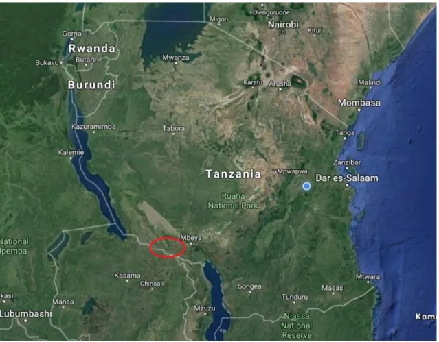
This study was a cross-sectional serological survey conducted in September 2018. The aim was to do a serologic screening close to the Zambia border in Tanzania, to see if PPR and FMD were present and to assess risk factors. Another study was also made on the Zambia side of the border, which is described in another thesis (Mitternacht, 2019). The larger PhD project, which this master is a smaller part of, is using this data to assess risk for transmission of different infectious diseases over the border to Zambia. Thus, the study area was the two districts of Tunduma and Momba in Mbeya region (Figure 1), in the southern part of Tanzania close to the Zambia border. Mbeya region was purposively selected because of two large transportation routes passing through the region, the Tazara railway and Tanzam highway.
Villages in the area were randomly selected from lists provided by the local District Livestock Officer or District Veterinary Officer. A total of 41 villages were selected, 33 in Momba district and 8 in Tunduma district. The difference in number was because Tunduma district only consists of 8 villages. Four households were selected in each village by snowball sampling. This was carried out by first visiting one random farmer and then asking him/her to be directed to the next a farmer by asking one of these criteria:
Someone who has more than 15 goats AND/OR sheep Someone who has 5-15 goats AND/OR sheep
Someone who has less than five goats AND/OR sheep
Three animals per household were sampled. The farmers were first asked if they agreed to contribute to the study, which all did. Farmers were then asked to catch three animals at random.
10
All animals over four months could be selected, to avoid maternal antibodies. In farms where they had sheep, at least two sheep were selected among the three animals.
Figure 1. Approximate location of the study area where goats and sheep were sampled.
Animal sampling
The samples were collected during two weeks in September. Blood was taken from the jugular vein on goats and sheep using a vacutainer system into serum tubes (BD Vacutainer, Plymouth, UK). After blood was taken, the serum tubes were carried in waist bags, when going around in the villages. The samples were put into cool boxes in the car during the day. Blood samples were left to separate during one night in refrigerators. After separation, serum was transferred into cryotubes and stored at approximately -9° C. All samples were then transported in cool boxes to the laboratory in SUA Morogoro, for analysis. The study was approved by an ethical committee (ILRI – IREC2018 - 04).
Sample size
When calculating the sample size a confidence interval of 95%, a margin of error of 5% and a prevalence estimate of 50% with infinite population were assumed to obtain the maximum sample size. The sensitivity and specificity numbers on the competitive enzyme-linked immunosorbent assay (cELISA) for rift valley fever (which was one of the diseases in the PhD project) gave the highest sample size when calculating and were therefore used. The
11
calculations were based on the formula in Humphrey, Cameron & Gunn (2004). According to this formula the calculated sample size was estimated to 461 samples.
Antibody detection
The samples were analyzed with commercial competitive enzyme-linked immunosorbent assays (cELISA) to identify presence of antibodies directed to the nucleoproteins of either PPRV or FMDV. Both kits were made by the company Innovative Diagnostics vet (ID.vet) and were used according to the manufacturer’s instructions.
PPR
The kit used to detect antibodies for PPR (ID Screen PPR Competition) can be used for serum or plasma on both sheep and goats. It had a sensitivity on 94.5 and a specificity on 99.4 (Libeau
et al., 1995). It is the only commercial ELISA available approved by FAO/OIE (IDvet website,
2012-10-14). The wells are pre-coated with purified recombinant PPR nucleoprotein (NP) which binds to anti-NP antibodies if present to form an antibody-antigen complex which masks the NP epitopes.
The manufacturer’s instructions were followed and below is a brief description. First, the reagents were allowed reach room temperature before use. Then 25µl of dilution buffer were put into each well. An amount of 25 µl of each sample were put into every well with the exception of the wells were the positive and negative control were added. The plate was then put into an incubator for 45 minutes at a temperature of 37°C. The contents were homogenized with small vibrations during the whole incubation period. After the incubation each well was washed three times with 300 µl wash solution. Between each washing step the plate was tapped onto paper to remove remaining fluid. After this, 100 µl of conjugate were added into each well and the plate was then left to incubate in room temperature for 30 minutes. After a washing step, 100 µl substrate solution were added to each well and the plate were left to incubate for 15 minutes in the dark at room temperature. After this, 100 µl stop solution were added to each well and the absorbance values were read and recorded directly at 450 nm with Erba LisaScan II (Erba Mannheim).
The validity of the positive and negative control was calculated. The mean value of the negative control had to be greater than 0.700 and the mean value of the positive control had to be less than 30% of the negative mean value. All assays were valid. For each sample the competition percentage (S/N %) was calculated. Three different outcomes were possible; positive, doubtful or negative. Samples presenting S/N % less than or equal to 50% were considered as positive. Samples presenting S/N % greater than 50% and less than or equal to 60% were considered as doubtful. Samples greater than 60% were considered as negative.
FMD
The kit used to detect antibodies for FMD (ID Screen FMD NSP Competition) is designed to detect the specific antibodies to the viral non-structural protein (NSP) of FMDV, by competitive ELISA. NSP is only expressed during virus replication and are unlike structural proteins not serotype-specific and the detection of these antibodies are not serotype restricted (OIE,
12
terrestrial manual 2012). Samples are put in the wells and if there are anti-NSP antibodies present they will form an antigen-antibody complex which masks the virus epitopes.
The manufacturer’s instructions were followed and below is a brief description. First, 50µl dilution buffer were put into each well. An amount of 30 µl of each sample were put into every well with the exception of the wells where the positive and negative control were added. The plate was then put into an incubator for 120 minutes at a temperature of 37°C. The contents were homogenized with small vibrations during the whole incubation period. After the incubation, each well was washed five times with 300 µl wash solution. Between each washing step the plate was tapped onto paper to remove remaining fluid. After this, 100 µl of conjugate were added into each well and the plate was then left to incubate in room temperature for 30 minutes. After another washing step, 100 µl substrate solution were added to each well and the plate were left to incubate for 15 minutes in the dark at room temperature. After this, 100 µl stop solution were added to each well and the absorbance values were read and recorded directly at 450 nm with Erba Lisa Scan II (Erba Mannheim).
The validity of the positive and negative control was calculated. The mean value of the negative control had to be greater than 0,700 and the mean value of the positive control had to be less than 30 % of the negative mean value. All assays were valid. For each sample the competition percentage (S/N %) was calculated by dividing the absorbance value of each sample with the mean negative control and then multiply it with 100. FMD outcomes were either positive or negative. Samples presenting S/N % less than or equal to 50% were considered positive. Samples with a S/N % greater than 50% were considered negative.
Questionnaire
Pre-prepared questionnaires were conducted to each farmer of every investigated herd. The questionnaire contained questions to assess possible risk factors according to management and transmission of diseases. The interview was performed by a PhD-student from SUA in Swahili and the answers were written down in English. The questionnaire was designed to focus on management, medical treatments, trade, animal and public health, details of goats/sheep owned and information about the farmer. In this master thesis only parts of the questionnaire are analyzed
where risk factors regarding PPR and FMD are the main focus. The parts of the questionnaire that were selected in this thesis contained the following questions:
Management routines
What grazing system are you utilizing?
How often are your sheep and/or goats in contact with sheep and goats from other herds?
How often are your sheep and/or goats in contact with cattle from other herds? How often are your sheep and/or goats in contact with wild ruminants?
Medicine
Do you vaccinate your sheep and/or goats?
When one or few of your sheep and goats are sick, do you keep it/them separated from the rest of the herd?
13
Trade
When was the last time you bought/bartered or in any other way acquired sheep and/or goats to include in your herd?
Where do you buy sheep and/or goats from?
Have you ever bought sheep and/or goats from other countries?
After acquiring new sheep and goats, do you let them mix with your original herd immediately?
When was the last time you sold sheep and/or goats?
Which sheep/goats diseases is it OK for a goat/sheep to have and it can still be sold? What diseases would you say that it is OK for the goat/sheep to have and you would
still buy it?
Animal health
What signs of diseases did you observe in your sheep and/or goats, in the last 12 months?
Details of goats/sheep owned
Herd size in goats and sheep (adult, males, females and kids/lambs)
Farmers’ details
How many years have you been in school? Age
Gender
Statistical analysis
The statistical analyses that were used was the chi-square calculation and the fisher calculation to compare risk factors between groups and see significant differences. A significant difference was defined as p-value < 0,05. Also, excel was used to calculate a 95% confidence interval on seroprevalence.
RESULTS
Study area and study design
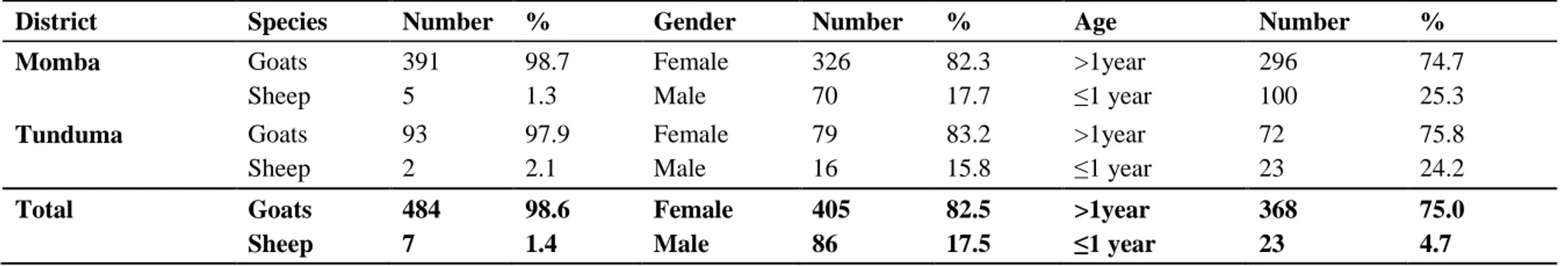
A total of 491 samples were taken in two different districts, Momba and Tunduma in Mbeya region. Samples were taken in 41 villages from 164 herds in total where four households were visited in each village. In Tunduma district, 32 herds in 8 villages were sampled in total. All villages in Tunduma were visited. In Momba district, 132 herds in 33 villages were sampled. One extra village was visited in Momba district because farmers there had experienced problems with several animals displaying clinical signs. See distribution of animals in table 1. Questionnaires were conducted to the farmer of every investigated herd which gave a total
14
Table 1. Distribution regarding species, gender and age of 491 goats and sheep sampled in Tunduma and Momba districts, Tanzania
District Species Number % Gender Number % Age Number %
Momba Goats Sheep 391 5 98.7 1.3 Female Male 326 70 82.3 17.7 >1year ≤1 year 296 100 74.7 25.3 Tunduma Goats Sheep 93 2 97.9 2.1 Female Male 79 16 83.2 15.8 >1year ≤1 year 72 23 75.8 24.2 Total Goats Sheep 484 7 98.6 1.4 Female Male 405 86 82.5 17.5 >1year ≤1 year 368 23 75.0 4.7
15
number of 164 interviews. Flock size varied between approximately 3 to 200 goats and the average herd size was 5-15 goats. Only seven farmers in total (in both districts) kept sheep and they had in average more than 15 sheep.
Animal sampling
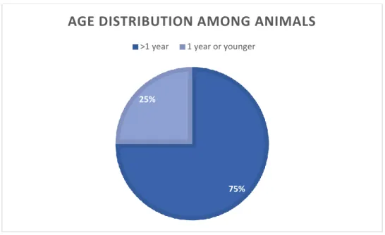
The animals were selected by random and all animals over 4 months could be sampled, to avoid maternal antibodies. The age distribution is presented in figure 2.
Figure 2. Age distribution among 491 goats and sheep sampled in Tunduma and Momba districts,
Tanzania.
75% 25%
AGE DISTRIBUTION AMONG ANIMALS
16
Antibody detection
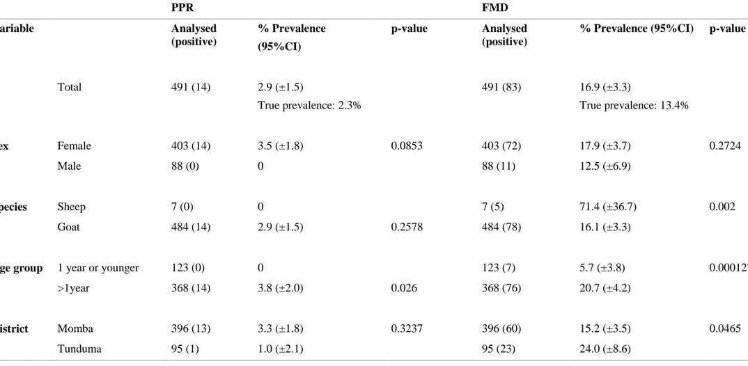
On herd level, 33.5% of investigated herds had at least one animal seropositive for FMD and 7.3% had at least one animal seropositive for PPR. The seroprevalence on individual level is displayed in table 2, with a total prevalence of 2.9% (true prevalence, 2.3%) for PPR, and 16.9% (true prevalence, 13.4%) for FMD. True prevalence is calculated by the known sensitivity and specificity of the ELISA kits.
Table 2. Seroprevalence on PPR and FMD at individual level according to sex, species, age and district among small ruminants in Tunduma and Momba
districts, Tanzania. P-value <0.05 is considered significant
PPR FMD Variable Analysed (positive) % Prevalence (95%CI) p-value Analysed (positive)
% Prevalence (95%CI) p-value
Total 491 (14) 2.9 (±1.5) True prevalence: 2.3% 491 (83) 16.9 (±3.3) True prevalence: 13.4% Sex Female 403 (14) 3.5 (±1.8) 0.0853 403 (72) 17.9 (±3.7) 0.2724 Male 88 (0) 0 88 (11) 12.5 (±6.9) Species Sheep 7 (0) 0 7 (5) 71.4 (±36.7) 0.002 Goat 484 (14) 2.9 (±1.5) 0.2578 484 (78) 16.1 (±3.3)
Age group 1 year or younger 123 (0) 0 123 (7) 5.7 (±3.8) 0.000127
>1year 368 (14) 3.8 (±2.0) 0.026 368 (76) 20.7 (±4.2)
District Momba 396 (13) 3.3 (±1.8) 0.3237 396 (60) 15.2 (±3.5) 0.0465
17
PPR
Analysis with cELISA for PPR had three different outcomes; positive, doubtful and negative. In total, 4 samples (0.8%) had the outcome of doubtful, but in the statistical analysis they have been considered as negative. The seroprevalence on individual level using cELISA for PPR on small ruminants (sheep and goats) was 2.9% (14/491) (table 2).
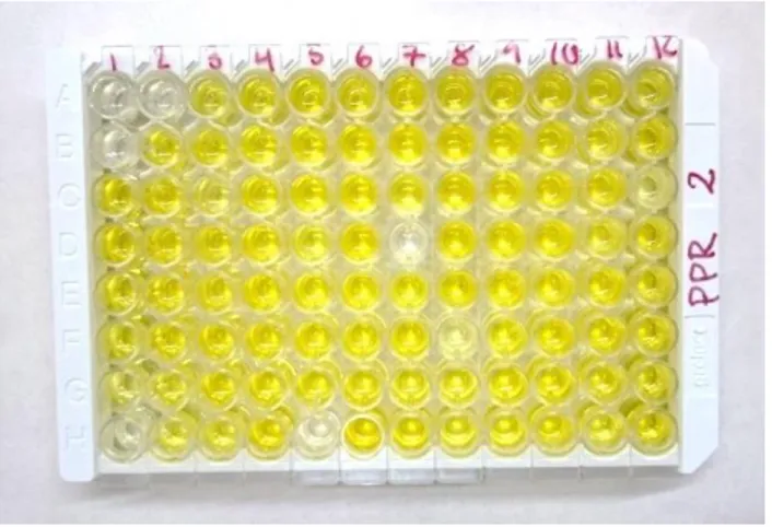
Figure 3. Ab-ELISA results on PPR. The positive controls are in wells A1 and B1. This plate displays 4
positive samples.
In tested villages, the household prevalence ranged from 0% to 25%. In Momba district, 30.3% (10/33) of the villages had at least one seropositive animal for PPR and in Tunduma district, 12.5% (1/8) had at last one seropositive animal. There was no significant difference between districts. Animals over 1 year of age had a significant larger risk of being seropositive for PPR than animals 1 year or younger. There was no significant difference in risk of being seropositive between the gender.
FMD
The ELISA kit used to analyze FMD had two outcomes; positive or negative. The seroprevalence on individual level when using cELISA for FMD was 16.9 % (Table 2). In tested villages, seroprevalence ranged from 0% to 58.3%. It was a significant difference in seroprevalence between the two districts, where it was higher risk of being seropositive for FMD in Tunduma district (24.0%) in comparison with Momba district (15.2%) on individual level. Within Tunduma district, no significant difference could be seen between villages located close (seroprevalence 22%, three villages) or further away (seroprevalence 25%, five villages) from Tanzam highway. Animals over 1 year of age had a significantly higher risk of being seropositive for FMD compared to animals 1 year or younger, and sheep had a significant higher risk of being seropositive compared to goats.
Questionnaire
All farmers (100%, 164/164) reported that they were using communal grazing as grazing system for their goats and sheep, where the animals could be in daily contact with other goats and sheep. The majority of these (97.6%, 160/164) also reported daily contact with cattle. In addition to communal grazing a small percentage of the famers also utilized herding (4%, 6/164) and tethering (1%, 2/164) but only during rainy season. Only two farmers reported that
18
their animals could be in contact with wild ruminants. Among the two farms who reported daily contact between their animals and wild ruminants, both had seropositive animals for FMD and one for PPR.
Regarding vaccination, 95.7% (157/164) reported that they did not vaccinate their sheep and goats. Of the seven farmers reporting vaccination, all were vaccinating against contagious caprine pleuropneumonia (CCPP).
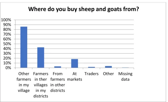
It was a significant difference in seroprevalence for FMD between farmers who bought animals outside of their district, (at markets and from farmers in other districts), compared to farmers who only bought animals from within their own district (table 3). Fig. 4. displays where farmers bought their sheep and/or goats from. Regarding how often they bought animals, 78.7% said it was more than one year ago. All farmers (100%) reported that they had never bought animals from other countries.
Figure 4. Where smallholder farmers in 164 investigated goat and sheep herds in Tunduma and
Momba district, Tanzania, bought their sheep and goats from.
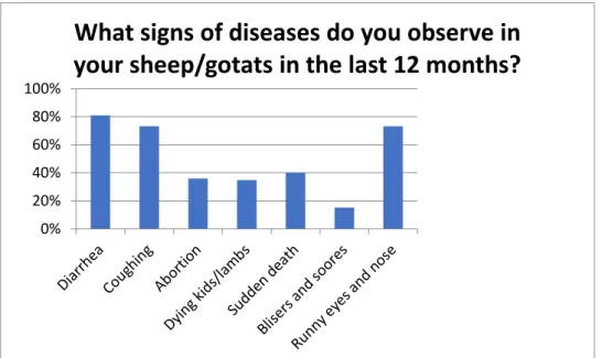
Among the farmers interviewed, 91% reported clinical signs in their animals within the last year (Table. 3). Fig. 5 displays the most commonly observed signs. All farmers (100%, 164/164) reported that they did not keep their sick animals separated from the rest of the herd.
0% 10% 20% 30% 40% 50% 60% 70% 80% 90% 100% Other farmers in my village Farmers in ther villages in my districts From farmers in other districts At markets
Traders Other Missing data
19
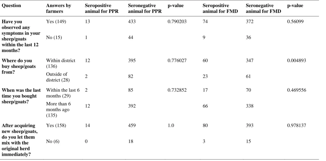
Table 3. Risk factor analysis based on answerers in a questionnaire compared to seropositive animals for PPR and FMD. P-value<0.05 is considered
significant Question Answers by farmers Seropositive animal for PPR Seronegative animal for PPR p-value Seropositive animal for FMD Seronegative animal for FMD p-value Have you observed any symptoms in your sheep/goats within the last 12 months? Yes (149) No (15) 13 1 433 44 0.790203 74 9 372 36 0.56099 Where do you buy sheep/goats from? Within district (136) Outside of district (28) 12 2 395 82 0.776027 60 23 347 61 0.004893
When was the last time you bought sheep/goats?
Within the last 6 months (29) More than 6 months ago (135) 2 12 85 392 0.732852 17 66 70 338 0.469556 After acquiring new sheep/goats, do you let them mix with the original herd immediately? Yes (158) No (6) 14 0 459 18 1.0 80 3 393 15 0.978137
20
Figure 5. Diseases observed the last 12 months among 164 investigated goat and sheep herds in
Tunduma and Momba districts, Tanzania.
Figure 6. Age distribution among 164 interviewed smallholder farmers keeping sheep and goats in
Tunduma and Momba districts, Tanzania.
The majority of the household heads were male over 51 years. Female represented 26.3% of the farmers interviewed, see figure 6.
Regarding education, 23.7% of all farmers were uneducated, had never gone to school. Educated farmers had attended school for at least 4 years. Of farmers having seropositive animals for FMD, 30.9% were uneducated and 69.1% were educated. Of those with at least one seropositive animal with PPR, 33.3% were uneducated and 66.7% educated. There was no significant difference in seroprevalence between uneducated and educated farmers.
0% 20% 40% 60% 80% 100%
What signs of diseases do you observe in
your sheep/gotats in the last 12 months?
17%
20% 63%
AGE DISTRIBUTION AMONG FARMERS
21
DISCUSSION
The aim of this study was to investigate the seroprevalence of the infectious diseases PPR and FMD in two districts in southern Tanzania bordering Zambia. Competitive ELISA kits were used to detect antibodies to specific proteins of PPRV and FMDV in serum samples from sheep and goats. In addition, questionnaires were administered to all farmers of investigated herds to get more information about management and to draw conclusions about risk factors of the diseases connected to holding of animals. In every herd 3, animals were sampled, selected by random with one criteria, that they were older than 4 months, to avoid maternal antibodies. In practice, this was hard to carry out because most of animals were scattered out in the fields and the ones sampled were the ones the farmer managed to capture.
PPR
In this study, an overall seroprevalence of 2.9% for PPR was measured in sheep and goats in the districts Tunduma and Momba. This is lower compared to previous reports in Tanzania, but strengthens previous studies in confirming that PPR is present in southern Tanzania (Torsson
et al., 2017). In earlier prevalence studies on PPR, seroprevalence in northern Tanzania has
been measured to 45.8% (Swai et al., 2009) and 22.1% (Kiviara et al., 2013). In southern Tanzania, in Tandahimba and Newala districts bordering to Mozambique, a seroprevalence of 31% (Muse et al., 2012b) were measured in 2011. A recent study conducted in 14 regions in Tanzania had an overall seroprevalence on 27.1% but it varied between 2.1% to 72.8% in the different regions (Kgotlele et al., 2016). In recent years, there have been vaccination campaigns in central and northern parts of Tanzania which might be an explanation for the differences in seroprevalence between studies.
No significant difference in seropositivity could be seen between the different species, but goats tended to have a higher prevalence (2.9%) compared to sheep (0%). Although, only seven sheep were sampled, which are a too few to draw any conclusions from. Earlier prevalence studies in Tanzania have suggested a significantly higher prevalence of PPR in goats in comparison with sheep (Swai et al., 2009). These findings differ between studies and several studies have found no difference in seroprevalence between sheep and goats (Muse et al., 2012b; Kgotlele et al., 2016).
It was a significant difference between the different age groups where the seroprevalence was highest in adult animals, over one year of age (seroprevalence 3.8%) compared to animals 1 year or younger (seroprevalence 0%). The age of animals over one year of age ranged from just over one year to ten years. Age was an expected risk factor because of the lifelong immunity after surviving an infection. No animal under one year of age where seropositive for PPR. This indicates that the animals have not been exposed to PPRV within the last year and that the virus might not have been present in this area during this time period. It was the farmers themselves who reported the age, no papers or documentation were showed for the animals, therefore it is a possibility that the answers not always were completely correct. Earlier studies have also observed a higher prevalence of PPR in adult animals (Muse et al., 2012b; Torsson et al., 2017). In this study, there was a tendency that female animals were at higher risk of being seropositive for PPR (female animal seroprevalence 3.5%, male animal seroprevalence 0%), but there was
22
no significant difference between the sexes. Sex has previously been identified as a risk factor for PPR where females usually tend to be at higher risk (Aziz-ul-Rahmanet al., 2016; Torsson et al., 2017). Other studies though, found no significant difference between sexes (Swai et al.,
2009; Muse et al., 2012b). It has also been discussed that females might be kept longer by their owner and therefore have a longer period when they could be exposed to the virus (Aziz-ul-Rahman et al., 2016).
FAO has constituted a framework containing 9 components which serve to provide a common vision for PPR prevention and control (FAO, 2013). One component is regarding public awareness and communication. Findings in this study indicate that there is a continued poor knowledge among farmers in how to prevent spread of diseases. On the question regarding use of quarantine, 95.7% reported that they let new animals mix with the original herd immediately and the majority thought it was okay to sell goats with signs of disease. No farmer reported a separation of the sick animals from the rest of the herd. All farmers in this study utilized communal grazing, which earlier has been identified as a risk factors contributing to spread of PPR (Muse et al., 2012b).
Previous studies have identified the introduction of PPR in a herd due to introducing animals purchased from live animal markets (Muse et al., 2012b). In this study farmers in 54.5% of the villages had at some point bought sheep or goats from markets, but there was no significant difference in seroprevalence whether the farmer had bought animals from within or outside of the home district.
FMD
FMD is a wide-spread disease and endemic in large parts of the world, including Tanzania. In this study, the overall seroprevalence of FMD was estimated to 16.9%. Most previous seroprevalence studies on FMD in Tanzania have been performed in cattle, where the disease has been found widely distributed in the country with a total prevalence of 46.1% (Kasanga, et
al., 2012). A recent study on sheep and goats in eastern and northern Tanzania estimated a
seroprevalence to 39.0% in 2014 and 14.2 % in 2015 (Torsson et al., 2017). Another prevalence study in the northern and central part of Tanzania, targeting different species, found the highest seroprevalence in bovine followed by sheep (52.4%) and goats (11.1%) (Mdetele et al., 2014). The present study strengthens previous studies (Allepuz et al., 2015) that FMD is present in the southern parts of Tanzania.
Tunduma district had a significantly higher prevalence of FMD (24.0%) compared to Momba district (15.2%). This is in agreement with an earlier study where FMD occurrence has been associated with closeness to main roads and international borders (Allepuz et al., 2015). Two main routes are passing through Tunduma district, the Tazara railway and Tanzam highway and Tunduma town is a bordering city to Zambia. Momba district is located further from the main transportation routes. No significant difference, though, could be seen between villages close or further away from Tanzam highway within Tunduma district. Other factors that could also have contributed to the difference in seroprevalence are different density of people, market places and movement of people and animals. Also, the location and density of water points, which the animals may or may not share, could contribute to spread of the disease.
23
Regarding species, sheep had a significantly higher risk of being seropositive for FMD (71.4%) compared to goats (16.1%). In this study it was hard to find farmers who had any sheep, so when sheep were found at least two of three animals sampled were sheep. In total seven sheep were sampled. This is too few to draw any reliable conclusions from. There are studies supporting a higher prevalence in sheep (Mdetele et al., 2014) but also studies showing the opposite results, where goats have been found at higher risk to be seropositive for FMD compared to sheep (Torsson et al., 2017).
Similar to the findings for PPR seropositivity, animals over one year of age had a significant higher risk of being seropositive compared to animals one year or younger. Of all seropositive animals, 91.6% were older than one year. These findings are in agreement with earlier studies where animals older than two years have showed increased seropositivity (Torsson et al., 2017). To be in consideration is that information regarding age in the present study was contained directly from the owner and might not always have been correct. In this study, there were no significant difference between sexes, although females had a tendency to have a higher prevalence (17.9%) compared to male (12.5%). Previous studies have found females to be significantly at more risk for FMD in comparison with males (Torsson et al., 2017).
Proximity to wildlife has been suggested to be a risk factor for spread of FMD (Mdetele et al., 2014). In this study, 97% reported that their animals never had any contacts with wild ruminants. Among the two farms that reported daily contact between their animals and wild ruminants, both had seropositive animals for FMD and one farmer had seropositive animals for PPR. Torsson et al., 2017 found no evidence that suggested proximity to wildlife as a risk factor for either PPR or FMD. Allepuz et al., 2015 suggested that risk factors for spread of FMD are more likely related to animal movement and human activity via communication networks rather than contact with wildlife or transboundary movements (Allepuz et al., 2015). Another study showed that serotypes A and O were mainly driven by human activities and trades while the origin of serotype SAT2 can be found in wildlife (Duchatel et al., 2018). The most likely risk factor for spread of FMD in this study-area is probably human activity and animal movement due to so few reported contacts between domestic animals and wild ruminants. Moreover, animals that lived close to the main routes, where trade is more likely to occur, had a higher seroprevalence.
The majority of farmers interviewed accepted to sell animals with clinical signs, such as runny eyes and nose, coughing, diarrhea, blisters and sores. Most farmers bought animals from other farmers in their own village, but 42.7% reported that they bought from other villages, 18.3% from markets and 1.8% from traders. There was a significantly higher risk to have seropositive animals for FMD if the farmer had bought animals from outside the district compared to a farmer who only bought from within his or her home district. The information on where the farmers bought animals were collected directly from the farmers and might not always have been entirely true.
To be in consideration in this study is that the findings might have been affected by sampling and analyzing techniques and season of sampling. The interviews were performed in Swahili and translated into English, and all information regarding management was acquired from owners. The answers could thus have been incorrect, for example if they did not remember or if the one
24
interviewed was not the same person who took care of the animals, and thus did not see clinical signs of disease or know how the animals were kept. Often, many people were gathering around when conducting the questionnaire which also could have influenced the answerers of that specific farmer.
In Tanzania, as well as other East African countries, small ruminants play an important role in food security and livelihood resilience for a large number of the population (Covarrubias et al., 2012). The importance of small ruminants in many low-income countries is one of the reasons why PPR has been notified in the World Organization for Animal Health’s (OIE) and Food and Agriculture Organization (FAO)’s joint program to eradicate PPR by 2030 (OIE & FAO, 2015). FMD is also a disease of devastating effects with severe socio-economic impact and is also listed by OIE as a disease of specific hazards (OIE website, 2018).
The PPR eradication strategy of FAO and OIE also recognizes that good quality veterinary services are indispensable for the successful prevention and control of PPR and other major transboundary diseases (OIE & FAO, 2015). Thus, strengthening the veterinary services and create more cost-effective opportunities to control other transboundary diseases are components to reach the final goal. It is difficult to control livestock movements in Tanzania and smuggling across borders have been reported to occur (Kivaria, 2003). Delivery of vaccines are not always effective and do not reach all small ruminant holders (OIE & FAO, 2015). FAO means that one of the first steps toward a global strategic approach is to assess the epidemiological situation to later be able to target at-risk populations. This study suggests that both PPR and FMD are present in the two districts Momba and Tunduma in Southern Tanzania. The occurrence of PPR in Tunduma and Momba, which are districts bordering to Zambia, upholds the risk of further spread southwards, even though there seems to have been no or only limited circulation of PPRV during the last year.
CONCLUSIONS
This study confirms the presence of antibodies for PPR and FMD among sheep and goats in the two districts Momba and Tunduma in Mbeya region in southern Tanzania. Presence of PPR in these districts bordering to Zambia, strengthens the concern of further spread of PPR into Zambia. Risk factor analysis on individual level identified age as a significant risk factor for being seropositive for both PPR and FMD, where animals over one year was at higher risk compared to younger animals. Sheep had a tendency of being at higher risk to be seropositive for FMD compared to goats. Closeness to main roads was also identified as a possible risk factor for occurrence of FMD, where Tunduma district, containing Tanzam highway and Tazara railway had a higher prevalence compared to Momba district, situated further from the main routes. The findings after analyzing the questionnaires revealed a continued poor knowledge about animal management routines and biosecurity measures among farmers.
25
ACKNOWLEDGEMENTS
I would like to thank everyone who has made this minor field trip possible. I especially want to thank my primary supervisor Dr Jonas Johansson Wensman for his help and support regarding planning and guidance throughout this master. Also, I am grateful to Sara Lysholm for good support and help with everything. I also want to thank my local supervisor professor Gerald Misinzo for all the help and organizing. I am also thankful for the help I got from Miriam Richards and Veronica (Mhoja Ndalawa) regarding everything from good support to planning, organizing and lab work. Thanks also to Professor Paul Gwakisa for the help in lab and use of your lab and equipment. Many thanks also to Edson Kinimi and our driver Swai for all help during the fieldtrip. For the funding, I want to thank Swedish International Development Cooperation Agency (SIDA), Swedish Research Council (Grant no. 348-2014-4293 and 2016-05667), the Swedish University of Agricultural Sciences and Michael Forsgrens stiftelse. Of course, I also want to thank my friend and fellow-MFS student Emelie Olovsson for sharing this great experience with me.