Research
Non-Ionising Radiation in
Swedish Health Care
2017:37
Authors: Jonna WilénKjell Hansson Mild Ronnie Lundström
SSM perspektiv
Bakgrund
Inom sjukvården används alltmera teknik som exponerar patienter för icke-joniserande strålning vid diagnostik och terapi. Ofta används sådan teknik som ett alternativ till och ibland i kombination med metoder som exponerar för joniserande strålning.
Huvudsyftet med studien var att identifiera och beskriva hur icke-jonis-erande strålning används inom hälso- och sjukvård, samt att undersöka förväntade exponeringsnivåer, identifiera eventuella hälsorisker och identifiera kunskapsluckor inom området. Icke-joniserande stråln-ing refererar här till icke-joniserande elektromagnetisk strålnstråln-ing med frekvenser från 0 Hz upp till 3,0 PHz, inkluderande elektromagnetiska fält, optisk strålning och mekaniska vågor såsom ultraljud.
SSM har saknat en samlad kunskap kring de metoder och exponerings-nivåer som använder icke-joniserande strålning inom sjukvården. Bland annat EU:s vetenskapliga råd SCENIHR (har numera bytt namn till SCHEER) har pekat på behovet av forskning om potentiella hälsoeffek-ter inom detta område. Studien var alltså viktig för att få ett underlag för att bedöma behovet av ytterligare säkerhetsåtgärder inom området, identifiera kunskapsluckor och också för att få ett bättre underlag för att besvara frågor angående säkerhet och eventuella risker.
Resultat
Inga hälsorisker eller allvarliga säkerhetsbrister har identifierats. Där-emot är de exponeringsnivåer som används många gånger på en nivå där det finns en tydlig biologisk påverkan och ibland kan även akuta symtom upplevas, detta är dock ofta helt avsiktligt för att få önskad effekt och av övergående natur.
Författarna har identifierat ett behov av att förbättra hälsovårdsper-sonalens kunskap om risker och säkerhet vid användning av icke-jonis-erande strålning samt pekar på vikten av tydlighet när det gäller vilken organisation/myndighet som ska tillhandahålla sådan information. Rapporten indikerar också att ett robust forskningsunderlag för när-varande saknas för att bedöma möjliga långsiktiga hälsorisker med användning av exempelvis MRI, TMS och ultraljud. Den kunskap och forskning som finns tillgänglig tyder dock inte på några betydande hälso risker med nu aktuella exponeringsnivåer.
Relevans
Rapporten stöder Strålsäkerhetsmyndighetens tidigare uppfattning att de tillämpningar med icke-joniserande strålning som används i sjukvården inte utgör några kända hälsorisker. När metoder som bygger på icke-joniserande strålning ersätter metoder som bygger på jonis-erande strålning ökar patientsäkerheten, under förutsättning att syftet med undersökningen eller behandlingen uppnås.
Behov av vidare forskning
Trots att det idag inte påvisats några hälsorisker med icke-joniserande strålning inom sjukvården (UV undantaget) anser myndigheten att ytterligare forskning är angelägen kring långsiktiga effekter på patienter vid i första hand de metoder som ger en exponering runt de nivåer där tydlig tillfällig biologisk påverkan eller akuta övergående symtom kan uppträda, detta gäller exempelvis MRI och TMS.
Mera kunskap är också önskvärd avseende långsiktiga effekter av ultraljudsexponering av foster.
Ytterligare forskning behövs också för att undersöka möjliga negativa effekter av ultraljud i kombination med kontrastmedia.
I takt med att det blir vanligare med hybridmetoder där olika strålslag kombineras blir det angeläget att följa forskning kring potentiella hälso-risker med detta.
Projektinformation
Kontaktperson SSM: Torsten Augustsson Referens: SSM 2016-498 / 7030054-00
2017:37
Authors: Jonna Wilén, Kjell Hansson Mild and Ronnie Lundström
Department of Radiation Sciences, Umeå University, Umeå, Sweden
Non-Ionising Radiation in
Swedish Health Care
This report concerns a study which has been conducted for the Swedish Radiation Safety Authority, SSM. The conclusions and view-points presented in the report are those of the author/authors and do not necessarily coincide with those of the SSM.
Non-Ionising Radiation in
Swedish Health Care
Jonna Wilén, Kjell Hansson Mild and
Ronnie Lundström
Department of Radiation Sciences,
Umeå University, Umeå, Sweden
Content
1. Sammanfattning ...4
2. Executive summary...6
3. Background...8
3.1. Aim of the study...8
4. Methods...9
5. Non-ionising radiation in health care...11
6. Electromagnetic fields in health care...13
6.1. Magnetic resonance imaging ...14
6.1.1. Static magnetic fields... 15
6.1.2. "Switched" gradient field... 16
6.1.3. Radiofrequency magnetic fields ... 18
6.1.4. Exposure near MRI scanners... 19
6.1.5. Limits and regulations... 20
6.1.6. Risks and effects of MRI exposure... 21
6.1.7. Organizational aspects of safety for MRI system... 24
6.1.8. Conclusion ... 25
6.2. Electro surgery...26
6.2.1. Indirect effects of surgical diathermy...28
6.2.2. Education... 29
6.2.3. Conclusion ... 29
6.3. Short-wave and microwave diathermy ...29
6.4. Transcranial Magnetic stimulation...30
6.4.1. Limit values and safety... 32
6.4.2. Use of TMS in healthcare... 32
6.5. Magnetic fields in incubators...33
6.6. Magnetic fields from induction loop pad...33
6.7. Active implantable medical devices ...34
6.7.1. Cochlear implants... 34
6.8. Other applications...34
6.9. General comments on the use of EMF in health care...35
7. Optical radiation ...37
7.1. Light treatments in health care...37
7.1.1. Neonatal phototherapy ... 37
7.1.3. Dental curing lamp... 38
7.1.4. Disinfection techniques using blue light. ... 39
7.2. Laser in health care...40
7.2.1. Basic types of lasers in medicine ... 40
7.2.2. Laser therapy... 41
7.2.3. Laser surgery... 42
7.2.4. Laser diagnostics... 42
7.2.5. Biological effects of laser radiation... 44
7.2.6. Laser applications in Swedish health care... 45
7.2.7. Safety regulations and guidelines ... 46
7.3. Ultraviolet light in medical health care...48
7.3.1. Medical UV phototherapy treatment... 49
7.3.2. UV disinfection... 50
7.4. Summary optical radiation...51
8. Ultrasound...53
8.1. Basic principles of ultrasound...53
8.1.1. Biological interaction... 55
8.2. Diagnostic ultrasound...56
8.3. Therapeutic ultrasound...59
8.4. Ultrasound in Swedish health care...61
8.5. Biological effects of ultrasound...63
8.6. Risk and safety guidelines...64
8.7. Research and development needs...68
8.8. Summary ...69
9. Discussion...70
10. General conclusion ...72
11. Recommendation for further work ...72
12. Acknowledgements...73
13. Abbreviations...74
1. Sammanfattning
Studien har gjorts på uppdrag av Strålsäkerhetsmyndigheten med huvudsyftet att identifiera och beskriva hur icke-joniserande strålning (NIR) används inom hälso- och sjukvård, men också att undersöka förväntade exponeringsnivåer, identifiera eventuella hälsorisker och identifiera kunskapsluckor inom området. NIR refererar här till icke-joniserande elektromagnetisk strålning med frekvenser från 0 Hz upp till 3,0 PHz, inkluderande elektromagnetiska fält, optisk strålning och mekaniska vågor såsom ultraljud.
I studien har vi kartlagt medicinska tillämpningar där NIR används inom Västerbottens läns landsting (VLL; två länssjukhus och över 30 vårdcentraler), Kalmar läns landsting (KLL och 3 länssjukhus) och Karolinska Universitetssjukhuset i Stockholm (KUH). Vi har använt publicerad litteratur och rapporter från myndigheter som International Commission on Non-Ionising Radiation Protection (ICNIRP), the Food and Drug Administration (FDA), Health Canada, och i vissa fall egna mätningar för att undersöka exponeringsnivåerna för patienter och personal och jämfört dem med dessa riktlinjer och vår nuvarande kunskap om möjliga hälsoeffekter. Även behandlingsrekommendationer från Läkemedelsverket och nationella riktlinjer från Socialstyrelsen har ingått i vår kartläggning. Vidare har vi konsulterat medicintekniska ingenjörer främst inom VLL för att få hjälp med att kategorisera medicinsk utrustning, och annan sjukvårdspersonal för eventuella riktlinjer för användning av utrustningen. Rapporten är indelad i tre sektioner; elektromagnetiska fält, optisk strålning och ultraljud.
Exponering för elektromagnetiska fält sker i vården främst vid diagnostik och terapi. Tre tillämpningar har identifierats som kan medföra hög exponering för patienter och personal, vilket innebär att akuta hälsoeffekter inte kan uteslutas: magnetisk
resonanstomografi (MRT), transkraniell magnetisk stimulering (TMS) och diatermi, där potentiella akuta effekter som t ex sensoriska effekter och värmeeffekter kan förekomma, ibland dock avsiktligt för diagnos/behandling.
När man använder optisk strålning såsom klass 3 och 4 lasrar för terapi eller kirurgiska ingrepp och UV behandlingar måste akuta effekter som oavsiktliga brännskador och fotoreaktion från laserexponering och erytem och påverkan på ögonen under UV behandlingen undvikas.
Ultraljud är i allmänhet att betrakta som ett säkert alternativ vid såväl diagnostiska som terapeutiska tillämpningar men risk för termiska och mekaniska effekter måste ändå alltid övervägas.
Kartläggningen av medicinsk utrustning hos de ingående sjukhusen/landstingen visade att i stort används samma typ av medicinteknisk utrustning används på de olika sjukhusen men med vissa undantag. Som ett exempel var antalet diagnostiska ultraljudsutrustningar som används på KLL, KUH och VLL, 98, 515 och 206, respektive. Motsvarande siffror för MRT var 2, 22 och 6. Enligt vår inventering var antalet diatermienheter 54, 259 och 202.
Vi har också försökt att uppskatta antalet MRI undersökningar i Sverige baserat på siffror från VLL. Vi har extrapolerat fram att ungefär 400 000 till 450 000 undersökningar utfördes under 2016. För andra applikationer var det inte möjligt att göra samma typ av uppskattning på grund av spridd användning av utrustning på många kliniker och avsaknaden av strukturerad rapportering.
Vår slutsats utifrån litteraturstudien och uttalanden från organisationer och myndigheter är att det finns en brist på kunskap om långsiktiga effekter av MRT och ultraljud. Eftersom det har blivit vanligare att använda hybridtekniker där kombinationen av
exempelvis joniserande strålning och magnetiska fält tillämpas (t ex PET-MR) behövs mer kunskap om möjliga hälsoeffekter vid kombination av olika exponeringar. Det finns också andra kombinerade tekniker såsom ultraljudsaspiration där tillämpningar finns som kombinerar skärande och koagulerande effekter från ultraljud och diatermi. Till exempel diskuterar både ICNIRP och SCENIHR behovet av mer kunskap om diagnostiska tekniker såsom MRI och ultraljud i kombination med kontrastmedel. Vi har inte täckt kombinationer explicit i rapporten, men hybridtekniker finns på vissa svenska sjukhus, t ex PET-MR och ultraljudsaspiration.
Vår slutsats är att regleringen av yrkesmässig exponering för NIR täcks väl av gällande arbetsmiljöföreskrifter och dessa gäller också för medicinsk användning. Tillämpningen och kunskapen om detta kan emellertid vara bristfällig inom sjukvården.
Det finns gott om europeiska standarder för medicintekniska produkter som bl a ställer krav på att visuellt rapportera patienternas exponeringsnivåer för till exempel MRI och ultraljud. Denna information används ofta vid MR undersökningar, särskilt när patienter med medicinska implantat eller andra situationer som kräver lägre exponering ska behandlas.
Vi har inte kunnat kartlägga hälsovårdspersonalens kunskap inom risker och säkerhet av NIR, men vi konstaterar baserat på litteratur och efter samråd med personalen, att detta är ett område som bör förbättras. ICNIRP anger i en sammanfattning från en workshop att utbildning av personal som använder NIR är nödvändig speciellt för de som använder klass 3 eller 4 laser. Deras slutsats är att särskilda riktlinjer för patienter kunde vara användbara, men att det inte är uppenbart att det krävs särskilda riktlinjer för personalen. De konkluderar också att information till användarna krävs, men att detta är komplicerat då användningen är utbredd på många olika kliniker.
Vi har till viss del funnit information om säkerhetsaspekter i samband med NIR från myndigheter som Strålsäkerhetsmyndigheten, Socialstyrelsen, och Läkemedelsverket men detaljerad information om specifika medicinska ingrepp saknas.
Slutligen drar vi slutsatsen att för att förbättra kunskaperna om säkerhetsaspekter för NIR i hälso- och sjukvårdssektorn, finns behov av tydlig, evidensbaserad information från tillförlitliga källor och det borde vara uppenbart för användaren vilken
2. Executive summary
The study was done as an assignment from the Swedish Radiation Safety Authority. The main aims of the study were to identify and describe methods that use non-ionising radiation (NIR) in health care, to examine anticipated exposure levels for each application and frequency range, to identify possible health hazards, and to identify knowledge gaps in the field. In this report NIR refers to electromagnetic radiation with frequencies from 0 Hz up to 3PHz. This range includes ultraviolet light, visible light, infrared light, radio waves, and mechanical waves such as ultrasound.
In this study we have mapped applications using NIR within Västerbotten County Council (VCC: two county hospitals and over 30 primary health care centres), Kalmar County Council (KCC: 3 county hospitals), and Karolinska University Hospital in Stockholm (KUH).
We have used published literature and reports from authorities such as the International Commission on Non-Ionising Radiation Protection (ICNIRP), the Food and Drug Administration (FDA), and Health Canada. In some cases, we have used our own
measurements to investigate the exposure levels for patients and staff, and have compared those with the present guidelines and our present knowledge of possible health effects. We have also used treatment recommendations from the Medical Product Agency and national guidelines from The National Board of Health and Welfare. We have consulted medical engineers, mainly at VCC, for help in categorization of medical equipment and other medical professionals for guidance on the use of the equipment. This report is divided into three sections: electromagnetic fields, optical radiation and ultrasound. Exposure to electromagnetic fields is mainly used in health care for diagnosis and therapy. Three applications were identified as “high exposure to patients and staff,” i.e., acute effects cannot be ruled out. These areas are: magnetic resonance imaging (MRI), transcranial magnetic stimulation (TMS) and electro surgery, where potential acute effects such as sensorial effects or heating effects might occur - sometimes intentionally for diagnosis.
When using optical radiation, such as class 3 and 4 lasers for therapy or surgical procedures, and when using UV treatments, acute effects such as unintentional burns, photo reactions from laser exposure, erythema and effects on the eyes during UV treatment need to be avoided.
Ultrasound is generally regarded as a safe alternative for diagnostic as well as therapeutic applications but the risk for thermal and mechanical effects still need to be taken into consideration.
Our survey of the medical equipment at the included hospitals showed that essentially the same type of medical equipment, with some exceptions, is used throughout Sweden. As an example, the numbers of diagnostic ultrasound apparatuses reported in use at KCC, KUH and VCC are 98, 515 and 206, respectively. The corresponding numbers for MRI are 2, 22 and 6. Our inventory showed that the number of electro surgical units (ESU), which seems to be one of the most common applications in health care using NIR, are 54, 259 and 202, respectively.
We have also tried to estimate the number of MRI procedures performed in Sweden based on figures from VCC. We extrapolated that approximately 400 000 to 450 000 procedures were performed in Sweden in 2016. For other applications, it was not possible to do the same type of estimation due to the widespread use of equipment at many clinics and lack of structured reporting.
We conclude, based on the literature review and statements from organisations and authorities, that there is a lack of knowledge regarding long-term effects of MRI and ultrasound. Since it has become more common to use hybrid techniques, i.e., where a combination of ionising radiation and magnetic fields (PET-MR) is used for diagnostics, more knowledge on the combination of different exposure levels is needed. There are also combined techniques, such as ultrasound aspiration where applications exists that
combine the cutting/coagulation effect of ultrasound and ESU. At an workshop arranged by ICNIRP workshop and also SCENIHR discussed the need for more knowledge about diagnostic techniques such as MRI and ultrasound in combination with contrast media. We have not covered combinations explicitly in this report, but some hybrid techniques do exist at some Swedish hospitals, e.g., PET-MR and ultrasound-aspiration techniques. We conclude that the regulation of occupational exposure to NIR is well covered. However, the appliance and knowledge about this might be sparse.
There are many European standards for medical devices that require the devices to visually report the exposure levels to the patients. e.g., MRI and ultrasound. This information is commonly used for MRI procedures, especially when patients with medical implants or other issues demand lower exposure.
We have not been able to survey the health care professionals’ knowledge about the risks and safety measures in NIR, but we conclude, based on literature and after consulting staff, that this is an area that should be improved. ICNIRP also states in a workshop summary of non-ionising radiation in medicine that training of staff using NIR is necessary and that training for class 3 or 4 laser is essential. They conclude that specific guidelines for patient exposure would be useful, but that it is not obvious that special guidelines for staff are needed. They also conclude that information to users is required, but due to the widespread use of NIR, this is a complex subject.
We have found information, to some extent, about safety issues in relation to NIR from the authorities such as the Swedish Radiation Safety Authority, The National Board of Health and Welfare, the Swedish Work Environment Authority and the Medical Products Agency. Unfortunately, detailed information on specific medical procedures is missing. Finally, we conclude that to improve the knowledge in safety aspects of NIR in the health care sector, there is a need for clear, evidence-based information from reliable sources, and it should be obvious to the user which source to address.
3. Background
Non-ionising radiation (NIR), e.g., ultrasound imaging, laser surgery, and UV-light treatments, have been used for a long time in health care. Some more recently introduced applications include Magnetic Resonance Imaging and Transcranial Magnetic
Stimulation which is used to treat depression.
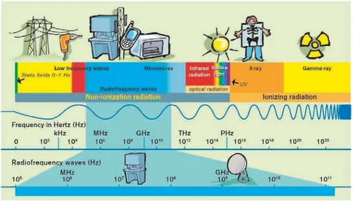
This document covers the use and safety of non-ionising radiation in health care. NIR refers to electromagnetic radiation with frequencies from 0 Hz up to 3,0 PHz, including ultraviolet light, visible light, infrared light, electric and magnetic fields, radio frequency fields, and mechanical waves such as ultrasound. ICNIRP [1] has recently published a statement on diagnostic devices using NIR, and they used the same division of NIR. In this report, we also include the use of NIR for therapeutic use.
Within the electromagnetic spectrum, NIR is situated below the ionising radiation band that includes X-rays (Figure 1). NIR has less energy than ionising radiation and cannot remove electrons from atoms, i.e., NIR cannot ionise (except for part of the UV band). NIR is sub-grouped into different frequency or wavelength bands. The different subgroups have different effects on the body and require different protection measures.
Figure 1. The electromagnetic spectrum. Illustration Gunilla Guldbrand
3.1. Aim of the study
This study was done as an assignment from the Swedish Radiation Safety Authority and the main aims of the study were:
• to identify and describe methods that use non-ionising radiation in health care. • to examine anticipated exposure levels for each application and frequency range. • to identify possible health hazards, and
• to identify knowledge gaps in the field.
In addition, the report discusses the need for continued activities in the area, such as: • To identify and stimulate research that aims to investigate possible health risks
with the use of NIR.
4. Methods
Initially we investigated applications using NIR within Västerbotten County Council (VCC, which has two county hospitals and over 30 primary health care centres) by the use of the VCC inventory database (Maximo). Search terms (translated into English) were magnet stimulation, MRI, surgical diathermia, laser, ultrasound, light treatment, and UV. We also recruited a reference group of biomedical engineers at VCC, who have the responsibility for customer support, product service and maintenance with respect to all equipment listed in the inventory and, thus, could help us identify applications using NIR. We have used published literature, both peer-reviewed scientific literature and reports mainly from authorities such as the International Commission on Non-Ionising Radiation Protection (ICNIRP), the Food and Drug Administration (FDA), Health Canada, and product datasheets from the manufactures. Our aim was to investigate the exposure levels and to compare those with the present guidelines and our present knowledge of possible health effects. When available, we have also used treatment recommendations from the Medical Product Agency and national guidelines from The National Board of Health and Welfare. In some cases, we have added data from our own measurements of the exposure (UV, MRI and surgical diathermia). From the literature search and from personal
communications with users and manufacturers we have tried to get a perspective over new or coming techniques using NIR. In this case we focused on applications, which are close to introduction or which have recently been introduced into the Swedish health care sector.
This project has primarily focused on the equipment routinely used in health care. Equipment that uses power in direct contact with the patient has been excluded, except for those that produce considerable electric or magnetic fields such as, e.g., surgical diathermy. This project has focused on the patient’s exposure, but when required, it has also examined exposure to the staff.
The work has been divided into these subareas:
Electromagnetic field (0-300 GHz) in health care including applications using static-magnetic field, low-frequency magnetic and electric fields, radiofrequency fields, e.g., for transcranial magnetic stimulation (TMS), MRI and electrosurgical devices.
Optical radiation including:
o Visible light (400-780 nm) such as laser and light treatments o Ultraviolet light (100- 400 nm) such as UV treatment and UV
disinfection units
Ultrasound for diagnostics and therapy in health care.
For the broader picture of the use of NIR in health care in Sweden, we also established contact with two other County Councils/hospitals, chosen to represent a larger and a smaller County Council in Sweden, i.e. Kalmar County Council (KCC; 3 county hospitals), and Karolinska University Hospital in Stockholm (KUH). We have also collected the same information as from VCC from their inventory databases. The products have then been grouped into:
UV therapy units
Neonatal phototherapy units Electro surgical units (ESU)
Transcranial magnetic Stimulation (TMS)
Laser (Laser doppler, surgical laser, laser welding) Magnetic resonance imaging (MRI) units
Ultrasound (diagnostic ultrasound, ultrasonic blood flow meters, surgical ultrasound, ultrasound heating, dental ultrasonic scalers and laboratory devices) An effort has also been made to collect reported accidents in relation to NIR in the deviation management system. Questions about the safety management systems at VCC, KCC, and KUH have also been asked. The questions were (translated to English):
1. How is the safety work organised for devices that emit non-ionizing radiation (including ultrasound)?
2. How do you think your safety work functions at the hospital today for NIR equipment? What works well or badly? Do you have any suggestions on how safety procedures should be improved?
3. Are safety issues concerning NIR included during the decision to acquire new equipment?
4. Are you performing your own measurements on how much NIR is emitted by the equipment to patients or to staff? If yes, how is this work organised? If not, do you engage external consultants, suppliers or someone else?
5. It is argued in the literature that knowledge about the risks for both patients and personnel in the use of NIR equipment is deficient? Do you agree with this? 6. Both technical and medical training for handling equipment is usually provided
by the supplier upon delivery. The question is how can staff skill level be maintained over time? What happens, for example, after technical updates, or when new personnel are introduced?
We have also investigated national as well as international guidelines on NIR that could be applied to patients and personnel. We have also searched for published information on safety aspects of NIR in health care from the Swedish National Board of Health and Welfare, Swedish Radiation Safety Authority and the Medical Products Agency. We have tried to briefly identify knowledge gap on safety and the use of NIR in health care mainly from published literature form authorities, but also in some cases from published
5. Non-ionising radiation in health care
The survey at the three hospitals/county councils with primary health care facilities showed rather similar types of medical products independent of the hospital. However, the number of devices within each product category varied considerably. A summary of the survey is given in Table 1. However, there might be misclassifications within each category due to different methods used to classify the medical products among the different hospitals. Some of the product categories that we would expect to find at each hospital have obviously not been included in the survey, probably due to other category names that were not included. These categories are marked with “not included” (n.i.).
Table 1. Summary of surveys at Kalmar County Council (KCC, Karolinska University Hospital (KUH), Stockholm and Västerbotten City Council (VCC)
KCC KUH VCC
UV therapy units 4 25 30
Neonatal phototherapy units 11 36 21
ESU 54 259 202
TMS - 5 5
Laser 5 28 27
Laser Doppler n.i. 4 7
Surgical laser 5 19 18
Laser welding (laboratory) - - 2
MR units 2 22 6
Ultrasound 109 624 853
Diagnostic ultrasound 98 515 206
Ultrasonic blood flow meters n.i. 68 166
Surgical ultrasound 7 26 40
Ultrasound heating 4 9 72
Laboratory devices - 6 3
A search within the VCC deviation management system database did not give any conclusive information about NIR-related accidents/deviations, but events such as ferromagnetic objects in the MRI scanner room, burns after UV phototherapy treatment and burns after surgical procedures due to metallic objects close to the patient while using ESU were found after closer scrutiny in the database. In general, NIR-related accidents are not easy to find within the system. We suspect an underreporting due to lack of routines that demand reporting and because it is not clear if the accident was in relation to NIR. Therefore, we did not go any further with these analyses.
The questions about the safety management systems, which were asked to biomedical engineers at VCC, KUH and KCC, gave mixed results. Some sites reported that the safety management works well, but that there are areas that could be improved, especially the transfer of knowledge to new colleagues. It was also pointed out that the biomedical engineers do not get enough education from the manufactures about safety issues. The engineers (N= 4) that answered these questions all had different areas of work (UV, laser, MRI, etc.), so their answers are not conclusive for the whole county council or hospital, but their answers give an indication of the situation. Below is a summary of the questions and answers.
1. How is the safety work organised for devices that emit non-ionizing radiation
(including ultrasound)?
From the answers we conclude that there are different routines in different hospitals. In some places a person from the medical technical department is responsible for the safety aspects for the use of US and laser, whereas in other hospitals it is not clear how safety work is handled.
2. How do you think your security works today at the hospitals for NIR equipment?
What works well or badly? Do you have any suggestions on how security work should be improved?
Again, a very diverse picture on the safety work was obtained. Some said it is working well, but in other places it is not clear where the main responsibility lies. 3. Are safety issues concerning NIR included in connection with the procurement of
new equipment?
In some cases the safety issues are addressed but not in all.
4. Are you performing your own measurements of how much NIR equipment emits
to patients or staff? If yes, how is this work organised? If not, do you engage external consultants, supplier or someone else?
In some hospitals regular controls are made for UV and light treatment, but others have no such controls.
5. It is argued in the literature that the knowledge of the risks for both patients and
personnel in the use of the equipment is insufficient. Do you agree with this?
In general they agree that the knowledge about risks are low with respect to UV, but safety in the use of laser and eye surgery is high.
6. Both technical and medical training for handling equipment is usually provided by the supplier upon delivery. The question is how the level of skill is maintained among the personnel over time. What happens, for example after technical updates, or when new users are introduced?
At the delivery of new equipment a short education by the manufacturer is usual. Older staff members educate the new staff members. It is pointed out that a continued education is also needed.
6. Electromagnetic fields in health care
Exposure to electromagnetic fields (EMF) is used in health care mainly for diagnostic and therapeutic purposes. The equipment used emits EMF that are much stronger than what is normally encountered in our daily environment. This leads to exposure of the patients undergoing examination or treatment and the staff, e.g., physiotherapists, surgeons, radiologists. Electronic equipment, including medical implants, can also be affected by the EMF.
The main interaction mechanism during exposure to EMF with frequencies up to 100 kHz in humans is the induction of electric currents. For frequencies between 100 kHz and 10 MHz, the effect is both induced currents and heat, and for frequencies above 10 MHz, the effect is seen only as heat.
To protect against these effects there is an EU directive [2] and a Swedish standard [3] which limit exposure to protect against known, immediate and negative effects in the staff and the patients. These limits are listed in the form of maximum induced electric field and the maximum heat load in the form of so-called SAR (Specific Absorption Rate). SAR is given in units of Watts per kilogram of tissue (W/kg). Since these limit values are not directly measurable quantities, the limits have also been expressed as action values which are directly measurable quantities, such as electric field and magnetic field.
The first tactile effects of exposure to EMF at low frequencies are nerve and muscle spasms. At higher frequencies, the effects are not as clear – such as a feeling of heat, discomfort and a behavioural impact, much like having a mild fever. The strength of the EMF when these effects begin to occur is well known and is the basis of the current limits.
If the EMF exposure is below the limits, according to our knowledge available today, there are no risks associated with the exposure, either in the long or short term. However, research has shown that EMFs can exert an effect on biological systems below these limit values. The mechanisms by which this occur, and if these effects are hazardous to
humans, are not known, see further SCENIHR [4]. Low-frequency magnetic fields, such as that from power lines, have been classified by the WHO's IARC cancer expert bodies as a possible carcinogen, class IIB [5]. Radiation from mobile phones is still a current research question. The IARC has stated that it is not possible to rule out the possibility that there is an increased risk of brain tumors with prolonged use of mobile phones and has also classified their use as a class IIB carcinogen [6].
The safety management with the use of EMF in healthcare attracted attention in a workshop organized by ICNIRP, and a summary is given by Sienkiewicz [7].
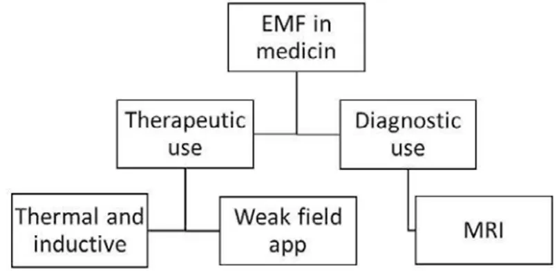
Figure 2. Division of electromagnetic field within the healthcare system.
The use of electromagnetic fields within the healthcare system can be divided into two main areas: therapeutic and diagnostic. This is illustrated in Figure 2. With regard to the
therapeutic division, low-frequency magnetic fields are used to induce currents in tissues and higher frequencies are applied to produce heat. For example, induced current is used for Transcranial Magnetic stimulation (TMS), and heat development is used in diathermy, either in the form of short-wave diathermy, microwave diathermy or electro surgery. Weak fields, with either low or high frequencies, are not used to any great extent in Sweden but are present in many places abroad. See further Markov [8] about bone and wound healing, cancer therapy, effect on microcirculation, and electroporation.
6.1. Magnetic resonance imaging
The Magnetic Resonance Imaging (MRI) technique plays a significant role in the diagnostic portion of health care today. This is because MRI is flexible in terms of what tissue properties need to be examined, it has high resolution and a very good soft tissue contrast. MRI is used for a wide range of applications, neuroimaging, cardiac imaging, musculoskeletal imaging, spectroscopy and functional imaging to mention a few. The number of areas where MRI is introduced increases and one of the recent areas where MRI has been introduced is modern radiation therapy where MRI is introduced for planning, positioning the patient, but also to monitor the treatment effect.
In Sweden today we have an estimated 150 MR cameras. We asked about the number of MRI investigations performed at the University Hospital in Umeå during 2016. Together on both cameras there were a total of 6,342 investigations performed, 3,374 on the 3 T system and 2,968 on the 1.5 T camera. It is estimated that during 2017 the total number of investigations may reach about 6,600 investigations.
At the county hospitals in Lycksele and Skellefteå there is one MR camera at each hospital and during 2016 they performed 2214 and 3817 investigations, respectively. A rough estimate would be that on each camera in clinical use about 10 patients could be scanned per day and with about 300 operating days per year a total number could be (150x3000) about 400,000-450,000 patients undergoing MR examination. The total number of patients may be somewhat less since some patients are getting multiple scans. A large number of people are exposed to the EMF associated with the camera. In addition to this, even the staff that handles the camera becomes exposed in different cases
depending on the role they have in the investigation. Besides from the cameras used in the daily diagnostic there are also units in use in research and in radiation therapy where a combination of PET and MR is used.
The electromagnetic fields associated with MR scanners have been studied closely [9], and have been discussed at length [10] and therefore only a brief summary is given here.
Figure 3. Preparation of a patient before entering the magnet for an MRI investigation. Photo: Jonna Wilén.
6.1.1. Static magnetic fields
MR scanners in clinical use have superconducting magnets, and these usually have a cylindrical opening or bore. The static magnetic field has a flux density of 1.5-3 T. A smaller number of systems are in use in research institutions worldwide, and these use static fields up to 9.4 T. In Lund, there is now a national common research MR working at 7 T. Due to the active shielding, especially for scanners with higher field strengths, the field declines rapidly with the distance from the scanner. The field is only significant within 0.5 m from the bore opening. However, this means that the static magnetic field gradient is steep, which can be of significance for the staff moving around the magnet. There is a requirement that the 0.5 mT contour (5 Gauss) around the magnet should be marked, or that access to that area is restricted. This is to prevent interference with the function of implanted pacemakers and cardioverter defibrillators. This contour is usually located inside the MRI scanner room.
When a person moves in the static field gradient in the MR room, electric currents will be induced in the body, and therefore the movement should be controlled and not too fast. So-called open systems provide far greater access to the patient and facilitate
interventional procedures. In such systems, the static field is usually around 0.2-1 T. The static magnetic field is always on, regardless of whether an MR scan is in progress or not. This means that anyone who moves around the scanner will be exposed to a room-varying magnetic field, caused by movement in the static field, and its gradient.
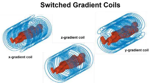
6.1.2. "Switched" gradient field
A “switched” gradient field is used for image coding, and this is emanating from three separate coils in the three directions within the scanner (Figure 4). The "switch mode" gradient field is only active when a scan is to be performed and it is switched on and off to indicate the area of diagnostic interest and spatially encode MR signals. Fast sequences are used to catch rapid biological events, like movement of the heart. The faster the sequence the larger time derivate of the gradient field is required. The amplitude is of the order of mT with fast rise and fall times, tens to hundreds of µs. Typically, the gradient in the region of interest may be 25-50 mT/m and the maximum value (maximum amplitude divided by the rise time, slew rate) can be 100-200 T/m/s within the picture area. The gradient field in modern systems can be as high as 100 mT/m with a slew rate of 800 T/m/s. The wave forms of the gradient field are complex and are not periodic, but can be characterized by the primary frequencies in the range of kHz. In the figures below we show some examples of waveforms of the applied current in the gradient coils. The limiting factor for the patient exposure is the electric potential induced in the patient’s nerve fibres that can lead to peripheral nerve stimulation (PNS). To avoid this a limit of about 20 T/s - which should not be exceeded - has been given as a rheobase value. However, for shorter pulse duration higher values can be allowed [3]. This also applies when the patient is moved into position in the camera. The speed of the movement must be limited in order not to cause nerve excitation.
Occupational exposure to the gradient field may be significant, especially near the bore opening. Wilen et al. [11] measured the rms value of the field up to 0.1 mT at 0.3 m from the centre of the magnet opening. From their data dB/dt values of 70 T/s could be
calculated in that position. The magnitude of the magnetic gradient field and its derivative time depend on the pulse sequence used.
Figure 4. Illustration of the three coils for the switched gradient magnetic field. Courtesy of Jeff Hand, King´s College, London.
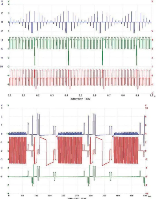
Figure 5. Measurements of current in the three gradient coils for two different sequences. The top sequence is a T2 weighted Turbo Spin Echo, and the bottom is an Echo Planar Imaging sequence. From Wilen et al. [11]
6.1.2.1. Ripple from the gradient field
Magnetic field exposure during an MRI scan is determined in large part by the gradient field from the three gradient coils. What was not known in the past was that there is a ripple overlaid on the signal with a frequency around hundreds of kHz. This gives a contribution to the time derivate, dB/dt, which can be higher than what is specified in the manual. Usually the values are up to tens of T/s, but the ripple can provide significantly higher values than that. Sundström et al. [12] measured the ripple on a Siemens Espree 1.5 T and found values of up to 56 T/s. When equivalent measurements were done on a Philips 3 T, the ripple was negligible. Thus, it is different between different machines. In order to fully characterize exposure, it is necessary to take into account the ripple effect.
6.1.3. Radiofrequency magnetic fields
An RF magnetic field is usually created with a body coil integrated into the scanner, which produces a circularly polarized magnetic field called B1. For systems with
cylindrical openings with 1.5 or 3 T, it is usually a coil in the form of a "bird cage" to provide an area around isocentre of the scanner where the B1-field is spatially
homogeneous. For open MRI scanners with the static field vertically, the RF B1field is
often produced by a pair of planar coils placed above and below the patient. Only the magnetic field component is required for the MRI, and the electric field, E, is generally low except in the vicinity of the coil windings. Exposure to RF B1outside the bore is
generally low since the field falls off quickly outside the transmit coil. An exception will be for employees who perform interventional procedures, especially in open scanners, where their hands and arms, and possibly head, can be exposed to levels that are like those to which the patients are exposed. The RF field has a frequency of about 42 MHz/T, meaning that for a 3-T scanner the frequency is approximately 126 MHz.
Various RF-pulse sequences are used depending on what contrast is required, and thus, the SAR value of each pulse sequence, is different. Usually, during clinical scanning, many different sequences are used to get the appropriate information. Based on our own measurements the peak values for the RF B1can reach 10 A/m and more. Since the duty
cycle can be about 1%, the SAR values in pulses can be rather high, in comparison with the limit values. With a whole-body average SAR value around 1 W/kg, the peak of the pulse can reach 100 W/kg. This type of exposure has not been studied with regard to health effects. There is very little information available on high peak pulses because nearly all research on RF has involved only the average values and thermal effects. The switch mode gradient field and the RF field is only activated during MRI procedure, and both the patients and personnel who are in the room during the scan will be exposed.
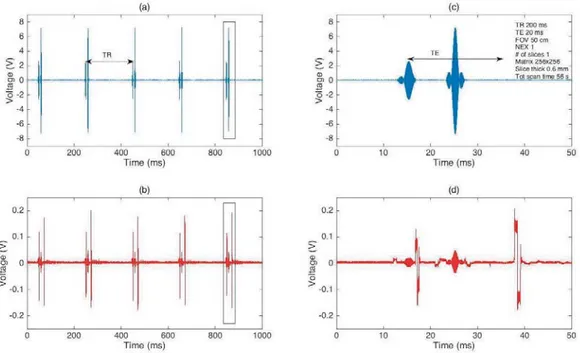
Figure 6. RF pulses are shown in the upper figure in blue, and the pulsed gradient field appears in the lower figure in red. Measurement is from a spin-echo sequence, oscilloscope photos show time derivate dB/dt, and the magnetic field in the form of induced voltage is measured in a pick-up coil. The pictures to the right show a magnified view of the 90 and 180 degree spin echo pulse pair with accompanying pulsed magnetic fields. From Frankel et al. [13]
Figure 7. RF Pulse is shown in the upper figure in blue and the pulsed gradient field in the lower in red. The measurements are from a "spoled gradient echo sequence". The picture to the right is an enlarged part of the sequence. From Frankel et al. [13].
6.1.4. Exposure near MRI scanners
Beside the patient also the operator will be exposed to the magnetic fields near the scanner, mainly the static magnetic field that is always switched on. In some
circumstances when personnel are present during scans, for instance during anaesthetic procedures when the patient needs to be monitored, the personnel are also exposed to stray fields from the switched gradient field and to some extent also the radiofrequency field. Several recent studies have examined the occupational exposure of people working with MRI. de Vocht et al. [14] measured both static and time-varying magnetic fields, and found that the time-weighted exposure levels were within the ICNIRP guidelines [15], but peak exposure limits were exceeded during certain procedures. Keevil [16] discussed the induced current density from movements in a static field, and he found that although the limit was set at 40 mA/m2according to [15] values up to 200--400 mA/m2
could be measured. The limit can be exceeded if one is closer than 0.5-1 m from magnet opening. For the gradient field the limit value was 10 mA/m2, and here values could be
measured over 200 mA/m2inside the magnet during scanning.
Karpowicz and Gryz [17] studied exposure to static magnetic fields (SMF) during scanning. Measurements near a 1.5 T MRI magnet revealed that SMF exposure from different scanners depends on both the SMF magnet, the scanner design and the work organization. During a routine examination of a patient, nurses were exposed to SMFs exceeding 0.5 mT for approximately 1.5-7 min, and up to 1.3 min for SMF exceeding 70 mT. The average values for exposure to SMF were 5.6-85 mT, with a mean of 30 mT. Chiampi and Zilberti [18] have developed a computational procedure to evaluate the internal E-field induced by movement in the static magnetic field gradient. For further details see Wang et al. [19]. ICNIRP [20] recently came with recommendations for mitigation of the induced electric field due to the movement of the static field. Working near an MRI scanner inevitably leads to an induced electric field in the body due to the
movement in a strong static magnetic field. The value given in the ICNIRP guidelines [20] is the same as that for induced field from ELF fields, namely 1.1 V/m. This is then translated to a reference value for the time derivative, dB/dt, as 2.7 T/s. Fatahi et al. [21] measured dB/dt in a study in which 5 MRI researchers moved around a 3 T and a 7 T scanner in a controlled way. They found large variations among the five people, but occasions that exceeded the limit were often similar, 30% of 60 scenarios. Further studies of this are needed to develop guidelines for adopting some simple precautionary rules for staff behaviour around MRI scanners to avoid exceeding the limits.
6.1.4.1. Reduced exposure
During most clinical scans, standard sequences are used, and often several sequences are needed for obtaining the required information. However, it is possible to reduce the exposure for the patient in some cases and many MRI scanners do have special protocols to reduce exposure for instance when the sound level needs to be lowered or also for some scanners when medical implants are present.
Figure 8. The change that occurs when the so-called Whisper mode is used on a Siemens MRI camera. By changing the rise and fall times of the fast gradient pulses, the exposure from dB/dt can be reduced with 1.5 times. An oscilloscope recording of current in one of the gradient coils during a Trufi sequence is shown. The scale is in Volts with converting factor 100 A = 1 V. From Wilen et al. [11].
6.1.5. Limits and regulations
Safety aspects for the use of MR have been described in several studies [22-25]. There are limits and rules for the exposure of patients as well as staff who may be exposed during an MRI examination. For the patient’s safety it is necessary to comply with the standard [3]. Its adoption was intended to ensure the safety of patients undergoing an MRI examination as well as the professionals who work with MR. The standard sets both technical requirements on the equipment and on the organization of the work with MR. For the staff, the new regulations from the Swedish Work Environment Authority on electromagnetic fields also apply [26].
When it comes to gradient fields, the patient is to be protected from peripheral nerve stimulation (PNS), and this is done by restricting the induced electric field in the body. For that, both pulse intensity and duration are taken into account. This can then be
translated into a restriction on the time derivative of the gradient magnetic field, dB/dt. The standard [3] also defines operating modes for MR systems: normal operating mode,
first level of control and second level of control based on level of SAR and dB/dt.
In normal operating mode the apparatus shall be set so that it does not exceed 80% of the threshold value of the PNS, and in a first level controlled mode it can go up to 100%. To go to this setting requires a specific action from the operator in order to clarify that the patient or operator now is entering a special risk position.
To protect the patient against undesirable temperature rise during an MR scan, there are also limits on how much the body temperature may rise. Normally, it should not be more than 0.5oC, but in first level controlled mode it can be allowed to reach 1°C, and for
special cases which require a permit (second level controlled mode), even higher levels can be allowed. These temperature rises have since been translated into the SAR values that are allowed, and in the normal operating mode, they should be kept at SAR < 2 W/kg. In the first level controlled mode, levels can go up to 4 W/kg whole body. These values are time averages over a 6-minute period. For times shorter than 10 s it is allowed to go up to two times these SAR values.
The standards also address the static magnetic field, and here the normal case is 3 T, and in special cases one can go higher. The audio level inside the machine is also restricted. The standard also discusses how to manage patients with various medical implants, but more on this in a separate section. Also the definition of controlled environment is defined by the standard were for instance the 0.5 mT limit apply due to potential risk to implants. To enter the controlled environment special authority need to be applied.
The Swedish Work Environment Authority has adopted the EU directive on regulations for protection from 1 July 2016. An exception is made for MR work so that exposure can exceed the limit values if the exposure occurs in connection with the installation, testing, operation, development and maintenance of, or research on, equipment for MRI for patients in healthcare. The exemption only applies to §§ 9--13 in the provision, and the other sections should be followed, and this applies in particular to the requirement that the employer must inform and educate the staff about the risks which can occur and how to work safely in order to avoid these [26].
For certain types of surveys, for example, when rapid processes are to be studied, it may happen that the patient can feel gentle nerve twitches in superficial nerves, which are caused by current induced in the body from the gradient field. The risk of nerve excitation is greatest when fast processes are applied, e.g., when rapidly changing gradients are used for functional imaging of the brain with MRI or for the diagnosis of stroke. Heating effects can occur when the number of RF pulses per unit time is high, such as in the anatomical image where detail resolution is important.
6.1.6. Risks and effects of MRI exposure
An MRI examination is not entirely risk-free. Because very strong magnetic fields are used, from static up to radio frequency, there are risks that require attention both before and during the investigation.
Static fields. A person inside the examination room moves in the strong gradient of the
static magnetic field, and this will induce currents in the body - the faster the movement the higher the current. These currents may lead to subjective feelings, depending on how sensitive the person is. Symptoms include dizziness, nausea, headache, and an experience
of “curved” space [27-30]. ICNIRP has issued guidelines for induced electric fields from movement in a static field [20].
These symptoms become more common the stronger the static field is [28]. Today the greatest strength of a human MR scanner in Sweden is 7 T. It is actively shielded for static field, which means that the static magnetic field does not reach far away from the bore, but it also has the implication that the gradient the patient passes through when inserted into the opening is stronger than that found in non-shielded scanners. This higher static magnetic field gradient therefore increases the risk of symptoms for the patient. To reduce the risk for patients the speed of the patient table is adjusted due to the static magnetic field gradient to avoid symptoms.
Switched gradient field. The gradient field used in the MR is in the range of what can be
tolerated before the experience of nerve excitation. Patients can experience slight nerve twitching on body parts that come closest to the solenoid coils, i.e. arms and legs that touch the walls of the magnet where the field is strongest. Setsompop et al. [31] have discussed the nerve stimulation thresholds and how different slew rates influences the quality of the picture, and a comprehensive study on nerve excitation and changing magnetic field has been done by Reilly [32].
Radiofrequency field. The intensity of the RF field is on the verge of what can be
tolerated in the form of heating of the body without an increase in body temperature more than 1oC. The strength is usually given in terms of how much energy is absorbed per
kilogram, SAR, and levels of single W/kg may lead to harmful local elevations of temperatures, and cause a general increase in body temperature. Therefore, the scanner warns when too high SAR values are being applied.
The risk for burn injuries is especially great if the person comes too close to the walls of the magnet, since the RF field will increase with distance to the coil. Therefore, it is recommended that an insulation of at least 2-cm thickness is placed near the wall to prevent burns. At high SAR levels it is also important not to form closed loops with body parts, such as the arms over the head and the hands or knees against each other. Special consideration must be given to patients with poor body control, such as babies who do not sweat, and others with impaired circulation.
Reddig et al. [33] have given a list of estimated absorbed energy during scanning with different MR protocols. The mean whole body SAR value varies from 0.3 W/kg in a cardiac scan with a contrast agent to 2.6 W/kg for a lumbar spine scan. Including the time of the scan the energy absorbed ranges from 182 J/kg to 2,818 J/kg.
Implant. Passive implants, such as orthopaedic implants, can lead to a concentration of
the radio frequency field and thus provide a very local harmful warming, something that is not detected until it is too late because the heat sensors in the body are superficial. For active implants, such as pacemakers and defibrillators, the risks are that they can concentrate the radio frequency field, but also that its function may be affected. For the whole area around the implants, it may be helpful to have access to a medical specialist who has a good overview of the area and can provide advice in cases of doubt.
All patients are reviewed for contraindications prior to MRI scanning where possible implants are investigated carefully. All implants are categorized as safe,
MR-Conditional or MR-Unsafe.
Implants that are categorised, as MR-Safe is completely non-magnetic, non-electrically conductive, and non-RF reactive, and it is safe to perform MR scanning. MR-Conditional implants may contain magnetic, electrically conductive or RF-reactive components that are safe for operations in special conditions such as for instance a certain level of SAR, dB/dt or static magnetic field strength. Some implants are categorised as MR-Unsafe
which means that due to their ferromagnetic material pose a significant risk to the patient and MR scans are prohibited.
This information is available through the manufacturer of the implants but it is a hard and time consuming task to investigate this for each patient. Therefore, many hospitals have developed their own implant data-bases where also routines for especially MR
conditional implants are stated. To keep these data bases updated is an important task for the safety organisation and might be especially challenging for smaller hospitals with few MR physicists. There have therefor been discussions about possible national databases to help especially the smaller clinics. There do exist some commercially available data bases but these are not complete. Also resources such as safety hand books are available, see for instance [34]
It is imperative that the first contact with the patient for an MRI examination includes a questionnaire on which implants the patient has, and this should then be followed up before the examination. This can cause problems with people who do not remember what implants they have and when the operation history is missing.
Mattei et al. [35] looked at the effect of the movement near the MRI scanner bore for people with a pacemaker or an implantable cardioverter defibrillator. They found that motion in and around an MR scanner can induce significant voltage, which would be enough to be misinterpreted by the implant and thus lead to inappropriate changes in its function. Persons with these implants, including MR-conditional systems, should not enter the MRI room, except in case of an examination under specified conditions.
Pregnancy and scanning. There are currently no established long-term effects of in
utero MR scans, but account must be taken of RF heating and noise level. In England it is
recommended that pregnant staff should avoid being in the room during scanning and that one should select sequences that minimize RF noise. This is also done in common
practice at Swedish hospitals, but there exist no guidelines for this from Swedish authorities.
6.1.6.1. Indirect effects
Static fields. In a static field there is the projectile risk. The field is so strong that
ferromagnetic objects can be dragged into the magnet with such great force that damage to the people who happen to be in the way can be life-threatening. It is important that all people entering the examination room such as staff, relatives and patients are controlled carefully so that no ferromagnetic objects are brought into the room. Incidentes have occurred when, e.g., trolleys, IV stands and office chairs, have been affected by the magnetic field, attracted to and stuck to the magnet [25].
This type of accident can be very dangerous in terms of injuries, but can also lead to a huge loss of investigative time. To remove the object from the magnet, it may be
necessary to turn off the magnetic field by releasing the helium gas that keeps the magnet cooled to the superconducting temperature. Replenishment of helium and rebooting takes at least a week.
It is also important that patients (and personnel who will be present in the MR room) have been screened for various implants. The static field can exert force on an implant that is somewhat ferromagnetic, and it can thus be moved out of position with unwanted consequences. It should be noted that the implants that are considered safe for 3 T may not be safe for a 7 T machine [36].
Gradient field. The most common problem that occurs due to the gradient field is the
level of noise caused by vibrations in the wires in the gradient coils. It is a requirement that personnel in the room during scanning have hearing protection, and patients must be
provided with protection. Sometimes a double hearing protection is put on the patient. Audio levels can range from 85 to 95 dBA.
6.1.6.2. Possible long term effects
Some studies have demonstrated genotoxic effects in cells after exposure to an MR scan [37-39], while others could not demonstrate any effects [40, 41] The MRI sequences used in those studies are clinically available and routinely used in heart and brain scans. In Reddig et al [33], patients undergoing clinical CT scans were used as positive controls, and the authors found nearly a doubling of DNA double strand breaks 5-30 minutes after the CT scan as compared with before the CT scan. There was no evidence of DNA damage after the MRI examinations.
In a recent paper by Foster et al. [42] those studies were criticized on the grounds that many lacked a positive control, sham exposure and blinding in the analysis work. They suggest that the results should be confirmed with studies with the same end points but with higher statistical power and more rigorous design.
In recent studies from Utrecht University [43-45] risk assessments for MRI workers have been carried out. They found an association between MRI-related occupational SMF exposure and an increased risk of accidents leading to injury, and for commute-related (near) accidents during the commute from home to work found that radiographers using intrauterine devices (IUDs) and were occupationally exposed to stray fields from MRI scanners reported abnormal uterine bleeding more often than their co-workers without an IUD, or nonexposed co-workers with an IUD. In particular, radiographers present inside the scanner room during image acquisition showed an increased risk. These findings points to the need for further research to find out if staff working close to MRI scanners are at increased health risk.
Recently a report from an EU Committee, the SCENIHR (Scientific Committee on emerging and newly identified health risks) [4], gave a review of the area of EMFs and health. They also give recommendations on health effects of MR fields. They recommend long-term prospective and retrospective cohort studies of personnel exposed to high-gradient fields in the operation of MRI units, and this is a high priority issue. As there have been reports on DNA damage in patients after an MRI scan, therefore additional studies investigating genotoxic effects of MRI examinations in either patients or volunteers should be done.
The challenges with the exposure assessment for epidemiological studies of MRI personnel have been discussed in a publication of a COST BM0704 group [46].
6.1.7. Organizational aspects of safety for MRI system
Personnel working with MR must receive adequate training on the various incidents that can occur in different contexts including direct effects such as movement induced symptoms, heating effects but also indirect effects such as possible risk for implants and ferromagnetic objects. For instance, if a patient starts feeling bad during a scanning and must quickly be taken out of the magnet, certain precautions are needed. If a fire is detected on the premises, the firefighters must be informed about the risks with the static field since they are likely to carry ferromagnetic equipment. If there is need for an emergency stop and a quench of the magnet, helium might leak out in the room, and the oxygen level can become low. Information about this must exist. At the MRI clinics, to our experience the safety management works quite well and the introduction of the regulation [26] will also improve this further. It is more challenging to make the safety management work for personnel that are not primary situated at the MRI clinics, such as
cleaning personnel, anaesthetist and other groups that rather seldom, enters the scanner room.
In a recently published document by Hand et al [47] recommendations on how to allocate the responsibility for safely working with MR are given. The organizations behind the document is European Federation of Organization of Medical Physics (EFOMP) and they recommend the introduction of a number of positions for the operative responsibility, such as:
MR Medical Director (MRMD) or MR Research Director (MRRD), MR Safety Officer (MRSO), and MR Safety Expert (MRSE). It is assumed that MRMD, MRRD and MRSO are part of the organization that performs the actual scanning, while MRSE can be an external person.
MRMD or MRRD shall, among other things, oversee the safe execution of the MRI scan on each patient/person being investigated in the system. MRMD/MRRD shall be
available to the operators of the MR system when the system is available. MRMD/MRRD shall ensure that appropriate human security and quality programs are implemented, as well as a risk assessment performed for MR facility.
MR Safety Officer (MRSO) is often carried out by the senior Radiographer. Multiple MRSOs can be appointed provided that only one is responsible at a given time. He/she shall be easily accessible to operators of MR system at all times when MR facility is accessible. The role also involves managing the risks posed by MR equipment and monitoring the measures taken to protect against such risks.
MR Safety Expert (MRSE). This position is expected to serve as a resource for the MRMD/MRRD or MRSO. MRSE is often an MR physicist, but others with the appropriate technical MR expertise could also fill this role.
MRSE roles include giving advice on construction, scientific, and administrative aspects of safe use of MRI equipment, to provide the Security Council with regard to non-routine MRI procedures for individual substances and groups of substances, such as implants, metallic foreign bodies, tattoos and other similar issues.
By implementing something like the above, then also requirements in the Swedish work environment authority's new regulation: electromagnetic fields, applicable from mid-2016, can be fulfilled.
6.1.8. Conclusion
There is an increasing number of patients, including children and young adults, who are examined with MRI, and MRI scanners with stronger magnetic fields have become more common. Therefore, the question of possible long-term effects on patients has been raised. There are no epidemiological studies on large groups of patients who have undergone MRI scans. MRI is used increasingly in paediatric imaging diagnosis. A study on the effects of MRI exposure in children is recommended by the SCENIHR [4] with high priority.
SCENIHR also recommends studies investigating possible cognitive effects of exposure to magnetic gradient field of employees working in the immediate vicinity of the MRI equipment.
The question about how to deal with patients with various types of medical implants is something that could be addressed in a national database which is accessible to all.
Overall there is a need for education and training of the personnel involved in MRI investigations. This should be organized in such a way that new staff members can easily get the training and information about the safety aspects. Implementing the safety management system suggested by [47] and described above, might be one way to go.
6.2. Electro surgery
Electro surgical units (ESU) is the most common medical-technical equipment in our hospitals using EMF. Only at VCC, there are 202 different ESU registered the corresponding figures for KUH and KCC are 254 and 54, respectively.
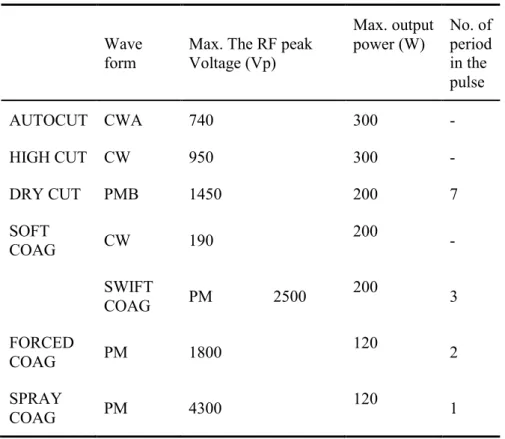
Radiofrequency energy is used in several surgical procedures, not only in medical care but also in dental care. In most cases, a small active electrode applicator and a flat electrode (also known as the ground electrode or return pad) are used. The ground electrode returns to the generator (monopolar configuration). The active electrode provides a high current density and serves as a cutting or coagulating instrument by applying a current with sinusoidal or pulsed waveform, and the frequencies are about 0.3 to 5 MHz. A widely used minimally invasive electro surgical procedure is radiofrequency ablation, which is routinely used in oncology, cardiology and otorhinolaryngology. In a study of 6 ESU, Marco and Maggi [48] found that close to the equipment the fields were quite high, but at a distance of 0.5 m from the machine, the electrical field strength fell to 32-57 V/m and the magnetic field strength fell to 0.2-0.8 A/m. In the worst case at maximum reading, the surgeon's hands were exposed to RF field with the magnetic field strength of 0.75 A/m and the electric field strength at 400 V/m. Significantly higher values were measured by Liljestrand et al [49] and they also noted that the fields are produced not only by the active electrode but also of the return pad and cable. The system uses different sequences depending on whether the intention is to cut or coagulate. Several different modes exist, and Table 2 gives examples of this. See also below from Wilén [50].
Measured peak values of the voltage at electrode tips are between 1 to 4 kV. The highest values are obtained when using coagulating mode. Wilén [50] also concludes by noting that when using ESU, both surgeons and other professionals that come close to the active cable are exposed to radiofrequency fields in excess of the guidelines specified by the ICNIRP [15]. They point out that further studies and calculations must be made to ensure that the ICNIRP specified "basic restrictions" are not exceeded.
The ICNIRP's values are now accepted as a Swedish regulation by the SWEA [26]. Laboratory tests show that shielding of the active conductor gives a substantial reduction of the electric field around the conductor and can thus be a possible way to move forward to reduce exposure using ESU. According to one of the manufacturers of diathermy equipment, there is one system on the market that has shielding, but the users think it is too clumsy to use and it lacks smoke exhaust. Therefore, on the Swedish market only non-shielded electrodes are used.
Based on measurements by Wilén [50] the use of ESU with unshielded electrodes leads to exposure to electric fields in excess of the action levels given in the EU directive on occupational exposure to EMF and thereby also the new Swedish regulation. The question then is raised if also the underlying limits are exceeded, and to answer this further studies and investigations need to be done.
The limits are set both as instant values not to be exceeded, and for the frequencies used here the E-field action value is 610 V/m. This should also be combined with a time
average value of 610/(frequency in MHz) for thermal effect. The first value is to protect against nerve stimulation and here the limit is set at 3.8 x 10⁻⁴x f (f in Hz). In the thermal
case the limit is set in SAR values: whole body 0.4 W/kg, head and torso 10 W/kg and extremities 20 W/kg.
It is not likely that the time averaged SAR values will be exceeded, but that needs to be shown. For induced E field, further investigations in form of measurements and
calculations are needed to ensure that the limit is not exceeded.
In the case study SWEA refers to in connection with the regulation: "the use of this device is likely not to exceed the Exposure Limit Value ( ELV) for the surgeon or other hospital staff "[51] . However, this assertion is completely at odds with the measurements made by Wilén [50], where she found a risk that the action values could be exceeded.
Table 2. Description of the evaluated VIO 300 d modes.
Wave
form Max. The RF peak Voltage (Vp)
Max. output
power (W) No. of period in the pulse AUTOCUT CWA 740 300 -HIGH CUT CW 950 300 -DRY CUT PMB 1450 200 7 SOFT COAG CW 190 200 -SWIFT COAG PM 2500 200 3 FORCED COAG PM 1800 120 2 SPRAY COAG PM 4300 120 1