ACTA UNIVERSITATIS
Digital Comprehensive Summaries of Uppsala Dissertations
from the Faculty of Medicine
1436
Clinical Consequences of Axonal
Injury in Traumatic Brain Injury
SAMI ABU HAMDEH
Dissertation presented at Uppsala University to be publicly examined in Auditorium minus, Gustavianum, Akademigatan 3, Uppsala, Saturday, 21 April 2018 at 09:15 for the degree of Doctor of Philosophy (Faculty of Medicine). The examination will be conducted in English. Faculty examiner: Professor Andreas Unterberg MD, PhD (Department of Neurosurgery, Heidelberg University).
Abstract
Abu Hamdeh, S. 2018. Clinical Consequences of Axonal Injury in Traumatic Brain Injury.
Digital Comprehensive Summaries of Uppsala Dissertations from the Faculty of Medicine
1436. 84 pp. Uppsala: Acta Universitatis Upsaliensis. ISBN 978-91-513-0251-5.
Traumatic brain injury (TBI), mainly caused by road-traffic accidents and falls, is a leading cause of mortality. Survivors often display debilitating motor, sensory and cognitive symptoms, leading to reduced quality of life and a profound economic burden to society. Additionally, TBI is a risk factor for future neurodegenerative disorders including Alzheimer’s disease (AD). Commonly, TBI is categorized into focal and diffuse injuries, and based on symptom severity into mild, moderate and severe TBI. Diffuse axonal injury (DAI), biomechanically caused by rotational acceleration-deceleration forces at impact, is characterized by widespread axonal injury in superficial and deep white substance. DAI comprises a clinical challenge due to its variable course and unreliable prognostic methods. Furthermore, axonal injury may convey the link to neurodegeneration since molecules associated with neurodegenerative events aggregate in injured axons.
The aim of this thesis was to study clinical consequences of axonal injury, its detection and pathological features, and potential link to neurodegeneration in severe TBI patients treated at the neurointensive care unit at Uppsala University Hospital. In paper I and IV DAI patients were studied for the relation of elevated intracranial pressure (ICP) and poor outcome to axonal injury on magnetic resonance imaging. In paper II, soluble amyloid-beta aggregates (oligomers and protofibrils), characteristic of AD pathology, were investigated in surgically resected brain tissue from severe TBI patients, using highly-selective Enzyme-Linked ImmunoSorbent Assays. In paper III, brain tissue biopsy samples from TBI patients with either focal injury or DAI were examined for differential proteome profiles using mass spectrometry-based proteomics.
The results provide evidence that axonal injury, located in the central brain stem, in substantia nigra and the mesencephalic tegmentum, is particularly related to poor outcome and increased ICP during neurointensive care of DAI patients. A novel classification system for prognostication after DAI is proposed. Furthermore, the thesis shows that severe TBI induces rapid accumulation of neurotoxic soluble amyloid-beta oligomers and protofibrils. In addition, DAI initiates unique proteome profiles different from that of focal TBI in structurally normal-appearing brain. These findings have implication for the clinical management of DAI patients, and provide new insight in the neuropathological consequences of axonal injury.
Keywords: Traumatic brain injury, diffuse axonal injury, intracranial pressure, magnetic
resonance imaging, amyloid-beta, tau
Sami Abu Hamdeh, Department of Neuroscience, Neurosurgery, Akademiska sjukhuset, Uppsala University, SE-75185 Uppsala, Sweden.
© Sami Abu Hamdeh 2018 ISSN 1651-6206
ISBN 978-91-513-0251-5
List of Papers
This thesis is based on the following papers, which are referred to in the text by their Roman numerals.
I Abu Hamdeh S., Marklund N., Lannsjö M., Howells T.,
Rain-inko R., Wikström J., Enblad P. (2017) Extended Anatomical Grading in Diffuse Axonal Injury Using MRI: Hemorrhagic Le-sions in the Substantia Nigra and Mesencephalic Tegmentum In-dicate Poor Long-Term Outcome. J Neurotrauma. 2017 Jan 15;34(2):341-352
II Abu Hamdeh S.*, Rollman Waara E.*, Möller C., Basun H.,
Alafuzoff I., Hillered L., Lannfelt L., Ingelsson M., Marklund N. (2017) Rapid amyloid-β oligomer and protofibril accumulation in traumatic brain injury. Brain Pathol. 2017 May 29. [Epub ahead of print]
III Abu Hamdeh S., Shevchenko G., Mi J., Musunuri S., Bergqvist
J., Marklund N. (2018) Proteomic differences between focal and diffuse traumatic brain injury in human brain tissue. Submitted
IV Abu Hamdeh S., Marklund N., Anders Lewén., Howells T.,
Raininko R., Wikström J., Enblad P. (2018) Intracranial pressure elevations in diffuse axonal injury are associated with non-hem-orrhagic MR lesions in central mesencephalic structures.
Submit-ted
* Contributed equally
Additional publications not included in this thesis
• Abu Hamdeh S., Lytsy B., Ronne-Engström E. (2014) Surgical site infections in standard neurosurgery procedures - a study of incidence, impact and potential risk factors. Br J Neurosurg. 2014 Apr;28(2):270-5
• Wicher G., Wallenquist U., Lei Y., Enoksson M., Li X., Fuchs B.,
Abu Hamdeh S., Marklund N., Hillered L., Nilsson G.,
Forsberg-Nilsson K. (2017) Interleukin-33 Promotes Recruitment of Micro-glia/Macrophages in Response to Traumatic Brain Injury. J
Neuro-trauma. 2017 Nov 15;34(22):3173-3182
• Tsitsopoulos PP., Abu Hamdeh S., Marklund N. (2017) Current Op-portunities for Clinical Monitoring of Axonal Pathology in Traumatic Brain Injury. Front Neurol. 2017 Nov 20;8:599. eCollection 2017.
Review
• Abu Hamdeh S., Virhammar J., Sehlin D., Alafuzoff I., Cesarini K.G., Marklund N. (2018) Brain tissue Aβ42 levels are linked to shunt response in idiopathic normal pressure hydrocephalus. J Neurosurg. 2018 Jan 19:1-9. [Epub ahead of print]
Content
List of Papers ... v
Abbreviations ... ix
Introduction ... 11
1.1 Classification of traumatic brain injury ... 12
1.2 Pathophysiology of traumatic brain injury ... 15
1.3 Multimodality monitoring in severe traumatic brain injury ... 16
1.3.1 Monitoring of intracranial pressure ... 17
1.3.2 Monitoring of cerebral perfusion pressure ... 17
1.3.3 Monitoring of cerebral blood flow ... 18
1.3.4 Cerebral microdialysis ... 19
1.4 Treatment algorithms in severe traumatic brain injury ... 19
1.5 Outcome measures in traumatic brain injury ... 20
1.6 Diffuse axonal injury ... 21
1.7 Biomechanics of axonal injury ... 22
1.8 Pathophysiology of axonal injury ... 22
1.9 Impact of axonal injury ... 24
1.10 Detection of axonal injury ... 24
1.10.1 Magnetic resonance imaging ... 25
1.10.2 Neuromolecular imaging ... 27
1.11 Classification of diffuse axonal injury ... 28
1.12 Consequences of axonal injury – clinical features during neurointensive care ... 29
1.13 Consequences of axonal injury – a link to neurodegeneration? ... 30
1.13.1 Amyloid-beta pathology ... 30
1.13.2 Tau pathology ... 33
Aims ... 34
2.1 Specific aims ... 34
Material and methods ... 35
3.1 Patient population ... 35
3.1.1 Severe traumatic brain injury cohorts and timing of MRI ... 35
3.1.2 Control subjects ... 36
3.2 Image acquisition and analysis ... 36
3.4 Sampling and preparation of brain tissue ... 38
3.5 Immunohistochemistry for Aβ plaques ... 38
3.6 Biochemical analysis of soluble Aβ species using ELISA ... 39
3.7 Proteome analysis using mass spectrometry ... 40
3.8 Outcome measures ... 41 3.9 Statistical methods ... 41 Results ... 43 4.1 Paper I ... 43 4.2 Paper II ... 44 4.3 Paper III ... 45 4.4 Paper IV... 49 General discussion ... 51 5.1 Paper I ... 51 5.2 Paper II ... 53 5.3 Paper III ... 54 5.4 Paper IV... 56 5.5 Statistical considerations ... 57 Conclusions ... 59 Future perspectives ... 60 Summary in Swedish ... 62 Acknowledgement ... 64 References ... 67
Abbreviations
ABP AD APOE Aβ BACE-1 βAPP BBB CBF Cho CPP CSF CT CTE DAI DTI DWI ELISA EVD FA FLAIR GCS GFAP GMT GOS GOSE ICP iNPH ISF m/z MAP MLSArterial blood pressure Alzheimer's disease Apolipoprotein E Amyloid-β
Beta-site APP-cleaving enzyme β-amyloid precursor protein Blood-brain barrier
Cerebral blood flow Choline
Cerebral perfusion pressure Cerebrospinal fluid
Computed tomography
Chronic traumatic encephalopathy Diffuse axonal injury
Diffusion tensor imaging Diffusion-weighted imaging
Enzyme-Linked ImmunoSorbent Assay External ventricular drain
Fractional anisotropy
Fluid-Attenuated Inversion Recovery Glasgow coma scale
Glial fibrillary acidic protein Good monitoring time Glasgow outcome scale
Glasgow outcome scale extended Intracranial pressure
Idiopathic normal pressure hydrocephalus Interstitial fluid
Mass-to-charge ratio Mean arterial pressure Midline shift
MRI MRS MS NAA NFL NFTs NI NIC PBtiO2 PD PET PiB PRx P-tau rCBF RLS ROS SjvO2 SPECT SWI T2*GRE TBI TSAH
Magnetic resonance imaging Magnetic resonance spectroscopy Mass spectrometry N-acetyl aspartate Neurofilament-light Neurofibrillary tangles Neurologically intact Neurointensive care
Brain tissue oxygen partial pressure Parkinson’s disease
Positron emission tomography Pittsburgh compound B Pressure reactivity index Hyperphosphorylated tau Regional cerebral blood flow Reaction level scale
Reactive oxygen species
Jugular venous oxygen saturation
Single-photon emission computed tomography Susceptibility-weighted imaging
T2*-weighted gradient echo Traumatic brain injury
Introduction
Traumatic brain injury (TBI) is a leading cause of mortality and morbidity. Annually, 262 per 100 000 inhabitants are admitted for TBI in Europe and it accounts for one third of all injury associated deaths in the United States (1, 2). In Sweden, TBI accounts for 9.5 deaths per 100 000 inhabitants (3). It affects all ages and income groups and survivors are frequently left with de-bilitating deficits in motor, sensory and cognitive functions with marked im-pact on quality of life (4-7). TBI is considered a silent epidemic since the in-cidence is not seldom underestimated (1). The consequences following TBI persist long after the trauma and are not always immediately recognized (5). Since its incidence is particularly high among children and young to middle-aged adults, TBI has profound socioeconomic impact (8). In young and mid-dle-aged adults, TBI is most frequently caused by motor-vehicle accidents, while in the pediatric and the elderly population falls account for the majority of TBI cases (1, 4, 9).
Historically, TBI has attracted the interest and fascination of ancient scientists, including Hippocrates (460–377 BC) who in his treatise “On Wounds in the Head” covered the contemporary knowledge of TBI thoroughly and exten-sively (10). In his treatise he described trepanation, a surgical method still in use today, for the treatment of skull fractures. Testimonies for this technique have been seen on trephined skulls from as far back as 10 000 BC, and in ancient times it was widely practiced in Western Europe, as well as in South America and Asia (11). Centuries later, Aulus Aurelius Cornelius Celsus (25 BC – AD 50), a scientist from the Alexandrian school of ancient Egypt, wrote one of the earliest known descriptions of severe TBI symptomatology, men-tioning unconsciousness, focal signs, post-traumatic seizures and signs of el-evated intracranial pressure (ICP) such as vomiting and blurred vision (11). However, the relation between altered level of consciousness and injury to the brain was not generally accepted until the 18th century (12). Modern
neurosur-gical management of TBI begun with the observations of Harvey Cushing on ICP (13) and the evolution of ICP monitoring techniques (14). Subsequently, TBI care evolved in combination with advances in neuroradiology (15). Dur-ing the last two decades, progress in the management of TBI patients has been achieved mainly with the introduction and successive refinements of neuroin-tensive care (NIC) and multimodality brain monitoring (16-19).
TBI is defined as an alteration in brain function, or other evidence of brain pathology, caused by an external force (20). Nonetheless, TBI may be consid-ered “the most complex disorder in the most complex organ in the body” (21). Therefore this definition, unified only by the externally afflicted brain dam-age, encompasses a markedly heterogeneous entity. The injury spectrum ranges from mild to severe and commonly, TBI is categorized into either focal or diffuse injuries. While focal injuries include contusion and epidural, sub-dural and intracranial hemorrhage, diffuse injuries encompass concussion and diffuse axonal injury (DAI), with widespread damage to axons mainly in the subcortical white matter, the corpus callosum and the brainstem.
The theme of this thesis, DAI, is a particularly challenging subtype of TBI. Typically, patients with severe TBI and DAI are deeply unconscious at the impact site, and when admitted to hospital care, their initial radiological im-ages using computed tomography (CT) display minimal findings. Further-more, DAI has a variable clinical course both in the acute as well as in the chronic phase, and in addition, existing prognostic methods are unreliable. Therapeutic options for DAI are non-existing. Therefore, increased under-standing of this condition is urgently needed.
1.1 Classification of traumatic brain injury
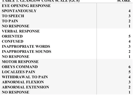
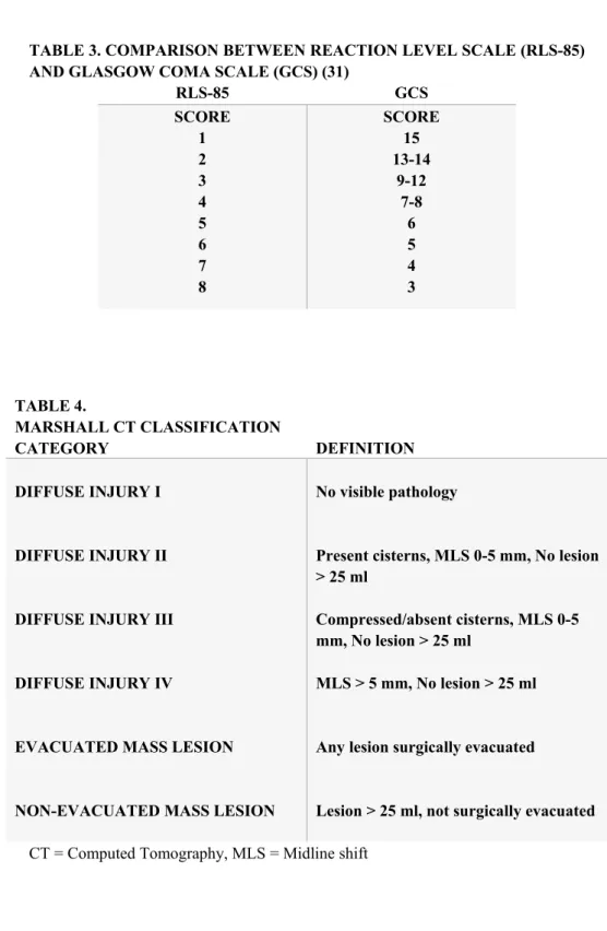
Traumatic brain injury is most often classified according to clinical severity indices, pathoanatomical type or physical mechanism (7). The Glasgow coma scale (GCS), a scale based on clinical assessment of level of consciousness using verbal and motor response and eye opening in response to stimuli (Table 1), is commonly used to classify TBI patients into broad categories of mild (GCS 15 – 13), moderate (GCS 12 - 9) or severe injury (GCS 8 or less) (22). The GCS has high inter-observer reliability, has proven to have excellent prognostic capabilities and is widely used in clinical trials (23). In most of Sweden, the similar Reaction Level Scale-85 (24) is used (Table 2). A com-parison between the two scales is provided in Table 3.
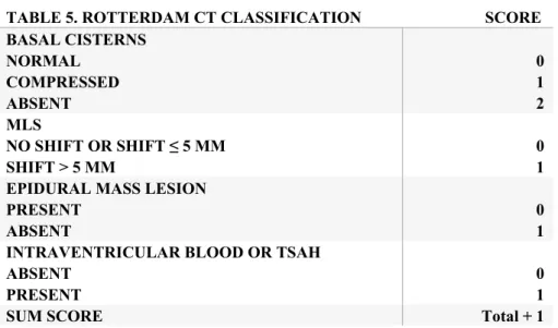
Since scales assessing clinical severity of symptoms do not provide any infor-mation about the pathoanatomical background for the deficits (25), additional classification of location and anatomy of lesions using radiological character-istics is needed. Frequently, such classification is performed using the Mar-shall CT classification of TBI based on the degree of compression of the mes-encephalic cisterns, the degree of midline shift, and the presence or absence of one or more surgical mass lesions (26) (Table 4). The Marshall classifica-tion has shown good predicclassifica-tion of ICP elevaclassifica-tions and outcome. More recently, the Rotterdam CT classification has been developed to better enable classifi-cation using a combination of findings (27) (Table 5), which overcomes some
limitations of the Marshall classification, including difficulties in classifying patients with multiple injuries (7). Furthermore, newer CT scoring systems include the Stockholm and the Helsinki CT scores (28, 29) and seem to pro-vide a more accurate outcome prediction (30).
TABLE 1. GLASGOW COMA SCALE (GCS) SCORE
EYE OPENING RESPONSE SPONTANEOUSLY TO SPEECH TO PAIN NO RESPONSE 4 3 2 1 VERBAL RESPONSE ORIENTED CONFUSED INAPPROPRIATE WORDS INAPPROPRIATE SOUNDS NO RESPONSE 5 4 3 2 1 MOTOR RESPONSE OBEYS COMMAND LOCALIZES PAIN WITHDRAWAL TO PAIN ABNORMAL FLEXION ABNORMAL EXTENSION NO RESPONSE 6 5 4 3 2 1
TABLE 2. REACTION LEVEL SCALE (RLS-85) SCORE CLINICAL RESPONSE
ALERT
DROWSY OR CONFUSED VERY DROWSY OR CONFUSED LOCALIZES PAIN WITHDRAWAL TO PAIN ABNORMAL FLEXION ABNORMAL EXTENSION NO RESPONSE 1 2 3 4 5 6 7 8
TABLE 3. COMPARISON BETWEEN REACTION LEVEL SCALE (RLS-85) AND GLASGOW COMA SCALE (GCS) (31)
RLS-85 GCS SCORE 1 2 3 4 5 6 7 8 SCORE 15 13-14 9-12 7-8 6 5 4 3 TABLE 4. MARSHALL CT CLASSIFICATION CATEGORY DEFINITION DIFFUSE INJURY I DIFFUSE INJURY II
DIFFUSE INJURY III
DIFFUSE INJURY IV
EVACUATED MASS LESION
NON-EVACUATED MASS LESION
No visible pathology
Present cisterns, MLS 0-5 mm, No lesion > 25 ml
Compressed/absent cisterns, MLS 0-5 mm, No lesion > 25 ml
MLS > 5 mm, No lesion > 25 ml
Any lesion surgically evacuated
Lesion > 25 ml, not surgically evacuated CT = Computed Tomography, MLS = Midline shift
TABLE 5. ROTTERDAM CT CLASSIFICATION SCORE BASAL CISTERNS NORMAL COMPRESSED ABSENT 0 1 2 MLS NO SHIFT OR SHIFT ≤ 5 MM SHIFT > 5 MM 0 1 EPIDURAL MASS LESION
PRESENT ABSENT
0 1 INTRAVENTRICULAR BLOOD OR TSAH
ABSENT PRESENT
0 1
SUM SCORE Total + 1
CT = Computed tomography, TSAH = Traumatic subarachnoid hemorrhage
Additionally, TBI may be classified by the physical mechanism of injury, i.e., whether the head strikes an object (impact loading) or injury develops follow-ing brain movement inside the skull (inertial loadfollow-ing). Impact loadfollow-ing typi-cally results in focal injuries, whereas inertial loading leads to subdural hem-orrhage and diffuse injuries such as concussion, and DAI.
1.2 Pathophysiology of traumatic brain injury
Traumatic brain injury is not regarded as one single event, but rather a disease process initiated at time of impact and subsequently exacerbated by a series of complicated events leading to secondary injuries, and ensuing progressive deterioration (5). The impact results in the primary injury caused by mechan-ical deformation of brain tissue, with direct damage to neurons, glial cells and cerebral vessels (6). In its mildest form, the primary injury will result in bio-chemical and molecular alterations without visible lesions, but as the severity of the brain injury increases, the primary injury can encompass damage to brain structures incompatible with life. Unfortunately, this initial injury is not possible to treat, but preventive measures such as improved traffic safety, hel-met use and prevention of falls in the elderly population can reduce individual suffering and related socio-economic cost (32-35).
Secondary injury processes may be initiated by the primary injury but may also be caused by a multitude of intracranial pathological events, secondary insults including hemorrhage expansion, cerebral edema, raised ICP and re-sulting ischemia. Furthermore, secondary insults may be due to systemic
dis-turbances such as electrolyte imbalance, hypo/hyperglycemia, pyrexia or hy-poxia and hypotension secondary to respiratory distress or circulatory failure (36). On the cellular and molecular level, the initial impact offsets a cascade of events, including influx of calcium ions, mitochondrial damage and in-crease in free radical production leading to major disturbances in energy me-tabolism and extensive damage to the cytoskeleton (37, 38). These processes continue over the course of hours to days, and additionally, evidence suggests that neuroinflammatory and neurodegenerative processes continue for years following TBI (39-42).
Secondary injury leading to neurological deterioration was recognized in re-ports of trauma victims who succumbed to TBI, but had talked at some time after injury (43). This raised awareness of preventive measures and surveil-lance to avoid secondary insults, and has eventually led to the monitoring and treatment algorithms used today in the management of TBI patients, focusing mainly on the avoidance of secondary injury (16-19).
1.3 Multimodality monitoring in severe traumatic brain
injury
Monitoring in severe TBI patients is aimed at avoiding clinical deterioration due to focal or diffuse neuronal distress (18). The brain is dependent on a con-tinuous supply of oxygen due to high consumption and limited storage capac-ity. Although the brain only comprises a small portion of the body weight, it utilizes 15 % of the cardiac output. Under normal circumstances, the cerebral blood flow (CBF) ranges between 20ml/100g/min in the white matter and 70ml/100g/min in the grey matter. Decreased CBF < 18ml/100g/min causes neuronal ischemia and reversible neuronal dysfunction, while CBF values < 10ml/100g/min results in neuronal death. However, cerebral oxygenation de-pends not only on CBF, but also on the arterial content of oxygen and the cerebral metabolic rate of oxygen (CMRO2) (44). In TBI, ischemia plays a
key role, and ischemic damage is observed in the vast majority of TBI patients at autopsy (45). Most commonly, cerebral ischemia following TBI is caused either by increased ICP or significantly decreased cerebral perfusion unable to match the brain’s metabolic demand. In addition to CBF reductions below critical values, neuronal distress may ensue despite seemingly adequate CBF. This occurs in situations where the brain’s metabolic demand is increased (e.g. seizures or hyperthermia) and not met by the cerebral perfusion or when mi-tochondrial dysfunction is a key feature, causing reduced oxidative metabo-lism (46). To aid the management of severe TBI patients, numerous tech-niques exist to monitor the different aspects of intracranial dynamics, in addi-tion to standardized monitoring of systemic parameters.
1.3.1 Monitoring of intracranial pressure
In the awake patient, monitoring of neurological status using the GCS score, pupillary light reflex, motor and sensory assessment is sufficient to determine if there is improvement or worsening in the patient’s clinical status. However, in severe TBI, where depressed level of consciousness mandates sedation and mechanical ventilation to secure the patient’s airway (47), monitoring of ICP is regarded as fundamental in patient care to guide medical and surgical deci-sions (48). Refractory ICP elevation is strongly related to mortality and poor neurological outcome (49, 50), and management of ICP elevations above a threshold of 20 – 25 mmHg is widely considered standard of care. Nonethe-less, in a trial comparing ICP monitoring to monitoring using clinical evalua-tions and neuroimaging to guide clinical decisions, similar outcomes were demonstrated (51). However, this study, conducted in Latin America where ICP monitoring is not regarded as standard of care, has insufficient external validity to be generalized (52) and ICP monitoring remains imperative to guide the treatment in severe TBI. Monitoring of ICP, usually performed via either an intraparenchymal monitoring device or an external ventricular drain-age (EVD), is considered safe with low frequency of complications (53). In contrast to intraparenchymal ICP monitors, the EVD also provides a therapeu-tic option by allowing drainage of cerebrospinal fluid (CSF) for ICP control (54).
1.3.2 Monitoring of cerebral perfusion pressure
Cerebral perfusion pressure (CPP) is a simple and established measure of cer-ebral perfusion. Calculated by subtracting ICP from the mean arterial pressure (MAP), it provides continuous monitoring of the pressure driving the blood flow in the brain (55). For accurate CPP, MAP should be measured with the blood pressure transducer referenced at the level of the foramen of Monroi (56). The preferred threshold level for CPP is controversial but current recom-mendations are between 60 – 70 mmHg (55). Under normal circumstances, the cerebral pressure autoregulation, a process involving vasoconstriction and vasodilatation of cerebral vessels, maintains CBF unchanged although the MAP may vary considerably, ranging between 50 – 150 mmHg. Outside these limits, caliber changes of the cerebral vascular bed cannot withstand the effect of the significantly reduced or increased CPP and the CBF will rise or fall in accordance with the CPP (57). In the severely brain injured, cerebral autoreg-ulation may be impaired, leading to inadequate CBF even with MAP well above 50 mmHg. Conversely, with elevations of MAP, CBF will rise sharply, causing hyperemia, increased cerebral blood volume and increased ICP. This may be aggravated by a disrupted blood-brain-barrier (BBB), causing leakage from the capillaries in the dilated vasculature, leading to brain edema (36), further ICP elevations and escalated cerebral ischemia (58). For this reason, it
is desirable to measure the status of cerebral pressure autoregulation to enable individualized treatment thresholds. By correlating the arterial blood pressure (ABP) to fluctuations in the ICP curve, measurement of the individual status of pressure autoregulation is possible (59). Additionally, by using the moving correlation coefficient between fluctuations in ICP and ABP, the pressure re-activity index (PRx) can be calculated and provides continuous monitoring possibilities of pressure autoregulation (60). The PRx, although strongly as-sociated to outcome, has not been widely adopted into clinical routine.
1.3.3 Monitoring of cerebral blood flow
Direct measurements of CBF are feasible using perfusion CT or xenon-CT (61). Perfusion CT is rapid and widely available, however transfer of the pa-tient from the NIC unit is usually a prerequisite (62). The xenon-CT utilizes inhalation of a gas mixture containing 28 % or 33 % xenon, a highly lipid soluble and radio opaque gas that rapidly diffuses into tissues, including the brain through the BBB (62). The xenon-CT has the advantage of being possi-ble to perform with a mobile CT scanner, allowing for bedside measurements of CBF in ventilated patients. On the other hand, disadvantages include low signal-to-noise ratio, large slice thickness and xenon’s tendency to raise CBF, as increases of 30-40 % have been recorded (63). Additional imaging tech-niques providing measurements of CBF include perfusion magnetic resonance imaging, positron emission tomography (PET), and single-photon emission computed tomography (SPECT). These are however usually impractical in the critically injured.
Noteworthy is that imaging techniques provide intermittent CBF measure-ments and transient ischemia may be difficult to reveal. Methods for continu-ous monitoring of CBF exist in the form of a thermal diffusion method and a laser Doppler method, both requiring the insertion of an intraparenchymal probe to allow CBF monitoring. These methods measure CBF in only a small volume of brain tissue and the experience in the NIC is still limited (64, 65). Apart from these direct CBF measuring devices, indirect methods to quantify CBF adequacy include jugular venous oxygen saturation (SjvO2) and brain
tissue oxygen partial pressure (PBtiO2). The SjvO2 can be quantified using a
fiberoptic probe placed in the jugular bulb to measure oxygen saturation. In the uninjured brain, SjvO2 ranges between 55 – 75 %. Low Sjvo2 values
indi-cate hypoperfusion and ischemia and episodes of desaturation are correlated with poor outcome (66). Additionally, high values > 75 % represent hypere-mia of brain tissue, but can also occur after large brain infarctions, since oxy-gen is not extracted from irreversibly injured brain tissue (67). PBtiO2 requires
a cerebral probe to be inserted, and allows regional measurements of cerebral oxygenation. Under normal circumstances, PBtiO2 values are > 20 mmHg and
critical hypoxia occurs with values < 10 mmHg. Also reduced PBtiO2 is
asso-ciated to poor outcome in TBI (68), and current treatment recommendations suggest interventions when PBtiO2 falls < 15 mmHg (55).
1.3.4 Cerebral microdialysis
Cerebral microdialysis, utilizing a double lumen catheter with a semipermea-ble membrane inserted in the brain tissue, enasemipermea-bles continuous neurochemical monitoring of the cerebral interstitial fluid (ISF). By perfusing the microdial-ysis catheter with a dialysate of artificial CSF, a concentration gradient is de-veloped over the semipermeable membrane allowing for metabolites to dif-fuse through the catheter. It was introduced as a neurochemical tool in the NIC in 1992 (69), is extensively studied in TBI and currently widely used (70). Applications exist for bedside monitoring of different brain metabolites. Through the measurement of neurochemical biomarkers, assessment of en-ergy metabolism (e.g. glucose, lactate and pyruvate including the lactate/py-ruvate ratio), excitotoxicity (e.g. glutamate) and cell membrane degradation (e.g. glycerol) is possible. In addition, cerebral microdialysis allows for sam-pling and quantification of protein biomarkers of e.g. axonal injury, including amyloid-β and tau, thoroughly reviewed elsewhere in this thesis (71).
1.4 Treatment algorithms in severe traumatic brain
injury
The Brain Trauma Foundation guidelines for severe TBI summarize the con-temporary evidence regarding treatment of the severely brain injured (55) and provide recommendations strongly supported by well conducted and docu-mented studies. However, it is noteworthy that many of the recommended therapies in severe TBI lack sufficient evidence from randomized controlled trials and are based on observational studies or expert consensus (72). In the developed world where resources exists, standardized treatment algorithms with focus on secondary injury prevention are the mainstay of TBI manage-ment in the NIC setting and have improved patient outcomes during the last decades (16, 73). With few exceptions, treatment algorithms are mainly based on ICP- and CPP-guided protocols, where stepwise escalation of medical or surgical therapeutic efforts is employed when the ICP is not sufficiently con-trolled, in accordance with the Brain Trauma Foundation guidelines (55).
In the NIC at the neurosurgical unit at the Uppsala University Hospital, treat-ment algorithms for severe TBI are based on the following principles; severe TBI patients presenting with GCS ≤ 8 are mechanically ventilated and sedated using a combination of intermittent intravenous (i.v.) morphine analgesia and
continuous i.v. propofol infusion. A CT scan is done at admission and repeat imaging is generously performed when needed. An ICP monitoring device is inserted for continuous measurements of ICP and CPP in all patients with se-vere TBI. All patients are initially mildly hyperventilated (PaCO2 30–35 mm
Hg; 4.0–4.5 kPa), kept with head of bed elevated 30°, and treated with volume expansion to normovolemia. Normoventilation is applied as soon as possible as determined by the ICP. Secondary brain injury is minimized by keeping ICP at < 20 mmHg and CPP at > 60 mmHg, using an organized secondary insult program. Surgical evacuation of mass lesions is rapidly performed. In-tracranial pressure elevations not controlled by standard therapy or CSF drain-age are treated with a pharmacologically-induced coma, using continuous propofol infusion or continuous sodium pentobarbital infusion therapy, and/or decompressive craniotomy (16).
1.5 Outcome measures in traumatic brain injury
When assessing outcome after TBI, care must be taken to choose measures that are clinically relevant. To gain statistical power, it is preferable to use end-points that are frequently distributed. One previously common end-point in TBI research is mortality. However advances in the care of TBI patients in developed countries have made mortality less prevalent. Instead the Glasgow Outcome Scale (GOS), an ordinal scale with five categories objectively as-sessing the degree of functional recovery, is the most widely used outcome measure (74) (Table 6). In addition, an extension of GOS with eight catego-ries, the Glasgow Outcome Scale Extended (GOSE), exists to overcome lim-itations of broad categories. The extended scale divides the good, moderate and severe categories of GOS into an upper and a lower class. The use of GOSE allows for potentiation of statistical power, as the increased number of categories in the ordinal scale theoretically provides increased sensitivity. Un-fortunately, statistical power is lost when dichotomization is performed, a commonly used approach in clinical research. The practice of dichotomization has the advantage that it allows for the division of patients into equally sized groups, but at the same time it underuses the information gained by the ordinal scale (75).
The GOSE evaluation is performed via a standardized questionnaire based on structured interviews with the patient or the patient’s closest relative (76). The interview covers symptoms including altered consciousness, independence in-side and outin-side home, ability to travel or shop, work status, social activities and relationships to family and friends as well as degree of return to pre-TBI lifestyle (77). The questionnaire approach enhances reliability and offers ob-jectivity with a high degree of agreement between assessors. The time-point when the outcome is assessed should preferably be at least 6 months
post-trauma, to allow for sufficient improvement to occur. This is especially rele-vant in patients with diffuse brain injuries, since recovery is considered to oc-cur over long periods of time. However, further extending the period post-trauma at which outcome is assessed carries the risk of causing patient drop-outs from follow-up. In addition, although some improvement is possible be-yond 6 months, it is likely the result of rehabilitative measures rather than true recovery (77).
TABLE 6.
GLASGOW OUTCOME SCALE (GOS) (74) GOS CATEGORY GOOD RECOVERY MODERATE DISABILITY SEVERE DISABILITY PERSISTENT VEGETATIVE STATE DEATH FUNCTIONAL STATUS Resumption of normal life, there may be minor neurological and/or psycho-logical deficit.
Independent in 'daily life'. Able to maintain self-care and 'activities of daily living'. Considerable family dis-ruption possible.
Dependent on daily support because of physical and/or mental causes.
Unresponsive and speechless for weeks or months after acute brain damage. Sleep-wake cycles return af-ter 2-3 weeks.
Ascribable to a particular incident and due to original brain damage.
1.6 Diffuse axonal injury
Widespread axonal injury was first reported by Sabina J. Strich in 1956 (78). In her paper, Strich described five victims of severe dementia following TBI who at autopsy 5-15 months post-injury revealed diffuse degeneration of cer-ebral white matter. Some decades later, the clinical entity of DAI was
charac-terized by the work of Adams, Genarelli and colleagues (79, 80), linking ro-tational acceleration-deceleration forces to diffuse shear injuries of white mat-ter structures. By duplicating findings in human TBI victims suffering from prolonged posttraumatic coma, it was concluded using primate models that widespread axonal damage is the major cause of posttraumatic unconscious-ness without focal lesions (79). Specifically, axonal injury was frequently found in predilection sites involving mainly subcortical white matter of the frontal and temporal lobes, the body and splenium of the corpus callosum and in the most severe cases, the upper rostral region of the brainstem (81). Today, DAI is considered a frequent finding in patients suffering from TBI, existing in approximately half of closed TBI cases in combination with focal lesions (82). Diffuse axonal injury in isolation, however, is less common (83). Still, DAI has a particularly negative impact on outcome, as patients frequently are left with severe motor, cognitive and behavioural impairments (84-86). Char-acteristically, severe TBI patients with DAI present with deep unconscious-ness but no focal lesions on the initial admission CT scan. Advanced neuroim-aging, using mainly magnetic resonance imaging (MRI), has enabled detec-tion of axonal injury dispersed diffusely in white matter structures (87-89).
1.7 Biomechanics of axonal injury
Under normal circumstances, brain tissue is compliant and resilient to me-chanical stretch, and can reshape to its original form (90). However, in DAI the principal force applied is rotational acceleration-deceleration leading to dynamic shearing, tensile and compressive deformation of the brain tissue (79, 91). The severe strain is instantly applied, causing the brain tissue to act stiff and brittle (92). The normally pliable axons will not withstand the uniaxial stretch or elongation, which cause damage to the axonal cytoskeleton. Addi-tionally, the large size of the human brain and the variable densities of gray and white matter cause mass effects at the moment of injury, resulting in ten-sion in the gray/white matter interface (91, 93). Although the deformations seldom lead to axonal disconnections at impact, these events trigger axonal pathology that eventually may lead to delayed axotomy (94). Additionally, damage is more profound in unmyelinated axons which seem vulnerable to injury and less likely to recover (95, 96).
1.8 Pathophysiology of axonal injury
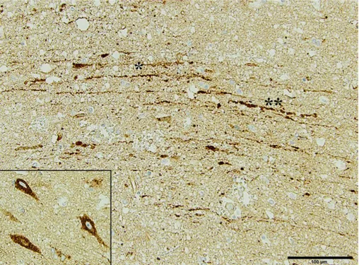
Histologically, DAI is characterised by β-amyloid precursor protein (βAPP) accumulations in injured axons as either a single large axonal swelling, the classical axonal bulb, or as periodic localised swellings, axonal varicosities (Figure 1) (97). βAPP is involved in axonal transport and it moves down the
axon via anterograde axonal transport. As the axon is injured, the axonal transport is interrupted, and thus βAPP accumulates. This produces reactive axonal swellings appearing as bead-like structures along the axonal length, representing disturbed but not totally interrupted axonal transport (94, 98).
Axonal stretch causes local mechanical dysregulation of sodium channels, re-sulting in calcium influx (99). The excess intracellular calcium leads to mi-crotubular loss and neurofilament impaction with damage to the cytoskeleton. In addition, neuronal depolarisation ensues as the cell membrane permeability is disrupted (100), while local mitochondrial damage leads to oxidative stress and disturbed energy metabolism (101). Calcium-mediated proteolysis of the cytoskeleton results in impaired axonal transport, swelling and ultimately dis-connection. In the most severe axonal injury, axonal poration leads to an un-controlled calcium surge, rapid breakdown of the axonal cytoskeleton and dis-tal axonal fragmentation and disconnection (94).
Figure 1. Immunohistochemical image from a TBI patient included in paper II of the thesis. Axonal injury is evident, here as pathological accumulation of beta-amyloid precursor protein (βAPP) in injured axons (*), and axonal swelling (**).
These neuropathological features may appear already within the first few hours and progress in size during 1-2 days following injury (81, 94, 97). Distal to the disconnection, Wallerian degeneration of the remaining axon occurs. Of interest, the βAPP can subsequently be cleaved by mainly presenelin-1 and
beta-site APP-cleaving enzyme (BACE-1) to yield β-amyloid (Aβ), the main component of insoluble aggregates observed in Alzheimer´s disease (AD) (102). Since TBI is a known environmental risk factor for AD (103-105), βAPP accumulations have caused DAI to be particularly appealing as a poten-tial link between the two diseases.
1.9 Impact of axonal injury
In TBI, structural lesions visualized with CT or conventional MRI fail to ex-plain the full extent of cognitive, emotional and behavioral symptoms ob-served (87, 106), mainly caused by disconnections in crucial brain networks due to axonal injury (107-109). In the human brain, neighboring and remote cortical regions are interconnected by a complex network of cortico-cortical axonal pathways (110), producing large-scale functional units. Patients suf-fering from DAI, in particular, tend to present severe deficits in cognition such as memory, attention and executive functions. Such complex cognitive func-tions are dependent on the integrity of widely distributed functional connec-tions (111). The brain displays a small world architecture, where local hubs are highly connected and integrated with spatially remote areas through long-distance white matter tracts, typically damaged in DAI. These large-scale con-nectivity networks are tightly coupled with high-level cognition. Conse-quently, axonal injury disrupts connectivity between brain structure and func-tion (108, 112). Cognitive deficits in TBI are associated with injuries in spe-cific white matter connections. Damage in white matter tracts connecting frontal to posterior regions, such as superior longitudinal fasciculus, corona radiata, internal capsule, and cingulum, leads to executive dysfunction (107, 113). Impaired connections in the thalamo-frontal network associate with at-tention and executive function deficits (114), while disruption in interconnect-ing fibers from the hippocampus, through the fornix to the basal forebrain and diencephalon relates to memory dysfunction and learning difficulties (107). In addition, damage to limbic fibers interconnecting temporal lobe structures to the medial frontal lobe is linked to memory deficiencies (108). Only re-cently have these disconnections been possible to visualize, with the develop-ment of advanced MRI methodology, particularly diffusion tensor imaging (DTI).
1.10 Detection of axonal injury
Generally in TBI, CT is the preferred initial imaging modality, due to its wide availability, rapid acquisition and sensitivity in visualizing acute hemor-rhages. However, its utility in DAI is limited, since CT poorly detects subtle axonal lesions in deep white matter structures (115, 116). Findings on CT are
mainly restricted to traumatic edema of the brain and petechial hemorrhage in the white matter suggestive of DAI. For diagnostic purposes, surveillance of axonal injury progression and adequate prognosis, other imaging modalities are more suitable.
1.10.1 Magnetic resonance imaging
Magnetic resonance imaging can detect microscopic amounts of blood (117) as well as non-hemorrhagic lesions in white matter associated with DAI (89), making this modality the primary method for detecting axonal injury in TBI patients. Additionally, MRI allows visualization, assessment and neurochem-ical analysis of critneurochem-ical white matter tracts.
1.10.1.1 Conventional MRI
Two MRI sequences sensitive to hemorrhagic lesions are widely available, the T2*-weighted gradient echo (T2*GRE) and susceptibility-weighted imaging (SWI). These sequences detect microhemorrhages from injury of small ves-sels running alongside axons. Paramagnetic properties of hemoglobin degra-dation products from the microhemorrhages produce distortion in the mag-netic field, and thus hypointense lesions appear (118). Susceptibility-weighted imaging is more sensitive than T2*GRE in detecting hemorrhagic lesions in deep seated white matter regions (119, 120). Taking advantage of susceptibil-ity differences between tissues (121, 122), it provides means to study micro-hemorrhages in deep brain regions previously difficult to detect. Importantly, both sequences sensitive to blood products depict lesions larger than their true size, and the lesion volume is strongly influenced by the MRI scanner’s prop-erties, in particular, the magnetic field strength (123). In addition, particularly the SWI sequence, may be difficult to interpret as deoxygenated blood in veins can mimic hemorrhagic lesions, and enlarged veins may result in areas of sig-nal loss (124).
Non-hemorrhagic lesions can be visualized with Fluid-Attenuated Inversion Recovery (FLAIR) and diffusion-weighted imaging (DWI). The FLAIR se-quence adds a long inversion time to a T2-weighted spin echo sese-quence to suppress CSF, causing it to appear dark in the images. Therefore, FLAIR aids in the detection of axonal injury near CSF spaces, particularly in the periventricular white matter, the corpus callosum and the brainstem (125, 126). The FLAIR sequence is however, unspecific to axonal injury and timing of the MRI acquisition strongly influences its ability to detect injury. Follow-ing TBI, lesions may in the acute phase represent tissue edema in white matter, but may also be due to old scars, since a similar signal can be caused by en-cephalomalacia or tissue gliosis (127). In addition, lesions seem to diminish with time (126), limiting its usefulness to some extent in DAI. The DWI se-quence is acquired by adding sequential gradient pulses to 90° and 180°
spin-echo sequences, and is sensitive to the microscopic motion of water molecules (128). It can therefore detect non-hemorrhagic lesions due to microstructural abnormalities following axonal shearing. The DWI sequence, in similarity with FLAIR, is influenced by the timing of the MRI, as the signal may evolve over time (129) and be significantly reduced already after 3 months post-TBI (126).
1.10.1.2 Diffusion tensor imaging
Diffusion tensor imaging is a technique where diffusion weighting is applied in different directions to estimate the amount of anisotropic water diffusion. This provides a quantitative measurement of axonal injury and enables precise anatomical reconstruction of white matter (130). Diffusion tensor imaging has enhanced sensitivity for axonal injury in comparison with conventional MRI, and accurately detects ultrastructural changes. By using post-processing tech-niques, DTI can be utilized to create detailed three-dimensional images of white matter tracts (131, 132). Parameters acquired with DTI include the mean diffusivity, which corresponds to the average diffusivity of water molecules, and the diffusion anisotropy or the fractional anisotropy (FA), measuring wa-ter molecules degree of directionality (130, 133). Additionally, the radial dif-fusivity measures water diffusion in two directions perpendicular to axons, while the axial diffusivity quantifies the diffusion along axons (133, 134). In DAI, reduced FA and increased diffusivity are observed (87, 135, 136), cor-relate to TBI severity (137, 138) and associate with cognitive and behavioral deficits (107, 139-144). Alterations in DTI parameters persist and may pro-gress until the chronic phase post-TBI, with continuously decreasing FA and increasing diffusivity over time, and measurable deterioration of axonal integ-rity beyond 24 months post-injury (136, 145-148). Diffusion tensor imaging is an extensively studied technique to visualize axonal injury. However, DTI is to some extent impractical in the acute phase in severe TBI, as post-pro-cessing statistics are required to produce the images. Additionally, variations in data acquisition and analysis techniques as well as the location of structures investigated in previous studies prevent generalized conclusions of the clinical utility (149).
1.10.1.3 Magnetic resonance spectroscopy
The chemical shift is a phenomenon caused by variations of proton resonance frequency due to the local chemical environment. This can be utilized to detect and quantify neurochemical alterations in the brain by a technique called mag-netic resonance spectroscopy (MRS) (150). The most studied metabolites in TBI are N-acetyl aspartate (NAA), a marker for neuronal and axonal integrity extensively found in neurons (151), and choline (Cho), which is increased af-ter damage to cell membranes (152). In TBI, a decrease in NAA is generally seen, whereas Cho is typically increased (153-159), and these findings asso-ciate with neurocognitive deficits (160-162) and global outcomes (154, 155,
163). Magnetic resonance spectroscopy can detect subtle axonal injury, not possible to visualize using conventional MRI. Furthermore, measurements of glutamate can indicate early excitotoxic injury (163), while lactate elevations can imply hypoperfusion and anaerobic metabolism (164, 165).
1.10.2 Neuromolecular imaging
Neuromolecular imaging techniques include SPECT and PET. These tech-niques can be used to assess the neurochemical and neurophysiological envi-ronment in the brain, either by assessing variables related to brain functional activity and energy metabolism, or by assessments of specific neurochemical metabolites (166).
1.10.2.1 Single-photon emission computed tomography
In SPECT, radiopharmaceutical agents are used to produce images of physio-logical or pathophysio-logical processes. Most commonly in TBI, [99mTc]
Hexa-methylpropylenamine oxime (HMPAO) and [99mTc] Ethylcisteinate dimer
(ECD) are used to measure the regional cerebral blood flow (rCBF) and indi-rectly, the regional cerebral metabolism (167). SPECT provides an adjunctive measure to anatomical imaging modalities of white matter injury and has the advantage of being both affordable and widely available. Specifically, SPECT allows an objective visualization of functional brain pathology, and thus is helpful in situation where deficits are discordant with visible structural inju-ries by MRI (168). Studies of rCBF following DAI have shown decreased blood flow in the frontal lobe and dysfunction in deep areas including the brainstem. Particularly, the cingulate gyrus is commonly involved despite a lack of visible abnormalities (169-171), a finding that is associated with neu-rocognitive deficits (170). Plausibly, this could be due to deafferentation of interconnecting white matter caused by widespread axonal damage (172, 173). In addition, research is currently focused on developing radiotracers for imag-ing of aggregates of Aβ and tau in AD (174, 175), efforts with potential im-plication also after DAI.
1.10.2.2 Positron emission tomography
In PET, radiopharmaceutical agents labelled with positron-emitting radioiso-topes, most commonly fluorine-18 [18F], carbon-11 [11C] and oxygen-15 [15O]
are used to image neurochemical and neurophysiological processes. The [15O]-radioisotope can be used to measure CBF, CBV and CMRO
2 with high
resolution (61). The commonly used [18F]-labeled fluorodeoxyglucose (FDG)
PET can measure local glucose metabolism, and thus regional neuronal activ-ity. Similar to SPECT, FDG PET studies have revealed regional hypometab-olism in medial frontal lobe structures including the cingulate gyrus in DAI (172, 176), findings associated with neuropsychological and cognitive
symp-toms (172, 176, 177). In addition [11C]PK11195, reflecting microglial
activa-tion, can be used to study neuroinflammation in the white matter following TBI (178). Furthermore, PET provides a method for imaging of substrates of axonal injury, particularly Aβ and tau, of special interest with regards to neu-rodegenerative aftermath following DAI. Imaging of Aβ using the [11C]
la-beled Pittsburgh compound B (PiB) have shown increased retention signals in the cortex and striatum in TBI, a finding similar to what is commonly observed in AD (179, 180). With regards to tau imaging, two small studies of retired contact sport athletes have revealed tau elevations in vivo (181, 182), however, the radiotracer used, [18F]FDDNP, binds both to tau tangles and Aβ
aggre-gates. New tracers such as [18F]T808, binding specifically to paired helical
fragments of tau, are currently being developed (183).
1.11 Classification of diffuse axonal injury
As previously mentioned, diffuse injuries can be classified using the Marshall CT classification, taking the degree of compression of the mesencephalic cis-terns, the degree of midline shift, and the presence or absence of one or more surgical mass lesions into account (26). However, this grading system is not specific for DAI. In 1989, Adams and colleagues proposed a histopathological grading for DAI based on autopsy findings in fatal TBI (81).The grading con-sisted of three grades and was based on the presence of axonal injury in the cerebral hemispheres with a predilection for the grey-white interface (Grade 1), the corpus callosum (Grade 2) and in the brainstem (Grade 3) (Table 7). With the development of advanced MRI, surrogate markers for the lesions found histopathologically in DAI became possible to visualize and therefore the Adams grading system has been adopted to neuroimaging (115). Today, this grading system is widely used. In severe TBI patients evaluated with MRI, at least 50 % are grade III DAI using this grading system, with evident lesions in brainstem structures (83). Nonetheless, it is important to recognize that MRI cannot appreciate the full extent of the histopathological findings.
TABLE 7.
ADAMS DAI GRADING (81) GRADE
I
II
III
PATHOLOGY Widespread axonal damage in white matter of cerebral hemispheres White matter damage extending to the
cor-pus callosum with tissue tear hemorrhages Pathology of grade I-II and in addition,
tis-sue tear hemorrhages in the brainstem
1.12 Consequences of axonal injury – clinical features
during neurointensive care
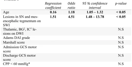
Patients with severe TBI and DAI in isolation exhibit highly variable clinical characteristics, distinct from patients with predominantly focal injuries. Alt-hough patients with focal injuries may present initially with a markedly de-pressed level of consciousness, this clinical feature is more commonly en-countered following DAI (93). In DAI, the deep coma seen at impact is caused mainly by damage to axons in diencephalic and brainstem structures (93, 184, 185). In addition, axonal injury in the pyramidal tract can cause motor deficit (186), without obvious radiological correlates on the initial CT scan. To date, methods used to prognosticate outcome following DAI in the acute setting are unreliable, and it is challenging to predict which patient will eventually regain consciousness and the time-point when this will occur (93, 184, 185). Fortu-nately, the advanced neuroimaging techniques previously described herein show promise in providing adequate prognostication in the near future. Some efforts have already been made to improve the prognostic accuracy with an increasing number of studies, and DAI-associated lesions in the corpus callo-sum, thalamus, and particularly the brainstem seem coupled to poorer out-comes (84, 125, 184, 187).
Moreover, the risk of developing elevated ICP in DAI is difficult to predict and studies evaluating the prevalence of high ICP show contradictory results (188-192). Already in 1982, Narayan and colleagues presented their experi-ence from ICP monitoring in 207 severe TBI patients, of which 61 had a nor-mal initial CT scan and were suspected to have DAI. Increased ICP was un-common in this patient category unless the patient was aged > 40 years, had unilateral or bilateral motor posturing or episodes of systolic blood pressure <
90 mmHg (189). Similarly, a low incidence of increased ICP has been ob-served by other authors (188, 192), although short elevations of ICP still exist and in some cases require treatment (188). On the contrary, one study found increased ICP in the majority of severe TBI patients, despite the absence of mass lesions, midline shift or compressed basal cisterns on the initial CT scan (190). This study has limitations however, due to the low sample size. Com-mon to all these studies, is that they were conducted before MRI was widely adopted in severe TBI management, and relied on CT scans with poorer qual-ity than today’s standards. Studies evaluating ICP in relation to MRI findings in DAI are scarce, however an association between the maximum ICP and the number of hemorrhagic lesions on T2*GRE has been implicated (191).
1.13 Consequences of axonal injury – a link to
neurodegeneration?
Compelling epidemiological evidence has linked TBI to the development of various neurodegenerative disorders (104, 105, 193-195). An episode of se-vere TBI has been observed to cause a fourfold increased risk for developing Alzheimer’s disease (AD) (193) and may also result in an earlier age at disease onset (196, 197). There is evidence suggesting that the risk of dementia rises with increased TBI severity (104, 193). Furthermore, large recent epidemio-logical studies have indicated an association between TBI and future onset of Parkinson’s disease (PD) (194, 195). Additionally, repetitive TBI has since long been linked to the development of cognitive and behavioral symptoms, a syndrome previously described as dementia pugilistica, but now termed chronic traumatic encephalopathy (CTE) (198, 199).
In AD, the hallmark histopathological findings are insoluble Aβ aggre-gates/plaques and neurofibrillary tangles (NFTs) consisting of hyperphos-phorylated tau (P-tau) protein (200). Aβ pathology has been strongly linked to the etiology of AD, and intracellular fibrillary deposits of tauare characteristic of a large variety of neurodegenerative disorders including AD (201). Accu-mulation of P-tau into NFTs is also the characteristic finding in CTE and has been observed in a majority of symptomatic athletes where this condition was diagnosed post-mortem. In addition, concomitant Aβ pathology is frequently seen also in CTE (40, 199), albeit with a different distribution than in AD (202).
1.13.1 Amyloid-beta pathology
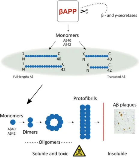
Following cleavage of βAPP, monomeric Aβ peptides of various lengths are produced. These peptides can either be N-terminally intact molecules or be
cleaved at the N-terminal, yielding N-terminally truncated forms of various lengths (203, 204). The Aβ monomers assemble as large soluble oligomers, defined as Aβ aggregates of variable molecular weights < 100 kDa, which may then assemble into protofibrils with a molecular weight > 100 kDa (205).
Figure 2. Evolution of Aβ aggregates begins with the enzymatic processing of βAPP that yields both N-terminally intact and truncated Aβ monomers, of which Aβ40 and Aβ42 are the most frequently found in Alzheimer’s disease related plaques. Monomers can then aggregate into soluble intermediary Aβ species, with potential toxic properties, and eventually aggregate further into insoluble Aβ plaques.
Further assembly of the soluble protofibrils can then form insoluble fibrils that constitute the Aβ plaques (Figure 2). Of particular interest for neurodegener-ative development are the monomeric Aβ40 and Aβ42 peptides, which are prone to aggregate and are the major Aβ species found in Aβ plaques. It is however not verified whether the Aβ plaques confer the neurotoxicity in AD, and more attention is currently attracted towards soluble intermediary Aβ spe-cies such as oligomers and protofibrils, which have been assigned neurotoxic properties (206-209).
In TBI, accumulation of βAPP is a feature observed in injured axons (97, 210), and detected within hours (211), where βAPP may co-accumulate with its Aβ converting enzymes presenelin-1 and BACE-1 (97). Furthermore, deposition of Aβ, both as soluble monomeric peptides and insoluble Aβ plaques have been consistently reported in brains of TBI patients (211-215). By using mi-crodialysis to analyze monomeric Aβ in ISF, it was also suggested that accu-mulations are more pronounced in patients with DAI, in comparison to focal injuries (213). Notably, immunohistochemical studies, as well as PiB PET im-aging, have shown that these accumulations may persist over time in a subset of patients (41, 180).
Aβ pathology is a general finding following TBI, however Aβ plaques seem restricted to about 30 % of patients across all age groups (211, 215, 216). Plausibly, this is due to genetic predispositions. Epidemiological data impli-cate the lipid transporter protein Apolipoprotein E ε4 (APOE ε4) allele as a risk factor for the development of AD pathology, particularly in APOE ε4 al-lele carriers who previously sustained a TBI (104, 105, 217, 218). The APOE allele distribution varies substantially among populations with ranges of 0-20 % for APOE ε2, 60-90 % for APOE ε3 and 10-20 % for APOE ε4 (219). In Caucasians, homozygote APOE ε4 genotype is prevalent in 1.2 – 3.2 % of the population while heterozygote APOE ε4 genotype ranges between 1.5 % in Italy to 30.6 % in Finland (220). Although the exact mechanism by which APOE ε4 conveys the increased risk of AD is unclear, APOE regulates Aβ proteolysis and clearance in an isoform-dependent manner and may modulate Aβ–induced oxidative stress (APOE ε4 < APOE ε3 < APOE ε2) (221-224). In early mild cognitive impairment, APOE ε4 may cause increased cortical Aβ deposition (225), implying an early role in Aβ dysregulation in AD progres-sion. Several meta-analyses show that the APOE ε4 allele is associated with a worse outcome following TBI, including an increased risk of seizures and de-mentia (226-228). Furthermore, the APOE ε4 allele has been associated with increased Aβ burden in TBI post-mortem brains and in CTE patients (202, 229). Experimental evidence also suggests that clearance of monomeric Aβ is impaired in APOE ε4 carriers following TBI (230).
1.13.2 Tau pathology
The microtubule-associated protein tau is an important structural element in the axonal cytoskeleton. It exists in six isoforms and under normal circum-stances its biological activity is regulated by phosphorylation. However, hy-perphosphorylation of tau may lead to aggregation in NFTs, which is associ-ated with a number of neurodegenerative diseases (201). In TBI, tau release is particularly linked to axonal injury (41, 231). Tau has been found elevated both in CSF and in ISF monitored by microdialysis in TBI patients (231-235), and ISF tau correlates to axonal injury depicted using DTI (232). Initial CSF tau levels seem to reflect injury severity and are associated with patient out-comes (236). In addition, high serum tau levels after TBI are associated with a worse outcome, probably reflecting a more severe axonal injury (237). Alt-hough tau deposited into NFTs has not been observed in the early phase post TBI, experimental evidence implies that P-tau develops rapidly after trauma (238). In young athletes who died within 6 months from a concussion, NFTs were observed in a subset (239). Furthermore, widespread NFTs were found in one-third of long-time survivors following a single TBI at autopsy (41), suggesting that NFTs take time to develop. The distribution of NFTs follow-ing moderate to severe TBI is similar to that seen in AD, however in TBI, NFTs were primarily seen in superficial cortical layers, as opposed to in deep layers in AD (41). This distribution of tau pathology is similar to the distribu-tion of NFTs in CTE following repetitive mild TBI/concussion observed in athletes (199). With the implementation of novel PET ligands specific for tau, further knowledge of temporal patterns of tau deposits will be acquired in the future (181, 182).
Aims
The general aim was to study axonal injury following severe TBI in humans, its detection in the clinical setting and its pathological features, with particular emphasis to neurodegeneration and potential long-term consequences.
2.1 Specific aims
I. To quantify the anatomical distribution of axonal in-jury using MRI, evaluate its impact on DAI patient out-comes and propose a novel grading system based on the widely used Adams histopathological grading (81).
II. To analyze Aβ deposition in surgically resected brain tissue post-TBI, including monomeric Aβ and soluble Aβ oligomers/protofibrils, and their relation to the APOE genotype.
III. To analyze the proteome profile of biopsies from struc-turally normal-appearing cortical tissue post-TBI, and compare proteome alterations in DAI vs focal TBI.
IV. To investigate the occurrence of increased ICP in DAI patients and to analyze whether anatomical distribu-tion of axonal injury on MRI associates with ICP ele-vations.
Material and methods
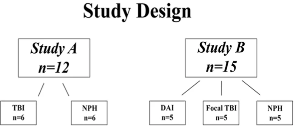
All research included was approved by the Regional Research Ethics Com-mittee at Uppsala University and was conducted in accordance with the ethical standards given in the Helsinki Declaration of 1975, as revised in 2008. For studies including human brain tissue (papers II and III), written informed con-sent was obtained. For patients with severe TBI, written informed concon-sent was obtained from the patients’ closest relatives and from the patients themselves when they had sufficiently recovered from their injury at > 6 months post-injury. Informed consent was also obtained from the idiopathic normal pres-sure hydrocephalus (iNPH) patients and neurologically intact (NI) subjects and AD patients (or their relatives) for post-mortem brain donations (paper II).
3.1 Patient population
3.1.1 Severe traumatic brain injury cohorts and timing of MRI
All TBI patients included in the papers had suffered a severe TBI, defined as post-resuscitation GCS scores ≤ 8. Patients were > 16 years of age, did not have any known neurological disorder and had a decreased level of conscious-ness mandating mechanical ventilation and ICP monitoring. Patients were treated in the NIC unit at the Uppsala University Hospital using the standard-ized protocol for severe TBI patients, previously described in the introduction.
The DAI cohorts (papers I, III and IV) included severe TBI patients with post-resuscitation GCS scores ≤ 8, but with no mass lesion ≥ 25 ml. There were no ischemic/vascular lesions explaining the decreased level of consciousness and the patients had DAI-associated lesions depicted on conventional MRI. In pa-per I, the aim was to study DAI-associated lesions in the early phase post-TBI. Timing of the MRI scan is of particular importance for non-hemorrhagic le-sions, since DWI lesions may become isointense to brain tissue after 5-7 days (129). To reduce the risk of missing lesions on DWI, the post-injury time to the MRI scan was chosen within one week. Preferably, the MRI scan should be performed within the first days post-injury. However, severe TBI patients are typically unstable during the first post-injury days and this would have caused the exclusion of a significant number of patients.
In papers III and IV the MRI was performed at 1 day – 8 weeks post-injury. In paper III, the objective was to confirm the DAI diagnosis. In paper IV, the rationale to also include patients who had performed MRI in the subacute phase was to decrease the risk of selection bias, since ICP instability may re-strict an early MRI in some patients. In paper II, severe TBI patients all suf-fered from life-threatening elevations of ICP and/or the presence of a space-occupying brain swelling or hemorrhage, mandating surgical focal decom-pression. Thus, the evacuated brain tissue could be stored, following informed consent, in an established tissue bank at our department (Uppsala Brain Bank-Trauma). Subsequently, tissue was extracted to allow the biochemical analysis performed in the study.
3.1.2 Control subjects
For obvious ethical reasons, fresh brain tissue from healthy control subjects was not possible to obtain. To allow comparison of TBI brain tissues with tissues lacking post-mortem alterations in papers II and III, we included pa-tients with iNPH, planned for ventriculoperitoneal (VP) shunt insertion. Prior to VP shunt insertion, these patients were evaluated clinically and lumbar CSF was obtained and analysed for Aβ42, total tau, P-tau and neurofilament-light (NFL). Care was taken to not include patients with clinical symptoms sugges-tive of AD and no iNPH patient had a CSF biomarker profile indicasugges-tive of AD or any other neurodegenerative disease. In addition to the iNPH patients in paper II, post-mortem temporal cortical tissue from five deceased patients with AD-related dementia, and post-mortem temporal cortex from four NI subjects were included. These tissues were obtained from the Uppsala Brain Bank at the Department of Pathology and Cytology.
3.2 Image acquisition and analysis
Patients with severe TBI were subjected to an admission CT, which was scored using the Marshall classification. When the clinical status had stabi-lized, patients with diffuse injury and suspected DAI underwent an MRI with a 1.5T scanner (papers I, III and IV). Imaging included three sequences, T2*GRE, DWI and SWI.
In paper I and IV, images were independently assessed by two raters, graded using Adams histopathological classification (81) and the number of DAI-as-sociated lesions were counted in different anatomical locations in the brain. In previous studies, anatomical description of DAI-associated lesions in the brainstem was not well defined in relation to visible anatomical structures (81), resulting in difficulties when comparing study findings. Therefore, an
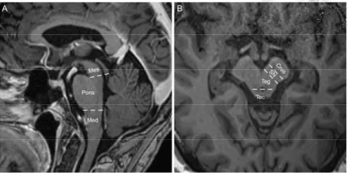
anatomical division of brainstem structures was performed to facilitate local-ization and grading (Figure 3). Anatomically, the mesencephalon was divided in three regions; (1) crus cerebri, (2) substantia nigra and mesencephalic teg-mentum, and (3) tectum including the superior and inferior colliculi. The pons and the medulla oblongata were divided into a ventral and a dorsal tegmental portion. Following the independent assessment of images by a neurosurgeon and a neuroradiologist, the inter-rater agreement was good in all assessed se-quences.
Figure 3. Definition of brainstem and mesencephalic structures. The mesencephalon was anatomically divided into the three regions; (1) crus cerebri, (2) substantia nigra and tegmentum, and (3) tectum including the superior and inferior colliculi, Mes = Mesencephalon, Med = Medulla oblongata, Crus = Crus cerebri, SN = Substantia nigra, Teg = Mesencephalic tegmentum, Tec = Mesencephalic tectum. Reprinted with permission from Journal of Neurotrauma.
3.3 Clinical and multimodality monitoring data
In all studies, clinical and demographic data were collected using the elec-tronic patient record system, the TBI NIC database, and the Uppsala TBI reg-istry, a registry established at our department in collaboration with Uppsala Clinical Research Center (UCR, www.ucr.uu.se, Uppsala University), includ-ing all severe TBI patients treated at the NIC (240). In papers I and IV, phys-iologic monitoring data were acquired using a computerized multimodality monitoring system collecting minute-by-minute average values (241). Data were assessed and validated manually and invalid data were withdrawn from the total monitoring time. Interruptions of data collection occurred when the patient was taken to the operating room or for radiologic evaluation and during network or software failures. The remaining monitoring data gave the good