Comparison of orthotic
interventions for
patients
with congenital talipes
equinovarus: a
systematic review
PAPER WITHIN: Orthotics
AUTHOR: Josefin Larsen & Matilda Strömbäck TUTOR: Cleveland Barnett
Sammanfattning
Syfte: Syftet med detta arbete är att undersöka olika ortos-lösningar med dess inkluderade resultat för patienter med kongenital idiopatisk klumpfot.
Metod: En systematisk sökning i databaserna MEDLINE, CINAHL, PubMed and Scopus genomfördes och väsentliga studier inkluderades utefter de förbestämda kriterierna. Inkluderade studiers validitet och möjliga partiskhet bedömdes samt att relevant data utifrån frågeställningen analyserades och besvarades.
Resultat: Resultatet presenterade 15 olika typer av ortoser, i 21 olika artiklar. De presenterade utfallsmåtten var inom komplians, återfall av deformationen, Pirani- och Dimeglio poäng, rörelseomfång samt inom funktionellt resultat.
Slutsats: Baserat på resultat går det inte att presentera en övervägande slutsats om vilken ortos-lösning som ger bäst resultat. Framtida forskning måste utveckla och framställa ortoser som tillfredsställer och möter patienternas behov.
Nyckelord: Klumpfot, PEVA, behandling, ortos, litteraturstudie
Summary
Aim: The aim with this study is to compare different orthotic solutions for pediatric patients with congenital talipes equnivarus.
Method: A search in the databases MEDLINE, CINAHL, PubMed and Scopus were made, and studies was included after the predefined criteria. The included studies were reviewed for risk of bias and validity, relevant data was extract and analyzed with regards to the research question.
Result: The result was presented on 15 different orthotic interventions in 21 different articles. The reported outcome measures were compliance, recurrence of deformity, Pirani- and Dimeglio score, range of motion and functional outcomes.
Conclusion: Based on the results a recommendation cannot be made on what orthosis that gives the best result. Future research should focus on developing and designing an orthosis that satisfies the patient's needs.
Table of content
Table of content ... 4
Previous research ... 7
Talipes equinovarus ... 8
Methods for correction ... 8
Terminology within orthotic device ... 8
Recurrence of deformity ... 9
Compliance with the brace ... 9
Pirani score ... 9
Dimeglio score ... 9
PBS- score ... 9
Functional outcome score ... 9
Research question ... 11
Research question... 12
Eligibility criteria ... 12
Search strategy and data collection ... 12
Screening process... 13
Outcome ... 13
Assessment risk of bias... 13
Internal validity, external validity and precision assessment ... 14
Analysis of data ... 14
Study selection ... 16
Dobb’s brace ... 18
Dennis Brown Brace ... 18
Foot abduction brace and Foot abduction orthosis ... 18
KAFO and AFO ... 19
Other orthotic solutions ... 19
Result of compliance ... 22
Result of Diméglio score ... 24
Result of functional outcome ... 25
Result of range of motion ... 25
Discussion ... 29
Effects of orthotic interventions ... 29
Translation of evidence into clinical practice ... 30
Limitations ... 31
Appendix list ... 33
References ... 34
Appendix 1: Search strategy ... 37
Appendix 2: Critical appraisal tool, in Swedish ... 39
a) Bedömning av icke-randomiserade studier av interventioner (effekt av att tilldelas en intervention (ITT)) ... 39
b) Bedömning av icke-randomiserade studier av interventioner (effekt av att fullfölja en intervention (per protokoll)) ... 41 c) Bedömning av randomiserade studier (effekt av att tilldelas en intervention (ITT)) ... 43 Appendix 3: SBU Bilaga 2: Granskningsmallar och checklistor för bedömning av studier... 44 Appendix 4: Form for Self-Assessment of Ethical Issues in Degree Projects1 at the School of Health and Welfare ... 51
Glossary
DBB= Dennis brown brace DB= Dobb’s brace
MS= Mitchell shoe
FAO= Foot abduction orthosis FAB= Foot abduction brace AFO= Ankle foot orhtosis
SFAB =Stenbeeck foot abduction brace MPB= Mitchell-Ponseti brace
Introduction
During our clinical placement at two different prosthetic and orthotic clinics in Sweden, different orthotic treatment for children with congenital clubfoot was observed. This made us question why different orthotic design is used and if there is any proven evidence if it’s leading to any different outcome. The common congenital deformity clubfoot occurs in somewhere between 1:250-1:1000 newborn children. Even if the misalignment of the foot or feet’s is classified as sever, by giving treatment early the most cases end up with a good result (Edinger et al., 2019). Untreated cases may, however, contain lifelong disability, deformity, and pain as describe by Ansar et al. (2018). The primary standard method used for clubfoot treatment is the Ponseti method. Which includes several parts as manipulation with serial casting, sometimes Achilles tenotomy and bracing. Foot-abduction braces, as the Dennis brown brace is considered to currently be the most used brace (Edinger et al., 2019). In Sweden there is no define national clinical guidelines for treatment of clubfoot. But the Swedish pediatric orthopedic quality register collect data about the currently used treatment method and its result, which is presented in their annual reports. When more sufficient data is collected, presented and analyzed that is thought to be used as a base for national clubfoot-treatment guidelines in Sweden (SPOQ, n.d). Several systematic reviews are published within the area clubfoot treatment, for example Ganesa et al. (2017), Gelfer et al.(2019), Jowett et al.(2011) and Zionts & Dietz (2010). While all these articles either compare the Ponseti method or/and different parts of the Ponseti method, the general conclusion is that the recurrency of deformity is generated by non-adherence with bracing. The recurrence rate of clubfoot treated with the Ponseti method differs in research, but it has previous been stated between 11%-48% (Haft et al., 2007; Morcuende et al., 2004). Despite all previous research, there seems not to be an agreement of which orthosis that should be used in clinical practice.
Previous research
Within the area of different methods for congenital clubfoot treatment some reviews are published. Ganesa et al. (2017) compare the Ponseti method with the Kite technique. The Ponseti method indicated an effective treatment method for correction of clubfoot, but the rate of recurrence was notable high. Ganesan et al. correlated this with a lack of brace adherence and socioeconomic factors. Systematic reviews that presents and analysis within the Ponseti methods in itself are for example published by Jowett et al.(2011) and Gelfer et al.(2019). In these studies, all the different parts of the Ponseti-treatment are included, such as the serial casting, surgery, and bracing. Both studies demonstrate that the Ponseti method is the currently best presented method in terms of successfully results, but as well in these studies non-compliance with the brace is stated as the effect creating relapse of deformity. Zionts & Dietz (2010) has in their published systematic review focus on the orthotic interventions that are presented within the Ponseti method. The authors include different alternatives of foot abduction braces as the Markell brace, Dobbs dynamic and Steenbeek brace. The authors does present different aspects associated with recurrence of deformity in these foot abduction braces such as nonadherence with the brace (Zionts & Dietz, 2010). Generally seen, whether it is a systematic review focusing on a comparison of the Ponseti treatment or an analyzation of the including parts of the treatment, the evidence in the last decade presents high rates of relapses since lack of brace compliance.
Background
Talipes equinovarus
Talipes equinovarus also known as Clubfoot affect between 1:250- 1:1000 newborn children and is classified as one of the most common congenital limb deformities. Approximately half of all the clubfoot cases is bilateral, and approximately 20% of the cases are related with other congenital abnormalities. The male to female ratio in the deformity is 2:1. No exact cause has been found why clubfoot occurs, but different factors as environment and genetic inheritance are suspect as contributory. The misalignment is characterised by sever deformity in forefoot, midfoot and hindfoot. Adductus occurs in the forefoot, cavus occurs in the midfoot and the hindfoot is affected by a rigid equinovarus. This results in medial navicular and cuboid subluxation and internal rotation of the calcaneus which displace it from the heel pad. Plantar, medial, and posterior contractions is created by shortening of the skeletal structure. The misalignment is also related with hypoplasia in the calf as well as potential leg length discrepancy (Edinger et al., 2019). Clubfoot is divided into three different types: idiopathic which means that there is no known reason for it, neurogenetic which means that it is secondary due to a neurological condition like cerebral palsy and syndromic which means that it is due to another underlaying syndrome. Often children get diagnosed with ultrasound during the pregnancy but can also be diagnosed at or after birth. Children born with clubfoot needs to be treated and the treatment generally is divided into two parts, casting followed by bracing (John Hopkins University, 2021).
Methods for correction
Nowadays the most common way for treating clubfoot is by using the Ponseti method but historically other methods such as the French functional method and the Kite technique has been used. The French method included stretching, assisted movement, and taping for maintenance. The Kite technique included long leg casting for manipulation and maintenance. But as these methods has been insufficient to reduce the deformity in a large proportion the Ponseti method has been introduced, and now used as a standard method (Edinger et al., 2019). The Ponseti method treats the affect foot or feet by manipulating and holding through plaster casting. The effected foot is in generally casted five to seven times and each time the foot is corrected more until it is fully corrected or even overcorrected since the chance of relapse is high. The last cast is used for about 3 weeks. This procedure takes about two months if the case is not classified as severe. In most cases an Achilles tenotomy is done in conjunction with the last casting procedure to allow full ankle dorsiflexion. Then as ending the treatment an orthosis should be used. The first three to four months the brace is used around 23-hours per day. Then during sleep for the following years, number of years varies individually, but often somewhere between 2-5 years. The function of the orthosis it to maintain and hold the affected foot. By using an orthosis, the chances of getting a deformity relapse are decreased (Edinger et al., 2019). The splint that is often used after the casting procedure is called a Dennis Brown splint and is named after an Australian surgeon. The orthosis is also referred to as the foot abduction brace. The splint is made from two boots and a bar. The boots are placed on the bar and should be shoulder length from each other (The Royal Children’s Hospital, 2007). A common setting could be 70° external rotation and 15° dorsiflexion (Edinger et al., 2019).
The Swedish pediatric orthopedic quality register describes 3 different orthotic interventions used for clubfeet in Sweden, the foot-abduction-orthosis, knee ankle foot orthosis and the ankle foot orthosis (SPOQ, 2018). The registry has in their annual reports not state if it is any correlation between used orthotic design and result (SPOQ, 2019). The biomechanical principal behind most of the designs and used orthoses includes external forces on the affected foot creating pronation, dorsiflexion, abduction, and an external rotation. In addition to the mentioned orthoses other interventions has been used, for example the shoes with straight or reverse lasts (Edinger et al., 2019).
Terminology within orthotic device
The international organization for standardization (ISO, 2020) has a standard of general terms that should be used for description within external orthoses. An orthosis should describe the body and joint that is encompassed. The orthosis should also be referred to as if it is prefabricated or custom fabricated and its biomechanical function. The biomechanical function can for example be to aim for prevention, maintenance or reduction of deformity (ISO, 2007).
Reported outcome following clubfoot correction
How researchers and clinicians use, measure, and evaluate the result of clubfoot treatment differ significantly. In Gelfer et al. (2020) systematic review the authors present and analyze the used outcome measures within idiopathic treatment according to the Ponseti method. In total the review present 36 different outcome measures (Gelfer et al., 2020). Below is a presentation of some of the commonly used outcome measures.
Recurrence of deformity
Recurrence of the clubfoot deformity that occurs at or after the initial treatment is commonly described as an outcome following the Ponseti method. The proportion of patients that suffer from recurrence differ, Morcuende et al.(2004) present a relapse rate of 11% and Haft et al. (2007) presented a relapse rate of 41%. Dobbs et al.(2004) also describe that the risk of recurrency is primarily during the orthotic procedure before the age of 4 years.
Compliance with the brace
There are different ways to investigate compliance, in the study done by Garg & Porter (2009) compliance is measured based on how adherent the patients and parents are to the treatment protocol based on reported hours of use. Other ways to measure compliance can be pressure sensors in the orthosis (Kuzma et al., 2020) and parents’ ability to take on and off the orthosis (Manousaki et al., 2016).
Pirani score
A way to measure the outcome of the Ponseti treatment is to use the Pirani scoring method. Pirani uses a score from 0-1 for each category, where 0 is normal, 0.5 is abnormal and 1 is severe abnormality. The maximum score can range from 0 to 6 where 0 is no deformity and 6 is severe deformity. When conducting this test six different characteristics for clubfoot is evaluated at and they are divided between signs in the hindfoot and signs in the midfoot. The hindfoot is scored in posterior crease, amount of flesh in the heel, rigidity of plantarflexion. The midfoot is scored based on crease on the medial side of the foot, curvature of the foot and how prominent the talus bone is (Dyer & Davis, 2006)
Jain et al. (2017) has done a study regarding the validity of the Pirani score and concluded that it is a reliable assessment tool. Gelfer et al. (2019) have demonstrated that the Pirani score is a reliable way for surgeons to assess a clubfoot during treatment, thereby not yet validated for assessment done by other healthcare professions.
Dimeglio score
Dimeglio score is used for an assessment of clubfeet, grading on a scale of 0-20, where 0 is a corrected foot and 20 is the most severe clubfoot. The scoring is divided into 4 different parts: equines, varus, rotation around the talus and adduction of forefoot. Each of these can get a score of 0-5, 0 being no deformity and 5 being severe deformity. The scores can later be divided into severity where 0-5 is grade 1, 5-10 being moderate deformity and grade II, 10-15 being severe feet and grade III and 15-20 being very severe feet and grade IV. This method includes a checklist and there is training material for new users of the Dimeglio score (Diméglio et al., 1995).
PBS- score
The Swedish pediatric orthopedic quality register present PBS-score as a way of scoring a foot based on its appearance and function. This is a validated test for children with clubfoot. This scoring system is explained as a way of assessing ambulating children that have a history of clubfoot and should therefore not be used when assessing an infant (Böhm & Sinclair, 2019).
Functional outcome score
Another way to assess clubfoot in children is the Functional outcome score. This assessment method is like the PBS-score, looking at ambulating children and should therefore be used on children that has already gone through correction of clubfoot and is not relevant when looking at assessing a foot during the treatment (Dietz et al., 2009).
These presented outcome measures are only a selection of several different outcomes that are used within research and clinical practice of clubfeet-treatment (Gelfer et al., 2020). All these outcomes provide good conditions for evaluation of the treatment. However, this also creates difficulties when it comes to comparisons between different studies since all the presented outcomes are not translatable to each other.
Aim
The aim of this thesis is to investigate how different orthotic interventions for pediatric patients with idiopathic congenital talipes equinovarus differ in outcomes, including maintenance of foot alignment, foot mobility as well as compliance-rate.
Research question
In pediatric patients with congenital idiopathic talipes equinovarus does the traditional treatment of foot abduction orthosis compared to other presented orthotic interventions create a different result in reported outcomes?
Method
As described by Dickson et al. (2017) a systematic review has the design to detect and assess the primary available evidence in a research area. Thereby a systematic review was chosen as study design to address the aim and research question. The PRISMA Checklist for systematic reviews from 2009 was followed for structure during the process (Liberati et al., 2009).
Research question
The research question is developed from the PICO-model, which is commonly used when structuring a clinical research. PICO is an acronym where the P stands for population, I stand for intervention, C stands for comparison and O stands for outcome. By using this method, it could easily be broken down into the different parts that was going to be investigated (Del Mar et al., 2017). In this project the population were children with congenital idiopathic talipes equinovarus and the intervention was foot abduction orthosis compared to other orthotic interventions and the outcome was not specified, instead all reported outcome measures was of interest.
Eligibility criteria
After deciding the research question and area of interest the criteria for inclusion and exclusion were determined. The different criteria´s were based on the main concepts from the research question. The included scientific articles all needed to be peer reviewed and written in English. All the articles also needed to be available in full text through Jönköping University library. The population of interested were participants with bilateral or unilateral congenital idiopathic talipes equinovarus, diagnosed before, at or after birth. All the patients needed to be treated according to the Ponseti method and thereby the treatment needed to include an orthotic intervention. The gender, age or ethnicity of the patients was not considered relevant as inclusion or exclusion. Reviews were excluded as well as articles published before the year 2000. The reason for only including evidence published after the year 2000 is based on the large research increase within this area that has taken place in the last two decades (Gelfer, et al., 2019).
Search strategy and data collection
An image of the final search can be seen in appendix 1, it includes all used terminology, MeSH-terms and number of hits for each database. Before the final search were compound and conducted, different scoping searches were made. Which are more simplified searches for prescribing the authors an overview over the published evidence, terminology and key issues withing the topic (Pilkington & Hounsome, 2017). Based on the scoping searches the final search begin to be constructed. The desired databases were MEDLINE, CINAHL, PubMed and Scopus as these are covering much in health science (Monash University Library, 2021). In discussion with Jönköping University Library these databases were confirmed as most relevant within the topic. The base in the final search was the research question that earlier were divided and structured according to the PICO-model. According to Booth (2017) a search can be conducted based on only the words in the PICO, but synonyms can also be included to make a broader search. It is common to only include the population (P) and the intervention (I) when conducting a search (Booth, 2017). As this project is based on a comparison between different orthotic interventions the authors desired to include the comparison (C) factors as well. In this case the search based on the desired PICO would be: P as children with congenital talipes equinovarus, I as foot abduction orthosis (often referred to Denis brown splint) and C as other orthotic solutions. In this project the O included all types of outcome measures but were desired to not include in the final search since the scoping searches identified a much smaller number of articles.
In all databases different searches was done for the synonymous of the population, intervention, and comparison. This was done separably to investigate the amount of result and its relevancy. Medical subject hedings (MeSH) were included in the allowing databases. As Dundar & Fleeman (2017b) describe, MeSH-terms is based on catalogues on different subject headings. By adding MeSH-terms different terminology within the same concept will be detected in the result, which gives the opportunity for a more comprehensive search (Dundar & Fleeman, 2017b). In MEDLINE and CINAHL MeSH-terms was marked by MH and brackets. The MeSH terms was put in quotation marks to indicate that the whole fraise should be coherent. Between synonyms OR was used and between different subjects AND was used. In PubMed the MeSH terms was identified by [MeSH] and the word in quotation marks to make sure that the words was coherent in the search. In Scopus the function of medical subheading
does not exist, the terms and synonymous were thereby separated and added for a broader search. Quotation mark was used for indication that the words needed to be coherent. When all the terms were combined the final search were made.
Articles that were written earlier than 2000 was directly excluded and results that was written in another language than English were excluded as well. By PubMed’s exclude function, non-peer reviewed articles were directly excluded. The final search in all different databases were conducted 2021-03-22.
Screening process
Duplications were removed by the authors manually before the screening process started, see detailed number of duplications in figure 1. The screening process was done in three parts, that includes screening of title, abstract and full-text separately (Dundar & Fleeman, 2017a). The first part included identification and application of the different article-titles against the predetermined inclusion/exclusion criteria's (see criteria under Eligibility criteria). As a result of that, many articles that were not consider relevant in terms of the aim could be sorted out. In the next step, the abstract was read by the authors. Bas on the pre-determined criteria's the different articles were either included or excluded. The last step was to read the whole study and determine if it was relevant based on the pre-determined criteria. If a study passed all the three steps whey were included further in the review for data extraction, quality assessment and data analysis. See detail number of excluded articles present in figure 1. All these steps were made by both authors independently. The program Rayyan was used to present the result of articles from the final search. The authors were in that program blinded from each other’s result when screening the title, abstract and full text. Any disagreement for either inclusion or exclusion that existed were primarily discussed between the two authors and secondarily with a third part, in this case the supervisor of the thesis.
Outcome
All the measured and presented outcomes related to the orthotic treatment were of interest in the analysis of the result. A specific outcome was not specified in this systematic review since this made the number of articles to limited, which was confirmed in the scoping searches.
Assessment risk of bias
SBUs critical appraisal templates are customized for systematic inspection of methods within different areas of healthcare (SBU, 2020). In support of identification with potential biases in the included articles, assessment templates published in (SBU, 2021) were used. Depending on the individually study design, suitable templates was applied.
In total 7 of the included articles did not state study design, see table 1. In these articles the SBU template for Assessment of non-randomized studies of interventions (effect of being assigned an intervention) were used. The authors of this thesis consider that template most appropriate accordingly to the individually information in the descriptive method sections. In total the three used templates were “Assessment of non-randomized studies of interventions (effect of being assigned an intervention (ITT))”, “Assessment of non-randomized studies of interventions (effect of completing an intervention (per protocol))” and “Assessment of randomized trials (effect of being assigned an intervention (ITT))”, see appendix 2 for templates. The assessment tables are written in Swedish, but are according to SBU either fully or partly translated of Cochrane risk-of-bias tools RoB:2 and ROBINS-1 (SBU, 2021). RoB:2 is design for randomized trials (Cochrane Methods Bias, n.d) and ROBINS-I are designed to use in non-randomized intervention studies (Sterne et al., 2016).
To ensure a common understanding of the included questions within the assessment tool the authors together conducted a test application of the different templates in three different non-included studies. When both authors agreed on the implication of all different questions, all 21 studies were appraised separately and independently. Different bias-related categories such as: bias from randomization or selection, confounding bias, classification of intervention bias and intended intervention bias, missing data bias, measuring outcome bias, reported result bias, reporting bias and conflict of interest were appraised and answered. The applicable answer was “yes”, “probably yes”, “probably no”, “no” or “information is missing”. An overall assessment as either high, moderate, or low risk of bias was decided in each category. The individual assessments were presented and discussed between the authors. Disagreements were investigated and a joint decision was made.
Internal validity, external validity and precision assessment
An assessment of the internal validity, external validity and precision in the included studies were made with support of the SBU “granskningsmallar” (translated = review templates), see appendix 3. The templates are based on question withing the categories internal validity, external validity, and precision. Each question was multi-answerable and had a defined score. The categories were defined by their total scores. To ensure a common understanding of the included questions within the assessment tool the authors together conducted a test application of the different templates in different non-included studies. When both authors agreed on the implication of all different questions, the assessment of the included articles was done individually. The individual assessments were presented and discussed between the authors. Disagreements were investigated and a joint decision was made.
Analysis of data
The primary outcome measures that were demonstrated in the included studies were recurrency of deformity, compliance with the brace, Diméglio score and Pirani score. Other outcomes that were included but only appeared in a small number of studies were functional outcomes and range of motion, see table 5 for a presentation of each studies and their outcome measures.
An analysis of each used and present orthotic intervention was made. Which included the orthosis design and function, based on the written description and if available also images and videos. The different orthosis that was equal and not equal in function and design could be identified and presented in the same category, see figure 2. Recurrence in relation to the used orthosis was in most of the included articles defined as recurrence of deformity needing additional treatment such as serial casting, Achilles tenotomy or/and re-bracing. The recurrence rate also described as relapse rate, were in the studies reported in either number of participants or number of feet. For clarification the results of recurrence for each article were decided to be presented in percentage of the total number of participants. To be able to analyze this data it was decided by the two authors to define different percentage limits for low, moderate, and high rate of relapse. 10% and below were decided as low rate of relapse, at 11% and up until 49% as moderate rate of relapse and at 50% and up till 100% as high rate of relapse. As previous studies investigating the recurrence rate of deformity within the Ponseti method has demonstrated big differences, the authors decided to define low rate of relapse as 10% and below, which approximately is the value that is presented in Haft et al.(2007) study. The other thresholds had no evidentiary support. The definition and how to measure compliance and non-compliance differed between the included studies. For example, some studies defined compliance on which proportion of time the orthosis was used, while other conducted interviews with the parents/caregivers. Thereby, an analysis of each individual study and measuring tool was made. Some of the articles also presented skin problems in relation to the orthotic use. In several of those studies, various skin problems were presented in a subcategory of compliance. Any reported skin problems were thereby decided by the authors to be included and analysed in the category compliance. In the same manner as in the analysis of relapse, the compliance rate was presented in percentage of the total number of patients. To be able to analyze and draw any conclusions from the individual compliance rate an individual definition of poor-, moderate- and good compliance was determined by the two authors. The definitions were: poor rate of compliance as 50% and below, moderate rate of compliance at 51% and up till 84% and good rate of compliance as at 85% and up till 100%. No evidence was found for guiding when defining the thresholds.
Several studies used the Pirani score or/and Diméglio score for severity grading of the feet before initial treatment. Only the articles that presented both a score before and after orthotic treatment were included in the data analysis of these scores. The value before and after bracing was compared individual since it differed between the studies when the measuring was completed. Since the studies that included functional outcome used different outcome scoring systems, the result was analysed individually within the used scoring system. Range of motion were analysed in the study that presented a value before and after bracing, this value was analysed individual in relation to the orthotic intervention.
Ethical consideration
According to the World Medical Association (2018) all research including human subjects must follow the ethical principles stated in Declaration of Helsinki. Before conducting this study, a dialog with supervisor regarding ethical consideration with this thesis have been made. Appendix 5 has been filled out for an understanding of the ethical needs for conduction of this study. As this is a systematic review, no human objects are used to conduct a study, instead it is dependent on other studies and
their research with human subjects. This means that it must be taken into consideration what kind of ethical approval the included studies have. This systematic review also includes 4 retrospective studies. A retrospective study looks back in time to find their participants and is in some countries therefore said not to be need an ethical approval, since is does not count as an invasive research. 7 out of 24 articles does not state that they have ethical approval, instead an informed consent from parents is stated.
Result
Study selection
When the first search was conducted 140 studies was identified. These studies have later been through a screening process which can be followed in the figure below. In the end of this screening process there was 21 studies left. Reasons for exclusion during the screening process was wrong population, wrong intervention, or systematic reviews as study design.
Figure 1. Flow chart of screened studies. Made from PRISMA template (Moher et al., 2009).
From: Moher D, Liberati A, Tetzlaff J, Altman DG, The PRISMA Group (2009). Preferred Reporting Items for Systematic Reviews and Meta-Analyses: The PRISMA Statement. PLoS Med 6(6): e1000097. doi:10.1371/journal.pmed1000097
For more information, visitwww.prisma-statement.org.
PRISM A 2009 Flow Diagram
Records identified through database searching (n = 140) Sc re e n in g In cl u d e d El ig ib ili ty Id en ti fi ca ti o n
Additional records identified through other sources
(n = 0)
Records after duplicates removed (n = 94)
Records screened (n = 94)
Records excluded (n = 61)
Full-text articles assessed for eligibility
(n = 33)
Full-text articles excluded, with reasons
(n = 12) - Wrong population (3) - Systematic review (1) - Wrong intervention (8) Studies included in critical
appraisal (n = 21)
Studies included for analysis of result
Study characteristics
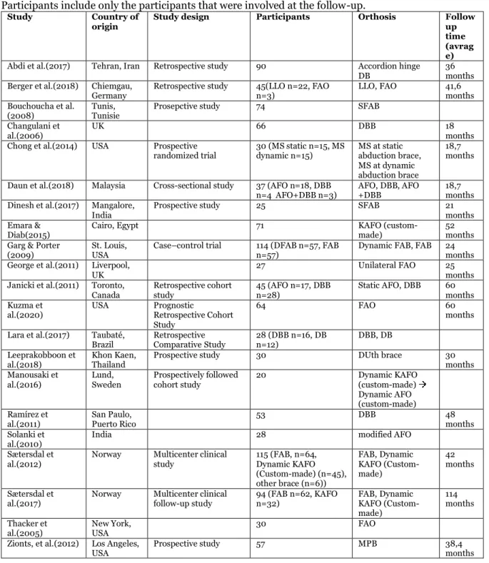
Table 1. Table of included studies and their characteristics. “ ” indicating change of orthosis. Participants include only the participants that were involved at the follow-up.
Study Country of
origin Study design Participants Orthosis Follow up
time (avrag e) Abdi et al.(2017) Tehran, Iran Retrospective study 90 Accordion hinge
DB 36 months Berger et al.(2018) Chiemgau,
Germany Retrospective study 45(LLO n=22, FAO n=3) LLO, FAO 41,6 months Bouchoucha et al.
(2008) Tunis, Tunisie Prosepctive study 74 SFAB Changulani et
al.(2006) UK 66 DBB 18 months
Chong et al.(2014) USA Prospective
randomized trial 30 (MS static n=15, MS dynamic n=15) MS at static abduction brace, MS at dynamic abduction brace
18,7 months Daun et al.(2018) Malaysia Cross-sectional study 37 (AFO n=18, DBB
n=4 AFO+DBB n=3) AFO, DBB, AFO +DBB 18,7 months Dinesh et al.(2017) Mangalore,
India Prospective study 25 SFAB 21 months
Emara &
Diab(2015) Cairo, Egypt 71 KAFO (custom-made) 52 months Garg & Porter
(2009) St. Louis, USA Case–control trial 114 (DFAB n=57, FAB n=57) Dynamic FAB, FAB 24 months George et al.(2011) Liverpool,
UK 27 Unilateral FAO 25 months
Janicki et al.(2011) Toronto,
Canada Retrospective cohort study 45 (AFO n=17, DBB n=28) Static AFO, DBB 60 months Kuzma et
al.(2020) USA Prognostic Retrospective Cohort Study
64 FAO 60
months Lara et al.(2017) Taubaté,
Brazil Retrospective Comparative Study 28 (DBB n=16, DB n=12) DBB, DB Leeprakobboon et
al.(2018) Khon Kaen, Thailand Prospective study 30 DUth brace 30 months Manousaki et
al.(2016) Lund, Sweden Prospectively followed cohort study 20 Dynamic KAFO (custom-made) Dynamic AFO (custom-made) Ramírez et
al.(2011) San Paulo, Puerto Rico 53 DBB 48 months
Solanki et
al.(2010) India 28 modified AFO
Sætersdal et
al.(2012) Norway Multicenter clinical study 115 (FAB, n=64, Dynamic KAFO (Custom-made) (n=45), other brace (n=6)) FAB, Dynamic KAFO (Custom-made) 42 months Sætersdal et
al.(2017) Norway Multicenter clinical follow-up study 94 (FAB n=62, KAFO n=32) FAB, Dynamic KAFO (Custom-made)
114 months Thacker et
al.(2005) New York, USA 30 FAO
Zionts, et al.(2012) Los Angeles,
Result of orthotic intervention
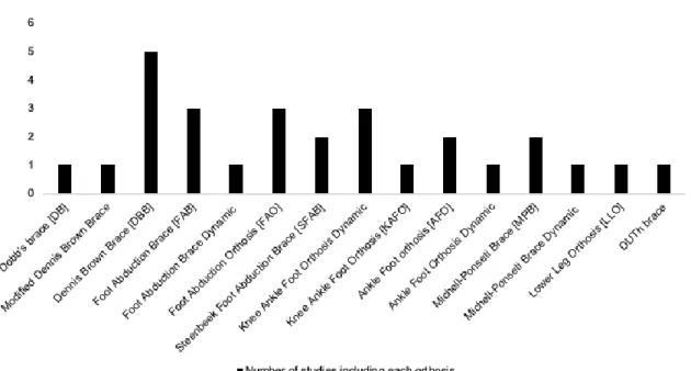
In this study 15 different orthotic solutions could be identified which is specified in figure 2. Orthotic design and relapse rate for all studies are presented in table 6.
Figure 2. Diagram that demonstrates number of each orthotic design presented in the included studies. Vertical axes illustrate how many times each orthotic was used.
Dobb’s brace
A traditional DB was used in one study (Lara et al., 2017) where they included 12 patients. The brace had the settings of 60°-70° external rotation on the effected side and 30°-40° external rotation on the non-effected side. They also had a dorsiflexion of 10°-15° on the effected side and the feet was placed as wide as the patients’ shoulders were. The orthosis was prescribed to be used for full time during the three first months and then for approximately 12 hours each day and be worn for three to four years. The rate of recurrence was 8.33%. The authors recommend further use of the orthosis (Lara et al., 2017). Dennis Brown Brace
DBB was used in seven studies (Reza Abdi et al., 2017; Changulani et al., 2006; Daun et al., 2018; Garg & Porter, 2009; Janicki et al., 2011; Lara et al., 2017; Ramírez et al., 2011). Garg & Porter (2009) had a historical control group using the DBB. All studies except Daun et al.(2018) and Garg & Porter (2009) stated that their settings were 70° external rotation on the effected side and 40°-45° external rotation on the non-effected side and that the feet was placed as wide as the patients shoulders. Lara et al.(2017) and Abdi et al.(2017) also included dorsiflexion of 10°-15° on both feet. The most common way was to prescribe the orthosis was for full time wear during the three first months and then during night and nap time until four years of age. Daun et al.(2018) did not report the prescribed wear time. Abdi et al.(2017) had a recurrence rate of 11% while Changulani et al.(2006) had a recurrence rate of 32%, Garg & Porter (2009) had a recurrence rate of 39%, Janicki et al.(2011) had a recurrence rate of 31%, Lara et al.(2017) had a recurrence rate of 8.33% and Ramírez et al.(2011)had a recurrence rate of 30%. Daun et al.(2018) did not report recurrence rate. All mentioned studies recommend to further use the DBB. Foot abduction brace and Foot abduction orthosis
Three studies used a FAB (Garg & Porter, 2009; Sætersdal et al., 2017; Christian Sætersdal et al., 2012; C. Sætersdal et al., 2012). Garg & Porter (2009) had 57 patients using the FAB while Sætersdal et al.(2012) and Sætersdal et al.(2017) had 64 patients using a FAB. All studies had the orthosis prescribed to be used full time for three months and then for night and nap time until age four. Garg & Porter (2009) had a recurrence rate of 19% and Sætersdal et al.(2012) had a recurrence rate of 23.28%. Sætersdal et al.(2012) and Sætersdal et al.(2017) recommend the use of FAB.
In total four studies (Berger et al., 2018; George et al., 2011; Kuzma et al., 2020; Thacker, Scher, Sala, van Bosse, et al., 2005; Thacker, Scher, Sala, Van Bosse, et al., 2005) used FAO as an orthotic treatment.
George et al.(2011) had a unilateral FAO. Thacker et al.(2005) used a FAO with MJ Markell shoes. Berger et al.(2018), George et al.(2011), Kuzma et al.(2020) and Thacker et al.(2005) prescribed the brace to be used for full time for three months and then 10 hours each day for three to four years George et al.(2011) and Thacker et al.(2005) had the setting of 70° of abduction on the effected side and the non-effected side was put in 45° of abduction. Both feet were put into 15° of dorsiflexion. George et al.(2011) had recurrence of 31.4%, Kuzma et al.(2020) had recurrence of 40% and Thacker et al.(2005) had 57% recurrence.
SFAB was used in two studies (Bouchoucha et al., 2008; Dinesh et al., 2017). Bouchoucha et al. (2008) brace was set to 60°-70° of external rotation on the effected foot with 15° of dorsiflexion. Dinesh et al.(2017) had the settings 70° abduction on the effected side and 45° abduction on the non-effected side and both feet were also put into 15° of dorsiflexion and as wide as the patients’ shoulders. Both studies prescribed the brace for full time for three months and then night and nap time, but it’s not stated for how long. Bouchoucha et al. (2008)had a recurrence rate of 11% and Dinesh et al.(2017) had 5.3%. Both studies recommend the use of SFAB.
KAFO and AFO
Four studies (Emara & Diab, 2015; Manousaki et al., 2016; Sætersdal et al., 2017; C. Sætersdal et al., 2012) used a KAFO in their treatment of clubfoot. Emara & Diab(2015) had a static KAFO while (Manousaki et al., 2016) had joints in the ankle and Sætersdal et al.(2012) and Sætersdal et al.(2017) had joints in both the ankle and the knee. Emara & Diab (2015) and Sætersdal et al. (2012) prescribed the orthosis full time for three months and then until the age of four during night and nap time. Manousaki et al. (2016) prescribed the orthosis for 18 hours each day for the first two months and then 12 hours each day for eight months. Emara & Diab (2015) had a recurrence rate of 17.46% and Manousaki et al. (2016) had a recurrence rate of 0%. Sætersdal et al. (2012) and Sætersdal et al.(2017) had a recurrence rate of 23.28% and in their follow up study (Sætersdal et al., 2017) they concluded that patients using a FAB had better outcomes after 8-11 years of follow-up but better compliance with the custom-made dynamic KAFO. The authors in the mentioned studies still recommend the FAB.
AFO was used in four studies (Daun et al., 2018; Janicki et al., 2011; Manousaki et al., 2016; Solanki et al., 2010). Janicki et al. (2011) had AFO as treatment until the year 2002 when they got new guidelines to use the DBB. Manousaki et al. (2016) used a custom made AFO with ankle joints. Manousaki et al. (2016) used the AFO after the initial ten months of using KAFO and for 10 hours each day. They had no recurrence but only recommend using it if there are problems adhering to a DBB. Janicki et al. (2011) used the AFO full time until the age of five and had a recurrence of 83%. They do not recommend using an AFO. Solanki et al. (2010) used a custom-made low temperature AFO that could be remolded when the patients grow. It was used until the patients started to walk. Solanki et al. (2010) also stated that their AFO could be used when there are problems with adhering to a DBB.
Other orthotic solutions
MPB static was used for two studies (Chong et al., 2014; Zionts et al., 2012) and MPB dynamic was used in one study (Chong et al., 2014). Chong et al. (2014) had 15 patients using the MPB dynamic and 15 patients using the MPB static while Zionts et al. (2012) had 57 patients using the MPB static. Both braces had a quick release mechanism to easier be able to take on and off the bar. Zionts et al. (2012) had the settings of 60° abduction on the effected side and 30° of abduction on the non-effected side. The shoes were placed as wide as the patients shoulders. Both studies prescribed the braces to be used full time for three months and then during nighttime and naps. Chong et al. (2014) reported recurrence in 27% of the patients and Zionts et al. (2012) reported 28% recurrence.
Berger et al. (2018) used a custom-made LLO on 22 patients. The settings of the LLO were 40° external rotation to begin with but then ended up with an external rotation of 20° for best result. The braces were prescribed to be used for 24h/day the first three months and then 10h/day until the age of 3-4. Relapse rate was not reported. The authors states that LLO can be an alternative for DBB when there are problems with adherence.
DUTh brace was used by Leeprakobboon et al. (2018). DUTh brace is a custom-made orthosis made from two footplates attached with a bar. The distance between the plates should be as wide as the patients shoulders. The setting of the plates is 70° external rotation on the effected side and 40° of external rotation on the non-effected side. Both feet have 10° of dorsiflexion. The brace is prescribed to
be used 24h/day for three months and then during night and nap time. Relapse rate was not reported. The authors recommend the DUTh brace as an alternative in treatment of clubfoot.
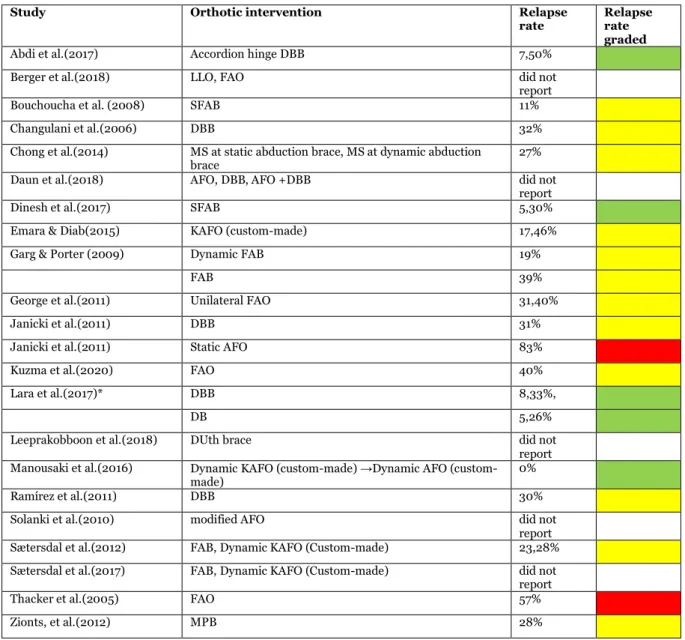
Table 6. Table of relapse rate in each study. Green= low relapse rate, yellow=moderate relapse rate, red=high relapse rate, white=could not define.
Study Orthotic intervention Relapse
rate Relapse rate graded
Abdi et al.(2017) Accordion hinge DBB 7,50%
Berger et al.(2018) LLO, FAO did not
report
Bouchoucha et al. (2008) SFAB 11%
Changulani et al.(2006) DBB 32%
Chong et al.(2014) MS at static abduction brace, MS at dynamic abduction
brace 27%
Daun et al.(2018) AFO, DBB, AFO +DBB did not
report
Dinesh et al.(2017) SFAB 5,30%
Emara & Diab(2015) KAFO (custom-made) 17,46%
Garg & Porter (2009) Dynamic FAB 19%
FAB 39%
George et al.(2011) Unilateral FAO 31,40%
Janicki et al.(2011) DBB 31%
Janicki et al.(2011) Static AFO 83%
Kuzma et al.(2020) FAO 40%
Lara et al.(2017)* DBB 8,33%,
DB 5,26%
Leeprakobboon et al.(2018) DUth brace did not
report Manousaki et al.(2016) Dynamic KAFO made) →Dynamic AFO
(custom-made) 0%
Ramírez et al.(2011) DBB 30%
Solanki et al.(2010) modified AFO did not
report Sætersdal et al.(2012) FAB, Dynamic KAFO (Custom-made) 23,28% Sætersdal et al.(2017) FAB, Dynamic KAFO (Custom-made) did not
report
Thacker et al.(2005) FAO 57%
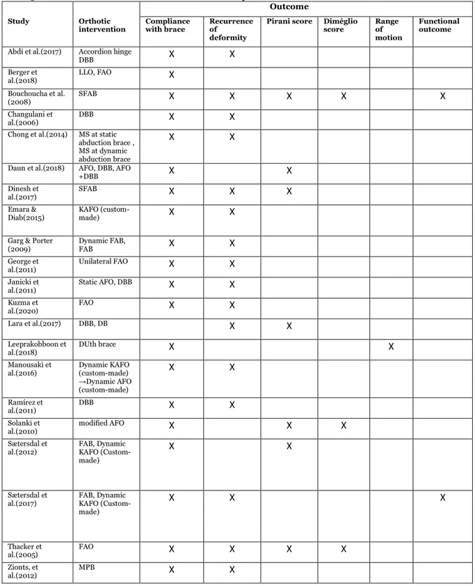
Result of presented outcomes
The present outcomes that could be identified in the included studies is Pirani score, Dimeglio score, compliance with brace, range of motion, recurrence of deformity, functional outcome, gait analysis and reported sin problems. Each individual study and its presented outcome are summarized in table 5.
Table 5. Table of the outcomes included in each study.
Outcome
Study Orthotic
intervention Compliance with brace Recurrence of
deformity
Pirani score Diméglio
score Range of
motion
Functional outcome
Abdi et al.(2017) Accordion hinge
DBB X X
Berger et
al.(2018) LLO, FAO X Bouchoucha et al.
(2008) SFAB X X X X X
Changulani et
al.(2006) DBB X X
Chong et al.(2014) MS at static abduction brace , MS at dynamic abduction brace
X X
Daun et al.(2018) AFO, DBB, AFO
+DBB X X
Dinesh et
al.(2017) SFAB X X X
Emara &
Diab(2015) KAFO (custom-made) X X
Garg & Porter
(2009) Dynamic FAB, FAB X X George et
al.(2011) Unilateral FAO X X Janicki et
al.(2011) Static AFO, DBB X X Kuzma et
al.(2020) FAO X X
Lara et al.(2017) DBB, DB X X
Leeprakobboon et
al.(2018) DUth brace X X
Manousaki et
al.(2016) Dynamic KAFO (custom-made) →Dynamic AFO (custom-made) X X Ramírez et al.(2011) DBB X X Solanki et
al.(2010) modified AFO X X X
Sætersdal et
al.(2012) FAB, Dynamic KAFO (Custom-made)
X X
Sætersdal et
al.(2017) FAB, Dynamic KAFO (Custom-made) X X X Thacker et al.(2005) FAO X X X X Zionts, et al.(2012) MPB X X
Result of compliance
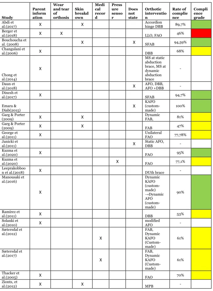
Table 7 presents those studies that have reported compliance, that includes how it has been measured, the result for each intervention, the measured rate and its grade of compliance. Four studies gave examples on how to improve compliance. Changulani et al., (2006) and Chong et al. (2014) stated that education for the parents including how the brace works and the risks with not using it would improve compliance. Daun et al. (2018) stated that frequent follow-up until the compliance is high decreases the risk for low compliance later during the treatment. Berger et al. (2018) tried to implement a bed night routine in the patients with non-compliance and this showed positive results. Abdi et al. (2017) and Emara & Diab (2015) both stated that the ability to move the legs independent lowers the risk of non-compliance.
Six studies (Chong et al., 2014; Dinesh et al., 2017; Garg & Porter, 2009; George et al., 2011; Manousaki et al., 2016; Ramírez et al., 2011) could find a relationship between non-compliance and recurrence while two studies (Sætersdal et al., 2012; Kuzma et al., 2020) stated that there was no relationship between non-compliance and recurrence. 2 participants using the dynamic FAB and 11 participants using the FAB developed skin complication such as skin ulceration and blistering (Garg & Porter, 2009). Ten studies (Bouchoucha et al., 2008; Dinesh et al., 2017; Chong et al., 2014; Daun et al., 2018; Garg & Porter, 2009; George et al., 2011; Janicki et al., 2011; Kuzma et al., 2020; Ramírez et al., 2011; Solanki et al., 2010) did not define compliance. But Bouchoucha et al. stated that 4 patients using the SFAB irregularly developed heel ulceration (Bouchoucha et al., 2008)
Nine studies (Abdi et al., 2017; Berger et al., 2018; Emara & Diab, 2015; Leeprakobboon et al., 2018; Manousaki et al., 2016; Sætersdal et al., 2012; Sætersdal et al., 2017; Thacker et al., 2005; Zionts et al., 2012) defined good compliance as when the patients adhered strictly to the included protocol. One study (Changulani et al., 2006) defined good compliance as wear time more than 10h/day. The participant using a FAO in Berger et al. (2018) that reported skin-problem were categorized as non-compliant with the intervention. The one patient that got affected by a serious cutaneous problem changed to a resin modified custom-made shoe on the FAO instead. None of the participants using an LLO reported any skin problems (Berger et al., 2018). One patient using the DUth brace got affected by skin breakdown (Leeprakobboon et al., 2018). 8 patients using the MPB developed skin problems in relation to the brace (Zionts et al., 2012).
Table 7. Table of how studies have reported compliance, which braces that were used and its rate of compliance. Green= good compliance, yellow=moderate compliance, red=poor compliance, white= could not be defined.
Study Parent inform ation Wear and tear of orthosis Skin breakd own Medi cal recor d Press ure senso r Does not state Orthotic interventio n Rate of complie nce Compli ence grade Abdi et
al.(2017) X X Accordion hinge DBB 89,7%
Berger et
al.(2018) X X LLO, FAO 46%
Bouchoucha et al. (2008) X X SFAB 94,59% Changulani et al.(2006) X DBB 68% Chong et al.(2014) X MS at static abduction brace, MS at dynamic abduction brace - Daun et
al.(2018) X AFO, DBB, AFO +DBB -
Dinesh et al.(2017) X SFAB 94,7% Emara & Diab(2015) X KAFO (custom-made) 100% Garg & Porter
(2009) X X Dynamic FAB, 81%
Garg & Porter
(2009) X X FAB 47%
George et
al.(2011) X Unilateral FAO 77,78%
Janicki et
al.(2011) X Static AFO, DBB -
Kuzma et
al.(2020) X FAO 95%
Kuzma et
al.(2020) X FAO 77,1%
Leeprakobboo
n et al.(2018) X DUth brace -
Manousaki et al.(2016) X Dynamic KAFO (custom-made) →Dynamic AFO (custom-made) 90% Ramírez et al.(2011) X DBB 53% Solanki et
al.(2010) X modified AFO -
Sætersdal et al.(2012) X FAB, Dynamic KAFO (Custom-made) 61% Sætersdal et al.(2017) X FAB, Dynamic KAFO (Custom-made) 61% Thacker et al.(2005) X FAO 70% Zionts, et al.(2012) X X MPB -
Result of Pirani score
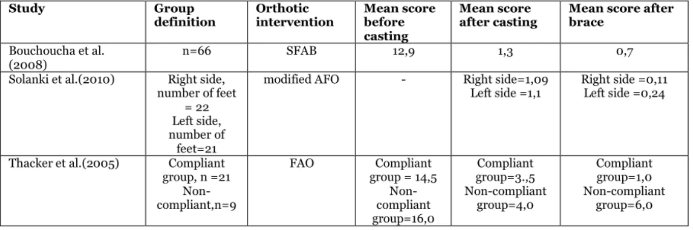
The 7 articles seen in table 8, presented Pirani score as an outcome, measured before and after bracing. In general, all the present articles demonstrated a decrease between the first and last measured Pirani score. Solanki et al. (2010) did not present a measured mean score before casting. Daun et al. (2018) and Sætersdal et al. (2012) did not present a mean score measured between the end of the serial casting and initial bracing. Dinesh et al. (2017) presented the mean score after bracing in two categories, the patient that experienced compliance with the brace and the patients that not experienced compliance with the brace. Compared with the mean score after casting, the compliant group demonstrated decrease in mean score of total 0,625. However, the score in the non-compliant group demonstrated an increase of 0,75.
The patient that used an FAO (Thacker et al., 2005) and DUth brace (Leeprakobboon et al., 2018) demonstrated a slightly score increase after bracing compared with the measured score after serial casting.
Table 8. This table present each study including Pirani score, with mean score before and after bracing. “-“ Indicates that data is not stated. n= number of participants
Study Orthotic
intervention Mean score before casting
Means score after
casting Mean score after bracing
Bouchoucha et al.
(2008) SFAB 3,4 0,45 0,3
Daun et al.(2018) AFO, DBB,
AFO+DBB 2,18 - 0,19
Dinesh et al.(2017) SFAB 3,79 0,65 Compliant group(n=36): 0,025 Non-compliant group (n=2): 1,4 Leeprakobboon et
al.(2018) DUth brace 5,6 0,07 0,1
Solanki et al.(2010) Modified AFO - 1,095 0,175 Sætersdal et
al.(2012) FAB 4,8 - 0,4
Sætersdal et
al.(2012) Dynamic KAFO (Custom-made)
4,8 - 0,6
Thacker et al.(2005) FAO 5,25 0,65 0,75
Result of Diméglio score
The present articles in table 9 included Diméglio score as an outcome measure, measured before and after bracing. How the authors of the studies have presented the result based on the participants differs. Bouchoucha et al. (2008) demonstrated a total mean score for all 66 participants. Solanki et al. (2010) presented the result for each individual affected foot. Thacker et al. (2005) divided the result into two groups, non-compliant and compliant.
Table 9. This table presents Diméglio score before casting and after bracing. n=number of participants, “-“ = data is not stated.
Study Group
definition Orthotic intervention Mean score before casting
Mean score
after casting Mean score after brace Bouchoucha et al.
(2008) n=66 SFAB 12,9 1,3 0,7
Solanki et al.(2010) Right side, number of feet
= 22 Left side, number of
feet=21
modified AFO - Right side=1,09
Left side =1,1 Right side =0,11 Left side =0,24
Thacker et al.(2005) Compliant group, n =21 Non-compliant,n=9 FAO Compliant group = 14,5 Non-compliant group=16,0 Compliant group=3.,5 Non-compliant group=4,0 Compliant group=1,0 Non-compliant group=6,0
Result of functional outcome
Bouchoucha et al. (2008) measured functional score with the functional classification of the Hospital for Joint Diseases. The mean score before casting were 13 and after full-time bracing 55,4. The follow-ups that were done in an average of 6,4 months after finished bracing, demonstrated a mean score of 54.
Sætersdal et al. (2017) used and evaluated two different clubfoot questionaries, Laaveg and Ponseti´s functional rating system and Roye´s Disease-specific Instrument. Both questionaries regarded the patient’s satisfaction, pain and function and was answered from the parents. The score in total could be divided between 0-100 in both questionaries. The patients that were using the bilateral FAB demonstrated higher functional outcome based on the used outcome measurement-tools.
Table 11. Presentation of Bouchoucha et al. (2008) functional outcome.
Study Orthotic
intervention Measurement tool before casting Mean score after full-time Mean score bracing Mean score after follow-up (mean 6,4 months) Bouchoucha et al.
(2008) SFAB classification of the The functional Hospital for Joint
Diseases
13 55,4 54
Table 12. Presentation of Sætersdal et al. (2017) functional outcome.
Study Measurement tool Orthotic
intervention Score
Sætersdal et al.(2017) Laaveg and Ponseti´s
functional rating system FAB 87 Sætersdal et al.(2017) Laaveg and Ponseti´s
functional rating system Dynamic KAFO 78 Sætersdal et al.(2017) Roye´s Disease-specific
Instrument FAB 82
Sætersdal et al.(2017) Roye´s Disease-specific
Instrument Dynamic KAFO 74 Result of range of motion
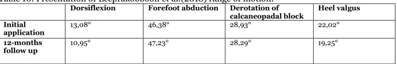
Leeprakobboon et al. (2018) included range of motion as an outcome, it included passive motions in the ankle dorsiflexion, forefoot abduction, derotation of calcaneopedal block and heel valgus. The abduction was measured in relation to the hindfoot and calcaneopedal block was measured in the horizontal plane.
Table 10. Presentation of Leeprakobboon et al.(2018)range of motion.
Dorsiflexion Forefoot abduction Derotation of
calcaneopadal block Heel valgus Initial
application 13,08° 46,38° 28,93° 22,02°
12-months
follow up 10,95° 47,23° 28,29° 19,25°
Result quality assessment
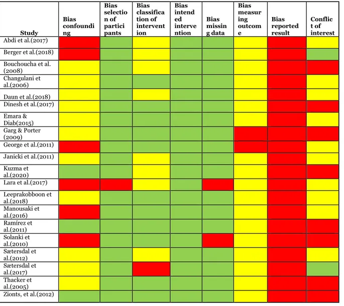
Bias is divided into two different sections, randomized studies, and not randomized studies. The randomized section only included one study. Conflict of interest was assessed as low if both questions was answered yes, moderate if either of the questions was answered no and high if both questions was answered no, see questions in appendix 2.
Chong et al. (2014) was the only randomized study included. The study was assessed as a randomized trial (effect of being assigned an intervention (ITT)) and the bias was assessed from the template that can be found in appendix 2. The study showed low risk of bias in both randomization and missing data but had moderate risk of bias based on intended intervention, in measuring outcome and in conflict of interest. Report bias is high because a study report or registration could not be found.
Table 2. Table of bias in randomized studies. Green= low risk of bias, yellow=moderate risk of bias and red=high risk of bias.
Study
Bias randomi
zation Bias intended intervention Bias missing data Bias measuring outcome Bias reported result Conflict of interest Chong et al.(2014)
There where 20 studies that was not randomized. These studies where assessed as either not randomized assigned interventions or as non-randomized studies of interventions (effect of being assigned an intervention (ITT)) or non-randomized studies of interventions (effect of completing an intervention (per protocol)). The templates used for assessing bias can be found in appendix 2. Confounders that could be identify in this review is different parental factors such as education, economics, and motivation to fulfill the treatment, social factors, esthetic factors with wearing a brace and the education and learning curve of the medical organization.
3 studies showed low bias of confounders. These studies presented a table of factors that could have affected the study. 11 studies showed moderate risk of bias. These studies presented possible confounders but did not discuss them or only had one. 6 studies showed high risk of confounding bias. These studies did not mention any confounders. 19 out of 20 studies shows low risk of bias based on selection of participants. Lara et al. (2017) is the only study that indicated high risk of bias regarding selection of participants. The reason for this is because they chose to exclude any participant that was not showing brace-compliance from the beginning and therefore might end up affecting the compliance-result. 11 out of 20 studies showed low risk of bias in classification of intervention. 8 studies had moderate risk of bias. Sætersdal et al. (2017) indicate high risk of bias in classification of intervention because it is a follow-up study based on their previous result and therefore part of the results already was known when conducting the study. 20 studies showed low risk of bias based on intended intervention. The bias based on intended intervention is hard to evaluate when it comes to retrospective studies since there is no way to know if the intervention was intended or changed. 18 studies showed low risk of bias based on missing data. Lara et al. (2017) and Solanki et al. (2010) showed high risk of bias based on missing data because they did not report the number of dropouts, or they had a high rate of dropouts. 18 out of 20 studies showed moderate risk of bias in measuring outcome. Two studies showed high risk of bias. The reason for the bias of measuring outcome is moderate or high in all studies might be because the intervention is something physical that depends on the design and can therefore not be changes in a way that blinds the intervention for the person assessing it. Reporting bias was observed for by inspection if the studies had a study report published before conducted the study or a study registry. This was done at ClinicalTrails.gov, International Clinical Trails Registry Platform and EudraCT. There could not be discovered a study report for any studies and therefore all studies are graded with high risk of report bias. Looking at conflict of interest two studies showed low risk of bias, 10 studies showed moderate risk of bias and 8 studies showed high risk of bias. This is summed up in table 3.
Table 3. Table presenting each studies and its assessed risk of bias. Green= low risk of bias, yellow=moderate risk of bias and red=high risk of bias.
Study Bias confoundi ng Bias selectio n of partici pants Bias classifica tion of intervent ion Bias intend ed interve ntion Bias missin g data Bias measur ing outcom e Bias reported result Conflic t of interest Abdi et al.(2017) Berger et al.(2018) Bouchoucha et al. (2008) Changulani et al.(2006) Daun et al.(2018) Dinesh et al.(2017) Emara & Diab(2015) Garg & Porter (2009) George et al.(2011) Janicki et al.(2011) Kuzma et al.(2020) Lara et al.(2017) Leeprakobboon et al.(2018) Manousaki et al.(2016) Ramírez et al.(2011) Solanki et al.(2010) Sætersdal et al.(2012) Sætersdal et al.(2017) Thacker et al.(2005) Zionts, et al.(2012)
3 studies showed clear external validity. 15 studies showed probable external validity and 3 studies showed uncertain external validity. 2 studies showed excellent internal validity, 8 studies showed great internal validity, 7 studies showed acceptable internal validity and 4 studies showed uncertain external validity. The result of the precision is assessed as probably underpowered study in 20 of 21 studies. This could be an effect of many studies being retrospective and therefore it is hard to calculate the study power. This is summed up in table 4.
Table 4. Table of internal validity, external validity and precision based on appendix 3. Dark Green= Clear external validity/Excellent internal validity. Green= Probable external validity/ Good internal validity/ Premeditated and sufficient study size. Yellow= Uncertain external validity/ Acceptable internal validity/ Sample size of uncertain adequacy. Red= External validity cannot be assessed/ Uncertain internal validity/ Probably underpowered study.
Study External validity Internal validity Precision
Abdi et al.(2017) Berger et al.(2018) Bouchoucha et al. (2008) Changulani et al.(2006) Chong et al.(2014) Daun et al.(2018) Dinesh et al.(2017) Emara & Diab (2015) Garg & Porter (2009) George et al.(2011) Janicki et al.(2011) Kuzma et al.(2020) Lara et al.(2017) Leeprakobboon et al.(2018) Manousaki et al.(2016) Ramírez et al.(2011) Solanki et al.(2010) Sætersdal et al.(2012) Sætersdal et al.(2017) Thacker et al.(2005) Zionts, et al.(2012)