Endocrine disrupting substances
–Impairment of reproduction and development
Per-Erik Olsson1 Bertil Borg2 Björn Brunström3 Helen Håkansson4 Eva Klasson-Wehler5
1Department of Cellular and Developmental Biology, Umeå University 2Department of Zoology, Stockholm University
3Department of Environmental Toxicology, Uppsala University 4Institute of Environmental Medicine, Karolinska Institute
Contact: TITUS KYRKLUND Telephone: +46 8 698 11 46
The authors assume sole responsibility for the contents of this report, which, therefore, cannot be cited as representing, the viewpoint of the Swedish Environmental Protection Agency. The report has been submitted to external referees for checking.
Illustrator: JOHAN WIHLKE, Figures 5.2–5.5, 5.8–5.9
Cover picture:
The moorfrog male (Rana arvalis) turns blue in the mating season – an example of naturally induced effects by hormones.
Photographer: Tero Niemi
Address for orders:
Swedish Environmental Protection Agency Customer Services
SE-106 48 Stockholm, Sweden
Telephone: +46 8 698 1200 Fax: +46 8 698 15 15 E-mail: kundtjanst@environ.se Internet: http://www.environ.se ISBN 91-620-4859-7 ISSN 0282-7298
P
REFACEThe Scientific Committee for Toxicology and
Environ-mental Health at the Swedish EnvironEnviron-mental Protection
Agency early brought up the question regarding hazards
related to environmental chemicals with hormone
dis-rupting properties. It was decided to make an up to date
review to be used by Swedish authorities and as a basis
for a research programme.
The present report gives a basis for the differences in
sexual development between different organisms and the
role of hormones. The aim is to explain the role of
for-eign chemicals with regard to endocrine disrupting
prop-erties in different organisms. Suggested areas for further
research have also been included.
C
ONTENTSS
VENSKSAMMANFATTNING... 7
E
NGLISHS
UMMARY... 9
1. I
NTRODUCTION... 11
2. H
ORMONESYSTEMS... 17
Steroid hormones ... 20 Thyroid hormone ... 29 Retinoids ... 343. S
EXDETERMINATIONANDDIFFERENTIATION... 44
Gonadal sex determination ... 44
Gential ducts ... 46
Other sexual differences ... 47
4. D
EMONSTRATEDORDISCUSSEDEFFECTSINHUMANSANDANIMALS... 48
Human studies ... 48
Other mammalian studies ... 53
Birds ... 54
Reptiles ... 56
Fish ... 56
Imposex in molluscs ... 58
Summary ... 60
5. E
NVIRONMENTALCONTAMINANTSWITHDEMONSTRATEDORDISCUSSEDHORMONALEFFECTS
... 68
Pesticides ... 69
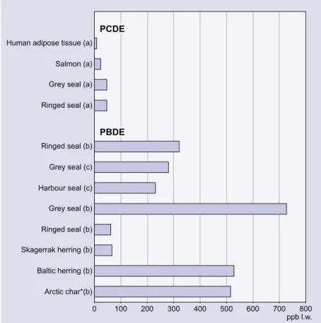
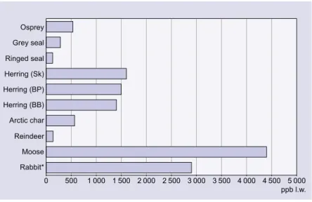
Halogenated industrial chemicals and unintentionally formed byproducts ... 74
Industrial chemicals ... 85
Additional compounds ... 87
Estimation of human exposure ... 89
6. E
XPERIMENTALSTUDIESOFDIFFERENTENDOCRINEMODULATORS... 102
Phytoestrogens and mycoestrogens ... 102
DDT ... 104 Lindane ... 106 PCDDs/Fs ... 107 PCBs ... 112 PBDEs ... 117 CPs ... 119
Halogenated phenols and benzenes ... 120
PCNs ... 121
Alkyl phenols ... 122
Phtalates ... 123
Bisphenol-A and Tetrabromobisphenol-A ... 125
Chemical interactions ... 125
Summary ... 126
7. G
ENERALCONCLUSIONSANDRECOMMENDATIONS... 143
S
VENSK
SAMMANFATTNING
D
en föreliggande rapporten avser att ge en sammanfattning av det nuvarande kunskapsläget vad gäller kända och befarade störningar av fortplantning hos ryggradsdjur. En utvärdering av möjliga samband mellan iakttagna effekter, på både djur och människor, och kemikalier som släpps ut i miljön har gjorts. Rekommenda-tioner för framtida forskning, med tonvikt på svenska problem, ges i slutet av rappor-ten.Det befaras att ett stort antal olika substanser, som släpps ut i miljön, kan påverka hormonsystem och därigenom störa fortplantning och utveckling hos ryggradsdjur. Att ett samband skulle föreligga mellan störd fortplantning och utsläpp av kemika-lier i miljön har indikerats i flera studier. Detta har lett till att ett flertal utredningar har genomförts för att sammanställa information om östrogen aktivitet hos kemika-lier i miljön. Förutom östrogena system kan dock även andra hormonsystem påver-kas och effekter har iakttagits på andra könshormonsystem, som androgener och progestiner, men även på hormoner som thyroxiner och retinoider. I många fall är det samma grupper av kemikalier som påverkar dessa olika hormonsystem. Flertalet av de substanser som befaras ha endokrint störande effekter omvandlas via en enzym-familj kallad cytokrom P450 (CYP). CYP är inblandat i bildning och omvandling av steroider men är även viktig för avgiftning av främmande substanser. Genom att CYP ändrar strukturen hos substanser finns även möjligheten att en substans om-vandlas från att påverka ett hormonsystem till att påverka ett annat. Komplexiteten i effekter som kan uppstå som ett resultat av exponering för en enskild substans gör att tolkningen av dess interaktioner med ett enskilt hormonsystem försvåras. Det är där-för nödvändigt att rikta uppmärksamheten på ett flertal hormonsystem.
R
APPORTENSMÅLRapporten är i huvudsak fokuserad på kemikalier som, genom att verka som agonis-ter eller antagonisagonis-ter, kan befaras störa hormonell signalering hos ryggradsdjur. För-utom iakttagelser av imposex (partiell hermafrodism hos honor) har inte effekter på ryggradslösa djur tagits med i utredningen. Väldigt lite är känt vad gäller miljögif-ters effekter på fortplantning hos ryggradslösa djur. Hormoner är inblandade i alla utvecklingsstadier hos ryggradslösa djur och avsaknaden av information om effekter på dessa organismer kan bero på avsaknad av studier på dessa och betyder därför inte att de inte kan påverkas. Vi bedömer därför att detta område snarare har varit negli-gerat än att det inte föreligger effekter.
D
EFINITIONAVENDOKRINTSTÖRANDEÄMNEN(
ENDOCRINEDISRUPTINGSUBSTANCES; EDS)
Det är nödvändigt att definiera vad som utgör en endokrint störande substans. Vid ett europeiskt möte angående ”impact of endocrine disrupters on human health and wildlife”, som hölls i december 1996 i Weybridge, UK gavs denna definition av EDS ”Ett endokrint störande ämne är en kroppsfrämmande substans som orsakar kritiska hälsoeffekter i en intakt organism, eller dess avkomma, som en konsekvens av änd-ringar i endokrin funktion”. Det definierades även att ”en potentiell EDS är en sub-stans som har egenskaper som kan befaras leda till endokrina störningar i en intakt organism”. Som dessa definitioner föreslår så är det viktigt att utveckla in vivo sys-tem för att klassificera substanser som varande EDS medan in vitro syssys-tem är viktiga för att välja ut substanser som varande möjliga EDS. I denna rapport använder vi oss av dessa definitioner.
R
APPORTENSINNEHÅLLHormoner är inblandade i regleringen av många olika och viktiga processer hos or-ganismer. Till dessa hör reglering av fortplantning, utveckling och ämnesomsätt-ning. Många kemiska ämnen kan störa dessa funktioner genom att påverka de hormonella systemen. För att förstå hur hormonsystemen störs krävs det kunskap om deras naturliga funktioner Utredningen har fokuserat på tre centrala hormon-system, könshormoner (östrogener, androgener och progestiner), thyroidhormon och retinoider. Dessa beskrivs i kapitel 2.
Könsbestämningen hos många ryggradsdjur styrs via könshormonerna. Detta gäller främst fiskar och kräldjur. Detta kan leda till att kemikalier i miljön kan påverka könskvoterna hos dessa djur. Utvecklingen av könsorgan och könsbeteende styrs av könshormon även hos människa. I kapitel 3 görs en genomgång av mekanismer för könsbestämning och utveckling hos ryggradsdjur.
En stort antal kemikalier befaras ge effekter på fortplantning och utveckling genom att störa hormonsystem som könshormoner, thyroidhormon och retinoider. I kapitel 4 görs en genomgång av olika iakttagna och befarade effekter på människor och djur. Därefter görs en genomgång i kapitel 5 av olika kemikalier som befaras ha hor-monella effekter. Ett mångfald av experiment har utförts på olika organismer för att utröna hur olika kemiska ämnen påverkar de olika hormonsystemen. I kapitel 6 görs en genomgång av experimentella studier som har gjorts på dessa kemikalier. I kapi-tel 7 ges en utvärdering av de iakttagna effekterna och rekommendationer för fort-satta studier.
E
NGLISH
SUMMARY
T
here is a current concern that an increasing number of substances, with possible endocrine disrupting potential, are being released into the environment and that these substances may have deleterious effects on vertebrate reproduction. Possible links between disturbed reproduction and exposure to environmental chemicals have been suggested in different studies. There are several international reports which address the estrogenic activity of such chemicals. Besides the estrogen system, many other endocrine systems may be affected by chemicals released into the environ-ment. Thus, effects have also been observed on other steroid systems, such as andro-gens and progestins, as well as on the thyroid and retinoid systems. In many in-stances several of these systems may be affected by the same groups of chemicals. Metabolism of many of these chemicals is controlled by different forms of cyto-chrome P450, and altered structure, due to metabolism of the chemical, may redirect the effect from one system to another. The complexity of effects which may be the result of exposure to any one substance render the interpretation of its interaction with any single system difficult.T
HEOBJECTIVESOFTHEREPORTThe present report aims to give an up to date account of the literature on known and suspected disturbances of reproduction in vertebrates and to examine the possible association between observed effects, in both humans and wild-life, and chemicals that are released into the environment, with special emphasis on Swedish conditions. Following the evaluation of existing knowledge, recommendations for future re-search are given.
In the present report we are primarily focusing on chemicals that have the potential of interfering, as agonists or antagonists, with hormonal signaling in vertebrates. We have not included effects on evertebrates, except for the development of imposex. Today there is very little known about the reproductive impairment of pollutants in arthropoda. This may be a reflection of the lack of studies on these organisms rather than that effects do not exist. It is known that the endocrine system plays important roles in all stages of development of arthropoda (e.g. growth, ecdysis, hibernation, sexual maturation) and that aquatic forms (e.g. crustacea) may be expected to be highly susceptible to endocrine disrupters. We conclude that this field has been ne-glected rather than that effects do not exist. Many chemicals may also interfere indi-rectly, as is the case with for instance metals and apart from tributyltin, and we have not included these in the report.
T
HECONTENTOFTHEREPORTHormones are involved in regulation of several different and important processes in organisms. These include regulation of reproduction, development and metabolism. Chemical substances may disturb these functions by affecting hormonal signalling. In order to understand how hormonal systems are disturbed it is essential to have knowledge of their natural functions. The report has focused on three hormonal sys-tems, the sex hormones (estrogens, androgens and progestins), thyroid hormones and retinoids. These are described in chapter 2.
Sex hormones regulate sex determination in many vertebrates, such as fish and rep-tiles. Exposure to chemical substances may therefore lead to changes in sex ratios in these species. The development of gonads and sexual behaviour is controlled by sex hormones in all vertebrates, including humans. The mechanisms for sex determina-tion and differentiadetermina-tion are discussed in chapter 3.
A large number of different chemical substances are suspected to affect reproduction and development by interfering with hormones such as sex hormones, thyroid hor-mones and retinoids. Chapter 4 deals with demonstrated and discussed effects in humans and animals. A number of chemicals with potential hormone modulating effects are presented in chapter 5. The selected compounds are mainly those that have been reported to have endocrine disrupting capacity but a few compounds lack-ing such data have been included because of their presence in the environment and structural similarity to hormones or to other endocrine disrupting compounds. A multitude of experiments have been conducted to determine how different chemical substances interfere with hormones. Experimental studies of different endocrine mod- ulators are discussed in chapter 6. At the end of the report, in chapter 7, an eva-luation of the observed and suspected effects are given together with recommenda-tions for future research.
1. I
NTRODUCTION
T
he present report aims to give an up to date account of the literature on known and suspected disturbances of reproduction in vertebrates and to examine the possible association between observed effects, in both humans and wild-life, and chemicals that are released into the environment, with special emphasis on Swedish conditions. Following the evaluation of existing knowledge, recommendations for future research are given.D
EFINITIONOFENDOCRINEDISRUPTINGSUBSTANCES(EDS)
It is recognized in this report that it is necessary to give a definition of endocrine disrupters. At the recent European workshop on the impact of endocrine disrupters on human health and wildlife, held in December 1996 in Weybridge, UK, (EUR 17549, 1996) it was agreed that “An endocrine disrupter is an exogenous substance
that causes adverse health effects in an intact organism, or its progeny, consequent to changes in endocrine function”. It was also agreed that “a potential endocrine disrupter is a substance that possesses properties that might be expected to lead to endocrine disruption in an intact organism”. In this report we use these definitions.
As these definitions suggest it is important to develop in vivo systems for deter-mination of such chemicals while in vitro systems may be helpful in the selection of candidate substances. In the present report we have adopted the Weybridge definition of endocrine disrupters.
B
ACKGROUNDThere are numerous incidences where humans or wildlife have been exposed to po-tential EDSs. In Taiwan, high levels of PCB and PCDF through contaminated oil resulted in several disorders in the children of exposed mothers (Rogan et al., 1988; Guo et al., 1993). In Florida, the alligators (Alligator mississippiensis) in Lake Apopka exhibit many reproductive and developmental abnormalities which have been suggested to be caused by EDSs (Clark 1990). In Western gulls (Larus
occidentalis) in southern California skewed sex ratios with increased numbers of
females and abnormal nesting behavior have been observed (Fox 1992). In fish out-side paper mill and sewage treatment plants, skewed sex ratios, decreased testoster-one levels and vitellogenin production in male fish have been reported from different parts of the world (Andersson et al., 1988; Munkittrick et al., 1991; Jobling and Sumpter 1993). In many parts of the world it has been observed that gastropods develop imposex (the development of a penis in females) when exposed to tributyltin from antifouling paint on ships (Bryan et al., 1986).
These observations have led to concern that environmental contaminants such as PCB, DDT, pentachlorophenol and alkylphenols may act as EDSs. Exposure to xeno- biotics with a potential to interfere with the normal hormone regulation may result in diminished fertility, altered sex differentiation, changes in behavior and altered cell differentiation leading to increased occurrence of cancer. Depending on the life stage when exposure occurs any or all of these effects may be induced. EDSs may thus induce changes in the normal development, cause different degrees of sex reversal and affect behavior.
Hormones are a diverse group of molecules that together with the nerve system coor-dinate different functions in the organism. The word hormone is derived from the Greek word horman, meaning to excite or stir up. Hormones are released by endo-crine cells/organs and exert their action on other cells/organs after having been trans-ported in the blood. Following synthesis in endocrine glands, hormones are released into the circulation and distributed throughout the body. While neural signaling sub-stances act over short disub-stances, generally moving a fraction of a micrometer across the synaptic cleft, hormones travel throughout the body in the circulation and may act far away from the site of release.
In the present report we consider the steroid hormones, the thyroid hormones and the retinoids. The steroid hormones are involved in the regulation of reproduction and the development of secondary sex characteristics. The thyroid hormones are in-volved in growth and differentiation and hypothyroidism leads to retarded growth and mental development. The retinoids are crucial for early embryonic differentia-tion and development and deficiency syndromes include effects on the reproductive systems. In addition to these hormones, many other signaling systems are involved during reproduction and developmental processes; and some of them act in concert with these hormones. Together these signaling systems form a central unifying regu-latory system used by cells and tissues to integrate information relating to their state of differentiation and proliferation. Hormones may act locally and are sometimes multifunctional mediators of cellular events and their functions cannot be fully un-derstood in isolation. Nevertheless, in the present evaluation we focus on the steroid, thyroid and retinoid signaling systems due to their proven critical roles in sex deter-mination, reproduction and/or embryogenesis, and to demonstrated effects on these signaling systems by chemical pollutants in the environment.
The focus on EDSs has been on their interaction with the hormone receptors and the subsequent regulation of target genes. Following binding to the receptor, the chemi-cals may either stimulate or inhibit gene transcription in a manner similar to the natural hormone or they may inactivate gene transcription by forming
receptor-lig-stances have, however, been found to exert both agonistic and antagonistic effects. Compounds having different mechanisms of action may cause similar biological changes. For instance, antagonists to the androgen receptor may give effects similar to those caused by estrogen receptor agonists. Besides interaction with hormone receptors, EDSs may interfere with transport proteins, alter the synthesis and bio-transformation of hormones, have direct toxic effects on the gonads or have adverse effects on the hypothalamus, the pituitary or endocrine glands.
H
ISTORICALBACKGROUNDTOTHEPRESENTRESEARCHONPERSISTENT ORGANICPOLLUTANTS,
CAUSINGREPRODUCTIVEDISTURBANCES,
INS
WEDENPersistent organic pollutants have long been indicated to be potential EDSs. Re-search on the occurrence and biological effects of persistent organic pollutants has a long tradition in Sweden. The Swedish pathologist Karl Borg suggested already in the 1950s that alkylmercury compounds could cause toxic effects in birds (Borg 1958). Seed-dressing with mercury salts had started around 1920 and alkylmercury was introduced as a dressing agent in the end of the 1940s. High mercury levels in dead white-tailed sea eagles (Haliaeetus albicilla) from the Baltic area were re-ported by Berg et al., (1966) and Henriksson et al., (1966). Westöö (1967) also iden-tified methylmercury in bird eggs and other biological material. In the 1940s DDT was introduced on a large scale. In the late 60s ecologists at the Swedish Museum of Natural History started collecting material for monitoring of PCB and DDT levels. At this time, Jensen identified PCB as a contaminant in biota (Jensen, 1966) and high levels of DDT and PCB were subsequently found in various organisms from marine environments, especially in white-tailed sea eagles from the Baltic area (Jensen et al., 1969).
Synthesis of radioactively labelled and unlabelled individual chlorobiphenyls was initiated by Carl-Axel Wachtmeister at the Wallenberg Laboratory, Stockholm Uni-versity (Sundström and Wachtmeister, 1973), which facilitated experimental studies of the toxicology of these compounds. By using the whole-body autoradiography technique developed by Ullberg (1954), tissue distribution of various chloro-biphenyls was studied by Berlin et al., (1975) and Brandt (1977). Following admin-istration of certain labeled chlorobiphenyls to mice, it was found that radioactive substances accumulated in the respiratory and reproductive tracts. The compounds in the airways were identified as methylsulfonyl metabolites of the chlorobiphenyls (Bergman et al., 1979). The presence of methyl sulfone metabolites of DDE and PCB in environmental samples had earlier been reported by Jensen and Jansson (1976). The high concentrations of PCB and DDT observed in the Baltic at the be-ginning of the 1970s were suspected to be correlated to the observed decrease in reproductive capacity of the animals (Olsson et al., 1975). In order to study the re-productive toxicity of these compounds, investigations on mice (Örberg et al.,
1972; Kihlström et al., 1975) and mink (Mustela vison) were carried out (Jensen et al., 1977). A number of studies on PCB toxicity in mink have been performed in Sweden (Kihlström et al., 1992; Brunström et al., 1994; Bäcklin 1996).
In 1981, several 2,3,7,8-substituted polychlorinated dibenzo dioxins/furans (PCDD/ Fs) were detected in grey seals (Halichoerus grypus) from the Baltic (Rappe et al., 1981). A broad survey of the dioxin contamination in Sweden was performed within a project called “The Swedish Dioxin Survey” which was administered by the Swed-ish Environmental Protection Agency and funded by the SwedSwed-ish government. Ex-perimental work on the toxicology of dioxins includes, e.g., studies of their terato-genic, immunotoxic, and tumor promoting effects (Hassoun et al., 1984, Dencker et al., 1985, Lundberg 1991, Flodström and Ahlborg 1989) as well as effects on retin-oid levels and metabolism (Thunberg et al., 1979, Håkansson 1988, Hanberg 1996). Nordic risk assessments of dioxins and polychlorinated biphenyls have been carried out at the Institute of Environmental Medicine at the Karolinska Institute (Ahlborg et al., 1988; 1992). Most of the research on persistent organic pollutants in Sweden has been supported by the Swedish Environmental Protection Agency.
R
EFERENCESAhlborg, U., Hanberg, A. and Kenne, K. (1992) Risk assessment of polychlorinat-ed biphenyls (PCBs), Nord 1992:26. Ahlborg, U., Håkansson, H., Wærn, F. and
Hanberg, A. (1988) Nordisk dioxinrisk-bedömning. Nordiska Ministerådet, Kö-penhamn, Miljörapport 1988:7. Nord 1988:49.
Andersson, T., Förlin, L., Härdig, J. and Larsson, Å. (1988) Physiological distur-bances in fish living in coastal water pol-luted with bleached kraft pulp mill efflu-ents. 45:1525-1536.
Berg, W., Johnels, A. Sjöstrand, B. and Wes-termark, T. (1966) Mercury content in feathers of Swedish birds from the past 100 years. Oikos, 17:71-83.
Bergman, Å., Brandt, I. and Jansson, B. (1979) Accumulation of Methylsulfonyl derivatives of some bronchial-seeking polychlorinated biphenyls in the respir-atory tract of mice. Toxicol. Appl. Pharmacol., 48:213-220.
Berlin, M., Gage, J. and Holm, S. (1975) Distribution and metabolism of 2,4,5,
Borg, K. (1958) Inverkan av betat utsäde på viltfaunan. VIII Nord. Veterinärmöte, Helsingfors, 394-400.
Brandt, I. (1977) Tissue localization of poly-chlorinated biphenyls chemical structure related to pattern of distribution. Acta Pharmacol. Toxicol., 40:1-108.
Brunström, B., Bergman, A., Bäcklin, B.M., Lund, B.-O. and Örberg, J. (1994) Effects of long-term exposure to PCB and PCB methylsulfones on reproduction in the mink. Organohalogen Comp., 20:471-480.
Bryan, G.W., Gibbs, P.E., Hummersonte, L.G. and Burt, G.R. (1986) The dceline of the gastropod Nucella lapillus around south-west England: Evidence for the effect of tributyltin from antifouling paints. J. Mar. Biol. Ass. U.K. 66: 611-640.
Bäcklin, B-M. (1996) Studies on reproduc-tion in femal mink (Mustela vison) ex-posed to polychlorinated biphenyls. Ph. D. Thesis, Swedish University of Agricul-tural Sciences, Uppsala.
and Wildlife Technical Report 29. Wash-ington D.C. U.S. Fish and Wildlife Ser-vice. pp. 1-37.
Dencker, L., Hassoun, E., d’Argy, R. and Alm, G. (1985) Fetal thymus organ cul-ture as an in vitro model for the toxicity of 2,3,7,8-tetrachlorodibenzo-p-dioxin and its congeners. Mol. Pharmacol., 27:133-140.
EUR 17549 (1996) European workshop on the impact of endocrine disrupters on human health and wildlife. Report of Pro-ceedings. pp 125.
Fox, G.A. (1992) Epidemiological and pathobiological evidence of contaminant-induced alterations in sexual develop-ment in free-living wildlife. In: Chemical-induced Alterations in Sexual and Func-tional Development: The wildlife/human conecction. Colborn, C.T. and Clement, C. (eds.) 23:147-158.
Flodström, S. and Ahlborg, U.G. (1989) Tumour promoting effects of 2,3,7,8-tetrachlorodibenzo-p-dioxin (TCDD)-ef-fects of exposure duration, administration schedule and type of diet. Chemosphere, 19(1-6):779-783.
Guo, Y.L., Lai, T.J., Ju, S.H., Chen, Y.C. and Hsu, C.C. (1993) Sexual development in biological findings in Yucheng children. DIOXIN 93:13th International Sympo-sium on Chlorinated Dioxins and Related Compounds, 14:235-238.
Hanberg, A. (1996)Effects of 2,3,7,8-tetrachlorodibenzo-r-dioxin on vitamin A storage in rat hepatic stellate cells. Ph. D. Thesis, Karolinska Institutet Stockholm. Hassoun, E., d’Argy, R. and Dencker, L. (1984) Teratogenicity of 2,3,7,8-tetra-chlorodibenzofuran in the mouse. J. Toxicol. Environ. Health, 14:337-351. Henriksson, K., Karppanen, E. and
Helmin-en, M. (1966) High residue levels of mer-cury in Finnish White-tailed eagles. Ornis Fennica, 43:38-45.
Håkansson, H. (1988) Effects of 2,3,7,8-tetrachlorodibenzo-p-dioxin on the fate of vitamin A in rodents. Ph. D. Thesis,
Ka-Jensen, S. (1966) Report of a new chemical hazard. New Sci., 32:612.
Jensen, S. and Jansson, B. (1976) Anthro-pogenic substances in seal from the Baltic: Methyl sulfone metabolites of PCB and DDE. Ambio, 5:257-260. Jensen, S., Johnels, A.G., Olsson, M. and
Ot-terlind, G. (1969) DDT and PCB in marine animals from Swedish waters. Nature, 224:247-250.
Jensen, S., Kihlström, J.-E., Olsson, M., Lundberg, C. and Örberg, J. (1977) Ef-fects of PCB and DDT on mink (Mustela
vison) during the reproductive season.
Ambio, 6:239.
Jobling, S. and Sumpter, J.P. (1993) Deter-gent components in sewage effluent are weakly oestrogenic to fish: An in vitro study using rainbow trout (Oncorhynchus
mykiss) hepatocytes. Aquatic Toxicol.
27:361-372.
Kihlström, J-E., Lundberg, C., Örberg, J., Danielsson, P.O. and Sydhoff, J. (1975) Sexual functions of mice neonatally ex-posed to DDT or PCB. Environ. Physiol. Biochem., 5:54-57.
Kihlström, J-E., Olsson, M., Jensen, S., Jo-hansson, Å., Ahlbom, J. and Bergman, Å. (1992) Effects of PCB and different fractions of PCB on the reproduction of the mink (Mustela vison). Ambio, 21:563-569.
Lundberg, C. (1991) Effects of 2,3,7,8-tetrachlorodibenzo-p-dioxin of the thy-mus and on the specific immune response in the mouse. Ph. D. Thesis, Uppsala University.
Munkittrick, K.R., Port, C.B., Van der Kraak, G.J., Smith, I.R. and Rokosh, D.A. (1991) Impact of bleached kraft mill effluent on population characteristics, liver MFO activity, and serum steroid levels of a Lake Superior white sucker (Catostomus
com-mersoni) population. Can. J. Fish. Aquat.
Sci. 48:1371-1380.
Olsson, M., Johnels, A.G. and Vaz, R. (1975) DDT and PCB levels in seals from Swe-dish waters. The occurrence of aborted
sium on the seal in the Baltic. June 4-6, 1974, Lidingö, Sweden. SNV PM, 591:43-65.
Rappe, C., Buser, H.-R., Stalling, D.L., Smith, L.M. and Dougherty, R.C. (1981) Identification of polychlorinated diben-zofurans in environmental samples. Na-ture, 292:524-526.
Rogan, W.J., Gladen, B.C., Hung, K.L., Shih, L.Y., Taylor, J.S., Wu, Y.C., Yang, D., Ragan, N.B. and Hsu, C.C. (1988) Con-genital poisoning by polychlorinated bi-phenyls and their contaminants in Taiwan. Science 241:334-336.
Sundström, G. and Wachtmeister, C.A. (1973) Synthesis of 14C-labelled and unla-belled PCB compounds. PCB Conference II, National Swedish Environmental Pro-tection Board, 4E:73-86.
Thunberg, T., Ahlborg, U.G. and Johansson, H. (1979) Vitamin A (retinol) status in the rat after a single oral dose of 2,3,7,8-tetrachlorodibenzo-p-dioxin. Arch. Toxi-col., 42:265-274.
Ullberg, S. (1954) Studies on the distribution and fate of S35-labelled benzylpenicillin in the body. Acta Radiol. (Stockolm), 118: 1-110.
Westöö, G. (1967) Determination of methyl-mercury compounds in foodstuffs. II. Determination of methylmercury in fish, egg, meat, and liver. Acta Chem. Scand., 21:1790-1800.
Örberg, J., Johansson, N., Kihlström, J.-E. and Lundberg, C. (1972) Administration of DDT and PCB prolongs oestrous cycle in mice. Ambio, 1:148-149.
TABLE 2.1. Nuclear receptors in various species. Note: C = receptor(s) cloned,
B = receptors indicated to be present by binding studies.
ORGANISM ESTROGEN ANDROGEN PROGESTIN THYROID RETINOID
Mammals C C C C C Birds C C C C C Reptiles C C C B Amphibians C C B C C Bony fish C B C C C Cartilaginous fish B B B
2. H
ORMONE
SYSTEMS
T
he genomic way of hormone action is to interact with nuclear receptors, which in turn bind to specific DNA-regions and activate or inhibit the transcription of specific mRNAs, leading to synthesis of specific proteins. Several intracellular hor-mone receptors have been isolated and characterized. They display a strong homo-logy between each other and are regarded as a receptor-superfamily. It has been shown that several different isoforms exist of each of the receptors in the steroid hormone superfamily. The receptors activate genes through binding to a variety of different gene response elements.Nuclear hormone receptors can be divided into steroid and non-steroid hormone re-ceptors. The steroid hormone receptors, including the estrogen, androgen and progester-one receptors, bind to DNA as homodimers, whereas the non-steroid hormprogester-one receptors, including the receptors for thyroid hormones and retinoids, bind to their response ele-ments either as homodimers or as heterodimers with the common partner, the retinoid X receptor (RXR). The genes for the receptors and binding proteins are highly conserved between species and show specific expressions in tissues and during development. The nuclear hormone receptors share a common structure and have a common mechanism of action. Receptors for many of these hormones have been cloned or identified from a number of different species (Table 2.1).
The nuclear hormone receptors in the steroid superfamily (except the glucocorti-coid receptor) are located in the nucleus even in the absence of bound ligands. Ster-oid, thyroid and retinoid receptors have all been shown to regulate gene activity in the absence of hormone (Lees et al., 1989, Graupner et al., 1989, Tzukerman et al.,
1990, Baniahmad et al., 1990, Zhang et al., 1991a, Bamberger et al., 1996). This suggests that the receptors may have functions independent of their specific hor-mones.
In addition to the classical signaling pathway, recent research indicates that steroid hormones, thyroid hormones and retinoic acid regulate cellular events through non genomic pathways, such as membrane-receptor mediated signaling (Wehling 1994). The presence of membrane receptors has been deduced from functional experi-ments, often where hormones exert actions too rapid to be accounted for by the gen-omic mechanisms.
The activity of hormonal systems may thus be regulated at a variety of different levels, including the synthesis, transport, receptor interaction and excretion of the hormones. The metabolism and action of hormones is outlined in Table 2.2.
In conclusion, hormone systems may regulate gene expression and physiological functions by a multitude of different mechanisms. The presence of different pathways may account for the high variation in the responses to different exogenous compounds as will be discussed in Chapter 6.
TABLE2.2. Overview of hormone metabolism and action.
SYNTHESIS Conversion from cholesterol to steroid hormone and from tyrosine to thyroid hormone. Retinoic acid is synthesized from dietary de-rived retinoids in situ.
TRANSPORT Lipophilic hormones are transported bound to specific plasma trans-port proteins. For the hormones discussed these include the steroid hormone binding globulin (SHBG), transthyretin, thyroid binding glo-bulin (TBG) and retinoid binding protein (RBP) In addition, dition, binding proteins transport retinol (CRBP) and retinoic acid (CRABP) in the cell.
CONVERSION For the steroid hormones and the retinoids there are several active metabolites.
GENOMIC Activation through the nuclear receptors. Differences between tis-PATHWAYS sues and organs exist. Slow effects.
NON-GENOMIC Activation through membrane receptors and other secondary pathways. PATHWAYS Differences between tissues and organs exist. Rapid effects.
EXCRETION Hydroxylation and or conjugation through phase I and phase II reac-tions leading to increased water solubility.
Nuclear receptors
The best known mechanism by which hor-mones act is by binding to specific recep-tors. These hormone receptors comprise the nuclear hormone receptor family, which in-cludes receptors for steroids, thyroid hormones, vitamin D3 and retinoids (Evans 1988).
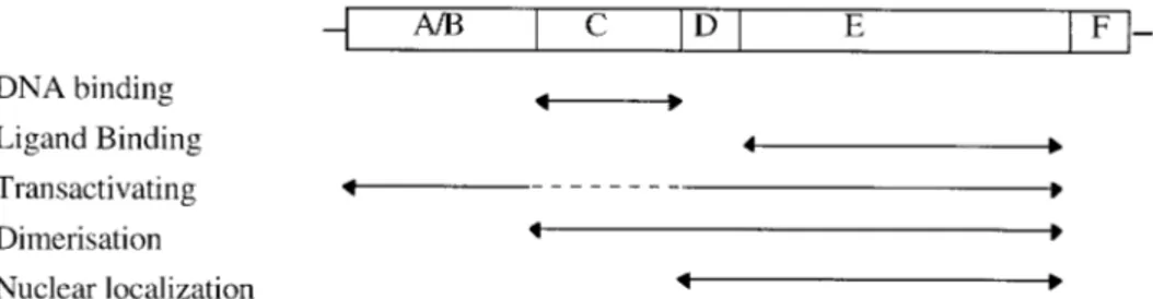
The receptors can be divided into six re-gions (A-F) with regard to amino acid simi-larities in different parts of the protein (Krust et al., 1986). The most conserved re-gion is the C rere-gion which is responsible for the DNA binding specificity. The ligand binding do- main is located in region E and is also con- served. The mechanisms of ac-tion are similar for these hormone receptors and below we use the estrogen receptor to exemplify the general mechanisms. A schematic representation of hormone recep-tors is given in Figure 2.1.
The main transcription-activating domain of these receptors is located in the ligand-binding domain (E-region) in the C-termi-nus (Webster et al., 1988). The receptors con-tain two inde- pendent acidic transcriptional activation function (AF) domains, AF-1 and AF-2 (Tora et al., 1989, Lees 1989). AF-1 is located in the N-terminal A/B-region and is responsible for constitutive activation, whereas AF-2 is hormone inducible and lo-cated within the E-region. The activities of
AF-1 and AF-2 are cell type-dependent. Tora et al., (1989) proposed that these two activate different stages in initiation of tran-scription. In that way an interaction between different activating domains and different components of the basic initiation complex would be possible. It is not known how these domains stimulate transcription but it is probably through interactions with the transcription complex.
The receptors are inducible transcription factors that modulate specific gene expres-sion by binding to short DNA sequences located on hormone regulated genes. These short DNA sequences are, in the case of es-trogens, termed the estrogen response ele-ments (ERE), and consist of inverted repeat sequences with the core sequence TGACCT (Parker 1990, Klein-Hitpass 1986, Klock et al., 1987). The consensus sequence of an ERE is 5'-AGGTCA nnnTGACCT-3'. Ideal-ized EREs are organIdeal-ized as 2 half-sites and mediate estrogen response. The half-sites for hormone receptor binding have been de-termined and show that the receptors can be divided into two major groups. Gluco-corticoids, aldosterone, progesterone and testosterone receptors all share the com-mon DNA sequence TGTTCT, while estro-gen, thyroid hormone, vitamin D3 and re-tinoid receptors all bind to TGACCT
half-Figure 2.1 Schematic drawing of the organization of receptors in the nuclear hormone superfamily. Regions are defined as; A/B - N-terminal domain; C - DNA-binding; D - Hinge domain; E - Ligand binding and F - C-terminal domain.
sites (Umesono 1989). While the half-sites are shared between different hormone re-ceptors, the spacing in-between the two half-sites may vary. It has been argued that the hormone itself interacts with DNA, thereby increasing the specificity of the DNA interaction (Hendry and Mahesh 1995).
The palindromic nature of the hormone response elements suggests that the receptors bind their targets as dimers (Kumar and Chambon 1988). A region within the hor-mone binding domain has been identifiedto be required for both hormone receptor dim-erization and high affinity DNA-binding (Fawell et al., 1990). Experiments using
point mutation of the murine ER has shown that there is a direct correlation between the ability of the different mutants to dimerize and their ability to bind to DNA (Fawell et al., 1990).
A recent hypothesis is that hormonal ligands in the steroid superfamily may act through insertion between base pairs in partially unwound DNA (Hendry and Mahesh 1995). This insertion hypothesis suggests that receptor bound ligands facili-tate DNA unwinding, stereospecific control of donor/acceptor functional groups on the DNA followed by insertion and release of the ligand between base pairs at 5'-TG-3'-5'-CA-3'.
S
TEROIDHORMONEST
he steroid hormone family consists of different groups that are recognized by their physiological functions. In this report we are addressing three systems. They include the female secondary characteristic inducing hormones, estrogens (mainly 17β-estradiol, but also estrone and estriol); the male counterpart, androgens (mainly testosterone and its derivative 5α-dihydrotestosterone) and the progestins essential for pregnancy (progesterone, 17-hydroxyprogesterone and 17,20-dihy-droxyprogesterone). In addition to regulating reproductive systems, the sex hor-mones also have effects on many other functions, such as growth, hemoglobin production and calcium metabolism in the skeleton.Besides being synthesized in gonads, a number of steroid hormones are synthe-sized by the adrenal cortex. In zona reticularis glucocorticoids and small amounts of adrenal sex hormones are produced. The major part of the adrenal sex steroids are androgens but small amounts of estrogens and progesterone are also produced. Adrenal androgens are of little physiological importance in the male, but in adult women they are thought to play a role for the sex drive. When secreted in abnormal amounts, as in patients with congenital enzyme deficiencies in the adrenal gland or in patients with adrenal tumors, they have effects that depend on the sex and age of the individual. In prepubertal males and in females the effects can be dramatic. Fe-males may develop a beard, a masculine pattern of body hair distribution and the clitoris may grow to resemble a small penis.
Estrogens
Estrogens have a central role in the control of both female and male reproduction. The estrogens are biosynthetically formed from androgens. They fulfill a range of roles in humans, some of which are only recently beginning to be understood. Estro-gens do not only occur in females, but are also present in the male though generally at much lower levels. The naturally occurring estrogens are 17β-estradiol, estrone and estriol. These three are all C18-steroids.
Estrogen synthesis is regulated via the hypothalamo-hypophyseal-gonadal axis. Gonadotropin-releasing hormone is secreted from the hypothalamus and stimulates the pituitary to release gonadotropins into the blood, which subsequently stimulates the follicle cells in the ovaries to synthesize and secrete estrogens.
Once estrogen has entered the cell, it may bind to the estrogen receptor. Following binding, the receptor-ligand complex elicits its effect in the nucleus by upregulating the synthesis of different gene products. The expression of the estrogen receptor is tissue-specific as well as developmentally regulated (Kuiper et al., 1996). It is in-teresting to note that a novel prostate and ovary specific estrogen receptor (ER) has recently been cloned from rat (Kuiper et al., 1996). It is possible that the different estrogen receptors may have different ligand binding and transactivating properties. Furthermore, estradiol stimulates the synthesis of its own receptors (Mommsen and Lazier, 1986).
Recent studies of estrogen receptor mediated gene regulation have revealed that estrogenic responses are mediated by a multitude of different pathways and that the estrogen receptor may regulate gene activity in the absence of estrogen (Tzukerman et al., 1990, Sukovich et al., 1994).
R
EPRODUCTIONANDDEVELOPMENTEstrogens regulate the growth of the ovarian follicles and increase the motility of the uterine tubes. They also increase uterine blood flow and increase the smooth muscle mass of the uterus. Estrogens regulate the growth of the endometrium. It has been suggested that this is due to upregulation of the progesterone receptor (Bamberger et al., 1996). Estrogens produce duct growth in the breast and are important for estrous behavior in most mammals, apparently through acting directly on certain neurons in the hypothalamus. During puberty, estrogen is partially responsible for the devel-opment of breasts, uterus and vagina. Other effects of estrogen include induction of pigmentation of the areolas and retention of salt and water prior to menstruation.
Estrogen is necessary for the differentiation of female secondary sex character-istics. In the fetal brain amortization of androgens to estrogens is believed to be of importance for male sexual brain differentiation and later on for sexual behavior. The estrogen receptor is present already at the blastula stage and estrogen is
pro-duced at blastula implantation (Dickman and Day 1973, George and Wilson 1978, Hou and Gorski 1993). These studies indicate that both estrogen and estrogen re-ceptors may be involved in early embryo development. It has been suggested that one effect of estrogen during early development may be regulation of Müllerian in-hibitory hormone (MIH) and this is supported by the presence of a putative estrogen response element in the MIH gene promoter (Guerrier et al., 1990).
In mice, it has been shown that the intrauterine position during fetal life has an impact on the behavior later in life (vom Saal 1989a). Female mice which developed between two males, in utero, were found to have a more aggressive behavior (more tail-rattling) than females which had developed between two females (Palanza et al., 1995). The steroid hormone levels were also observed to be dependent on the positioning in
utero. Thus, for instance, a male that developed between two males had higher
testos-terone levels than a male that developed between two females (vom Saal 1989a) and was also found to be more aggressive. Several other behavioral effects were also seen in these experiments, including effects on sexual activity, infanticide and reproductive capacity (vom Saal and Moyer 1985; vom Saal 1989a, vom Saal 1989b, Palanza et al., 1995). Since mouse embryos share a uterus, the steroids are able to pass between fetuses via diffusion through the amniotic fluid across the fetal membranes. The diffusion of steroids is believed to cause the changes in behavior observed in these mice, showing the im-portance of steroids in behavioral development.
Androgens
Testosterone is synthesized from cholesterol in the Leydig cells of the testis but also from androstenedione secreted by the adrenal cortex. All natural androgens are C19-steroids. The naturally occurring androgens are testosterone and 5α -dihydrotestos-terone in mammals. In teleosts 11-ketotestos-dihydrotestos-terone is also present and has been shown to stimulate development of male characteristics. Androgens increase protein synthesis and stimulate growth leading to increased growth rate.
Like estrogen, testosterone works primarily through interaction with nuclear re-ceptors. The classic androgen receptors in mammals are intracellular and exert their effects via the genom (Bolander, 1989). The androgen receptor is regulated by endo-genous levels of hormones. There are some conflicting data on the effect of an-drogens on androgen receptor mRNA levels (Jänne and Shan 1989; Abdelgadir et al., 1993; Gonzales-Cavadid et al., 1993). In vivo it appears that both testosterone and 5α-dihydrotestosterone down-regulate the androgen receptor mRNA levels. How-ever in vitro it appears that testosterone down-regulates the androgen receptor while 5α-dihydrotestosterone upregulates it (Lin et al., 1993; Gonzales-Cavadid et al., 1993). The exact mechanism for androgen regulation of the androgen receptors still remains to be elucidated.
Teleosts utilize 11-ketotestosterone in addition to the mammalian type androgens. The hor-mone systems vary markedly between different teleost species. Testosterone is produced in the gonads of both sexes and displays increas-ed plasma levels in prespawning and spawning fish. For a detailed account of the teleost hor-mone systems there are reviews available on gonadal steroids and androgens in teleosts (Fostier et al., 1983, Borg 1994). In most stud-ied fish, including salmonids, the testosterone levels are higher in females than in males. However, the 11-oxygenated androgens, main-ly 11-ketotestosterone occur at higher levels in
males than in females. They are effective in sti-mulating male secondary sexual characteris-tics and at least in some fish, spermatogenesis and male reproductive behavior.
Receptors for testosterone have been found in some organs in fish, but the physiological function of testosterone is largely unknown. No traditional steroid receptors have been found for ketotestosterone. As far as is known, 11-oxygenated androgens appear to be important hormones only in teleosts and sturgeons. In ad-dition gonadal steroids and/or their derivatives have been shown to have a pheromonal func-tion in many fish.
A
NDROGENSINTELEOSTFISHR
EPRODUCTIONANDDEVELOPMENTAndrogens stimulate the appearance of masculine traits such as differentiation of male reproductive tracts, secondary male characteristics and spermatogenesis. 5α -dihydrotestosterone is more potent than testosterone in stimulating virilisation of the outer genitalia and growth of the prostate gland (Wilson 1992). The major conver-sion site of testosterone is the prostate gland where over 90% is converted to 5α -dihydrotestosterone by 5α-reductase. The production of 5α-dihydrotestosterone can be blocked with the aid of specific inhibitors (Mellin et al., 1993). Testosterone is responsible for male reproductive behavior and castration has been shown to dimin-ish this behavior.
Production of testosterone during early development is necessary for the develop-ment of male genitals from the primordial genital ducts. Absence of testosterone leads to default development of female genitals. Testosterone is produced from the Leydig cells of the fetal testis while the Sertoli cells produce Müllerian inhibiting hormone. Both are necessary for development of male secondary sex characteris-tics. The development of male behavior is believed to occur in response to a peak in testosterone following birth. In rats it has been shown that exposure of females to androgens during the first days following birth results in male behavior.
Progesterone
Progesterone, a C21-steroid, is formed from cholesterol and is converted from preg-nenolone by 3β-hydroxysteroid dehydrogenase. It is an important intermediate in steroid hormone synthesis. Progesterone is secreted by the corpus luteum, placenta, the follicles and apparently also by the testes and adrenal cortex. The function of progesterone in males may only be as a biosynthetic intermediate. The hormone acts primarily on the uterus, breasts and brain.
There are two progesterone receptors and they have been shown to be generated from one single gene by alternative promoter usage (Kastner et al., 1990). The pro-gesterone receptors are regulated by estrogen and possibly by glucocorticoids as well (Jeltsch et al., 1990). It has been shown that the B-isoform of the human pro-gesterone receptor contains an N-terminal autonomous transactivation domain that is lacking in the A-isoform (Sartorius et al., 1994). This indicates that the two iso-forms may perform different functions.
R
EPRODUCTIONProgesterone secretion is increased by luteinizing hormone and prolactin. It is re-sponsible for the progestational changes in the endometrium and the cyclic changes in mucus secretion in the cervix and vagina. Progesterone has antiestrogenic effects on the myometrial cells and also decreases the number of estrogen receptors in the endometrium. In breasts, progesterone stimulates the development of lobules and alveoli, induces differentiation of the ducts and maintains secretory functions during lactation.
Progesterone is involved in the regulation of endometrial proliferation and dif-ferentiation. Progestins have been shown to block estrogen induced uterine growth in rats (Kraus et al., 1993). Furthermore, the addition of antiprogestins, such as RU 486, prevent this progesterone mediated inhibition of uterine growth (Kraus et al., 1993). Estrogen has been shown to upregulate the progesterone receptor in the uterus (Manni et al., 1981) and both progesterone and estrogen have been shown to down-regulate the uterine estrogen receptor (Manni et al., 1981, Hsueuh et al., 1976).
In a study using an endometrial adenocarcinoma cell line derived from epithelial cells it was shown that transfection with the progesterone receptor resulted in gene activation in the absence of progesterone (Bamberger et al., 1996). Addition of pro-gesterone inhibited this activation. The estrogen receptor did not activate the studied genes, and estrogen did not block the progesterone receptor induced gene activity. The effect of progesterone receptors on endometrial proliferation in the absence of progesterone may thus be to regulate the hyperplasia of the endometrium during the early parts of the menstrual cycle. The increase in progesterone levels during the later part may explain the reversal of this effect.
Steroid hormone
biochemistry
S
YNTHESISANDCATABOLISMSteroid hormones are synthesized from cholesterol that, in turn, may be synthesized in steroidogenic tissues from acetate. Most of the cholesterol is however derived from low density lipoproteins or from high den-sity lipoproteins. Free cholesterol is insolu-ble in the cytosol and is transported into the mitochondria by sterol carrier protein. Once it has reached the mitochondria, cholesterol can be cleaved into pregnenolone by cyto-chrome P450scc (side chain cleavage). This first conversion of cholesterol is a rate lim-iting step in steroid hormone synthesis. Fol-lowing this step, pregnenolone can be modi-fied to either progesterone or to 17OH-pregnenolone. Regardless of which of these pathways that is used, both cytochrome P450c17 and 3 -hydroxysteroid dehydroge-nase are needed for the production of an-drostenedione. Testosterone is then synthe-sized from androstenedione. Finally a
cyto-In salmonid fish the main progestin is 17-hydroxy-20-dihydroprogesterone while other fish often have other types. These hormones occur in both sexes. In salmonids
17-hydroxy-P
ROGESTINSINTELEOSTFISH20-dihydroprogesterone stimulates ovulation in females and spermiation (production of running milt) in males. 17-Hydroxy-20-dihydroprogeste-rone exerts its effects via membrane receptors.
chrome P450 enzyme (aromatase) converts testosterone into estradiol. Aromatase is of particular interest since regulation of its ac-tivity influences the ratio of testosterone to estrogen which in turn is important for the development of sex specific traits in many vertebrates.
Finally the hormones need to be removed from the circulation and excreted. This is usually done by sulfate conjugation leading to inactivation and excretion via the urine. Sulfatation is thus an important pathway in the biotransformation of steroid and thyroid hormones. In humans there are several forms of sulfotransferase enzymes. These include the dehydroepiandrosterone transferase and two forms of phenol sulfo-transferase. Dehydroepiandrosterone-sulfo-transferase catalyses the sulfation of ster-oids such as dehydroepiandrosterone, es-trone and estradiol.
Transport
Following secretion the steroid hormones bind to circulating plasma proteins. Since the steroid hormones are lipophilic they re-quire protein binding in the plasma in order to be distributed throughout the body. The steroids bind specifically to two high affinity steroid-binding proteins, the sex-hormone-binding globulin and corticoster-oidbinding globulin (Hammond 1993; Hammond and Bocchinfuso 1995), and nonspecifically to albumin. Be sides bind-ing to the above mentioned transport pro-teins, progesterone also binds to cortico-steroid-binding globulin. Testosterone has a higher affinity to sex-hormonebinding glo-bulin than either estrogen or progesterone. The free, or unbound, fraction of the steroid hormones is considered to represent the
bio-Cytochrome P450s (CYP) are key enzymes in steroid synthesis but are also involved in the metabolism of other endogenous compounds, such as retinoids, thyroid hormones, fatty acids, prostaglandins, leukotrienes and bio-genic amines. Many forms of CYP are also in-volved in the metabolism of foreign chemicals (xenobiotics) such as drugs, environmental pollutants and natural plant products. CYP comprises a multigene family of hemoproteins found in large abundance in the endoplasmatic reticulum of the liver, but can also be found in other tissues, although to a considerably lesser extent. CYP constitutes the monooxygenase (MO) system together with the membrane bound NAD(P)H-cytochrome c reductase, NAD (P)H-cytochrome b5 reductase and cyto-chrome b5. The oxidative biotransformations performed by the P450 MO system are also
C
YTOCHROMEP450
MEDIATED METABOLISMreferred to as phase I reactions. These reac-tions are normally followed by conjugation reactions (also called phase II reactions) with a polar endogenous compound such as gluta-thione, glucuronic acid, sulfate or amino acids to facilitate excretion of the compounds.
A number of compounds are known to indu-ce or inhibit the CYP system and/or the phase II enzyme reactions and can thus interact with the synthesis and biotransformation of hormones. Different compounds may however induce dif-ferent forms of CYP and phase II conjugating enzymes. The inducible forms of CYP belong to the gene families CYP1 (polyaromatic hydro-carbon, PAH), CYP2 (phenobarbital/ethanol), CYP3 (steroid) or CYP4 (clofibrate). Examples of inducers of the different families are given in parenthesis.
logically active fraction since it may enter target tissues/organs through diffusion from the capillaries.
Mechanism of action
D
IRECTCONTROLOFGENE EXPRESSIONSteroid hormones regulate gene expression by interacting with specific intracellular re-ceptors. These receptors are specific for the different hormones but may exist in mul-tiple isoforms. Thus, there are at least two different isoforms of the estrogen receptor, the ERα and the ERβ, that are derived from different genes (Kuiper et al., 1996). The different isoforms of androgen and pro-gesterone receptors have on the other hand
been shown to be different translation pro-ducts from the same gene. The presence of different isoforms of steroid hormone re-ceptors may be important for tissue specific functions.
Steroid hormones have been suggested to have a dual function in the mechanism by which the receptors activate transcription. As seen for estrogen, it may be an “un-masking” of the DNA-binding domain of the receptor since both ER-estrogen-com-plexes and ER-antiestrogen-comER-estrogen-com-plexes pro-mote tight nuclear binding (Webster et al., 1988). Estrogen also appears to be required to activate a transcription-activating domain located within the hormone-binding domain (Webster et al., 1988). The antagonistic effects of the antiestrogens may be based on their inability to induce the transcription activation function of the ER (Webster et al., 1988).
Some substances have been found to exert both agonistic and antagonistic effects. This is most notable with many pharmaceutical chemicals such as tamoxifen and raloxifene. It is believed that tamoxifen induces estro-genic responses through the N-terminal activation domain in the estrogen receptor, while it inhibits by blocking the C-terminal activation domain. The activation through the N-terminal domain is enough to give a weak estrogen response and tamoxifen has for instance been found to induce vitel-logenesis in fish (White et al., 1994). How-ever, when tamoxifen competes with est-radiol for the binding to the receptor it down-regulates the vitellogenin response (White et al., 1994).
While tamoxifen is effective in treatment of hormone-dependent breast cancer, due to its estrogen antagonistic effects, the agon-istic effects observed in the uterus indicate that tamoxifen treatment may not be with-out side effects. In a study of estrogen-in-duced calbindin-D gene expression Blin et al., (1995) observed that tamoxifen had both agonistic and partial anti-agonistic
ef-fects in ovariectomized rat uterus. Since tamoxifen is a weak estrogen-agonist in ute-rus there has been concern that tamoxifen treatment may promote uterine cancer.
Besides tamoxifen, raloxifene has also been shown to be an estrogen antagonist in breast tissue. However in contrast to tam-oxifen, raloxifene is an antagonist in the uterus as well. Both substances function as agonists in bone tissue (Sato et al., 1996; Yang et al., 1996). There are thus distinct tissue specific patterns in the effects of estrogen, tamoxifen and raloxifene on breast, uterus and bone. Recent research has shed some light on the possible regulatory mechanisms behind these tissue specific ef-fects of different pharmaceuticals and en-vironmental endocrine disrupters.
The effects of estrogen in bone have been suggested to be independent of the classical genomic estrogen response elements (Yang et al., 1996). Instead of activating tran-scription through EREs, raloxifene confers its activity in bone through a new class of response elements, called raloxifene re-sponse elements (RRE) found in for in-stance the TGF-b3 gene. Yang and co-work-ers (1996) found that estrogen did not bind to RRE and activate TGF-b3 transcription. However, an intermediate metabolite of estrogen, 17-epiestriol did activate TGF-b3 in bone. This indicates that estrogen by-products may be the active forms in some tissues and that activation may be achieved through alternative genepromoter elements. Since raloxifene is antagonistic in breast yet agonistic in uterus tissue there may be yet other mechanisms governing the tissue specificity of estrogens. It is therefore inte-resting to note that a novel prostate and ovary specific estrogen receptor (ERβ) has recently been cloned from the rat (Kuiper et al., 1996). Different estrogen receptors may have different ligand binding and trans-activating properties. The presence of diffe-rent pathways may account for the high variability in responses to different
exoge-nous compounds as will be discussed below. The observed differences in tissue specific responses to estrogen antagonist and agon-ists may be dependent on which estrogen re-ceptor that is being expressed, on which form of estrogen that is active and on which gene-promoter element that the receptor or the receptor-complex interacts with.
Another mechanism by which hormone receptors may be regulated is phosphory-lation. Phosphorylation of the human ER by MAP kinase pathways may influence recep-tor action by a mechanism other than the estradiol-dependent phosphorylation of hu-man ER by casein kinase II (Arnold et al., 1995.). Phosphorylation of the ER has been correlated with nuclear retention and specific DNA binding (Denton. et al., 1992). Steroid receptors may therefore not only regulate gene-expression following hormone-binding, but also in response to changes in the cellular milieu, through activated MAP kinase or AP-1.
It has been shown that the ER may bind to ERE and regulate gene activity in the absence of ligand (Lees et al., 1989, Tzuker-man et al., 1990). TzukerTzuker-man and co-workers (1990) described two novel regu-latory functions of the human ER, one con-stitutive activator function and one DNA binding function which enables the ER to bind and repress other activators of the ERE in the absence of estrogen. Thus, by binding to ERE in the absence of estrogen the recep-tor may function as a silencer of certain genes while allowing a constitutive basal activity of other genes.
Indirect control of gene expression
In some instances steroid hormones have been shown to exert effect on cells and tis-sues void of nuclear steroid hormone re-ceptors. In many cases the responses in these regions have been too rapid to be attributed to transcriptional activation. These effects are believed to occur via membrane receptors. Thus, besides the
classical activation pathway involving cyto-solic receptors, steroids have also been in-dicated to exert effects at the cellular mem brane. Of special interest are the effects of estrogens and androgens on the develop-ment of the brain. Testosterone has been indicated in numerous studies to affect both behavior and neuroendocrine function of the brain. In the rat, it has been indicated that testosterone alters the pattern of estro-gen receptor expression in the brain (Küh-nemann et al., 1995).
Progesterone was reported to be active as a sedative already in 1941 by Seyle (1941). It has since been found that the inhibitory effect of steroids results from interaction with the gamma-aminobutyric acid-A (GABAA) receptor, a class of ligand-gated Cl- channels, mediating inhibitory activity in the brain (reviewed by Paul and Purdy 1992; Sieghart 1992). In the hypothalamus, estradiol increases the turnover and release of GABA as well as the activity of GAD (glutamic acid decarboxylase) (Mansky et al., 1982; Duvilanski et al., 1983; Jarry et al., 1986).Thus, steroid hormones appear to be involved in GABAergic neuron activity by increasing the transcription of GAD mRNA thereby increasing the GABA syn-thesis, by increasing the presynaptic release of GABA and by prolonging the opening time of the post synaptic GABAA receptors. Taken together these effects indicate a dra-matic effect of steroid hormones on the GABA activity. Recently a membrane pro-gesterone has been identified in the plasma membrane of Atlantic croaker
(Micropogo-nias undulatus) and spotted seatrout (Cyno-sion nebulosus) (Thomas et al., 1997). It has
been indicated to be of importance for sperm motility in fish and recent evidence suggests that mammalian sperm also pos-sess progeserone receptors (Thomas et al., 1997).
Several studies have now shown that ster-oids may bind to different membrane recep-tors including calcium channels, chloride
channels, muscarinic receptors and hista-mine receptors (Ben-Baruch et al., 1982; Brandes and Bogdanovic 1986; Hardy and Valverde 1994; Mermelstein et al., 1996).
NIH3T3 fibroblast cells, permanently transfected with MDR1 (multi drug resis-tant) cDNA, become insensitive to colchi-cine. When these NIH 3T3MDR cells were treated with estradiol, progesterone or tamoxifen in the presence of colchicine, it was found that estrogen inhibited chloride channels while antiestrogens activated them when the compound was added in the media (Hardy and Valverde 1994). This suggests that estrogens and antiestrogens may inter-act with the large conductance chloride channels.
17β-estradiol has been shown to reduce the calcium current in primary cultures of
neostriatal neurones from rats (Mermelstein et al., 1996). Using whole-cell patch-clamp techniques 17β-estradiol was found to re-versibly reduce Ba2+ entry through Ca2+ channels. The reduction of Ba2+ entry through Ca2+ channels is greater in female than in male rat neurones (Mermelstein et al., 1996). By conjugating 17β-estradiol to bovine serum albumin and thereby inhibit-ing cellular uptake, it was shown that the signals were mediated at the membrane sur-face. The hormonal effect on the calcium current was indicated to occur via G-protein activation (Mermelstein et al., 1996).
The above observations thus indicate that steroids have specific, rapid and non-geno-mic effects at the cellular membrane through interaction with binding sites distinct from the nuclear receptor.
T
HYROID HORMONEST
he actions of thyroid hormones in higher organisms are critical for normal growth, differentiation and metabolic regulation (Legrand, 1986). Some of the most prominent effects of thyroid hormones occur during fetal development and early childhood. In humans, the requirements for thyroid hormones during develop-ment are shown dramatically in the syndrome of cretinism in which fetal hypo-thyroidism, often combined with maternal hypohypo-thyroidism, causes irreversible mental retardation and growth retardation if not treated early. Similarly, childhood hypothyroidism is characterized by striking impairment of linear growth. In adults, the primary effects of thyroid hormones are apparent by alterations in metabolism. Clinical features of hypothyroidism such as slowed mentation and speech, depres-sion, hypothermia, skin changes, bradycardia, constipation, and reproductive dys-function serve as reminders that thyroid hormones cause pleiotropic effects on many different organ systems.The thyroid hormones are largely transported in the blood bound to thyroid bin-ding globulin and transtyretin (Robbins et al., 1978). In adult rodents lacking thyroid binding globulin, more thyroid hormone is free of protein binding and therefore will be metabolized and excreted more easily. As a result, the half-life of thyroxine (T4) in
the rat is only about 12–24 hr in contrast to 6–7 days in humans. To compensate for increased turnover of thyroid hormone and to maintain physiological levels, the rat pituitary secretes more thyroid-stimulating hormone: As a comparison, the human baseline serum thyroid-stimulating hormone level is about 2.5 µU/ml, whereas in rats it ranges between 55 and 65 µU/ml in males and between 36 and 41 µU/ml in females. The resulting serum T4 levels are 15–20 times higher in rats compared to humans.
The control of thyroid hormone synthesis and excretion is affected by a sensitive feedback mechanism that responds to changes in circulating levels of T4 and triiodo-thyronine (T3). The regulation system includes in addition to the thyroid gland also
the hypothalamus and the anterior pituitary of the brain. An important peptide in this feedback system is the thyroid-stimulating hormone, which is secreted by the an-terior pituitary gland and causes the thyroid to initiate new thyroid hormone synthe-sis and excretion. The effects of TSH on the thyroid appear to be the consequence of binding to cell-surface receptors and activation of adenyl cyclase and protein kinase with subsequent phosphorylation of cellular proteins. Cyclic adenosine monopho-sphate (cAMP) can mimic most of the actions of thyroid-stimulating hormone on thyroid cells. The rate of thyroid-stimulating hormone released from the pituitary is controlled by the amount of thyrotropin-releasing hormone secreted by the hypo-thalamus, and by the circulating levels of T4 and T3. If the levels of thyroid hormones
are decreased, this will stimulate the secretion of thyroid-stimulating hormone lead-ing to restoration of thyroid hormone levels; on the other hand, if exogenous thyroid hormone is administered, thyrotropin-releasing hormone secretion is suppressed and the thyroid will become inactive and eventually regress.
Studies on the regulation of thyroid-stimulating hormone output from the pituitary have indicated that a link exists between T3 nuclear receptor occupancy and the
mRNA levels for the thyroid-stimulating hormone subunit chains; administration of exogenous T3 resulted in decreases in thyroid-stimulating hormone mRNA levels in
the pituitary.
Furthermore, thyroid hormone responsive tissues contain a variable number of nu-clear receptors for thyroid hormones (mainly T3), usually several thousands per cell.
Under euthyroid conditions in the rat, usually about 30–50% of the sites are occupied by T3, although in the pituitary about 80% of the sites are filled. The T3
Biochemistry
Synthesis
The thyroid hormones are synthesized in the thyroid gland and are stored as amino acid residues of thyroglobin, a complex gly-coprotein constituting most of the colloid in the thyroid follicles. (The thyroid follicles consist of epithelial cells, thyrocytes, that surround a noncellular, proteinaceous sub-stance, the colloid.) The first stage in the synthesis of the thyroid hormones is the up-take of iodide from the blood by the thyroid gland. The uptake requires energy and is ef-fected by the so-called “iodide pump”. Un-der normal conditions the thyroid may centrate iodide up to about 50-fold its con-centration in blood. Iodide uptake may be blocked by several anions (e.g. thiocyanate and perchlorate) and, since iodide uptake in-volves concurrent uptake of potassium, it can also be blocked by cardiac glycosides that inhibit potassium accumulation. In the next step, iodide is oxidized to an active iod-ine species that in turn iodinates the tyrosyl residues of thyroglobin. The reaction is ca-talyzed by a heme-containing peroxidase in the presence of hydrogen peroxide. The ma-jor products consist of diiodotyrosyl (DIT) residues, but monoiodotyrosyl (MIT) pep-tides are also formed. Additional reactions (which are thought to be catalyzed by the same peroxidase enzyme) involve the coup-ling of two DIT residues or of one DIT and one MIT residue, and lead to peptides con-taining residues of the two major thyroid hormones, i.e. thyroxine (T4) and triiodo-thyronine (T3).
The release of T4 and T3 from thyroglo-bulin is effected by endocytosis of colloid droplets into the follicular epithelial cells and subsequent action of lysosomal proteas-es. The free hormones are subsequently transported out from the epithelial cells into the circulation. Although T4 is by far the major thyroid hormone excreted by the
thy-roid (normally 8 to 10 times the rate of T3), it is usually considered to be a prohormone. Thus, T3 is about fourfold more potent than T4, and about 33% of the T4 secreted under-goes 5'-deiodination to T3 in the peripheral tissues; another 40% undergoes deiodina-tion of the inner ring to yield an inactive material, reverse triiodothyronine (rT3).
Transport
After entering the circulation, both T4 and T3 are transported bound, although not co-valently, to plasma proteins (Robbins, 1991, De la Paz et al., 1992). In man, the major carrier protein is thyroxine-binding globu-lin (TBG), a glycoprotein that forms a 1:1 complex with the thyroid hormones. TBG has a high affinity for T4(Ka about 1010 M) and a lower affinity for T3. This specific car-rier protein is not present in rodents, cats and rabbits. Thyroxine-binding prealbumin, more often called transthyretin (TTR), and albumin also transport thyroid hormones in the blood; TTR has Ka values of about 107 and 106 M for T
4 and T3, respectively. In the blood, TTR forms a complex with retinol-binding globulin, and this complex will therefore transport both thyroid hormones and retinol. In the normal situation, only ca 0.03% of the T4 in the circulation is free and available for cell membrane penetration and thus hormone action, metabolism, or excre-tion. The levels of free thyroid hormones in the circulation may be changed through competitive binding interactions of certain drugs and other foreign compounds.
As indicated above, TTR is the major thy-roid hormone transporter in rodents. How-ever, during early stages of development TBG is expressed also in (at least some of) these animals (demonstrated in the euthy-roid mouse). Between day 16 of fetal and day 60 of postnatal life, T4 and T
3 binding in sera shows a striking ontogenic pattern, which largely could be explained by the pre-sence of TBG: in fetuses, plasma protein