List of Publications
This thesis is based on the following publications, which are referred to in the text by their Roman numerals.
I Lovmar L, Ahlford A, Jonsson M, Syvänen A-C. “Silhouette” scores for assessment of SNP genotype clusters. BMC
Genom-ics. (2005) 6:35.
II (a) Chen D*, Ahlford A*, Schnorrer F*, Kalchhauser I, Fellner M, Viràgh E, Kiss I, Syvänen A-C, Dickson BJ. High-resolution, high-throughput SNP mapping in Drosophila
melanogaster. Nature Methods. (2008) 5(4),323-9.
(b) Schnorrer F*, Ahlford A*, Chen D*, Milani L, Syvänen A-C. Positional cloning by fast-track SNP-mapping in
Droso-phila melanogaster. Nature Protocols. (2008) 3(11),1751-65.
III Ahlford A, Kjeldsen B, Reimers J, Lundmark A, Wolff A, Ro-mani M, Syvänen A-C, Brivio M. Dried reagents for multiplex genotyping by tag-array minisequencing to be used in micro-fluidic devices. Analyst. (2010) Jul 29. (Published online ahead of print)
IV Kiialainen A, Karlberg O, Ahlford A, Sigurdsson S, Lindblad-Toh K, Syvänen A-C. Performance of microarray and liquid based capture methods for target enrichment for massively parallel sequencing and SNP discovery. Submitted manuscript.
* These authors contributed equally to this work
Additional publication by the author
Li J.B, Gao Y, Aach J, Zhang K, Kryukov GV, Xie B, Ahlford A, Yoon JK, Rosenbaum AM, Zaranek AW, LeProust E, Sun-yaev SR, Church GM. Multiplex padlock targeted sequencing reveals human hypermutable CpG variations. Genome
Supervisors
Prof. Ann-Christine Syvänen, Department of Medical Sciences, Uppsala University, Uppsala, Sweden
Associate Prof. Mats Nilsson, Department of Genetics and Pathology, Uppsala University, Uppsala, Sweden
Chair
Prof. Eva Tiensuu Janson, Department of Medical Sciences, Uppsala University, Uppsala, Sweden
Faculty opponent
Prof. David Schwartz, Department of Chemistry, Laboratory of Genetics, University of Wisconsin-Madison, WI, USA
Review board
Prof. Pål Nyrén, School of Biotechnology, Albanova, KTH (Royal Insti-tute of Technology), Stockholm, Sweden
Associate Prof. Mattias Mannervik, Wenner-Gren Institute, Developmen-tal Biology, Stockholm University, Stockholm, Sweden
Prof. Lars Engstrand, Swedish Institute for Infectious Disease Control and Department of Microbiology, Cell and Tumor Biology, Karolinska Insti-tutet, Stockholm, Sweden
Contents
Introduction ... 13
The chemical structure of DNA ... 13
Sequence variation ... 14
Analysis of sequence variation ... 15
Polymorphisms as genetic markers ... 15
Gene mapping in model organisms ... 17
Resources for sequence analysis ... 19
Methods for variant detection... 20
Complexity reduction ... 21
Reaction principles ... 25
Assay formats ... 35
Coding and decoding ... 37
Data analysis and quality control ... 38
Aim of this thesis ... 39
The present study ... 40
Tag-array minisequencing ... 40
Reaction steps and assay set-up ... 40
Silhouette scores for quality assessment of genotype data ... 43
Silhouette scores ... 44
Enzyme performance ... 45
Tools for gene mapping in Drosophila melanogaster ... 47
SNP map and FLYSNPdb ... 48
Genome-wide genotyping assays for gene mapping ... 48
SNPmapper for simplified data analysis ... 49
Resources for fine-mapping ... 50
Mapping of genes underlying muscle development ... 50
Tag-array minisequencing in a lab-on-a-chip... 51
Freeze-drying of reagents ... 51
Systematic genotyping comparison ... 52
Genotyping in micro-fabricated structures ... 54
Sample complexity reduction for re-sequencing candidate genes... 56
Study design ... 56
Capture quality ... 57
Concluding remarks ... 59 Acknowledgements ... 62 References ... 64
Abbreviations
bp Base pair
CNV Copy number variant
dNTP Deoxynucleotide triphosphates ddNTP Dideoxynucleotide triphosphates
ExoI Exonuclease I
GA Genome Analyzer
InDel Insertion-deletion polymorphism
kb Kilobase pair
Mb Megabase pair
MIP Molecular inversion probe MPS Massively parallel sequencing nt Nucleotide PCR Polymerase chain reaction
RFLP Restriction fragment length polymorphisms SAP Shrimp alkaline phosphatase SBE Single base primer extension
SBS Sequencing by synthesis
SNP Single nucleotide polymorphism µ-TAS Micro-total analysis systems
Introduction
The advances in the field of biomedical research have the last decades been driven by the rapid development of technology. Dideoxy “Sanger” sequencing (Sanger et al. 1977) for the determination of the base order of DNA mole-cules, together with polymerase chain reaction (PCR) (Mullis et al. 1986) for targeted amplification of DNA, are the two most important examples. These tools have revolutionized molecular biology and made it possible to determine the complete sequence of the human genome, of which the first draft cele-brates its 10 year anniversary this year (Lander et al. 2001; Venter et al. 2001). The trend has continued through the “post-genomic” era with the development of large-scale genotyping techniques for the study of genetic sequence varia-tion (Syvänen 2005). More recently, principles for massively parallel sequenc-ing (MPS) have followed (Mardis 2008; Shendure and Ji 2008; Metzker 2010). These tools enable extensive genetic research. In addition, hopes are high that the technology may have an impact on clinical genetics and its tran-sition into healthcare. This thesis addresses some of the features related to the analysis of sequence variation in the genetic model organism Drosophila
melanogaster and in humans.
The chemical structure of DNA
The deoxyribonucleic acid (DNA) molecule contains the code for the inherited information of all living organisms (Avery et al. 1944). Commonly, DNA consists of two long polymers made up of four different types of units called nucleotides. Each nucleotide is composed of the sugar molecule deoxyribose, a phosphate group and one of the four nitrogen bases, adenine, thymine, cyto-sine and guanine (Levene 1919; Astbury 1947). The nucleotides are ordered in a linear sequence in a predetermined way. The two molecules are complemen-tary to each other and twined together to form a double helix where the bases pair in a specific manner (Franklin and Gosling 1953; Watson and Crick 1953) . In each cell, these large molecules are organized into structures called chro-mosomes. In the fruit-fly Drosophila melanogaster the genetic information is encoded in ~180 million base pairs (bp) (Celniker and Rubin 2003) packed into four pairs of chromosomes: the X/Y sex chromosomes and the autosomes 2, 3 and 4. In humans the complete genetic code, the genome, is about 20 times bigger (Lander et al. 2001) and organized in 23 pairs of chromosomes
(Tjio and Levan 1956). The portions of DNA that contains the genetic infor-mation are called genes. These are read and transcribed from DNA to RNA, which in turn can be translated into amino acids to form proteins with differ-ent functions, according to the cdiffer-entral dogma of molecular biology stated by Francis Crick (Crick 1970). Surprisingly, the number of protein coding genes in more complex organisms, like humans, is only slightly higher than in the fruit fly. It is therefore suggested that the complexity lies in the plasticity and regulation of expression of the human genome. Many studies have shown that the large fraction of non-protein coding DNA sequences have important bio-logical functions (Mattick et al. 2010).
The comparisons of DNA sequences from a great number of different organ-isms have revealed the common features of life. Fundamental genetic princi-ples and functions are amazingly conserved throughout evolution considering the wide spectrum of organisms on earth. This commonality makes it possible to use model organisms for understanding principles that can be applied to more complex systems. Drosophila has been one of the most studied organ-isms in biological research already for almost a century, particularly in genet-ics and developmental biology (Burdett and van den Heuvel 2004). The fruit fly serves as an important research model and has in many aspects contributed to knowledge important for human health such as the understanding of human development and basic functions. About half of the protein coding genes in
Drosophila have human homologues and 75% of known human disease genes
have a homolog in the genetic code of the fruit fly (Fortini et al. 2000; Reiter et al. 2001).
Sequence variation
The increasing amount of genetic sequence information from multiple indi-viduals has facilitated the study of sequence variation both between and within species. It is estimated that the haploid genome of two human beings on aver-age differ by 0.5-1% (Levy et al. 2007; Pang et al. 2010). Together with envi-ronmental factors, these inherited signatures contribute to the phenotype of each individual and are part of what makes each of us unique.
All variation in the genome has occurred due to mutation events in the DNA sequence, either caused by copying errors during cell division or by the expo-sure to environmental factors such as toxic compounds, radiation or viruses. Variations that arise in the germline are passed on to the offspring while so-matic mutations only affect the individual organisms. Genetic sequence varia-tions range from large chromosomal alteravaria-tions to substituvaria-tions of individual nucleotides. The most abundant form is single nucleotide polymorphisms (SNPs), which are germline mutations that are fixed in the population. They
occur approximately every 1,200 bp in the human genome (Sachidanandam et al. 2001) and every 200 bp in the Drosophila genome (Moriyama and Powell 1996). The complete sequenced individual diploid genomes show that each human carries around 3 million SNPs compared to the human genome refer-ence sequrefer-ence, which constitute 0.1% of the total sequrefer-ence (Levy et al. 2007; Bentley et al. 2008; Wang et al. 2008; Wheeler et al. 2008). In addition to the single nucleotide variations, approximately half of the human genome consists of repetitive nucleotide sequences (Lander et al. 2001), (Sachidanandam et al. 2001) (Korbel et al. 2007). Examples of structural alterations are large copy number variants (CNVs), deletions or insertions, and small (1-5 bp) insertion-deletion polymorphisms (InDels). Structural variation in total constitutes a bigger fraction of nucleotides than SNPs (Conrad et al. 2010; Pang et al. 2010) and recent studies suggest that non-SNP variation, and in particular small CNVs also arise more frequently than previously expected (Stankiewicz and Lupski 2010). Short tandem repeats (STRs) or microsatellites consist of re-peated units, 2-6 nucleotides in length (Toth et al. 2000). They are often highly polymorphic but less abundant in the genome. SNPs that create or dis-rupt a restriction enzyme cleavage site are called restriction fragment length polymorphisms (RFLPs) (Schmidtke et al. 1984). Alternatively, a repeated sequence or insertions, deletions, translocations and inversions, can also lead to RFLPs. In the studies included in this thesis we have focused on the detec-tion and analysis of small sequence variants including SNPs and InDels.
Analysis of sequence variation
The impact of sequence variation is dependent on its genomic location. Vari-ants occurring in the coding sequence may alter amino acids of proteins and changes in intragenic regions and in introns can affect the stability of tran-scripts and the regulation of gene expression. The variants may be neutral, lead to an altered phenotype, or give rise to disease.
Polymorphisms as genetic markers
Sequence variants serve as markers in genetic studies. Variant analysis for indentifying disease genes commonly relies on one of two different ap-proaches; linkage studies in family pedigrees, and candidate gene or genome-wide associations studies (GWAS) on sets of affected and unaffected control individuals. In linkage studies the co-segregation of a characteristic with a marker is traced based on their physical proximity on a chromosome with limited recombination between them. Chromosomal segments that are shared between affected individuals in a family are identified. The altered cleavage pattern caused by RFLPs were the first type of markers to be used in linkage analysis and enabled the positional cloning of the cystic fibrosis gene in 1989
(Kerem et al. 1989; Riordan et al. 1989). The disease is an effect of a 3-bp deletion (delF508) in the CFTR gene, resulting in a non-functional protein product.RFLPs were later replaced by microsatellites which are multi-allelic and more informative as genetic markers. Both RFLPs and microsatellites are however difficult to analyze in a high-throughput way and recently SNPs have gained importance in linkage studies. The bi-allelic nature of SNPs makes them less informative but technically easy to analyze at a large scale in an automated way. SNPs also provide high resolution due to their high density in the genome. Linkage studies have been very successful in identifying highly penetrant rare alleles with monogenic patterns. Most diseases that are inherited in a Mendelian way have now been identified and many are analyzed in pa-tients in routine diagnostic laboratories today
Many types of genetic variants are predisposing factors in common multifac-torial disorders. Recently SNPs have been widely applied as markers in asso-ciation studies which rely on linkage disequilibrium (LD) or the non-random association between the loci influencing the disease and the tested marker. GWAS were initiated to identify the high frequency genetic variants underly-ing common disease in an unbiased way. These studies have resulted in the detection of genes for many complex disorders such as Crohn’s disease, where more than 30 loci have been identified (Barrett et al. 2008). In addition, these studies have identified novel cellular pathways important for the pathogenesis of different diseases which can pinpoint the disease mechanisms and suggest new drug targets. Despite some success, common diseases have turned out to be complicated to map and the functional variants difficult to identify. The reason for this is that they are caused by multiple genetic factors with low effect sizes in combination with environmental features. The causal alleles may also be rare and differ between individuals or among populations. Differ-ent approaches are currDiffer-ently discussed and tested in medical genetics to fur-ther elucidate the genetic component of complex disease (Bodmer and Bonilla 2008; Manolio et al. 2009; Cirulli and Goldstein 2010). Re-sequencing of interesting regions or whole genomes, of patients and individuals from the general population using MPS technology, is one promising means. The new sequencing technologies and “paired-end” analysis will also play an important role for precisely mapping and exploring more complex sequence variants in relation to disease and other phenotypes (Stankiewicz and Lupski 2010). Complex variants often lead to severe pathogenic phenotypes, and large chromosomal aberrations are for example commonly found in tumour cells. Many studies have shown that CNVs give rise to different mental disorders like schizophrenia (Need et al. 2009; Tam et al. 2009), but that they are fre-quent also in healthy individuals (Sebat et al. 2004; Korbel et al. 2007). Additionally SNPs are important as markers in population genetics, evolution-ary studies, and in pharmacogenetics (Ge et al. 2009). RFLPs and
microsatel-lites have also been frequently used as markers for gene mapping and in fo-rensic investigations (Moretti et al. 2001).
Gene mapping in model organisms
Forward genetic screens are performed by selecting individuals who possess a phenotype of interest and subsequently identifying the unknown underlying gene (Figure 1) (St Johnston 2002). In model organisms such as Drosophila
melanogaster, mutations can be induced in individuals by exposure to a
mutagen, such as a chemical or radiation. Mutants that harbour a desired phe-notype are then isolated. Other mutagens, such as random DNA insertions by transformation or active transposons, can also be used to generate new mu-tants. An advantage with these techniques is that the new alleles are tagged by a known molecular marker that can facilitate the identification of the gene. The mutated gene is next located on its chromosome by first crossbreeding the mutants with individuals carrying other traits with known locations, and fi-nally by estimating how frequently the traits and the mutant phenotype are inherited together in recombinant individuals. Large-scale genetic screens in the multi-cellular model organism Drosophila are powerful for gaining knowledge about cellular and developmental processes. The possibility of performing clonal screens enables the discovery of mutations underlying the affect of a given process, and the identification of phenotypes in practically any cell, or stage of development.
Figure 1. In gene mapping the line carrying an induced mutation (indicated by a star) is crossed to a divergent mapping line to generate recombinants. The phenotype of the recombinants are scored as mutant of wildtype, and by combining this information with the SNP genotype the recombination breakpoints are defined and the location of the mutation is identified.
The identification of the mutated gene is usually the rate limiting step in ge-netic screens. In classical screens phenotypic traits are used to map the new mutant alleles. Traditional mapping strategies in Drosophila are recombina-tion mapping, deficiency mapping (Parks et al. 2004) or mapping with the help of P-elements (Spradling et al. 1999; Ryder et al. 2004), which mainly rely on genetic and cytological markers that are not easily linked to the mo-lecular map. Moreover, phenotypic traits are not very frequent, or evenly spread throughout the chromosomes. Large gene regions are “cold-spots” for P-element insertions which result in a bias for mutagenesis of certain loci. These mapping traits also suffer from variability in the genetic background. With an increasing amount of genomic sequence information available, muta-tions in model organisms are frequently mapped by using small molecular polymorphisms as mapping traits. Examples of such are microsatellites, In-Dels and most commonly SNPs. In contrast to the classical mapping tech-niques mentioned above, SNP mapping offers many advantages including phenotypic neutrality, defined genetic background, high resolution and even distribution in the DNA sequence between common laboratory strains, and the feasibility to analyze all mutational classes (Martin et al. 2001). In several model organisms, mapping of genes with the help of polymorphisms has thus proven to be the preferred method. SNP mapping in Drosophila has relied on traditional genotyping methods like analysis of RFLPs, PCR product-length polymorphism (PLP) assays, and denaturing high performance liquid chroma-tography (DHPLC) (Hoskins et al. 2001) (Berger et al. 2001). These methods are well established and easy to perform with standard equipment. They are however hampered by the low throughput, and the genotyping procedure has therefore been a bottle-neck in mapping studies. Recently there has been a enormous technology development in the genotyping field, and large-scale systems are available for parallel genotyping of large SNP sets at a low cost per genotype (Syvänen 2005), described later on in this thesis. The current systems are however available mainly for fixed human applications and often involve huge investments of specialized instrumentation. As stated by Mac-donald S. et al (MacMac-donald et al. 2005) it is difficult to identify an available system for efficient, cost-effective genotyping at an intermediate scale outside of human genetics, and there is a need for open-source and off-the-shelf solu-tions to fill this gap. In Study II we developed a cheap microarray-based geno-typing system to facilitate SNP mapping in Drosophila.
In reverse genetic screens, mutations are instead generated in known genes or at known genome positions, and the phenotypic consequence is subsequently studied. With the genome sequence available it has become possible to silence genes using RNA interference (RNAi) in model organisms such as
Caenor-habditis elegans (Fire et al. 1998) and Drosophila melanogaster (Pal-Bhadra
Resources for sequence analysis
The advances in molecular biology and the large genome projects have re-sulted in publicly available resources, including sequence databases and analysis tools, for a great number of organisms. Human and Drosophila are among the organism with the most comprehensive tools available. The tional Center for Biotechnology Information (NCBI) was initiated by the Na-tional Institutes of Health (NIH) already in 1988, and is today the largest re-source for biomedical and genomic information (http://www.ncbi.nlm. nih.gov/). The Ensembl project is a joint effort between EMBL-EBI and the Wellcome Trust Sanger Institute since 2000 to establish genome databases for vertebrates and other eukaryotic species including automatic sequence annota-tions (http://www.ensembl.org). A similar resource is provided by the Univer-sity of California Santa Cruz (UCSC) (http://genome.ucsc.edu).
One of the databases associated with the NCBI is the Single Nucleotide Poly-morphism database, dbSNP (http://www. ncbi.nlm.nih.gov/SNP/) (Sherry et al. 2001). It serves as a central repository of SNPs and short InDels, currently for 58 different organisms. The majority of the polymorphisms are human and build 131 (April 2010) comprised 23.7 million variants of which 14.7 million have been validated. There has been a tremendous increase in the number of variants reported in the last years, primarily due to the increased amount of human sequence data generated by MPS projects. By the effort of the interna-tional Haplotype mapping (HapMap) project, a catalogue of common human variation (>1%) in individuals from different populations has been established (The International HapMap Consortium 2005; Frazer et al. 2007). The Hap-Map phase III data was released at the end of May 2010 and in total the re-source today comprises data for 1301 individuals from 11 populations (http://hapmap.ncbi.nlm.nih.gov). The goal of the HapMap project has been to identify the chromosomal regions where genetic variants are shared in the different populations, the common haplotype blocks, and to identify so called "tag" SNPs that uniquely identify each of these blocks. The 1000 genomes project was initiated in 2008 with the aim of sequencing the complete ge-nomes and gene regions of a large number of individuals using the new MPS technology (http://www.1000genomes.org). The goal is to provide a compre-hensive resource on human genetic variation, and to discover and characterize rare single nucleotide alleles that are present at minor allele frequencies around 1% or less.
FlyBase is an extensive bioinformatics database for the genome of the model organism Drosophila melanogaster and related species (http://flybase.org). It includes the highest-quality genome sequences and annotations available for any organism (Ashburner and Bergman 2005; Tweedie et al. 2009). Despite the big toolbox available for researchers in the Drosophila community
(Venken and Bellen 2005) it has until recently lacked a large-scale catalogue of small molecular polymorphisms, similar to the human dbSNP. Study IIa of this thesis includes an effort to indentify sequence variants in common
Droso-phila strains and to establish a high resolution variant database (Chen et al.
2008; Chen et al. 2009).
Methods for variant detection
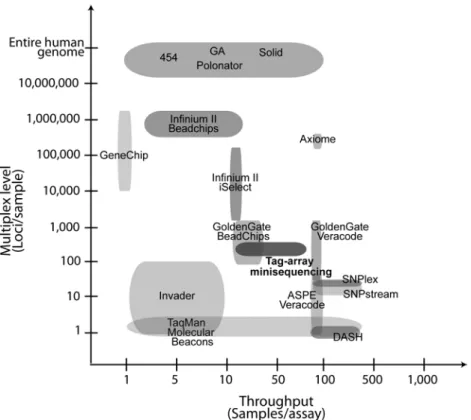
The large-scale genomic efforts mentioned above have provoked a rapid de-velopment in technology for the analysis of sequence variation, primarily SNPs. Today there are many genotyping systems available, both commercial and open source, for scoring known SNPs in the whole complexity range from single markers to genome wide analysis. Figure 2 compares different genotyp-ing methods in terms of sample and marker throughput. Recently there have been significant efforts to establish large-scale genotyping systems where key aspects have been cost efficiency per genotype, automation, and parallelisation of the biochemical reactions and the assay format. As a result, commercially systems for genetic analysis on a very large scale, and based on microarray technology, are now available in human genetics (Syvänen 2005). The largest assay for highly multiplexed SNP genotyping, provided by Illumina (Steemers and Gunderson 2007), allows parallel scoring of 2.5 million SNPs, to be in-creased to 5 million before long (www.illumina.com).
Nevertheless, the genotyping systems available today still suffer some limita-tions. With the growing numbers of SNPs identified it is desirable to further lower the cost per genotype to facilitate wide-spread large-scale genotyping. Additionally, the chips should be extended to comprise not only common SNPs but also rare variants. The largest, genome-wide genotyping approaches are primarily developed for fixed panels of markers to identify disease predis-posing SNPs that are associated to complex diseases in humans (Hirschhorn and Daly 2005) and require significant investments in equipment with high reagent costs. Moreover, the systems are inflexible for adaptation to special-ized and novel applications. Therefore, cost-efficient genotyping systems on an intermediate scale are needed. These will be required for replication of GWAS findings, for follow-ups of sequencing studies, as well as for future genetic analysis and diagnostics of previously identified risk variants. More-over, genotyping in non human systems has a need for good solutions and better intermediate scale systems in an open-source manner. Although mi-croarray-based methods have been successfully adapted for the analysis of CNVs (Redon et al. 2006; Craddock et al. 2010), large-scale analysis of more complex structural variants needs to be addressed further by new approaches (Teague et al. 2010).
The existing genotyping platforms all combine different reaction principles for allele discrimination with a number of assay formats and strategies for label-ling and for read-out of individual signals, in a modular fashion (Kwok 2001; Syvänen 2001). Below follows a description of these modules and examples of technology.
Figure 2. Comparison of SNP multiplexing levels and sample throughput in common SNP genotyping systems. In the Axiome system Affymetrix has increased the sample throughput of their hybridization-based method.
Complexity reduction
Direct analysis of sequence variants in the complexity of the genome of higher organisms is technically challenging. To achieve a sufficient sensitivity and specificity, most techniques for genotyping or sequencing require either am-plification of the starting material for further analysis, and/or amam-plification of the read-out signal after processing. Despite the recent improvements in MPS technology, it is costly and time consuming to sequence the whole genome of complex organisms in a large number of individuals (Summerer 2009; Turner et al. 2009; Mamanova et al. 2010). Additionally, the amount of data gener-ated and the data handling is overwhelming for most laboratories today. Hence, in many MPS studies it is desirable to reduce the complexity of the
template to perform targeted analysis on portions of the genome in multiple individuals, and utilize the sequencing capacity of MPS machines in a more optimal way.
Polymerase chain reaction
Two commonly used approaches for amplification of specific genomic frag-ments are cloning (Cohen et al. 1973) or amplification by polymerase chain reaction (PCR) (Mullis et al. 1986). PCR is powerful for increasing sensitivity and specificity for downstream assays. With PCR a locus-specific region of the genome is amplified, which increases the number of template copies and reduces the complexity of the DNA to be analysed. The principle requires very low amounts of input material. For over a decade it has been a golden standard for copying of DNA. Recently, long range PCR has been adapted for targeting smaller regions for MPS (1-100 kb) (Mamanova et al. 2010), and applied to sequence candidate genes in pools of a larger number of individuals (Nejentsev et al. 2009). The increasing amounts of genes, SNPs, and se-quences to be analyzed, require procedures for parallel amplification. How-ever, two problems with multiplex PCR are uneven amplification efficiency due to sequence-dependent differences of the fragments, and an exponentially increase of amplification artefacts as the number of added primer-pairs grow. Presently, multiplex PCR reactions are a bottleneck in highly multiplex geno-typing methods, since careful design and optimization of PCR assays at multi-plexing levels exceeding 10-20 amplicons is difficult and time consuming, even with better design software. One way to circumvent the difficulties with multiplex PCR is to physically separate individual DNA molecules before clonally amplifying them. PCR colonies, or “polonies” (Mitra and Church 1999) enable parallel amplification of DNA by separating PCR reactions in a thin polyacrylamide film, and in emulsion PCR (Dressman et al. 2003) clonal amplification of fragments from single DNA molecules is performed in a wa-ter-in-oil emulsion. Recently, micro-droplet based PCR was applied for the parallel amplification of 457 fragments (172 kb total genomic size) (Tewhey et al. 2009) for subsequent MPS. It is currently limited to ~4000 primer pairs and a throughput of 8 samples per day. Additionally it requires dedicated in-strumentation and as much as 7.5µg of input material.
One example of an alternative PCR-strategy is rolling circle amplification (RCA) and circle-to-circle amplification, presented by Dahl et al (Dahl et al. 2004), where a circularized target molecule is repeatedly amplified to generate a long linear molecule containing tandem repeats of the target sequence. Re-duction of genome complexity has also proven successful by the amplified fragment length polymorphism (AFLP) approach. In AFLP, genomic DNA is cleaved by restriction digestion followed by PCR-amplification with universal linker-primers and fragment size selection. This approach has been used for
SNP genotyping (Vos et al. 1995) and the same principle is employed in the Affymetrix GeneChip system (Kennedy et al. 2003; Matsuzaki et al. 2004). A reversed approach is taken by methods which are based on initial allele detection directly in genomic DNA, followed by PCR with universal primer pairs for signal amplification (Oliphant et al. 2002; Baner et al. 2003). Illu-mina applies this solution in their GoldenGate assay for multiplexed genotyp-ing in large-scale projects (Fan et al. 2003).
For undirected DNA amplification, a number of whole genome amplification (WGA) strategies in different forms are available (Lovmar and Syvänen 2006). These are powerful for increasing the total amount of DNA and to se-cure the source material. Multiple displacement amplification (MDA) is the most commonly used approach, which is based on an isothermal and branch-like amplification using the DNA polymerase of the bacteriophage phi29 and random primers (Dean et al. 2002). Illumina has in their Infinium II assay implemented an initial step of MDA followed by allele discrimination and finally signal magnification with an antibody sandwich strategy (Steemers and Gunderson 2007).
Capture by circularization
Enzymatic reactions and oligonucleotides are employed for target capture by circularization. Padlock probes/molecular inversion probes (MIPs) (Nilsson et al. 1994) are linear single stranded oligonucleotides consisting of two target specific sequences flanking a universal linker for amplification. They require a highly specific dual recognition event when hybridized to their target mole-cule, which enables a filling of the “gap” and a ligation to generate a circular library molecule. MIPs have been applied for large-scale target capture at Mb scale and MPS for; analysis of 10,000 human exons (Porreca et al. 2007), identification of genetic variation in hypermutable CpG regions (Li et al. 2009) methylation profiling (Ball et al. 2009) or to screen predicted RNA edit-ing sites (Li et al. 2009), usedit-ing array-released oligonucleotide libraries. The principle eliminates the need for additional shot gun library preparation. Since it is solution-based it is easily scaled up and automated using standard molecu-lar laboratory equipment. The main limitations are the capture uniformity and the upfront cost of probes for large target regions and the difficulty to obtain large amounts of each probe species. Double stranded Collector probes (Fredriksson et al. 2007) and Selector probes (Dahl et al. 2005; Dahl et al. 2007) follow a similar reaction principle for target capture. These reactions include an initial multiplex PCR or restriction digestion after which the probes are used to guide the circularization of the target molecules. They have been applied for sequencing the coding sequence of 10 human cancer genes (Dahl et al. 2007; Fredriksson et al. 2007) but currently only enables intermediate scale enrichment.
Capture by hybridization
Targeted pull-down reactions based on hybridization of target molecules to oligonucleotide microarrays have been performed for enriching ~5Mb exonic regions per reaction for MPS (Albert et al. 2007; Hodges et al. 2007; Okou et al. 2007). This approach, provided by Roche/Nimblegen (http://www.nim blegen.com), requires an initial library preparation step which is followed by hybridization to target specific oligonucleotide arrays containing 385,000 probes (Figure 3A) and recovery and amplification of the captured targets. Recently the principle has been further scaled up to facilitate the capture of up to 34Mb on a single array and applied for targeting the whole human exome for genetic diagnosis of Bartter syndrome (Choi et al. 2009). It is available for exon capture, and can be designed for custom and continuous target regions. The ability to capture large regions is a strength while the array-based format requires expensive hardware and gets laborious for larger number of samples. Additionally, it requires rather high amount of starting material, regardless of the total size of the target region. A similar approach in a fully automated mi-crofluidic format has also been used to capture a 480 kb exome subset of 115 cancer-related genes (Summerer et al. 2010). This format is still only adapt-able for targets up to 1Mb in size but has the advantage of the integrated mi-crofluidic format, promising for scaling up and for the adaption to clinical applications.
Gnirke et al. presented a hybridization-based capture method in solution for MPS using long (170-mer) RNA probes to capture >15,000 coding exons and four continuous regions, 4.2 Mb in total size (Gnirke et al. 2009) (Figure 3B). Today Agilent Technologies (http://www.home.agilent.com) are providing the SureSelect target capture method in solution for MPS both for exon sequenc-ing and for customized capture. The method involves the same reaction steps as the array capture but overcomes many of the obstacles related to the array format. The reactions are easily parallelized and automated for increased sam-ple throughput and are performed using standard molecular biology instru-mentation. Compared to the array format the hybridization is driven further to completion by an access of probe molecules per target molecule and the reac-tion time is decreased. The probe cost decreases with the number of samples analysed, making the system suitable for large-scale analysis. For smaller studies the cost per capturing experiment is however favourable with the Nimblegen arrays.
Figure 3. In the first step a sequencing library is generated through DNA fragmenta-tion, end-repair and universal adapter ligation. (A) The Nimblegen sequence capture method uses probes that are synthesized on microarray slides, with a varying probe length (60-90nt) to yield a uniform melting temperature. Library molecules are hy-bridized to 385K feature arrays in a custom incubation station for 65 hours, after which unbound fragments are washed away and the enriched portion is eluted. (B) The SureSelect method uses biotinylated RNA capture probes, 120 nucleotides in length, to which the library molecules are hybridized in solution for 24 hours in a regular thermocycler. The reaction is mixed with magnetic, streptavidin-coated beads to capture probe bound library molecules, followed by washing and elution. In both methods, the capture products are finally amplified by universal PCR by means of the adapter sequences.
Reaction principles
Most biochemical reaction principles used to determine the identity of a given nucleotide in a DNA sequence, are based on short oligonucleotide probes or primers. The methods rely either on allele-specific hybridization or enzyme-assisted allele discrimination. Some of these principles are discussed below and shown in Figure 4.
Allele-specific oligonucleotide hybridization (ASH)
In many approaches the hybridization between complementary DNA strands is employed for allele discrimination. Two allele-specific oligonucleotides are designed to differ in the variable base position and hybridized to the target DNA under stringent conditions (Wallace et al. 1979). The difference in ther-mal stability between the perfectly matched and the mis-matched probe is used to distinguish between SNP alleles. Amplified target DNA can be hybrid-ized to high-density oligonucleotide arrays containing allele-specific primers for high-throughput genotyping (Pease et al. 1994; Chee et al. 1996). Perlegen Sciences (closed operations in 2009) developed a system where long-range PCR and array hybridization was combined for multiplex genotyping of ~50,000 SNPs (Hinds et al. 2005), which among other things was used in the HapMap project. The GeneChip system by Affymetrix allows genotyping of up to 1.8 million SNP and CNP markers by array hybridization. The hybridi-zation characteristics of the probes are largely influenced by the sequence surrounding the SNP and the reaction conditions. This requires careful SNP selection and up to 40 different oligonucleotides per SNP to obtain high speci-ficity. In dynamic allele-specific hybridization (DASH) (Howell et al. 1999) the template-probe duplex stability is instead monitored during a temperature increase. DASH has only been multiplexed for two SNPs per reaction and used in small genotyping projects (Jobs et al. 2003). Hybridization-based ap-proaches are simple and do not require expensive enzymes.
Allele specific probes for real-time detection
DNA amplification during PCR can be monitored directly in an allele-specific way using double-labelled, allele-specific oligonucleotide probes. The 5’-exonuclease or TaqManTM assay uses specialized probes and the 5’-3’ nu-cleolytic activity of Taq DNA polymerase for allele discrimination (Holland et al. 1991; Lee et al. 1993; Livak et al. 1995). The probe contains a fluorescent reporter label in its 5’-end and a quencher in its 3’end. The signal from the reporter is suppressed when the two molecules are in proximity of each other. During the extension step of PCR the probe is displaced and cleaved by the polymerase, whereby the fluorophore is released and a detectable fluorescence signal is generated. This assay has been frequently used for single marker genotyping and serves as standard method. Molecular beacons are another type of double-labelled probes that form a hairpin structure in solution leading to quenching of the signal (Tyagi and Kramer 1996; Tyagi et al. 1998). Hy-bridization of the probe to a PCR product breaks the hairpin shape and sepa-rates the probe ends which gives rise to a signal.
Allele-specific primer extension (ASE)
The strategy is based on the ability of a DNA polymerase to only extend al-lele-specific primers with a perfectly matched 3’-end (Gibbs et al. 1989; Wu et
al. 1989), either directly on the target sequence or on already amplified sam-ples. The discrimination between matched and mis-matched probes by the polymerase is however not perfect which leads to low specificity. One ap-proach to increase the selectivity of the reaction is to include the enzyme apyrase. It inactivates the nucleotides and competes with the primer extension action of the DNA polymerase (Ahmadian et al. 2001). A beneficial feature of the principle is a cost reduction due to the use of un-labelled probes, although it still involves some visualization of the genotyping results. Allele-specific extension has been applied for interrogating low numbers of variants, as in Illumina’s custom genotyping system using cylindrical glass microbeads (Ve-racode beads), and in specialized assay formats (Mitani et al. 2007).
Single base primer extension (SBE)
The incorporation of a single nucleotide in an allele-specific manner is called single base primer extension (SBE) or minisequencing (Syvänen et al. 1990). One detection primer that anneals directly adjacent to the SNP position in the target molecule is used per reaction. The detection primer is extended with a nucleotide by a DNA polymerase, guided by the complementary template strand. The accuracy of the enzyme governs the high specificity of the reac-tion. The extension reaction is independent of the nature of the variable nu-cleotide and the sequence context surrounding it, which allows most SNPs to be analysed at the same reaction conditions. Furthermore, the high specificity makes the reaction principle suitable for multiplexing. In an early comparison, a microarray-based minisequencing assay showed approximately one order of magnitude better genotype discrimination than allele-specific hybridization in the same assay format (Pastinen et al. 1997). The robust underlying principle of minisequencing is the basis for its successful integration into numerous assays at different levels of complexity, in various reaction formats and with different labelling strategies, for the analysis of nucleic acids. It is the basis of the SNPStream system from Beckman Coulter (Bell et al. 2002) and the Infin-ium II platform provided by Illumina (Steemers and Gunderson 2007). Further the minisequencing principle has also shown excellent performance in quanti-tative applications, especially with tritium-labelled nucleotides, or when using mass spectrometric detection (Syvänen et al. 1993; Buetow et al. 2001; Milani et al. 2007). Minisequencing is the underlying reaction principle in the tag-array based genotyping system which was used in Studies I-III of this thesis. This system is discussed in more detail in the present study section.
Figure 4. Hybridization-based and enzymatic principles for allele discrimination. The reporter and quencher of the dual-labelled probes are denoted R and Q, respectively.
Oligonucleotide ligation (OLA)
As for the polymerase-mediated extension reaction, the joining of two per-fectly matched DNA strands by DNA ligase is a highly specific event. DNA ligase has hence been used to discriminate between two allele-specific probes that are hybridized at a SNP site in a PCR product. When there is a perfect match, the ligase seals the nick between one of two allele-specific probes and a third, locus specific probe binding upstream of the SNP site. This principle, denoted oligonucleotide ligation assay (OLA) was first described in 1988 (Alves and Carr 1988; Landegren et al. 1988) and is applied in the SNPlexTM system from Life Technologies (Applied Biosystems, http://www.lifetech nologies.com) for 48-plex SNP genotyping. The concept was adapted in the development of the padlock probe (Nilsson et al. 1994). The padlock probe is a linear oligonucleotide which contains the sequence from both OLA probes in either end, joined together with a linker sequence. The highly specific reac-tion requires two adjacent recognireac-tion events followed by an enzymatic liga-tion to form a circular molecule. The circle is amplified by rolling circle am-plification or by universal PCR by sequences in the linker before signal read-out. Padlock probes have recently been used for single molecule detection in
situ (Larsson et al. 2010). Single base extension was combined with ligation of
molecular inversion probes for high throughput SNP genotyping of 12,000 markers (Hardenbol et al. 2003; Hardenbol et al. 2005), commercially offered by ParAllele Biosciences, now part of Affymetrix. The polymerase extends the “gap” that is created at the SNP position before sealing by ligation. This approach requires only one probe per queried SNP but separate extension re-actions with one type of nucleotide present in each. Un-circularized probes are digested by exonuclease following amplification of the circles and signal read out on arrays.
In the GoldenGate assay, developed by Illumina (Fan et al. 2003), the enzy-matic discrimination is combined with hybridization for a highly specific genotyping assay for up to 1,536 SNP. Hybridization of allele-specific primers directly to genomic DNA is combined with an extension reaction and a subquent ligation to a locus-specific probe. All oligonucleotides contain se-quences for universal PCR amplification of the generated template with la-belled primers, and the locus-specific probe carries a barcode for capturing of the ligated probe complex to bead-arrays, Veracode beads, or bead-chips. Invasive cleavage
In the invasive cleavage method, the Invader assay, a structure-specific flap endonuclease (FEN) is used to cleave a complex formed by two allele-specific overlapping oligonucleotides that are hybridized to a target DNA containing a polymorphism (Lyamichev et al. 1999). The three-dimensional structure trig-gers the cleavage of the oligonucleotide and the difference in cleavage rate
between complementary and non-complementary probes is the basis for allele discrimination. The Invader assay principle has been developed for a variety of assay formats and detection strategies (Olivier 2005). Its advantages in-clude high accuracy, the isothermal reaction which does not require thermal cycling and the possibility to perform genotyping directly on genomic DNA. Despite the improvements, the method uses fairly large amounts of input DNA and was only successfully multiplexed for the analysis of ~100 SNPs (Ohnishi et al. 2001).
Sequencing
DNA sequencing is nowadays the most commonly used method to identify specific nucleic acid sequences. It has played a great role in the identification of novel sequence variants but is also used in small scale genotyping studies and for validation. Sequencing technology was pioneered in the mid seventies by Maxam and Gilbert through their chemical sequencing method (Maxam and Gilbert 1977). At the same time, the less cumbersome chain-terminating method, based on incorporation of labelled terminating dideoxynucleotide triphosphates (ddNTPs) and electrophoretic separation of DNA fragments of varying length, was published by Sanger et al (Sanger et al. 1977). The method uses transfected cloning vectors or PCR products as template for se-quencing. In the early versions the sequencing primers were radioactively labelled and only one out of four terminating nucleotides was present in each reaction in addition to unlabelled deoxynucleotide triphosphates (dNTPs). The development of this method has been remarkable over the years. Today Sanger sequencing is performed in a single reaction with all four, fluores-cently labelled nucleotides present (Smith et al. 1986; Prober et al. 1987). Other improvements include the development of whole-genome shotgun se-quencing and separation of the sese-quencing products on automated high-throughput capillary DNA sequencers (Drossman et al. 1990). The sequence of Drosophila melanogaster was published in 2000 as the first application of the whole-genome shotgun approach to sequencing of an animal genome (Adams et al. 2000).With sequencers such as the ABI3730xl DNA Analyzer from Life Technology, up to 1000 bases can be read in 96 or 384 samples per run. The technique has been crucial in the large genome projects but has also played a major role as a standard method for many genetic research ap-proaches.
Pyrosequencing is an alternative principle for sequencing (Ronaghi et al. 1996; Ronaghi et al. 1998). Different systems applying the principle are today commercially available through the company Qiagen (http://www.pyrose quencing.com). Its underlying reaction principle is sequencing by synthesis (SBS) (Melamede 1985), in which SBE has been further developed to deter-mine the sequential nucleotide incorporation during strand synthesis by the DNA polymerase. In pyrosequencing the nucleotides are resolved through an
enzymatic cascade resulting in a detectable signal. Nucleotide incorporation initiates a release of pyrophosphate (PPi) which together with adenosine phos-phosulphate (APS) act as substrates for ATP generation by ATP sulfurylase. The energy in ATP is converted to detectable light by luciferin and the signal is directly proportional to the number of incorporated nucleotides in a nucleo-tide flow. Each nucleonucleo-tide cycle contains one of four dNTPs and between dispensions the unincorporated nucleotides and the remaining ATPs are de-graded by apyrase. The degradation is crucial to avoid washing procedures in between cycles and decrease accumulation of light signals that lead to high background levels at longer length. In an effort to increase the read-lengths, up to 100 bases could be determined (Gharizadeh et al. 2002). Reli-able quantification of the signal is a problem when reading homopolymeric sequences. The usage of multiple enzymes makes the assay rather expensive. Pyrosequencing has been used in small scale genotyping projects and is fre-quently used for clinical applications.
Massively parallel sequencing
Despite the tremendous improvements, Sanger sequencing has reached its limits with respect to cost and parallelization (Shendure et al. 2005), and is too expensive for whole genome sequencing of large genomes. To stimulate inno-vation in the field, a sequencing race was initiated by the J. Craig Venter Sci-ence Foundation in 2003, to award the first complex (human or corresponding complexity) whole-genome sequenced for less than 1000 USD. In 2004 this strive was further encouraged through a large grant program for sequence technology development by the NIH. The rationale was that 1000 USD would be a reasonable cost to make multiplex large-scale sequencing a standard method. Among numerous promising applications it would enable compre-hensive analysis of the mutational spectrum of an organism. The efforts have indeed led to the development of several emerging sequencing technologies and new innovative solutions with the ability to process millions of sequence reads in parallel; so called next or second generation sequencing technology (Mardis 2008; Shendure and Ji 2008; Metzker 2010).
Ensemble-based methods involve multiple clonally amplified molecules. The SBS strategy described above using DNA polymerases and ligases, has been implemented for parallel analysis of DNA strands (Fuller et al. 2009). Nucleo-tides or short oligonucleoNucleo-tides are used for determining the base type. The established methods all employ synchronized sequencing cycles in an iterative fashion by adding a single kind of nucleotide at a time or by adding nucleotide substrates that are reversibly blocked. Although the need for vector-based cloning strategies is eliminated, all of the methods require an initial sequenc-ing library preparation step. This involves fragmentation of the molecules and incorporation of general priming motifs, i.e. adapter sequences, to both ends of a fragment for amplification and sequencing. The platforms can determine
either one end, or the paired-ends of a given molecule. They also allow the inclusion of barcodes for sample indexing and pooled sequencing approaches. 454 sequencing, today commercially provided by Roche (http://www. roche.com), was the first MSP technique described, published in 2005 (Margulies et al. 2005). It is based on emulsion PCR, where library molecules are clonally amplified en masse on agarose beads, and the pyrosequencing chemistry for nucleotide discrimination conducted in a picotiter plate format (Leamon et al. 2003). Currently the Genome Sequencer FLX system generates more than one million high-quality reads per each 10 hour instrument run with a read length of about 500 bases. One drawback with the technique is the problem of reading homopolymers, which is an effect of the pyrosequencing reaction. The sequential flow of nucleotides however diminishes the occur-rence of substitution errors in the sequence. The long reads simplifies the alignment and makes the system ideally suited for de novo sequencing of whole genomes and transcriptomes, including partially non-unique sequences. The second MPS system was developed by the company Solexa and intro-duced on the market in 2006 (Bennett 2004; Bennett et al. 2005) (Figure 5). Today the Solexa technology is commercially available in the Genome Ana-lyzer (GA) platform from Illumina. Library molecules are bound to oligonu-cleotide arrays in a flowcell and amplified by solid phase bridge amplification to generate millions of clonal template clusters. Each round of SBS involves the addition of four differently labelled and reversibly terminating nucleotides and four-color imaging of the incorporated base. The 3’-terminus of the incor-porated base is de-blocked and the fluorophore is removed before the next sequencing cycle is initiated. Each flow cell can hold eight separate reactions or samples. The current throughput of the GAIIx system is 15-20 million paired-end reads per sample of ~2x100 bp length, adding up to a total of 20 Gbp generated in each ten day machine run. In 2010, the company announced the launch of an updated version of the system (HiSeq2000) that will generate approximately ten times more data in each run, mostly due to better usage of their flow cells which increases the surface area. The advantage with the Solexa technology is the large amount of data generated at a fairly low price, while the relatively short read-length still is an obstacle for some applications and made it initially suitable for re-sequencing applications. With the constant increase in read-length it has also been applied for whole human genome se-quencing (Bentley et al. 2008) and it is the technology used by the 1000 ge-nomes project.
The mutiplex polony sequencing by ligation method (Shendure et al. 2005), uses mate-pair library molecules that are constructed by circularization of template molecules, RCA, restriction cleavage and adapter ligation. The linear library molecules contain two genomic target sequence tags of 17-18 bp, each
separated and flanked by universal sequences. The fragments are captured to beads and amplified in an emulsion PCR step which is followed by immobili-zation in an acrylamide gel to form a monolayered array of beads. The se-quencing strategy utilizes an anchor primer that hybridizes directly 5’ or 3’ of one of the genomic sequence tags. Differently labelled degenerate nonamers are added to the reaction, followed by a selective ligation between the anchor primer and the complementary nonamer. The base identity at the query site in the nonamer correlates with its label. After signal read-out the primer-probe complexes are stripped away and the next cycle begins with the addition of a new population of nonamers with a different query position. This permits se-quencing with high specificity of 13 bp (6+7 from each end) in each genomic tag and a total of 26 determined nucleotides per amplicon. Proof of principle was shown by sequencing the genome of an Escherichia coli. It has been fur-ther developed in the Polonator G.007 (http://www.polonator.org), an open platform provided by Dover Systems with open-source software and protocols and off-the-shelf reagents at a very competitive price.
Figure 5. Schematic drawing of the Genome Analyzer system using Solexa technol-ogy for MPS. (A) Sequence library molecules are via their universal adapter se-quences randomly hybridized to tags in one of eight lane of a microfluoidic flowcell. This is followed by bridge amplification on the surface to form millions of clonal molecule clusters (B). (C) The sequence of the molecules is deduced by SBS with four differently labeled, reversibly terminating nucleotides present. Each incorporation of a complementary base is followed by washing and (D) four colour imaging.
The sequencing by oligonucleotide ligation principle has also been applied in the SOLiDTM system from Life Technologies. Library molecules are captured
to oligonucleotides on magnetic beads, amplified by emulsion PCR and at-tached to glass slides. The sequencing process starts with the annealing of an adapter specific sequencing primer and a set of four fluorescently semi-degenerate labelled di-base probes. A probe match results in a specific ligation event and a fluorescent read-out where the label corresponds to the first two positions in the probe. The last two bases of each probe including the label are removed to enable the next sequencing cycle. Multiple cycles of ligation, de-tection, and cleavage are performed before the extension product is removed, and the template interrogated in a second round of ligation cycles with a primer complementary to the n-1 position. By this reset approach each base is interrogated twice which enables a read accuracy check. Two drawbacks with the technology are the short read length and the somewhat complex data out-put format in colour space. The current version of the instrument (SOLiDTM4) can generate up to 100 Gb of mappable sequence or about 1.4 billion reads of 50 bp length per ~11 day run. With the SOLiD™ 4hq System upgrade this scales to 300 Gb of sequence in 2.4 billion reads of up to 75bp in length per run.
The three major players described above have all introduced smaller systems during 2010 to better suit the needs of individual laboratories or for clinical applications. These are convenient for targeted re-sequencing, pathogen detec-tion and de novo sequencing of small genomes. General features include high speed, easy workflows and simplified data analysis. In addition to the systems mentioned above the company Complete Genomics (http://www.complete genomics.com) offers whole-human-genome service sequencing with a tech-nique that resembles the principles employed in Polony sequencing (Drmanac et al. 2010).
Many of the problems related to ensemble-based sequencing methods can be overcome with single molecule sequencing, also referred to as third generation sequencing. These include the difficulty of generating long reads because of phasing problems of signals from populations of molecules, the need for tem-plate amplification, and the fairly large consumption of sequencing reagents and template material for library preparation. The company Helicos Biosci-ences (http://www.helicosbio.com) developed an amplification-free method based on sequential SBS of single molecules attached to glass slides and sig-nal readout using fluorescence microscopy (Braslavsky et al. 2003). The aver-age read-length today is 35 bases. Pacific Biosciences and Life Technology both have real-time techniques using a “free running” DNA polymerase for single molecule SBS on their way. Pacific Biosciences (http://www.pacific biosciences.com) are currently launching their SMRTTM method where DNA polymerase molecules are attached to zero-mode waveguides (Levene et al.
2003). After the addition of single molecule templates the incorporation of labelled nucleotides by each individual DNA polymerase molecule is detected. The waveguide enables specific detection of incorporated labels among a background of thousands of labelled nucleotides. The DNA polymerase incor-porates 1-3 bases per second in the initial commercial release and generates a long DNA chain in minutes. Life Technologies single molecule sequencing system employs a similar but reversed approach. Here the single molecule templates are attached to a solid support to which individual DNA polymerase molecules are captured for sequence determination. This enables addition of new enzymes when the polymerase activity decreases. The incorporation of nucleotides is detected by using quantum dots and FRET chemistry. Ion Tor-rent use semiconductor technology for determining the identity of natural nu-cleotides that are sequentially incorporated by a DNA polymerase (http://www.iontorrent.com). When a base is added to a DNA stand a hydro-gen ion is released. The ion charge changes the pH of the solution which can be detected by a sensor. Currently this technique is not at the single molecule sensitivity level.
Exonuclease–mediated cleavage of labelled nucleotides can also be utilized for sequencing (Werner et al. 2003). The same principle has been combined with nanopores which can recognize single unmodified deoyribonucleoside monophosphates (dNMPs) (Astier et al. 2006). The pores act as base sensors when a dNMP interacts with the binding site and an adapter in the pore, result-ing in a measurable change in ion current.
Assay formats
The most common format for conducting biochemical reactions has been in solution in test tubes. Homogeneous assays, where all reactions take place in the same test tube, require less laboratory handling but are limited in the mul-tiplexing level of the reactions and are less sensitive due to lack of separation possibilities. This format is among others applied in TaqMan assays (Holland et al. 1991). Different solid supports have however gained importance due to the possibility of parallelisation of samples and reactions. Solid-phase assay formats enable immobilization of targets by capturing to oligonucleotides or to chemically activated surfaces, which is convenient for multi-step reactions that require multiple separation steps. The microarray format, first presented in 1992 (Southern et al. 1992), consists of a miniaturized solid-support com-monly made of glass. It facilitates parallel and miniaturized analysis with re-duced costs and usage of DNA. Since the arrayed format was introre-duced a number of supports and strategies have been developed for target organisation. These supports include microtiter plates, micro-beads in solution or assembled on microwells of fibre-optic bundles (Michael et al. 1998) or silica slides (Gunderson et al. 2004), gel films (Dufva et al. 2006), and various types of
micro-structures (Summerer et al. 2010). Apart from wide usage in RNA-expression analysis, microarrays are commonly used in DNA analysis and are gaining importance for analysis of proteins, antibodies and tissue. Microarrays have been essential in the development of large-scale SNP genotyping sys-tems. Oligonucleotide arrays can be produced by in situ synthesis of the mole-cules on the surface, and Affymetrix use a photolithographic process for their on-chip synthesis. More commonly, oligonucleotides are immobilized to acti-vated surfaces by different printing techniques. The early microarrays for genotyping contained immobilized SNP-specific probes (Shumaker et al. 1996; Pastinen et al. 1997; Pastinen et al. 2000). “Tags”, on the contrary, are barcode sequences complementary to sequence motifs included in the detec-tion primers of enzyme-assisted genotyping assays. When combined with genotyping reactions in solution, tags allow flexible assay design. In addition, their generic characteristic enables the development of universal capture ar-rays, which simplifies manufacturing. Tag-arrays were initially applied to SNP genotyping in a ligase assay (Gerry et al. 1999) and further combined with SNP genotyping by minisequencing (Fan et al. 2000; Hirschhorn et al. 2000). This assay format has been utilized in Studies I-III of this thesis. Miniaturization
Genetic analysis is becoming increasingly important in clinical genetics and pharmacogenetics. Genetic variants that are found to be risk factors for inher-ited diseases will be analysed to predict the susceptibility and onset of disease, or the treatment response and clinical outcome. In addition, the identification and scoring of somatic changes that initiate cancers and the identification of the allelic spectra of human pathogenic microbes, will also be important. There is great hope that the enhanced understanding of the effect of genetic variation may be implemented for diagnostic purposes to enable better, more individual treatment of each patient.
Large-scale genotyping techniques require massive equipment and time-consuming, labour intensive procedures and analysis. These methods provide high-throughput analysis with a low price per genotype. Progress in the field of clinical genetics is however dependent on new assay formats and robust, yet flexible technology that is fast, sensitive and inexpensive per sample, when smaller numbers of clinical samples are to be analyzed (Ohno et al. 2008; Weigl et al. 2008). Miniaturization has been an efficient means for en-hanced and portable information technology. Also in life-science, micro-total analysis systems (µ-TAS) or lab-on-a-chip have the capability of integrating miniaturized components and assay functionalities of multiple reaction steps, at pL-nL volume scale, into a single microfluidic device. The advantageous features of these systems are automation, low risk of contamination, low re-agent and sample consumption, decreased assay time, increased sensitivity, and potentially high level of parallelization (Liu and Mathies 2009). Hence,
these technologies offer promising tools and analysis solutions for efficient point-of-care DNA analysis. µ-TAS systems commonly consist of a microflu-idic network for biochemical reactions in a device often made of glass or polymers. In addition, they require a number of control components such as heaters and temperature sensors for thermocycling, microvalves for separating functional units, actuators and micropumps for sample transportation and flow control and magnets for sample manipulation using beads.
The adoption of lab-on-a-chip assays to routine DNA-based diagnostics has been slow. This is mainly due to the difficulty of integrating multiple func-tional steps required in genotyping assays, in particular sample treatment and signal detection (Whitesides 2006). The simple assay formats and short reac-tion times of single-reacreac-tion assays make them attractive. Consequently, the commercially available biochips use electrophoretic separation and/or ho-mogenous amplification methods for analysing single/low numbers of mark-ers. Two such examples are the chips provided by Agilent Technologies for high resolution capillary electrophoresis in the Bioanalyzer, and Fluidigm’s (http://www.fluidigm.com) digital real-time PCR assays for quantification of DNA sequencing libraries for subsequent MPS (White et al. 2009). Integrated multiplex genotyping assays often rely on separation of several homogenous reactions in different channels (Summerer et al. 2010). Genetic tests for com-plex diseases will require multicom-plex genotyping of multiple SNPs in multiple genes in each DNA sample. Ultimately, targeted clinical analysis of genes/mutations will be replaced by large-scale sequencing of disease gene pathways and networks. Thus, there is a need for completely automated mi-crofluidic devices with several reaction steps and a solid-phase support for separation of reagents and analytes during the process. To increase assay complexity while reducing hardware and system complexity as well as costs per test, is the true challenge. In Study III we have developed a multiplexed genotyping assay for cancer diagnostic in a biochip using freeze-dried re-agents for simplified reaction integration.
Coding and decoding
Previously, radioactivity and/or gel electrophoresis was the primary detection method of nucleic acid assays (Syvänen et al. 1993). Today, commonly used features for labelling are fluorescent molecules (Pastinen et al. 1996), specific mass of molecules (Karas and Hillenkamp 1988; Kim et al. 2004), the use of chemiluminescent enzymes (Nyren et al. 1993) and various sequence tags. The assay format influences the choice of allele label. Since homogenous as-says do not include any separation steps, the label must indicate when an al-lele-specific reaction has occurred. These molecular changes can with favour be detected using interacting fluorophores and observed by for example fluo-rescence resonance energy transfer (FRET). To be able to score different
al-leles of multiple products, unique coding of each molecule is necessary. The integration of a variant label and a locus label can be achieved during the process of the experiment and during assay design. Parallel read-out of indi-vidual signals depends on physical separation of products by for example cap-illary electrophoresis or on microarrays. Detection methods include mass spectrometry and microscopes or high-resolution scanners with charged cou-pled device (CCD) cameras or photomultiplier tubes (PMT).
Data analysis and quality control
Large-scale genetic studies such as genotyping, sequencing, and gene expres-sion analysis generate large amounts of data. In particular the output from MPS machines is increasing dramatically. This is accompanied by challenges for massive scale data storage and information technology to ensure proper data management and interpretation of huge data quantities (Pop and Salzberg 2008; Horner et al. 2010). Computational analyses of complex data sets re-quire (i) management of data including image analysis, signal processing and background subtraction in an automated manner with minimal levels of man-ual input and visman-ual inspection (ii) good tools and analysis strategies for base calling, read alignment and variant identification or allele scoring (iii) auto-mated and objective quality control. Further, downstream and application spe-cific interpretation of data is needed for finding biological relevant answers. In Study I and II of this thesis, tools for automated allele calling and data quality control were developed.
Aim of this thesis
The overall aim of this thesis was to develop and apply strategies based on single base primer extension (SBE) to study sequence variation in the fruit fly and in humans. Specifically the aims were:
• To improve the performance of the tag-array minisequencing sys-tem for SNP genotyping and implement it in novel applications.
• To use the Genome Analyzer for massively parallel sequencing to evaluate two hybridization-based methods for targeted sequence capture.