temporomandibular joint cavity: Clinical comparison with
the conventional puncture technique
Kunihito Matsumoto, DDS, PhD,aTore Bjørnland, DDS, PhD,bYukiko Kai, DDS,c Masahiko Honda, DDS, PhD,dYoshiyuki Yonehara, MD, PhD,eand
Kazuya Honda, DDS, PhD,fTokyo, Japan, Oslo, Norway, and Malmö, Sweden NIHON UNIVERSITY, UNIVERSITY OF OSLO, AND MALMÖ UNIVERSITY
Objective. The objective of this study was to compare an image-guided puncture technique (IGPT) with conventional puncture technique (CPT) with respect to accuracy of needle entry, maximal mouth opening, and pain in pumping manipulation treatment of internal derangement of the temporomandibular joint (TMJ).
Study design. The subjects comprised 178 patients with internal derangement of the TMJ with closed lock. Treatment was provided using CPT in 102 cases and IGPT in 76 cases. Three variables, number of repunctures, maximal mouth opening distance, and pain threshold according to a visual analogue scale, were measured and compared between IGPT and CPT groups.
Results. Access to the superior joint cavity was achieved without correcting the puncture point in 97% of patients who underwent IGPT and 82% of patients in the CPT group. Significant differences were seen in 1-week maximal mouth opening and pain threshold between IGPT and CPT groups (P⬍ .05 each) and resetting of the puncture point was significantly less frequent using IGPT compared with CPT (P⬍ .05).
Conclusions. IGPT is effective for pain mitigation and improves mouth opening during the early postoperative period after pumping manipulation treatment. (Oral Surg Oral Med Oral Pathol Oral Radiol Endod 2011;xx:xxx)
Many modalities have been established for diagnostic imaging of the temporomandibular joint (TMJ), includ-ing plain radiography, arthrography, magnetic reso-nance imaging (MRI), computed tomography (CT),
cone-beam CT (CBCT), and arthroscopy.1-7 Since the emergence of MRI, arthrography and arthroscopy of the TMJ have been performed less frequently in pa-tients with TMJ disorders because of the invasiveness and technical difficulty of these procedures.7 Punctur-ing the TMJ cavity is a basic technique used for diag-nosis and treatment of joint cavity lesions in TMJ disorders, and is frequently used clinically. Puncturing the TMJ cavity permits contrast studies, arthroscopy, pumping manipulation, arthrocentesis, drug injection, and collection of fluids from the joint cavity8-13; how-ever, puncturing the TMJ cavity may cause complica-tions, such as facial nerve damage, penetration of the middle cranial fossa, or irreversible changes to the TMJ itself.14-17In disorders such as severe temporomandib-ular osteoarthritis and synovial chondromatosis, TMJ disk examination, arthrocentesis, arthroscopic surgery, and other techniques are useful and significantly influ-ence the clinical course.8-12,16-18 Safer, more reliable, and easier puncture of the TMJ cavity would thus improve quality of life for the patients. The effective-ness of image-supported surgical procedures has often been reported in the medical field, and safer methods have undergone continued development,19,20 but few such reports have been presented in dentistry.21-23We have already reported the effectiveness of the image-guided puncture technique (IGPT),21 but no clinical
This study was supported by the Sato Fund, the Uemura Fund, a grant from the Dental Research Center, Nihon University School of Den-tistry, and a grant for the Promotion of Multidisciplinary Research Projects titled “Translational Research Network on Orofacial Neuro-logical Disorders” from the Japanese Ministry of Education, Culture, Sports, Science, and Technology.
aAssistant Professor, Department of Oral and Maxillofacial
Radiol-ogy, Nihon University, School of Dentistry, Tokyo, Japan.
bProfessor, Department of Oral Surgery and Oral Medicine, Faculty
of Dentistry, University of Oslo, Norway; Professor and Chairman, Department of Oral and Maxillofacial Surgery, Faculty of Odontol-ogy, Malmö University, Sweden.
cPostgraduate Student, Department of Oral and Maxillofacial
Radi-ology, Nihon University, School of Dentistry, Tokyo, Japan.
dLecturer, 2nd Division, Department of Oral and Maxillofacial
Sur-gery, Nihon University, School of Dentistry, Tokyo, Japan.
eProfessor and Chairman, 2nd Division, Department of Oral and
Maxillofacial Surgery, Nihon University, School of Dentistry, To-kyo, Japan.
fProfessor and Chairman, Department of Oral and Maxillofacial
Radiology, Nihon University, School of Dentistry, Tokyo, Japan. Received for publication Mar 16, 2010; returned for revision Jan 1, 2011; accepted for publication Jan 8, 2011.
1079-2104/$ - see front matter © 2011 Published by Mosby, Inc. doi:10.1016/j.tripleo.2011.01.019 e1 3 4 5 6 7 8 9 10 11 12 13 14 15 16 17 18 19 20 21 22 23 24 25 26 27 28 29 30 31 32 33 34 35 36 37 38 39 40 41 42 43 44 45 46 47 48 49 50 51 52 53 3 4 5 6 7 8 9 10 11 12 13 14 15 16 17 18 19 20 21 22 23 24 25 26 27 28 29 30 31 32 33 34 35 36 37 38 39 40 41 42 43 44 45 46 47 48 49 50 51 52 53
comparisons of IGPT with the conventional puncture technique (CPT) have yet been described.
The purpose of this study was to compare IGPT with CPT with respect to accuracy of needle entry, maximal mouth opening, and pain in pumping manipulation treatment of internal derangement of the TMJ.
MATERIAL AND METHODS
We studied 178 subjects with internal derangement of the TMJ and closed lock, based on both clinical and MRI examinations, using either the CPT technique (n ⫽ 102; 23 men, 79 women; mean age, 36 years; range, 16-62 years) or IGPT (n ⫽ 76; 12 men, 64 women; mean age, 38 years; range, 18-58 years) for pumping manipulation therapy (Table I). Patients were randomly divided into either group without consider-ation of disease severity. Anatomical landmarks were used in CPT, and CBCT (3DX multi-image micro CT; Morita Manufacturing, Kyoto, Japan) was used in IGPT to safely puncture the superior TMJ cavity. All the procedures were performed by the same oral radiologist (K.H.) who was experienced in TMJ puncture.
The use of patients for this study was reviewed and approved by the Bioethics Committee at Nihon Uni-versity School of Dentistry, Japan. All patients pro-vided written informed consent to participate in this study.
CPT Method
In the CPT method, positions of the mandibular fossa and head were preoperatively confirmed by palpation as anatomical indicator points, based on a point 10 mm from the antilobium on the line between the midpoint of the antilobium and the lateral angle of the eye. The puncture point was indicated on the skin by tracing the outline with a marker. The superior joint cavity was therefore punctured in the fossa or along the poste-rior slope of the tubercle. A wide area of skin around the preauricular region was disinfected using isodine and 8% hypoalcohol. Infiltration anesthesia was ad-ministered by subcutaneous injection of 2% xylo-caine (AstraZeneca, Osaka, Japan), using a 26-G needle. Arthrography was performed from the supe-rior joint cavity under fluoroscopy, using CT with a
21-G needle. The contrast medium was nonionic isovist (Bayer Schering Pharma, Osaka, Japan).
IGPT Method
In the IGPT method,21to permit safe insertion of the puncture needle, an outline of the condyle and fossa was elicited on palpation of the TMJ area and drawn using a medical marker on the skin. In addition, refer-ence lines (Frankfort horizontal [F-H] plane) and axial planes passing through the top of the condyle were drawn on the skin (Fig. 1, A). A small metal ball (diameter, 1.5 mm) was placed on the skin surface covering the top of condyle using medical adhesive (Aron alpha A; Daiichi-Sankyo, Tokyo, Japan) (Fig. 1, B). The patient was positioned in the F-H plane, parallel to the floor, and CBCT was then performed. Images were not rotated for preoperative image evaluation.
First, the point to be reached by the needle tip was set according to the posterior slope of the articular tubercle on the CBCT monitor (Fig. 2). Based on this set point and the 3-dimensional features of the individual mor-phology of the mandibular fossa, the insertion point of the needle on the skin was decided. Next, puncture angles with respect to the reference plane (axial plane) were measured using the image tool in I-view 3D software (Morita Manufacturing) (Fig. 3). In addition, the distance between the insertion point and end point, and the distance between the metal ball and insertion point (vertical and horizontal distance) were measured using the same image tool (Fig. 3).
The measured coordinate point of the insertion point with respect to the center of the metal ball was drawn using a marker (Fig. 1, B). A wide area of skin around the preauricular region was disinfected using isodine and 8% hypoalcohol. Infiltration anesthesia was admin-istered by subcutaneous injection of 2% xylocaine us-ing a 26-G needle.
To reproduce the measured angle and depth of punc-ture needle (21-G needle), we used a plastic semicir-cular protractor that had been sterilized using ethylene oxide gas. First, the protractor was set to the reference plane (axial plane). The puncture needle was set and adjusted according to the angle measured with respect to the axial plane (Fig. 1, C), then a puncture with the needle was made along the determined angle. The depth of puncture was reproduced using a rubber stop-per for endodontic therapy (GC, Tokyo, Japan) (Fig. 1, D). The depth of puncture is often changed and requires appropriate coordination, owing to bulging of the skin surface caused by infiltration anesthetic or indentation of the skin produced by astriction.
When the needle puncture had proceeded to the measured depth, pumping action of the syringe was performed, irrespective of whether the needle tip had
Table I. Distribution of patients (n⫽ 178) by sex and
puncture technique
Male Female Total
IGPT 12 62 76
CPT 23 79 102
CPT, conventional puncture technique; IGPT, image-guided puncture
technique. 54 55 56 57 58 59 60 61 62 63 64 65 66 67 68 69 70 71 72 73 74 75 76 77 78 79 80 81 82 83 84 85 86 87 88 89 90 91 92 93 94 95 96 97 98 99 100 101 102 103 104 105 106 107 108 T1 54 55 56 57 58 59 60 61 62 63 64 65 66 67 68 69 70 71 72 73 74 75 76 77 78 79 80 81 82 83 84 85 86 87 88 89 90 91 92 93 94 95 96 97 98 99 100 101 102 103 104 105 106 107 108 F1 F2 F3
reached the joint cavity. If back-flow of xylocaine was not confirmed, the depth was adjusted and rechecked using the same manipulation. After confirming that the joint cavity had been reached, arthrography was per-formed from the superior joint cavity under fluoros-copy, using CBCT. Nonionic isovist was used as the contrast medium.Figs. 3and4show CBCT arthrogra-phy and images obtained using the double-contrast method for a patient treated using IGPT. Figs. 2to4 show images of the same patient treated using the IGPT method.
Pumping manipulation procedure
In all subjects during both puncture methods, pump-ing manipulation treatments were performed after con-firming the flow of contrast media into the superior joint cavity. The joint cavity was alternately washed with xylocaine and saline. Finally, 1.0 mL of sodium hyaluronate was injected into the cavity.
Explanation of variables
The effectiveness of IGPT was assessed by measur-ing 3 variables: (1) the number of repunctures, i.e., whether access to the superior joint cavity could be achieved without correcting the puncture points; (2) maximal mouth-opening, measured as the distance be-tween the central incisors (this variable was defined
according to the opening distance at which the patient could no longer stand the pain); and (3) the pain thresh-old, determined by self-assessment using a visual ana-logue scale (VAS), with responses ranging from 0 mm (no pain) to 100 mm (the most intense pain imagin-able).
Statistical analysis
Statistical analysis of changes in the puncture point between IGPT and CPT was performed with Mann-Whitney U test using SPSS for Windows version 11.0 (SPSS Japan, Tokyo, Japan). The unpaired 2-group t test was used to compare maximal mouth opening and pain thresholds during IGPT and CPT, using SPSS for Windows. Values of P less than .05 were considered statistically significant.
RESULTS
Access to the superior joint cavity was achieved without correcting the puncture points in 97% of pa-tients who underwent IGPT and 82% of papa-tients who underwent CPT. A significant difference in the fre-quency of changing puncture points was seen between IGPT and CPT (P⬍ .05) (Table II).
In the IGPT group, mean maximal mouth opening was 24 mm before treatment and 40 mm at 1 week after treatment. Maximal mouth opening improved
signifi-Fig. 1. IGPT method. A, The basic shapes of the condyle, fossa, and reference lines are drawn on the skin. B, A small metal ball is bonded to the skin surface. Arrowhead shows the insertion point as set by preoperative CBCT evaluation. C, The puncture needle is set and adjusted using a sterile plastic semicircular protractor according to the angle measured with respect to the axial plane. D, The depth of puncture is reproduced using a rubber stopper for endodontic therapy.
109 110 111 112 113 114 115 116 117 118 119 120 121 122 123 124 125 126 127 128 129 130 131 132 133 134 135 136 137 138 139 140 141 142 143 144 145 146 147 148 149 150 151 152 153 154 155 156 157 158 159 160 161 162 163 F4 109 110 111 112 113 114 115 116 117 118 119 120 121 122 123 124 125 126 127 128 129 130 131 132 133 134 135 136 137 138 139 140 141 142 143 144 145 146 147 148 149 150 151 152 153 154 155 156 157 158 159 160 161 162 163 T2
cantly to 44 mm after 3-months of observation time and 45 mm after 1-year of observation time. In the CPT group, mean maximal mouth opening was 26 mm be-fore treatment and 33 mm 1 week after treatment. Maximal mouth opening in this group improved signif-icantly to 40 mm after 3 months and 43 mm after 1 year. A significant difference in 1-week maximal mouth opening was apparent between IGPT and CPT (P⬍ .05) (Table III).
In the IGPT group, mean VAS rating was 54 mm before treatment and 25 mm at 1 week after treatment. Pain rating improved significantly to 8 mm after 3-months and 6 mm after 1-year follow-up. In the CPT group, mean VAS rating was 52 mm before treatment and 35 mm at 1 week after treatment. Pain rating in this group improved significantly to 10 mm after 3-month follow-up and 4 mm after 1-year follow-up. A signifi-cant difference in 1-week VAS was apparent between IGPT and CPT (P⬍ .05) (Table IV).
DISCUSSION
Puncturing the TMJ cavity is the most basic skill in TMJ surgery. Surgical treatment of the TMJ should be the choice when conservative treatments have yielded only minimal effectiveness. In any case, the invasive-ness of the surgical procedure needs to be reduced and less invasive treatments for patients need to be devel-oped; pumping manipulation is one of the least invasive procedures for TMJ treatments. Accuracy of the punc-ture for pumping manipulation may influence the ther-apeutic outcomes and prognosis, because the pumping manipulation procedure itself is very simple. The pres-ent study compared 2 puncture techniques during pumping manipulation treatment in patients with TMJ disorders.
In 2 (3%) of our 76 cases using IGPT and 18 (18%) of 102 cases using CPT, puncture needles did not reach the superior TMJ cavity on the first attempt. Statistical analysis in the present study showed that IGPT was
Fig. 2. Preoperative CBCT evaluation. Patients were positioned in the F-H plane, parallel to the floor. Images were not rotated during evaluation. Arrowhead shows the point reached by the needle tip according to the posterior slope of the articular tubercle.
A, Axial image; B, oblique coronal image; C, oblique sagittal image.
164 165 166 167 168 169 170 171 172 173 174 175 176 177 178 179 180 181 182 183 184 185 186 187 188 189 190 191 192 193 194 195 196 197 198 199 200 201 202 203 204 205 206 207 208 209 210 211 212 213 214 215 216 217 218 T3 T4 164 165 166 167 168 169 170 171 172 173 174 175 176 177 178 179 180 181 182 183 184 185 186 187 188 189 190 191 192 193 194 195 196 197 198 199 200 201 202 203 204 205 206 207 208 209 210 211 212 213 214 215 216 217 218
more accurate than CPT for puncture point settings. This supports the notion that IGPT using CBCT is suitable for image-guided puncture of the TMJ with minimal invasiveness to the patient. Of note was that the 2 cases in which IGPT failed were attributable to incorrect angle measurement following movement by the patient. Ancillary equipment to hold the head or needle in place should thus be developed to avoid such problems in the future and to ensure a correct puncture angle. Understanding of the morphologic features of the individual patient, including the shape of bone components as well as the length and angle between puncture points of the skin and cavity, will contribute to accurate needle insertion.
Using CT, ultrasonography and MRI, image-guided puncture has been reported in medical fields, particu-larly for needle biopsies,19,20and the efficacy of image-guided puncture for deep and difficult sites has been
studied. In the dental field, our previous report and a few others regarding image-guided TMJ puncture have been published.21-23We have reported the effectiveness of image-guided puncture of the TMJ cavity using CBCT,21whereas Fritz et al.23reported a 100% punc-ture success rate using real-time MRI planning. Yeung et al.22introduced an automatic puncture technique for the superior and inferior joint cavities of the TMJ, assisted by 3-dimensional MRI using a special naviga-tion system; clinical outcomes of that method have yet to be shown.
As CBCT cannot visualize soft tissue, including disk and retrodiscal tissue, MRI-assisted methods may be more suitable for planning TMJ puncture. However, almost all patients requiring TMJ surgical treatments have been diagnosed by prior MRI examinations for the position, form, and size of disk and joint components. Only arthrography or invasive arthroscopy can reveal
Fig. 3. Setting insertion point, vertical angle, and depth of puncture needle using CBCT image. Based on the set point and the 3-dimensional features of individual morphology of the mandibular fossa, the insertion point of the needle on the skin is decided in the same slice image (arrowhead). The distance between the metal ball and insertion point (a), the angle of puncture (b), and the depth of the puncture were measured using image analysis software. A, Axial image; B, oblique coronal image; C, oblique sagittal image. 219 220 221 222 223 224 225 226 227 228 229 230 231 232 233 234 235 236 237 238 239 240 241 242 243 244 245 246 247 248 249 250 251 252 253 254 255 256 257 258 259 260 261 262 263 264 265 266 267 268 269 270 271 272 273 219 220 221 222 223 224 225 226 227 228 229 230 231 232 233 234 235 236 237 238 239 240 241 242 243 244 245 246 247 248 249 250 251 252 253 254 255 256 257 258 259 260 261 262 263 264 265 266 267 268 269 270 271 272 273
the presence of perforation and adhesion of the disk. We thus believe that puncture planning and image evaluation by CBCT is adequate, safe, and valuable.
This clinical trial has demonstrated that pumping manipulation using IGPT is effective for pain mitiga-tion and improvement of mouth opening during the
early stages of treatment. Early pain relief in IGPT cases may be assisted by short treatment time and accurate puncture, and early pain relief should therefore contribute to early improvement of mouth opening. In addition, sodium hyaluronate may also contribute to improvement of symptoms. Sodium hyaluronate has been widely used for patients with various joint disor-ders.24,25 However, injection of sodium hyaluronate into the TMJ cavity as well as drug administration and physiotherapy after pumping manipulation treatment may have biased the study results.
To the best of our knowledge, no previous reports have compared postoperative pain and puncture meth-ods using TMJ puncture. Our results suggest the pres-ence of a strong correlation between postoperative pain and TMJ puncture, and that TMJ puncture itself can cause evanescent synovial and soft tissue inflammation, although such inflammation may improve after 1 week. On the other hand, no significant difference was
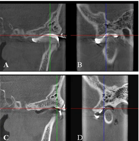
appar-Fig. 4. Arthrography and double contrast method on CBCT images. Needle puncture is performed according to the preoperative plan. CBCT provides clear high-resolution arthrographic images of the TMJ and contributes to sensitive diagnosis of patients. A, Oblique coronal image from arthrography; B, oblique sagittal image from arthrography; C, oblique coronal image from double-contrast arthrography; D, oblique sagittal image from double-contrast arthrography.
Table II. Number of resettings of the puncture point in 178 TMJs
Change of puncture point
0 1 2
IGPT (n⫽ 76) 74 2 0
CPT (n⫽ 102) 84 15 3
Frequency of change of puncture points differed significantly be-tween IGPT and CPT (P⬍ .05).
CPT, conventional puncture technique; IGPT, image-guided puncture
technique; TMJ, temporomandibular joint. 274 275 276 277 278 279 280 281 282 283 284 285 286 287 288 289 290 291 292 293 294 295 296 297 298 299 300 301 302 303 304 305 306 307 308 309 310 311 312 313 314 315 316 317 318 319 320 321 322 323 324 325 326 327 328 274 275 276 277 278 279 280 281 282 283 284 285 286 287 288 289 290 291 292 293 294 295 296 297 298 299 300 301 302 303 304 305 306 307 308 309 310 311 312 313 314 315 316 317 318 319 320 321 322 323 324 325 326 327 328
ent between IGPT and CPT groups in terms of pain or mouth opening after the 1-month follow-up.
In conclusion, the present investigation has shown that IGPT may be the most reliable method for TMJ puncture. IGPT is effective in pain mitigation and im-proves mouth opening after pumping manipulation treatment in patients with internal derangement. The present study indicates that IGPT using CBCT is a safe procedure for treating internal derangement of the TMJ. Methods for automating, standardizing, and obtaining reproducible results for puncturing the joint cavity need to be improved in future studies.
REFERENCES
1. Krisjane Z, Urtane I, Krumina G, Bieza A, Zepa K, Rogovska I. Condylar and mandibular morphological criteria in the 2D and 3D MSCT imaging for patients with Class II division 1 subdi-vision malocclusion. Stomatologija 2007;9:67-71.
2. Larheim TA. Current trends in temporomandibular joint imag-ing. Oral Surg Oral Med Oral Pathol Oral Radiol Endod 1995;80:557-76.
3. Brooks SL, Brand JW, Gibbs SJ, Hollender L, Lurie AG, Omnell KA, et al. Imaging of the temporomandibular joint: a position paper of the American Academy of Oral and Maxillofacial Radiology. Oral Surg Oral Med Oral Pathol Oral Radiol Endod 1997;83:601-18.
4. Westesson PL. Reliability and validity of imaging diagnosis of temporomandibular joint disorders. Adv Dent Res 1993; 7:135-51.
5. Honda K, Arai Y, Kashima M, Takano Y, Sawada K, Ejima K, et al. Evaluation of the usefulness of the limited cone-beam CT (3DX) in the assessment of the thickness of the roof of the glenoid fossa of the temporomandibular joint. Dentomaxillofac Radiol 2004;33:391-5.
6. Honda K, Larheim TA, Maruhashi K, Matsumoto K, Iwai K. Osseous abnormalities of the mandibular condyle: diagnostic reliability of cone beam computed tomography compared with helical computed tomography based on an autopsy material. Dentomaxillofac Radiol 2006;35:152-7.
7. Lewis EL, Dolwick MF, Abramowicz S, Reeder SL. Contempo-rary imaging of the temporomandibular joint. Dent Clin North Am 2008;52:879-90.
8. Ohnishi M. Clinical application of arthroscopy in the temporo-mandibular joint diseases. Bull Tokyo Med Dent Univ 1980;27:141-50.
9. Murakami K, Matsuki M, Iizuka R, Ono T. Recapturing the persistent anteriorly displaced disc by mandibular manipulation after pumping and hydraulic pressure to the upper joint cavity of the temporomandibular joint. J Craniomandib Pract 1987; 5:17-24.
10. McCain JP. Arthroscopic surgery of the human temporomandib-ular joint. J Oral Maxillofac Surg 1988;46:648-55.
11. Nizan DW, Dolwick MF, Martinez GA. Temporomandibular joint arthrocentesis: a simplified treatment for severe, limited mouth opening. J Oral Maxillofac Surg 1991;49:1163-7. 12. Kobayashi K, Yuasa M, Imanaka M, Igarashi C, Yamamoto A.
Development of interventional radiology for the internal de-rangements of the temporomandibular joint. Dent Jpn 1999;35:97-9.
13. Matsumoto K, Honda K, Ohshima M, Yamaguchi Y, Nakajima I, Micke P, et al. Cytokine profile in synovial fluid from patients with internal derangement of the temporomandibular joint: a preliminary study. Dentomaxillofac Radiol 2006;35:434-41. 14. Westesson PL, Eriksson L, Liedberg J. The risk of damage to
facial nerve, superficial temporal vessels, disk, and articular surfaces during arthroscopic examination of the temporomandib-ular joint. J Oral Surg 1986;62:124-7.
15. Green MW, Hackney FL, Van Sickels JE. Arthroscopy of the temporomandibular joint: an anatomic perspective. J Oral Max-illofac Surg 1989;47:386-9.
16. Sugisaki M, Ikai A, Tanabe H. Dangerous angles and depths for middle ear and middle cranial fossa injury during arthroscopy of the temporomandibular joint. J Oral Maxillofac Surg 1995; 53:803-10.
17. Bjørnland T, Rørvik M, Haanæs HR, Teige J. Degenerative changes in the temporomandibular joint after diagnostic arthros-copy. An experimental study in goats. Int J Oral Maxillofac Surg 1994;23:41-5.
18. Honda K, Hamada Y, Ejima K, Tsukimura N, Kino K. Interven-tional radiology of synovial chondromatosis in the temporoman-Table III. Change in mean maximal mouth opening over time
Mean maximal mouth opening, mm
Before TMJ injection 1 wk after TMJ injection 3 mo after TMJ injection 1 y after TMJ injection
IGPT 24⫾ 5.2 40⫾ 6.1* 43⫾ 5.5 44⫾ 5.5
CPT 26⫾ 5.0 33⫾ 5.9* 40⫾ 5.6 42⫾ 5.9
Values are given as mean⫾ standard deviation.
CPT, conventional puncture technique; IGPT, image-guided puncture technique; TMJ, temporomandibular joint.
*A significant difference was identified in 1-week maximal mouth opening between IGPT and CPT (P⬍ .05). Table IV. Changes in mean pain evaluation over time using VAS (0-100)
Before TMJ Injection 1 wk after TMJ injection 3 mo after TMJ injection 1 y after TMJ injection
IGPT 54⫾ 9.0 25⫾ 12.2* 8⫾ 10.4 6⫾ 7.7
CPT 52⫾ 9.6 35⫾ 13.5* 10⫾ 12.2 4⫾ 6.5
Values are given as mean⫾ standard deviation.
CPT, conventional puncture technique; IGPT, image-guided puncture technique; TMJ, temporomandibular joint; VAS, visual analog scale.
*A significant difference was observed in 1-week VAS between IGPT and CPT (P⬍ .05). 329 330 331 332 333 334 335 336 337 338 339 340 341 342 343 344 345 346 347 348 349 350 351 352 353 354 355 356 357 358 359 360 361 362 363 364 365 366 367 368 369 370 371 372 373 374 375 376 377 378 379 380 381 382 383 329 330 331 332 333 334 335 336 337 338 339 340 341 342 343 344 345 346 347 348 349 350 351 352 353 354 355 356 357 358 359 360 361 362 363 364 365 366 367 368 369 370 371 372 373 374 375 376 377 378 379 380 381 382 383
2008;37:232-5.
19. Seibel RM. Image-guided minimally invasive therapy. Surg En-dosc 1997;11:156-62.
20. Perrin DP, Vasilyev NV, Novotny P, Stoll J, Howe RD, Dupont PE, et al. Image guided surgical interventions. Curr Probl Surg 2009;46:736-66.
21. Honda K, Bjørnland T. Image-guided puncture technique for the superior temporomandibular joint space: value of cone beam computed tomography (CBCT). Oral Surg Oral Med Oral Pathol Oral Radiol Endod 2006;102:281-6.
22. Yeung RW, Xia JJ, Samman N. Image-guided minimally inva-sive surgical access to the temporomandibular joint: a prelimi-nary report. J Oral Maxillofac Surg 2006;64:1545-52. 23. Fritz J, Thomas C, Tzaribachev N, Horger MS, Claussen CD,
Lewin JS, et al. MRI-guided injection procedures of the tem-poromandibular joints in children and adults: technique, accu-racy, and safety. AJR Am J Roentgenol 2009;193:1145-54.
intra-articular injection of sodium hyaluronate in patients with osteoarthritis of the knee. Curr Med Res Opin 1988;11:201-13. 25. Bjørnland T, Gjærum AA, Møystad A. Osteoarthritis of the temporomandibular joint: an evaluation of the effects and com-plications of corticosteroid injection compared with injection with sodium hyaluronate. J Oral Rehabil 2007;34:583-9.
Reprint requests:
Kunihito Matsumoto, DDS, PhD
Department of Oral and Maxillofacial Radiology Nihon University School of Dentistry 1-8-13 Kanda-Surugadai, Chiyoda-ku Tokyo 101-8310, Japan matsumoto@dent.nihon-u.ac.jp 384 385 386 387 388 389 390 391 392 393 394 395 396 397 398 399 400 401 402 403 404 405 406 407 408 409 410 411 412 413 414 415 416 417 418 419 420 421 422 423 424 425 426 427 428 429 430 431 432 433 434 435 436 437 438 384 385 386 387 388 389 390 391 392 393 394 395 396 397 398 399 400 401 402 403 404 405 406 407 408 409 410 411 412 413 414 415 416 417 418 419 420 421 422 423 424 425 426 427 428 429 430 431 432 433 434 435 436 437 438