This is an author produced version of a paper published in Journal of
Biomaterials Applications. This paper has been peer-reviewed but does not include the final publisher proof-corrections or journal pagination.
Citation for the published paper:
Galli, Silvia; Jimbo, Ryo; Tovar, Nick; Yoo, Daniel; Anchieta, Rodolfo; Yamaguchi, Satoshi; Coelho, Paulo G. (2015). The effect of osteotomy dimension on osseointegration to resorbable media-treated implants : a
study in the sheep. Journal of Biomaterials Applications, vol. 29, issue 8, p. null
URL: https://doi.org/10.1177/0885328214553958
Publisher: Sage
This document has been downloaded from MUEP (https://muep.mah.se) / DIVA (https://mau.diva-portal.org).
The effect of osteotomy dimension on osseointegration to resorbable media treated implants: a study in the sheep.
Authors: Silvia Galli†* DDS, Ryo Jimbo† PhD, Nick Tovar¶ PhD, Daniel Y. Yoo¶, Rodolfo B. Anchieta¶ PhD, Satoshi Yamaguchi ¶ PhD, Paulo G. Coelho¶ PhD.
Affiliation:
† Department of Prosthodontics, Faculty of Odontology, Malmö University, 205 06 Malmö, Sweden;
¶
Department of Biomaterials and Biomimetics, New York University, New York NY 10010, USA
Corresponding author: *Silvia Galli,
Department of Prosthodontics, Faculty of Odontology, Malmö University, 20506 Malmö, Sweden.
telephone: +46 (0) 40 665 8614 email: silvia.galli@mah.se
Running head: Ideal osteotomy for resorbable media blasted surface
Key words: implant surface; resorbable surface – blasted surface; removal torque; histology; osseointegration.
Abstract
The drilling technique and the surface characteristics are known to influence the healing times of oral implants. The influence of osteotomy dimension on osseointegration of microroughned implant surfaces treated with resorbable media blasting (RBM) was tested in an in vivo model.
Ninety-six implants (ø4.5 mm, 8 mm in length) with RBM treated surfaces were placed in the ileum of six sheep. The final osteotomy diameters were 4.6 mm (Reamer), 4.1 mm (Loose) 3.7 mm (Medium) and 3.2 mm (Tight). After 3 and 6 weeks healing the implants were biomechanically tested and histologically evaluated. Statistical analysis was performed using Page L trend test for ordered and paired sample and linear regression, with significance level at p<0.05.
An overall increase in all dependent variables was observed with the reduction of osteotomy diameter. In addition, all osseointegration scores increased over time. At 3 weeks, the retention was significantly higher for smaller osteotomies. The histological sections depicted intimate contact of bone with all the implant surfaces and osteoblast lines were visible in all sections.
The RBM microroughed surfaces achieved successful osseointegration for all the instrumentation procedures tested, with higher osseointegration scores for the press-fit insertion group.
Key words: implant surface; resorbable surface – blasted surface ; removal torque; histology; osseointegration.
1. Introduction
Implant surface has long been identified as one of the crucial factor for successful osseointegration1-3. Surface modifications plays an important role in the early phases of implant healing, when osteogenic cells are recruited and migrate directly onto the implant surface. It has been demonstrated that the physical and chemical properties of titanium surfaces modulate platelet activation, growth factor release and the blood cloth stabilization due to a direct interaction of fibrin fibers with the implant interface 4.
Roughened surfaces were found to provide better conditions for the cell attachment and cell spreading due to their increased surface area5 and, in addition, these have been shown to influence cell proliferation and differentiation6, 7. Accordingly, numerous in vivo studies presented a stronger bone response to rougher surfaces compared to turned surfaces8-10. Similar evidences were found clinically, in which implants with half turned and half treated surface were placed in humans11. When the implants were retrieved for histological analysis after 6 months, the implant portion with the roughened surface presented a significantly higher bone-to-implant contact than the smooth surface11 .
Besides surface topography, surface chemistry has been modified by incorporation of different bioactive substances, such as calcium, magnesium, phosphates or fluorides, to further promote bone regeneration, proteins adsorption and cell attachment on the implants12-16. One method to modify the texture of titanium surfaces while deposing hydroxyapatite or calcium phosphate (CaP) on them, is blasting with resorbable biocompatible bioceramics (resorbable blasting media or RBM) 17-20(Coelho and Lemon 2009, Marin et al 2008 and 2010). The bioceramic
blasting particles produce a moderately rough topography and they can be incorporated into the titanium surfaces and be released in bone after implant placement, which has been suggested to increase the bioactivity.
Extensive surface characterization has shown that RBM-treated surfaces are less rough than titanium surfaces blasted with alumina particles and possess a non-homogenous distribution of CaP, depending on several factors, such as the composition of the basting media, the particle size and other processing parameters21. This incorporation technique overcomes the drawbacks of the thick plasma-sprayed HA and CaP coatings, which exhibited a low clinical survival in the long-term due to debonding of the coating at the implant interface22, 23. It has been reported that RBM obtained a higher and faster biomechenical fixation, after placement in dogs24, 25 and newly formed bone was observed in close contact with the surfaces24.
A great amount of experiments focused on the impact of the different surface modifications on osseointegration, while a significantly smaller body of literature exists regarding the influence of the drilling protocol in the host response to specific surfaces. Osteotomy preparation, in combination with implant macrogeometry and the quality of the implant bed, determines the initial mechanical interaction between the implant and the bone (primary stability) and it is one of the factors that ultimately determine the biological stabilization of the implant26-28.
Testing the outcome of implants with bioactive surfaces in sites prepared with different surgical procedures would be of great interest if one is attempting to maximize osseointegration at the shortest period of time possible. Thus, the objective of the current investigation was to compare the bone healing around resorbable blasting media treated implants, placed under different drilling protocols in a sheep model, by means of biomechanical and histological testing.
2. Material and Methods
Implants and surgical procedures
Implants treated with resorbable media blasting where used in this study (BioHorizon, 4.5 mm in diameter by 8 mm length).
The implants were placed in 6 Finnish Dorset cross-bread sheep, each one weighting approximaly 150 punds and being 1.5 years old, under the ethical permit released by the Ethics Committee for Animal Research at the École Nationale Vétérinaire d’Alfort (Maisons-Alfort, Val-de-Marne, France). The sheep were acclimatized 2 weeks in the facility before the beginning of the experimental procedures.
At day 0 of the experiment, the sheep where anesthetized with Sodium Pentothal (15-20 mg/kg) in Normasol solution in the jugular vein. The hips were shaved, to expose the skin of surgical site for the implant placement. The area was disinfected with Poviodine solution and then delimitated by aseptic drapes. The animals were constantly monitored for vital parameters during the surgical procedure, by an automated system that provided ventilation and sustain of the anesthesia with Isoforane (1.5-3%) in O2/N2O (50/50). Antibiotic (500 mg Cefazolina) was administered pre-operatively intravenously.
The surgery started with skin incision and muscles and the iliac crest was exposed by blunt dissection. Following 4 different drill sequences, four osteotomies were prepared, so to obtain implants sites with different final diameters: Reamer (R)
4.6 mm (final instrumentation hand reamer), Loose (L) 4.1 mm, Medium (M) 3.7 mm and Tight (T) 3.2 mm. Each sheep received 4 implants on one side at day 0 of experiment and 4 implants on the other side 3 weeks after the first surgery. A total of 48 implants were placed, corresponding to 4 experimental groups (n=6 per healing time).
Removal torque and histomorphometry
Six week after the first surgery (day 0), the animals were euthanized with an overdose of Sodium Pentothal and the hips were dissected. Half of the implants had been in situ for 3 weeks and the other half for 6 weeks. Twenty-four implants undergone biomechanical test by means of removal torque (Tonhichi, torque gauge wrench) while the other twenty-four implants were harvested with the surrounding bone and were processed for non-decalcified histology. The bone blocks were embedded in resin (methylmethacrylate, MMA) after dehydration in increasing ethanol concentrations. Sections were cut and grinded down to thin slices, following the cutting-grinding standard procedure29 ,
After staining with toluidine blue dye, the samples were observed using the optical microscopy with magnifications from ×50 to ×200 (Leica DM2500M, Leica Microsystems GmbH, Wetzlar, Germany). The bone-to-implant contact (BIC) and the bone area fraction occupancy (BAFO) within the implant threads were calculated by means of specialized computer software (Leica Application Suite, Leica Microsystems GmbH, Wetzlar, Germany)..
To evaluate the effects of drilling techniques on the osseointegration parameters at two healing times the Page-L trend test for related and ordered samples was used. This test is appropriate when it is reasonable to predict an order of the treatment effects, as it applies to the current case, and when the samples are related (different samples in the same animal). The null hypothesis was that the scores of the different instrumentation techniques were equals . To evaluate the effects of instrumentation over time linear regression analysis was used. Statistical significance was set at 5% (α = 0.05).
3. Results Animals
All the animals recovered well from the surgery and showed no signs of complications, infection or disease during the whole observation period. All the implants were stable when examined immediately after euthanasia; therefore no implant was excluded from the study.
Removal Torque
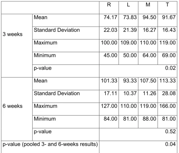
The removal torque (RTQ) values and the related descriptive statistics are presented in Table 1 and Figure 1. In general, an overall increase in removal torque values was observed between 3 and 6 weeks.
The Page trend statistical test showed a significant trend in RTQ values among the different instrumentation procedures, ordered from the widest to the tightest osteotomy (p<0.05). At 3 weeks of healing the RTQ values were statistically significantly ordered, with higher torques correlating to tighter osteotomies. After 6 weeks, RTQ values for the M and T groups were the highest, but no statistical significance was detected. When considering the change in removal torque values
over time, a significant increase in torque values was observed for group R between 3 and 6 weeks (p<0.05).
Histologic and histomorphometric evaluation
The histological analysis presented new bone formation in close contact with the implant surfaces for all group at both observations times (Figure 2). At 3 weeks, the implants inserted in tight osteotomies (group T) were in close contact mainly with cortical bone, while the implants in the group R were surrounded by newly formed bone and loose acellular tissue. For all groups, areas of bone apposition were visible in the area within the threads and newly formed woven bone, with numerous highly stained osteocytes were present in all sections. At 6 weeks, less differences among and between groups were visible as new and mineralized bone was in contact along a substantial portion of the bone-implant interface.
The mean bone to implant contact (BIC) percentages ± SD for each group are presented in table (Table 2) and graph (figure 3a). In general, an increase in bone contact over time was noticed, although the increase was significant only for group M (p<0.05). Higher BIC values corresponded to the different instrumentation procedure, order from the loosest to the tightest preparation. The mean differences among the groups did not reach significant value sat 3 or 6 weeks, but when the BIC values of both time points were pooled, a significant trend of increasing bone-contact for the ordered groups was found (p<0.05).
Descriptive statistics for the bone area fraction occupancy (BAFO) are presented in Table 3 and Figure 3b. Again, a general increase over time was observed for BAFO values and a tendency of increased values for the groups ordered from R to T. At 3 weeks of healing the trend of higher bone filling between instrumentation
groups ordered from R to T was statistically significant (p<0.05), while the difference was not significant between groups at 6 weeks (p>0.05). When the BAFO results at both time points were pooled, the level of significance increased between groups (p<0.05).
4. Discussion
This experiment investigated the effect of different osteotomy preparations on the osseointegration of an implant with microrough surfaces obtained with resorbable blasting media (RBM).
This RBM surface treatment induces topograghical changes in combination with chemical alterations, due to the incorporation of CaP particles onto the titanium surfaces30. Previous studies have demonstrated improved host-to-implant responses for implant treated with resorbable blasting media to be comparable to other moderately rough surfaces18.
Besides microgeometry, implant macrogeometry has a significant effect on the healing process through implant primary stability. For each unique combination of modified implant surface and different microgeometry, implant companies suggest a specific surgical protocol. The initial distance created between the implant surface and the bone bed is thought to induce different bone healing phenomena31. When the osteotomy is smaller than the implant diameter, the mechanical friction is high and the implant micromotion is reduced32. The predominant process in the bone regeneration is the remodeling of the original bone through osteoclasts activity and the substitution with new bone. During the uninterrupted healing, there is a gradual shift from the implant primary stability, the early mechanical interaction between the bone bed and the implant, and the secondary stability given by biological
integration33. The least stable moment in this process is weeks after implant placement, when mechanical interlocking drops due to viscoelastic relaxation of the bone along with remodeling of the bone close to the implant interface 26.
On the other hand, when the implant is placed after preparation with full drilling sequence, the difference in diameter between the implant inner thread diameter and drilling outer diameter results a closed chamber that is filled with blood and blood clot, which interact with the whole implant surface, and bone formation proceeds for contact osteogenesis without extensive interfacial remodeling4.
In the present study, an increase in osseointegration measurable parameters such as BIC and BAFO from the loosest to the tightest preparation was observed. On the other hand, the removal torque testing showed significantly higher retention for implants inserted in tighter osteotomies after 3 weeks of healing. This finding is consistent with the higher bone-area fraction occupancy (BAFO%) of the press-fit groups (Medium and Tight), which had significantly more bone within the implant threads. The removal torque scores further increased at 6 weeks for all groups, indicating that the implant stability was maintained at a high level during the healing process and the shift from primary to secondary stability did not undermine the implant anchorage. However, only for the R group, the increase of RTQ scores from 3 to 6 weeks was statistically significant, indicating that new bone successfully formed around the implant in this drilling condition.
Histologic slides displayed an intimate contact between bone and the implant surfaces, at both time points. The osteoid secretion by active osteoblasts was evident in the histologic sections, and they surrounded the implants along the entire surface. Newly formed woven bone visible on the sections at 3 weeks was entirely substituted by mineralized bone in the 6-weeks sections. The high bone-contact rates indicate the
biocompatibility and osteoconductivity of the moderately roughed and chemically modified RBM surface regardless of drilling protocol. Furthermore, the potential presence of residual CaP particles from the surfaces may have inducted osteogenic cell recruitment and activation contiguously to the implant.
These results confirmed that a simplified drilling protocol does not have negative indications and it achieves at least comparable or faster osseointegration than the conventional drilling procedure, with gradual expansion of the implant site34, 35 .
Conclusion
The results of the study confirmed the biocompatibility and successful integration of microrough and CaP-doped titanium surfaces obtained by resorbable media blasting. Moreover, it can be concluded that a simplified surgical procedure, which reduces the drill sequence and generates a press-fit situation, achieved high primary stability and led to successful bone regeneration and stable biological fixation of the implants.
Table 1: RTQ values at 3 and 6 weeks (values in cNm) for the four test groups (R, L, M and T). The p-values were obtained with Page´s L trend test for ordered related samples. R L M T 3 weeks Mean 74.17 73.83 94.50 91.67 Standard Deviation 22.03 21.39 16.27 16.43 Maximum 100.00 109.00 110.00 119.00 Minimum 45.00 50.00 64.00 69.00 p-value 0.02 6 weeks Mean 101.33 93.33 107.50 113.33 Standard Deviation 17.11 10.37 11.26 28.08 Maximum 127.00 110.00 119.00 166.00 Minimum 84.00 81.00 88.00 81.00 p-value 0.52
Table 2: BIC values (%) at 3 and 6 weeks for the four test groups (R, L, M and T). The p-values were obtained with Page´s L trend test for ordered related samples.
R L M T 3 weeks Mean 45.07 45.86 51.77 54.81 Standard Deviation 17.54 12.76 14.80 14.20 Maximum 58.51 62.82 66.59 74.44 Minimum 10.70 30.19 24.58 37.50 p-value 0.2 6 weeks Mean 55.69 57.44 66.43 65.79 Standard Deviation 8.64 11.42 5.91 5.89 Maximum 69.90 69.18 74.80 72.80 Minimum 46.55 43.04 59.85 55.25 p-value 0.08
Table 3: BAFO values (%) at 3 and 6 weeks for the four test groups (R, L, M and T). The p-values were obtained with Page´s L trend test for ordered related samples
R L M T 3 weeks Mean 37.56 41.08 45.55 49.49 Standard Deviation 13.43 13.52 10.14 15.18 Maximum 58.60 52.60 56.70 68.17 Minimum 19.00 21.55 31.67 25.07 p-value 0.03 6 weeks Mean 44.69 41.35 57.52 60.83 Standard Deviation 12.48 14.17 15.14 9.97 Maximum 64.69 58.61 75.14 74.06 Minimum 28.90 22.33 40.59 43.26 p-value 0.3
Figure Legends Figure 1
Removal torque (adjusted estimates ± 95% CI) as a function of instrumentation and healing time.
Figure 2
Histologic observations (bars: 200 µm) of the effect of 4 different instrumentation procedures. (a) After 3 weeks. (b) After 6 weeks.
Figure 3
(a) Bone-to-implant contact (BIC, adjusted estimates ± 95% CI) as a function of instrumentation and healing time. (b) Bone area fraction occupancy (BAFO, adjusted estimates ± 95% CI) as a function of drilling technique and healing time.
Reference list
1. Albrektsson T, Branemark PI, Hansson HA and Lindstrom J. Osseointegrated titanium implants. Requirements for ensuring a long-‐lasting, direct bone-‐to-‐implant anchorage in man. Acta orthopaedica Scandinavica. 1981; 52: 155-‐70.
2. Le Guehennec L, Soueidan A, Layrolle P and Amouriq Y. Surface treatments of titanium dental implants for rapid osseointegration. Dental materials : official
publication of the Academy of Dental Materials. 2007; 23: 844-‐54.
3. Wennerberg A and Albrektsson T. On implant surfaces: a review of current knowledge and opinions. The International journal of oral & maxillofacial implants. 2010; 25: 63-‐74.
4. Davies JE. Understanding peri-‐implant endosseous healing. Journal of dental
education. 2003; 67: 932-‐49.
5. Mustafa K, Wennerberg A, Wroblewski J, Hultenby K, Lopez BS and Arvidson K. Determining optimal surface roughness of TiO(2) blasted titanium implant material for attachment, proliferation and differentiation of cells derived from human mandibular alveolar bone. Clinical oral implants research. 2001; 12: 515-‐25.
6. Sammons RL, Lumbikanonda N, Gross M and Cantzler P. Comparison of
osteoblast spreading on microstructured dental implant surfaces and cell behaviour in an explant model of osseointegration. A scanning electron microscopic study. Clinical
oral implants research. 2005; 16: 657-‐66.
7. Zhao G, Zinger O, Schwartz Z, Wieland M, Landolt D and Boyan BD. Osteoblast-‐ like cells are sensitive to submicron-‐scale surface structure. Clinical oral implants
research. 2006; 17: 258-‐64.
8. Gotfredsen K, Wennerberg A, Johansson C, Skovgaard LT and Hjorting-‐Hansen E. Anchorage of TiO2-‐blasted, HA-‐coated, and machined implants: an experimental study with rabbits. Journal of biomedical materials research. 1995; 29: 1223-‐31.
9. Piattelli M, Scarano A, Paolantonio M, Iezzi G, Petrone G and Piattelli A. Bone response to machined and resorbable blast material titanium implants: an experimental study in rabbits. The Journal of oral implantology. 2002; 28: 2-‐8.
10. Novaes AB, Jr., Souza SL, de Oliveira PT and Souza AM. Histomorphometric analysis of the bone-‐implant contact obtained with 4 different implant surface
treatments placed side by side in the dog mandible. The International journal of oral &
maxillofacial implants. 2002; 17: 377-‐83.
11. Lazzara RJ, Testori T, Trisi P, Porter SS and Weinstein RL. A human histologic analysis of osseotite and machined surfaces using implants with 2 opposing surfaces.
The International journal of periodontics & restorative dentistry. 1999; 19: 117-‐29.
12. Sul YT, Johansson C, Wennerberg A, Cho LR, Chang BS and Albrektsson T.
Optimum surface properties of oxidized implants for reinforcement of osseointegration: surface chemistry, oxide thickness, porosity, roughness, and crystal structure. The
International journal of oral & maxillofacial implants. 2005; 20: 349-‐59.
13. Sul YT, Kwon DH, Kang BS, Oh SJ and Johansson C. Experimental evidence for interfacial biochemical bonding in osseointegrated titanium implants. Clinical oral
implants research. 2013; 24 Suppl A100: 8-‐19.
14. Fini M, Cigada A, Rondelli G, et al. In vitro and in vivo behaviour of Ca-‐ and P-‐ enriched anodized titanium. Biomaterials. 1999; 20: 1587-‐94.
15. Franco Rde L, Chiesa R, Beloti MM, de Oliveira PT and Rosa AL. Human osteoblastic cell response to a Ca-‐ and P-‐enriched titanium surface obtained by anodization. Journal of biomedical materials research Part A. 2009; 88: 841-‐8.
16. Cecchinato F, Xue Y, Karlsson J, et al. In vitro evaluation of human fetal osteoblast response to magnesium loaded mesoporous TiO coating. Journal of
biomedical materials research Part A. 2013.
17. Coelho PG and Lemons JE. Physico/chemical characterization and in vivo evaluation of nanothickness bioceramic depositions on alumina-‐blasted/acid-‐etched Ti-‐ 6Al-‐4V implant surfaces. Journal of biomedical materials research Part A. 2009; 90: 351-‐ 61.
18. Bonfante EA, Marin C, Granato R, et al. Histologic and biomechanical evaluation of alumina-‐blasted/acid-‐etched and resorbable blasting media surfaces. The Journal of
oral implantology. 2012; 38: 549-‐57.
19. Marin C, Granato R, Suzuki M, et al. Biomechanical and histomorphometric analysis of etched and non-‐etched resorbable blasting media processed implant
surfaces: an experimental study in dogs. Journal of the mechanical behavior of biomedical
materials. 2010; 3: 382-‐91.
20. Marin C, Granato R, Suzuki M, Gil JN, Piattelli A and Coelho PG. Removal torque and histomorphometric evaluation of bioceramic grit-‐blasted/acid-‐etched and dual acid-‐etched implant surfaces: an experimental study in dogs. Journal of periodontology. 2008; 79: 1942-‐9.
21. Coelho PG, Suzuki M, Guimaraes MV, et al. Early bone healing around different implant bulk designs and surgical techniques: A study in dogs. Clinical implant dentistry
and related research. 2010; 12: 202-‐8.
22. Ong JL, Carnes DL and Bessho K. Evaluation of titanium plasma-‐sprayed and plasma-‐sprayed hydroxyapatite implants in vivo. Biomaterials. 2004; 25: 4601-‐6. 23. Yang Y, Dennison D and Ong JL. Protein adsorption and osteoblast precursor cell attachment to hydroxyapatite of different crystallinities. The International journal of oral
& maxillofacial implants. 2005; 20: 187-‐92.
24. Coelho PG, Marin C, Granato R, Giro G, Suzuki M and Bonfante EA. Biomechanical and histologic evaluation of non-‐washed resorbable blasting media and alumina-‐
blasted/acid-‐etched surfaces. Clinical oral implants research. 2012; 23: 132-‐5.
25. Marin C, Granato R, Bonfante EA, Suzuki M, Janal MN and Coelho PG. Evaluation of a nanometer roughness scale resorbable media-‐processed surface: a study in dogs.
Clinical oral implants research. 2012; 23: 119-‐24.
26. Halldin A, Jimbo R, Johansson CB, et al. Implant Stability and Bone Remodeling after 3 and 13 Days of Implantation with an Initial Static Strain. Clinical implant dentistry
and related research. 2012.
27. Halldin A, Jimbo R, Johansson CB, et al. The effect of static bone strain on implant stability and bone remodeling. Bone. 2011; 49: 783-‐9.
28. Tabassum A, Meijer GJ, Wolke JG and Jansen JA. Influence of surgical technique and surface roughness on the primary stability of an implant in artificial bone with different cortical thickness: a laboratory study. Clinical oral implants research. 2010; 21: 213-‐20.
29. Donath K. Preparation of Histologica Sections by the Cutting-‐Grinding Technique for Hard Tissue and other Material not suitable to be sectioned by routine methods.
EXAKT-‐Kulzer, Norderstedt. 1993: 1:16.
30. Witek L, Marin C, Granato R, et al. Surface characterization, biomechanical, and histologic evaluation of alumina and bioactive resorbable blasting textured surfaces in titanium implant healing chambers: an experimental study in dogs. The International
journal of oral & maxillofacial implants. 2013; 28: 694-‐700.
31. Berglundh T, Abrahamsson I, Lang NP and Lindhe J. De novo alveolar bone formation adjacent to endosseous implants. Clinical oral implants research. 2003; 14: 251-‐62.
32. Freitas AC, Jr., Bonfante EA, Giro G, Janal MN and Coelho PG. The effect of implant design on insertion torque and immediate micromotion. Clinical oral implants research. 2012; 23: 113-‐8.
33. Norton M. Primary stability versus viable constraint-‐-‐a need to redefine. The
International journal of oral & maxillofacial implants. 2013; 28: 19-‐21.
34. Jimbo R, Giro G, Marin C, et al. Simplified drilling technique does not decrease dental implant osseointegration: a preliminary report. Journal of periodontology. 2013; 84: 1599-‐605.
35. Jimbo R, Tovar N, Yoo DY, Janal MN, Anchieta RB and Coelho PG. The effect of different surgical drilling procedures on full laser-‐etched microgrooves surface-‐treated implants: an experimental study in sheep. Clinical oral implants research. 2013.
Fig. 1
Fig. 2a
Fig. 3