© 2012 S. Karger AG, Basel
This is the author's version of the work. It is posted here by permission of Karger AG, Basel for your personal use. Articles may not be used for commercial purposes. The definitive version was published in Caries Research, vol.46, iss.2,
pages: 156-160
http://dx.doi.org/10.1159/000337390
Fluoride-Supplemented Milk Inhibits Acid
Tolerance in Root Caries Biofilms
J. Neilands, L.G. Petersson, D. Beighton, G. Svensäter
Abstract
In this study we investigated the effect of fluoride on plaque acid tolerance. The test group consumed 200 ml of milk supplemented with 5 mg F/l as NaF once a day, the milk control group drank 200 ml of unsupplemented milk, and the nomilk control group did not consume milk in this manner. Plaque samples were taken at baseline and after 15 months. The proportion of acid-tolerant bacteria in plaque was estimated using LIVE/DEAD®BacLight™ staining after exposure to pH 3.5 for 2 h. The fluoride group
showed a statistically significant decrease in plaque acid tolerance compared to baseline. This study shows that daily intake of fluoride in milk reduces plaque acid tolerance.
Key Words: Milk, Plaque, Root caries
Microbial fermentation of dietary carbohydrates results in the production of organic acids, in particular lactic acid, causing demineralization of enamel and exposed root surfaces leading to the development of caries [Takahashi and Nyvad, 2008]. In response to repeated periods of low pH some microorganisms are able to initiate an acid tolerance response (ATR) which involves phenotyp ic changes. The ATR allows the bacteria to survive and continue to produce acids at low pH values, resulting in an acid-tolerant microflora [Hamilton and Buckley, 1991; Svensäter et al., 1997; Lemos et al., 2005]. According to the ‘ecological plaque hypothesis’, the selection of cariescausing bacteria is mediated by changes in the oral environment, and any species with relevant traits can contribute to the disease process [Marsh, 1994, 2003]. Therefore in the search for caries-causing bacteria, it is more important to search for bacteria with the ATR phenotype rather than attempting to identify specific ‘cariogenic’ bacterial species.
Fluoride affects the physiology of oral bacteria by inhibiting enolase in the glycolytic pathway and membranebound proton-pumping H+/ATPases, thus reducing sugar transport
and acid production [Kanapka and Hamilton, 1971; Hamilton, 1977; Marquis, 1977; Sutton et al., 1987]. Fluoride also inhibits ATR induction in newly formed biofilms of Streptococcus mutans, making the bacteria acidsensitive [Welin-Neilands and Svensater, 2007]. Although much is known about the effect of fluoride on bacterial physiology, data on the effect of fluoride on acid tolerance in plaque in vivo is limited. The aim of the present study was therefore to investigate the effects of fluoride included in the diet as a supplement to cow’s milk on plaque acid tolerance and the composition of the predominant culturable acid-tolerant microflora of root caries lesions.
Subjects and Methods Subjects
Patients with at least one active root caries lesion were invited to participate. All patients regularly attended a private dental specialist clinic in Ljungby, Sweden. Patients with severe chronic diseases and lactose-intolerant individuals were excluded from the study. 30 subjects were selected, 21 men and 9 women. They were randomly divided into three groups of ten and the groups were then randomly allocated to the three intervention groups. Gender was evenly distributed between the groups and mean age were 61 years for all three groups. All primary root caries lesions were examined on buccal/labial sites of the teeth by one of the staff members. One primary root caries lesion recorded as ‘leathery active’ or ‘soft active’ [Beighton et al., 1993] per subject was randomly selected for plaque sampling. At baseline, there were no significant differences in the frequency of different clinical severity grades of the root caries lesions in the groups. The test group (F group) consumed 200 ml of cow’s milk supplemented with 5 mg F/l as NaF as a single dose once per day, the milk control group (M group) drank 200 ml of unsupplemented cow’s milk, and the no-milk control group (C group) did not consume milk in this manner. The length of the study was 15 months. Compliance was checked every 3 months by a dental hygienist when the patient received new capsules, containing milk or milk and fluoride, at the local dental clinic. The intervention received by each group was unmasked only after all data had been collected and analyzed statistically at the end of the study. The study was approved by the Research Ethics Committee at Umeå University (§562/03, dnr 03- 475). During the study, one subject from each group dropped out, leaving 27 subjects for final analysis. All subjects were instructed to continue consuming their normal diet and not to alter their oral hygiene practices, thus all subjects used F-containing toothpastes throughout the study period. All subjects lived in an area with low fluoride levels (0.1 ppm) in the drinking water.
Plaque Sampling Procedures and Culture Media
Three biofilm samples were taken with a sterile Teflon loop (cat. no. 612-9356, VWR, Stockholm, Sweden) from the central part of the root caries lesion avoiding soft dentine. Each sample was transferred into a separate sterile microfuge tube. Samples were collected at the start and after 15 months. For the estimation of bacterial acid tolerance, a minimal medium (MM4) containing 40 m M phosphate/citrate buffer (pH 7.5 or 3.5) and 20 m M glucose was used [Hamilton and Svensäter, 1998]. The agar medium used for the isolation of acid-tolerant microorganisms was Todd- Hewitt broth (Difco Lab, Becton Dickinson, Sparks, Md., USA) supplemented with 17 g/l Bacto agar and 20 m M glucose [Svensäter et al., 2003]. Following autoclaving and cooling to 45 ° C, sterile phosphate/citrate buffer pH 5.0 was added and pH adjusted to 5.2. Lactobacillus -selective agar (SL agar) was also used (Difco Lab, Becton Dickinson).
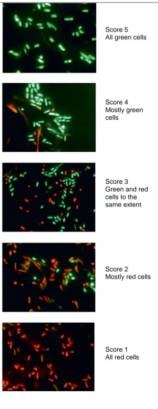
Score 5 All green cells
Score 4 Mostly green cells
Score 3 Green and red cells to the same extent
Score 2 Mostly red cells
Score 1 All red cells
Fig. 1. LIVE/DEAD _ BacLight TM stained
plaque bacteria. Green cells are acid-tolerant while red cells are non-acid-tolerant. Pictures show representative plaque samples from subjects in the study representing different scores.
Estimation of Plaque Acid Tolerance
Two plaque samples from each individual were suspended in 100 µl MM4 pH 7.5 and 3.5, respectively and incubated in a CO2
incubator (5% CO2) at 37 ° C for 2 h. The
cells kept at pH 7.5 were used as a control to ensure that the cells were viable after transport to the laboratory. Cells were stained using LIVE/DEAD®BacLight™
Fluorescent Stain (Molecular Probes, Eugene, Oreg., USA) [Welin-Neilands and Svensater, 2007] and each plaque sample was examined using a fluorescent microscope (Aristoplan, Leitz). Seven random images (×1,000) taken with a digital camera were recorded. The images were analyzed using a modification of the method described previously [Rundegren et al., 1992]. Briefly, each image was given a score from 1 to 5 (fig. 1), depending on the degree and type of stain and the median value for each sample, and the median values for each study group were calculated at baseline and after 15 months.
Composition of the Cultivable Acid-Tolerant Microflora
One plaque sample was resuspended in 100 µl prereduced dilution medium [Holdeman et al., 1977] and vortex-mixed with glass beads. Another 200 µl of prereduced dilution medium was added and the sample mixed again. Samples were plated on pH 5.2 agar and on SL agar using the pour plate technique. The pH 5.2 plates were incubated anaerobically for 5 days. The SL agar plates were incubated in a CO2 incubator (5% CO2)
for 4 days. All
procedures were carried out as previously described [Svensater et al., 2003].
Identification of Microbial Groups
Representatives of all microbial colonies with different morphologies were isolated and Gram stained. Depending on the results of Gram staining, different procedures were carried out for identification. Gram-positive catalase-negative cocci in chains were considered streptococci and identified at species level [Beighton et al., 1991; Whiley and Beighton, 1998]. Isolates of Gramnegative diplococci were identified as Veillonella based on the following characteristics: size of the cells in the Gram stain, obligatory anaerobic growth
and growth stimulated by lactate but not by glucose. Gram-positive pleomorphic rods were identified to species level with 16S rRNA sequencing using universal primers as previously described [Mantzourani et al., 2009]. Gram-positive catalase-negative rods on SL agar were allocated to the genus Lactobacillus. Yeasts were identified according to morphology on pH 5.2 agar and Gram staining.
Statistical Analyses
Data were analyzed using InStat (version 3.0, Graph Pad, San Diego, Calif., USA). Image analysis data were analyzed using Wilcoxon matched-pairs signed-rank test.
Results
Plaque Acid Tolerance
The median score value for all the samples kept at pH 7.5, both at baseline and 15 months, was 5, indicating good viability of the cells after transport to the laboratory. At baseline, the median score for acid tolerance in all three groups was 4 (p = 0.5), indicating mostly green, acid-tolerant cells in the samples. In the F group all subjects had a less acid-tolerant microflora after 15 months of intervention compared to baseline, with a median score value of 2 (p<0.01). For the M and C groups there was no statistical difference in acid tolerance at the end of the study compared to baseline (p = 0.2 and p = 0.4, respectively). The F group differed significantly (p<0.01) from both the M and C group by having a lower proportion of acid-tolerant bacteria at the end of the study.
Composition of the Acid-Tolerant Microflora at Baseline
Streptococcal species were present in all 27 subjects. A wide range of other genera – Bifidobacterium dentium, Veillonella, Lactobacillus, Propionibacterium acnes, Parascardovia, Scardovia, Actinomyces and yeasts – were isolated from different subjects. However, the dominant genus was Streptococcus which constituted more than 50% of the acid-tolerant microflora in 20 of the 27 subjects and more than 25% in 24 of the subjects. Fourteen different species of streptococci were isolated, with Streptococcus gordonii, Streptococcus oralis and Streptococcus anginosus being the most frequent.
Effect of Fluoride on the Composition of the Acid-Tolerant Microflora
In the F group, streptococci were still the dominating genus after 15 months. As at baseline, there was a variety in the streptococcal composition among the different subjects. In one case the dominating streptococcal species was the same after fluoride treatment as at baseline. In all other cases there was a change in the dominating streptococcal species. The observations seen in the F group were the same for the M and C group. There was a slight decrease in isolation frequency of Lactobacillus in the F group, while in the C and M group there was an increase. The lactobacilli however still only represented, in all cases but one, less than 1% of the acid-tolerant microflora. After 15 months there was still great species diversity among the different subjects in all three groups.
Discussion
While some organisms are constitutionally acid-resistant, others are able to mount an ATR when exposed to an acidic environment [Hamilton and Buckley, 1991; Svensäter et al., 1997; Lemos et al., 2005]. Microbial acid adaptation and subsequent acid selection of low-pH bacteria play a critical role in destabilizing the homeostasis of plaque and lead to a shift in the demineralization/remineralization balance and dental caries [Takahashi and Nyvad,
2008]. By inhibiting the ATR, caries development may be prevented and carious lesion progression inhibited. In this study, we were interested in the effect of fluoride on the development of acid tolerance in vivo. Fluoride, provided to the biofilm using milk as a vehicle, significantly reduced the acid tolerance of the plaque biofilm. Acid tolerance was tested in vitro after dispersal of the plaque biofilm in MM4 medium. This environmental change might have affected the physiology of the bacteria, since it is known that planktonic cells are less acid-tolerant than biofilm cells [Welin et al., 2003]. Therefore the differences between the different groups might be even more profound in vivo.
In S. mutans, acid adaptation involves several different mechanisms, including the synthesis of stress response proteins and increased ATPase activity and proton impermeability which in turn makes the bacteria more acidtolerant [Hamilton and Buckley, 1991; Svensäter et al., 2000; McNeill and Hamilton, 2004]. Fluoride inhibits membrane-bound ATPases which pump protons out of the cell [Marquis, 1977; Sutton et al., 1987]. Protons will thereby accumulate and acidify the cytoplasm, which affects acid-sensitive enzymes, e.g. the glycolytic enzymes. Fluoride also acts by inhibiting enolase in the glycolytic pathway, which leads to decreased acid production [Kanapka and Hamilton, 1971; Hamilton, 1977]. With a lower acid production among the bacteria in plaque, the plaque pH would be raised and ATR would not be induced. This would provide an explanation for the decreased acid tolerance observed in this study. Alternatively, fluoride may affect ATR induction directly [Welin-Neilands and Svensäter, 2007]. The relatively low test pH (3.5) in this study was used to distinguish between acidtolerant and non-acid-tolerant organisms and is not proposed to reflect in vivo conditions. Due to the higher influence of fluoride at lower pH, the patients did not consume any fluoridated milk at the day of plaque sampling to avoid high fluoride concentrations in the plaque during the acid tolerance testing. The effect of fluoride was also investigated after 1 month (data not shown). No difference in plaque acid tolerance compared to baseline was seen in the fluoride group. Had the effect of fluoride in this study been a result of the toxic effects of fluoride on bacterial viability at low pH, a decrease in acid tolerance most probably would have been seen after 1 month of exposure as well.
The composition of the culturable acid-tolerant microflora at baseline varied greatly between individuals. Although S. gordonii was the most frequently isolated species both at baseline and after 15 months, the dominance/frequency ratio was quite evenly spread among the 14 streptococcal species. In samples of infected dentin from soft active root caries lesions, the aciduric microflora has been reported to be dominated by lactobacilli and Actinomyces israelii , with few species of streptococci [Brailsford et al., 2001]. In our study lactobacilli were present in 46% of the samples but did not dominate, while Actinomyces were only present in samples from a few subjects. The differences may be a reflection of the sampling procedure or the methods used to isolate the aciduric microbiota. While Brailsford et al. [2001] investigated the aciduric flora in infected dentin, the plaque in this study was sampled with a Teflon loop and represented the biofilm on the surface of the caries lesion sampled. Van Houte et al. [1991, 1994, 1996] reported that non-mutans streptococci from sound sites showed high aciduricity and were better able to generate pH values below 4.2 after growth in glucose broth, supporting the concept that the phenotype of the isolate is more important than its identity. In microcosmic dental plaque grown in artificial saliva supplemented with milk or fluoridated milk (5 ppm), the presence of fluoride reduced the proportion of streptococci [Pratten et al., 2000]. However no such trend was seen in this study, at least for acid-tolerant streptococci; neither did 5 mg/l fluoride seem to influence the composition of the acid-tolerant microflora per se. In this study we have shown that a low concentration of fluoride in milk has the ability to affect bacterial physiology in vivo.
Future studies will be directed towards larger clinical studies to provide more evidence for the effect seen here as well as to investigate how this affects the caries process.
References
Beighton D, Lynch E, Heath MR: A microbiological study of primary root-caries lesions with different treatment needs. J Dent Res 1993; 72: 623–629.
Beighton D, Russell RR, Whiley RA: A simple biochemical scheme for the differentiation of Streptococcus mutans and Streptococcus sobrinus. Caries Res 1991; 25: 174–178.
Brailsford SR, Shah B, Simons D, Gilbert S, Clark D, Ines I, Adams SE, Allison C, Beighton D: The predominant aciduric microflora of root-caries lesions. J Dent Res 2001; 80: 1828– 1833.
Hamilton IR: Effects of fluoride on enzymatic regulation of bacterial carbohydrate metabolism. Caries Res 1977; 11(suppl 1):262–291.
Hamilton IR, Buckley ND: Adaptation by Streptococcus mutans to acid tolerance. Oral Microbiol Immunol 1991; 6: 65–71.
Hamilton IR, Svensater G: Acid-regulated proteins induced by Streptococcus mutans and other oral bacteria during acid shock. Oral Microbiol Immunol 1998; 13: 292–300.
Holdeman LV, Cato EP, Moore WEC (eds): Anaerobe Laboratory Manual, ed 4. Blacksburg, Virginia Polytechnic Institute and State University, 1977.
Kanapka JA, Hamilton IR: Fluoride inhibition of enolase activity in vivo and its relationship to the inhibition of glucose-6-P formation in Streptococcus salivarius . Arch Biochem
Biophys 1971; 146: 167–174.
Lemos JA, Abranches J, Burne RA: Responses of cariogenic streptococci to environmental stresses. Curr Issues Mol Biol 2005; 7: 95–107.
Mantzourani M, Fenlon M, Beighton D: Association between Bifidobacteriaceae and the clinical severity of root caries lesions. Oral Microbiol Immunol 2009; 24: 32–37.
Marquis RE: Inhibition of streptococcal adenosine triphosphatase by fluoride. J Dent Res 1977; 56: 704.
Marsh PD: Microbial ecology of dental plaque and its significance in health and disease. Adv Dent Res 1994; 8: 263–271.
Marsh PD: Are dental diseases examples of ecological catastrophes? Microbiology 2003; 149: 279–294.
McNeill K, Hamilton IR: Effect of acid stress on the physiology of biofilm cells of Streptococcus mutans. Microbiology 2004; 150: 735–742.
Pratten J, Bedi R, Wilson M: An in vitro study of the effect of fluoridated milk on oral bacterial biofilms. Appl Environ Microbiol 2000; 66: 1720–1723.
Rundegren J, Hvid EB, Johansson M, Astrom M: Effect of 4 days of mouth rinsing with delmopinol or chlorhexidine on the vitality of plaque bacteria. J Clin Periodontol 1992; 19: 322–325.
Sutton SV, Bender GR, Marquis RE: Fluoride inhibition of proton-translocating ATPases of oral bacteria. Infect Immun 1987; 55: 2597–2603.
Svensäter G, Borgstrom M, Bowden GH, Edwardsson, S: The acid-tolerant microbiota associated with plaque from initial caries and healthy tooth surfaces. Caries Res 2003; 37: 395–403.
Svensäter G, Larsson UB, Greif EC, Cvitkovitch, DG, Hamilton IR: Acid tolerance response and survival by oral bacteria. Oral Microbiol Immunol 1997; 12: 266–273.
Svensäter G, Sjogreen B, Hamilton IR: Multiple stress responses in Streptococcus mutans and the induction of general and stress-specific proteins. Microbiology 2000; 146: 107–117. Takahashi N, Nyvad B: Caries ecology revisited: Microbial dynamics and the caries process. Caries Res 2008; 42: 409–418.
van Houte J, Lopman J, Kent R: The predominant cultivable flora of sound and carious human root surfaces. J Dent Res 1994; 73: 1727–1734.
van Houte J, Lopman J, Kent R: The final pH of bacteria comprising the predominant flora on sound and carious human root and enamel surfaces. J Dent Res 1996; 75: 1008–1014. van Houte J, Sansone C, Joshipura K, Kent R: Mutans streptococci and non-mutans
streptococci acidogenic at low pH, and in vitro acidogenic potential of dental plaque in two different areas of the human dentition. J Dent Res 1991; 70: 1503–1507.
Welin-Neilands J, Svensater G: Acid tolerance of biofilm cells of Streptococcus mutans . Appl Environ Microbiol 2007; 73: 5633–5638.
Welin J, Wilkins JC, Beighton D, Wrzesinski K, Fey SJ, Mose-Larsen P, Hamilton IR, Svensater G: Effect of acid shock on protein expression by biofilm cells of Streptococcus mutans. FEMS Microbiol Lett 2003; 227: 287–293.
Whiley RA, Beighton D: Current classification of the oral streptococci. Oral Microbiol Immunol 1998; 13: 195–216.