Involvement of mitogen-activated protein
kinases in mast cell mediator induced
airway hyper-responsiveness in guinea
pig trachea
Author: Maria Belikova Institute of Environmental Medicine
Maria Belikova
Study Program in Medicine KI Degree project 30 credits Spring 2016
Supervisor: Mikael Adner Co-supervisor: Jesper Säfholm
Betydelse av mitogen-aktiverade proteinkinaser i mastcell-mediator inducerad luftvägshyperresponsivitet i marsvinstrakea
Bakgrund: Allergenexponering orsakar aktivering av mastceller och frisättning av deras mediatorer,
vilket leder till sammandragning av luftvägar. Luftvägshyperresponsivitet (LHR) är en av grundpelarna i astma och idag finns ingen fullständig förståelse för orsakerna bakom detta fenomen. Det finns teorier som förklarar LHRs uppkomst med upprepade sammandragningar, inflammation, och förändringar i osmolalitet. En annan teori är att LHR beror på förändringar i signalering via mitogen-aktiverade proteinkinaser (MAPK). Syfte: Syftet med detta projekt var att undersöka om ovalbumin (OVA) kan orsaka LHR mot mastcellsmediatorer och klarlägga om MAPK bidrar till denna process.
Material och Metoder: Marsvin sensitiserades mot OVA genom en intraperitoneal injektion varpå
cirkulära segment av marsvinstrakea dissekerades ut och inkuberades med eller utan OVA och hämmare av c-Jun N-terminal kinase (JNK), extracellular-signal-regulated kinase/MAPK (MEK) och p38. Trakeasegmenten monterades i ett organbad i en myograf för att mäta tensionen i dessa efter tillsättning av mastcellsmediatorer och karbakol. Data från varje segment och agonist korrelerades till den maximala kraftutvecklingen detta segment åstadkommit och presenterades som medelvärde±medelfel. Resultat: exponering för OVA orsakade LHR mot histamin i trakeasegmenten (p<0.05; pEC50 6.5±0.1) jämfört med ingen exponering för OVA (pEC50 6.2±0.1). Inkubation med
JNK MAPK hämmare, men inte med p38 och MEK hämmare motverkade LHR mot histamin.
Slutsats: LHR mot histamin kan induceras i en ex vivo marsvinsmodell och kan motverkas med
hämning av signalvägen JNK.
Involvement of mitogen-activated protein kinases in mast cell mediator induced airway hyper-responsiveness in guinea pig trachea
Introduction: Exposure to allergens causes activation of mast cells and release of its mediators leading
to airway constriction. Airway hyper-responsiveness (AHR) is a major characteristic of asthma but understanding of its causes is incomplete. Theories include repetitive airway constriction, inflammation or changes in osmolality. Another theory is that AHR is mediated through changes in mitogen-activated protein kinases (MAPK) signalling pathways. Aims: To determine the ability of ovalbumin (OVA) to induce AHR to mast cell mediators and to determine the contribution of MAPK to AHR. Material and Methods: Guinea pigs (GP) were sensitized to OVA by intra-peritoneal injection, thereafter circular segments of GP trachea were dissected and incubated in presence or absence of OVA and inhibitors of c-Jun N-terminal kinase (JNK), extracellular-signal-regulated kinase/MAPK (MEK) and p38. Thereafter, segments were mounted on a myograph in organ baths in order to measure the tension in trachea after addition of mast cell mediators and carbachol. The data from each segment and agonist was related to maximal contraction of that segment and presented as mean±SEM. Results: Exposure to OVA induced AHR to histamine in tracheal segments (p<0.05; pEC50 6.5±0.1) as compared to no OVA exposure (pEC50 6.2±0.1). Incubation with MAPK JNK
inhibitor, but not with MAPK p38 and MEK inhibitors reduced OVA-induced AHR to histamine.
Conclusions: AHR to histamine can be induced in a GP trachea ex vivo model and it can be reduced
by inhibition of MAPK JNK.
Keywords: asthma, mitogen-activated protein kinases, p38 mitogen-activated protein kinases, JNK
mitogen-activated protein kinases, extracellular signal-regulated MAP kinases, respiratory hypersensitivity.
Abbreviations
5-LOX – 5-lipoxygenaseAHR – airway hyper-responsiveness ASM – airway smooth muscle DMSO – dimethyl sulfoxide
ERK - extracellular-signal-regulated kinase GP – guinea pig
GPCR – G-protein coupled receptor GPT – guinea pig trachea
IgE – immunoglobulin E JNK – c-Jun N-terminal kinase LTE4 – leukotriene E4
MAPK – mitogen-activated protein kinases MEK – ERK/MAPK
1
Introduction
Asthma is a globally prevalent heterogeneous conductive airway disease characterized by variable and recurring symptoms, reversible airflow obstruction, chronic airway inflammation and airway hyper-responsiveness (AHR) (1).
Epidemiology and global impact
The asthma diagnosis is based on the clinical characteristics of wheezing, shortness of breath, chest tightness and cough that vary over time and in intensity. The prevalence of doctor diagnosed disease is today around 300 million people worldwide (1). Since the middle of the 20th century the prevalence has been reported to be increasing, though most recent studies indicate that the increase in prevalence among adolescents is in fact abating. In contrast, in countries with a high urbanization rate or areas with low income, the prevalence is increasing (2). This indicates that while the differences in global prevalence are decreasing, the total burden of the disease worldwide is increasing. However, time trends have been difficult to evaluate due to varying inclusion criteria and questionnaires, but also due to increased awareness and improved diagnostic activities (2).
The lifetime risk of developing asthma is around 35%, comparable to the risk of developing cancer or coronary heart disease. In contrast with other chronic diseases, asthma tends to develop early and causes life-long suffering (3). Estimates show that asthma is responsible for about 1 in 250 deaths worldwide. For asthmatics the number of lost disability-adjusted life years is comparable to patients with diabetes or liver cirrhosis (4). In conclusion, asthma is a highly prevalent disease that burdens both patients and global health.
Pathogenesis of asthma
Although mechanisms of the disease are not fully understood, a complex interaction between environmental and genetic factors is considered to contribute to the development of asthma. Airway inflammation
The stimulus that activates a symptomatic asthmatic attack can be inhalation of allergens, pollutants or viral infections (5). Acute inflammation starts when the allergen activates mast cells via crosslink of the cell-bound allergen-specific Immunoglobulin E (IgE) molecule with a receptor on the mast cell (6). This activates several intracellular cascades inside the mast cell, causing it to de-granulate. The granula contain several preformed biologically active
2 mediators, among them histamine and proteases (6). Histamine is known to act on four different receptors, activation of which causes bronchoconstriction and increase in mucus secretion (7). Another process that is induced in the mast cell is the activation of the enzymes cyclooxygenase and and 5-lipoxygenase (5-LOX) yielding increased production and release of prostaglandins and leukotrienes (6). This part of the inflammation process is called the early phase and results in acute airway smooth muscle (ASM) contraction and mucus production within minutes, leading to limitation of airflow (8).
In some patients the early phase resolves within hours, but around half of asthmatics experience a second wave of obstruction. Referred to as the late phase, it develops within 2-6 hours and peaks at 12 hours after the early phase (9). This phase is characterized by recruitment of other inflammatory cells (eosinophil, neutrophil and T-helper type 2 cells) (10) through chemokines and cytokines, IgE production leading to a slowly developing ASM contraction (6). This response can be prevented by prophylactically blocking IgE (11) and leukotrienes (12), which suggest a contribution of mast cells.
Repetitive or continuous exposure to allergens can lead to chronic inflammation (6). Typical changes in the airways are referred to as airway remodelling and include an up-regulated inflammatory environment (6), loss of epithelial integrity, mucus cell metaplasia, fibrosis and increased ASM mass. The result is constant airway narrowing due to increased ASM size, mucus plugging and airway hyper-responsiveness (13). The role for the mast cell in these processes is not yet defined.
Airway hyper-responsiveness
AHR is, together with inflammation, a central attribute of asthma, defined as an excessive airway narrowing due to stimuli that normally do not affect healthy individuals. The stimuli may be physical (cold air or exercise), pharmacological substances or pollutants (14). Clinical assessment of AHR is done by provocation with histamine or methacholine, which cause an acute bronchoconstriction in affected individuals (15). Carbachol is, like methacholine, a nonselective muscarinic receptor agonist, which is in contrast to acetylcholine is not degraded by acetylcholinesterase, which results in a long-lasting and sustainable contraction (16). AHR is considered to have a persistent component, described as a baseline AHR that most chronic asthma patients suffer from, which is connected to the remodelling phenomenon. Superimposed on that, there is a variable component that can be induced by the acute airway
3 inflammation. Although both components have been defined, the driving mechanisms behind AHR remain unclear (14).
As described above, allergens and infections can induce airway inflammation in asthmatic patients. However, there is evidence that a bronchospasm can occur on its own without inflammation (17) and mast cells can be activated through osmotic stimuli (18), adenosine or cytokines, independent of IgE activation (19). Moreover, isolated bronchoconstriction alone, without inflammation, can induce airway remodelling (20), which shows, that though AHR and airway inflammation are characteristic for asthma, both processes can occur independently.
Airway smooth muscle contraction
ASM cells, which encircle the airways throughout the entire bronchial tree, are considered to be deeply involved in AHR because of the increased sensitivity of the airways to constricting agents. For a long time ASM cells were considered as simply the effector cells in asthma. However, recent studies suggest that the ASM itself contributes to the development and sustainment of asthma and presumably AHR by releasing cytokines and growth factors that maintain airway inflammation (21).
Methods of in vitro quantification of the smooth muscle contraction and characterization of different contractile and relaxant agents have been developed over many years. Tension measurements in ex vivo tissue containing smooth muscle using a myograph were first done on vessel rings (22, 23). The method was then adapted for use on bronchiole rings (24) and trachea rings (25). Adner et al. has shown that tracheal rings from mice can survive in culture for as long as 32 days without changing their physiopharmacological properties, making them a useful model for asthma research (25).
Animal model for studying AHR
Guinea pig (GP) is the most correct animal model for studying airway pharmacology. Many studies have shown that anatomy, physiology and pharmacology of the GP lung closely resembles that of human (26, 27). As an illustration, during mast cell degranulation, mice and rats release serotonin while both humans and GP release histamine (8). The ovalbumin GP sensitization model used in this project has been developed during the past 30 years and has been shown to induce acute airway inflammation, late phase inflammation and AHR in the animals, modelling an asthmatic airway (26). The first encounter with the allergen does not
4 produce any symptoms. However, it induces the process of IgE-production to the specific allergen. When the animal encounters the same allergen again, the produced IgE-molecules activate the early phase of inflammation (6).
Mitogen-activated protein kinases
Mitogen-activated protein kinases (MAPK) are ubiquitous and well conserved cellular proteins, whose function is to transduce signals from outside of a cell, like inflammation or stress, into the cell by protein phosphorylation (28). MAPK are phosphorylated in the cytosol and activated by MAPK kinases, which are activated by MAPK kinase kinases, which in turn are activated at the level of G-proteins at the cell membrane (29). The existence of several intermediates allows amplification of the signal as the amount of substrates increases for each level. Moreover, it allows for interplay between the different signalling pathways. The most well-studied signal pathways are ERK, JNK and p38 (28).
Extracellular-signal-regulated kinase (ERK) signalling pathway
ERK1 and ERK2 are two highly similar ubiquitous ERK isoforms at the MAPK level of the ERK signalling pathway, activated by different types of stimuli like growth factors, ligands for G-protein coupled receptors (GPCR), cytokines and osmotic stress (30). MAPK/ERK, shortly named MEK1 and MEK2, are two similarly efficient isoforms at the MAPK kinase level. MEK1 and MEK2 both phosphorylate and fully activate ERK1/2. ERK pathway is involved in regulating cell functions, like motility, proliferation and differentiation and are highly expressed in well differentiated cells, where they have been found to contribute to long-term potentiation of cell signalling in neurons and other highly differentiated cells(28). c-Jun N-terminal kinase (JNK)signalling pathway
JNK1, JNK2 and JNK3 are structurally highly similar isoforms of JNK at MAPK level of JNK pathway, but have functional differences. JNK1 and JNK2 are ubiquitous, while JNK3 is restricted to the central nervous system. They are activated by cytokines, growth factors, DNA damage and environmental stressors. There are two MAPK kinases that activate JNKs, MEK4 and MEK7, which also have the ability to activate the p38 signalling pathway at higher concentrations. Activated JNK pathway contributes to the production of cytokines, inflammatory response, cell metabolism and apoptosis (30).
5 p38 signalling pathway
p38 is a family of four kinases α, β, γ and δ, at MAPK level of p38 pathway, which have been shown to have different, sometimes opposite, actions (28). p38α and p38β are ubiquitously expressed, while γ and δ seem to be restricted to specific tissues outside of the airways (30). p38 pathway can be activated by signals such as cytokines, hormones, ligands of GPCR and environmental stressors. MAPK kinases of p38 pathway have the ability to activate the JNK pathway at high concentrations (28). Due to structural and functional differences of p38 family members, activation of p38 pathway leads to a variety of responses, such as induction of cell cycle checkpoints and regulation of proliferation and differentiation (30).
Inhibitors of MAPK
In order to block specific MAPK signalling pathways, more or less specific inhibitors have been developed. PD98,059 is a selective inhibitor in the ERK pathway, acting on MEK1 and MEK2, preventing MEK1/2 from being phosphorylated (31). SB203580 is a selective inhibitor of p38 pathway, acting on p38α and p38β, but not on the δ and γ isoforms (32). 1 µM of SB203580 lowers p38α and p38β activity down to 9-13% of the original, however, it has also been shown to inhibit JNK2 down to 60 % of its original activity at the same concentration (32). SP600125 is an inhibitor of the JNK pathway, acting on JNK1 and JNK2 isoforms. 1 µM of SP600125 lowers JNK1 and JNK2activity down to 23-24% of the original, however, it has also been shown to inhibit p38α and p38βactivity down to 86-89% of its original activity at the same concentration (32). Unfortunately, both SB202580 and SP600125 inhibit several other, less studied, protein kinases (32, 33).
MAPK and AHR
Recent research has shown that MAPK JNK and ERK are involved in modulating the calcium response to contractile mediators in human ASM cells (34). JNK has been shown to mediate the elevation of extracellular calcium influx in rat bronchial segments after stimulation with contractile agents (35). In addition, JNK has been shown to be involved in ozone-induced AHR in GP (36, 37), as has p38 (36). A JNK inhibitor, SP600125, has been shown to inhibit AHR to OVA in sensitized mice (38). Similarly, inhibition of ERK prevented AHR to methacholine in mice sensitized and challenged with OVA (39). These findings point towards the involvement of MAPK in the formation of AHR. Yet, it is still unknown whether MAPK
6 are involved in AHR that is mediated by the most common mast cells mediators, which are clinically relevant in asthma.
Aim
The aim of the project was to study AHR in an ex vivo GPT model by determining the ability of OVA to induce AHR to mast cell mediators and to determine the contribution of MAPK to that process.
Material and Methods
Chemicals
Albumin from chicken egg white (OVA) grade V, aluminium hydroxide, Krebs-Henseleit buffer modified (#K3753), sodium bicarbonate, dimethyl sulfoxide (DMSO) (#D5879), PD98,059, SB203580, SP600125, indomethacin, histamine, carbachol, leukotriene E4 (LTE4),
potassium chloride, papaverine were purchased from Sigma-Aldrich. Sterile phosphate buffer solution (PBS) (#10010015), Dulbecco's Modified Eagle Medium: Nutrient Mixture F-12 (DMEM-F12) (#11320074), Penicillin-Streptomycin (#15140122) were purchased from Thermo Fischer Scientific. Calcium chloride dihydrate was purchased from Merck Millipore. All stock solutions were made according to manufacturer’s instructions.
Organ bath
Tissue extraction
Male albino GP (Dunkin-Hartley) (445-676 gram) were sensitized by a single intra-peritoneal injection containing 100 µg OVA and 0.1 g aluminium hydroxide dissolved in 0.8 ml sterile PBS. Animals were sacrificed not earlier than 10 days later by inhalation of CO2 followed by
removal of the trachea. Each trachea was transferred to cold Krebs-Henseleit buffer modified by addition of 25 mM sodium bicarbonate and 2.5 mM calcium chloride dehydrate. The trachea was dissected from the surrounding connective tissue and cut into eight rings of equal length containing between two to three cartilage rings.
Tissue culture
The rings were placed in a 48-well culture plate containing DMEM-F12 and 1% penicillin-streptomycin and treated with one of the inhibitors: PD98,059 (50 µM), SB203580 (50 µM), SP600125 (5 µM), all dissolved in DMSO, or the vehicle, DMSO. After 40 minutes the rings were challenged with 0.1 mg/ml OVA and kept at 37°C for 18-20 hours.
7 Myograph measurements
Tracheal rings were mounted and lowered into a 5 ml tissue organ bath containing Krebs-Henseleit buffer modified with addition of sodium bicarbonate to the concentration of 25 mM and calcium chloride dihydrate to the concentration 2.5 mM. The buffer was kept at 37°C and bubbled with carbogen (5% CO2 in O2). Changes in smooth muscle force were detected using
an isometric force-displacement transducer linked to a grass polygraph. The response was displayed on a monitor using the IOX data acquisition system (EMKA, Paris, France).
First, the rings were allowed to equilibrate in the new milieu with washes every 15 minutes and the resting force was gradually adjusted to 30 mN in order to create a baseline mechanical tension (Fig. 1). Second, 3µM of a non-selective COX-inhibitor indomethacin (dissolved in ethanol) was added to inhibit synthesis of prostaglandins in the tissue, since it has been established that they are responsible for upholding the basal tone in GPT (40). Third, histamine (dissolved in distilled water), a muscarinic receptor antagonist carbachol (dissolved in distilled water) and LTE4 (dissolved in ethanol) were added in cumulative concentrations (1
nM to 0.1 mM) with washing and addition of indomethacin between the three substances in order to elicit contractions. Last, viability was assessed by contraction with 1mM histamine and 60 mM potassium chloride (dissolved in distilled water) followed by relaxation induced by 1 mM papaverine (dissolved in distilled water).
Figure 1. A typical force-time trace of an experimental protocol from one tracheal segment.
LTE4 – leukotriene E4, OVA – ovalbumin
8 A successful sensitization was evaluated by a combination of two methods. One was the presence of white intra-peritoneal clots composed of aluminium hydroxide which indicated successful injections. The second was addition of OVA to the baths at the end of myograph measurements – if a contraction was evoked in the controls, but not in the segments incubated with OVA, the sensitization was considered successful (Fig. 1).
Statistics
The data measured in the organ baths was the force (in Newton) of the tracheal smooth muscle contraction and relaxation. Data collected for each segment was normalized in relation to the value of its strongest contraction and presented as mean ± standard error of mean. Emax
shows the maximal response an agonist can elicit and is a measure of an agonist’s efficacy. pEC50 is the –log10 agonist concentration that produces 50% of the agonists maximal effect
and is a measure of agonist’s potency. For all agonists, a non-linear regression with a variable slope was used to calculate Emax and pEC50. Data comparison between two groups was
assessed by a parametric unpaired t-test. All statistical analysis was done using GraphPad Prism® (GraphPad Software Inc., San Diego, CA).
Ethical considerations
More research is needed to understand the physio-pharmacological properties of AHR in asthmatic individuals in order to improve the treatment options. A potential ethical problem with this model is that it requires whole tissue samples, implying the need to sacrifice the GP to be able to obtain the trachea. In order to reduce the suffering, pure CO2 was used for
euthanasia and analgesia since it leads to very fast progressing unconsciousness and coma. Unconsciousness was evaluated using the pain reflexes, eyelid shutter reflex and pupil size. Each acquired trachea was divided to 8 pieces and several agonists were tested on the same tissue sample to minimize the use of animals.
The advantage of this method is the possibility to examine the contractile pattern of the tracheal segments in real time using inhibitors of MAPK signalling pathways. Another advantage is that the outcome is pharmacological properties of mast cell mediators and the signalling pathways that are activated in AHR. The advantages of this study outweigh the ethical issues, since obtained values are clinically relevant in assessment of airway tone in humans and thus might lead enhanced understanding of mechanisms behind AHR. An ethical
9 permit has been acquired (N143/14) from the regional Ethical Committee on Animal Experiments, Stockholm.
Results
Tracheal ring segments were dissected from the previously sensitized GP and incubated with OVA and inhibitors of different MAPK signalling pathways or vehicle, i.e. DMSO. The segments were mounted in organ baths where changes in ASM tone was measured by a myograph.
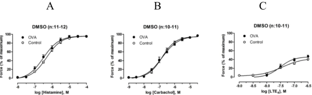
Sensitization of GP to OVA caused AHR to histamine, but not carbachol and LTE4
Segments incubated with OVA and DMSO were compared to those incubated without OVA and with DMSO. Histamine evoked a contraction in all segments (not shown), but segments incubated with OVA and DMSO had a significantly higher pEC50 (p<0.05) than those
incubated without OVA (Fig. 2:A) (Tab. 1).
A
B
C
Figure 2. Concentration-dependent constriction of tracheal segments incubated with DMSO in presence or absence of OVA. The panels show dose-response curves for histamine, carbachol and
LTE4. (A) Histamine was more potent in segments incubated with OVA than those incubated without
OVA, n:11-12. There was no change in potency or efficacy for carbachol (B) or LTE4 (C) in
segments treated with or without OVA, n:10-11. DMSO – dimethyl sulfoxide, OVA – ovalbumin, LTE4 – leukotriene E4. Data plotted from Table 1.
There was no significant difference in Emax evoked by histamine between control segments
and segments incubated with OVA (Fig. 2:A) (Tab. 1). Carbachol and LTE4 similarly evoked
contractions in controls incubated with DMSO, though there was no significant difference in Emax or pEC50 between the controls and segments incubated with OVA (Fig. 2:B,C) (Tab. 1).
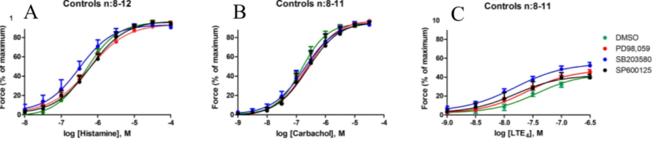
10 pEC50 and Emax values for histamine, carbachol and LTE4 in controls incubated with different
inhibitors were compared to controls incubated with DMSO alone. There was no significant difference for these values for any of the inhibitors compared to DMSO (Fig. 3) (Tab. 1).
Figure 3. Contraction induced by histamine, carbachol and LTE4. The panels show dose-response
curves for controls incubated with inhibitors or DMSO. No significant difference in either potency or efficacy was detected between the control segments treated with either MAPK inhibitors or DMSO, induced by (A) histamine, (B) carbachol or (C) LTE4. DMSO – dimethyl sulfoxide, OVA –
ovalbumin, LTE4 – leukotriene E4. PD98,059 – MEK1/2 inhibitor, SB203580 – p38 inhibitor,
SP600125 – JNK inhibitor. Data plotted from Table 1.
Use of SP600125(JNK) but not PD98,059 (MEK1/2) and SD203580 (p38) affected the ASM contraction
pEC50 and Emax values for histamine, carbachol and LTE4 in segments incubated with OVA
and DMSO were compared to segments incubated with OVA and inhibitors. There was a significant difference (p<0.05) in pEC50 values induced by histamine between segments
Table 1. pEC50 and Emax values for segments incubated with DMSO or inhibitors in presence or absence of OVA.
Treatment Histamine Carbachol LTE4 pEC50 Emax pEC50 Emax pEC50 Emax
Control + DMSO 6.2±0.1 96.9±1.5 6.7±0.2 97.3±1.6 7.6±0.2 42.8±2.3 OVA + DMSO 6.5±0.1* 95.3±2.6 6.7±0.2 96.7±1.7 7.5±0.1 45.8±2.5 Control + SP600125 6.2±0.1 97.5±1.6 6.6±0.3 96.9±1.8 7.8±0.2 44.3±5.0 OVA + SP600125 6.2±0.1 92.5±2.4 6.6±0.2 99.5±0.8 7.7±0.2 49.6±4.8 Control + PD98,059 6.2±0.1 94.5±5.4 6.6±0.2 98.2±1.6 7.6±0.1 49.1±4.7 OVA + PD98,059 6.5±0.1* 94.0±4.0 6.6±0.2 95.8±2.4 7.8±0.1 52.6±3.7 Control + SB203580 6.3±0.1 92.0±1.8 6.7±0.2 94.8±4.4 7.8±0.1 56.2±4.4 OVA + SB203580 6.7±0.1* 96.5±1.2 6.8±0.2 96.1±1.8 8.0±0.1 56.2±5.2 Data presented as mean ±SEM. * represents p<0.05 compared to respective control without OVA. DMSO – dimethyl sulfoxide, OVA – ovalbumin, LTE4 – leukotriene E4, PD98,059 – MEK1/2
inhibitor, SB203580 – p38 inhibitor, SP600125 – JNK inhibitor. Emax - maximal response an
agonist can elicit, pEC50 is the –log10 agonist concentration that produces 50% of the agonists
maximal effect.
11 incubated with OVA and DMSO and segments incubated with OVA and SP600125 (Fig. 4) (Tab. 1), while Emax for histamine between these two groups was not affected (Fig. 4) (Tab.
1). There was no significant difference in pEC50 and Emax induced by histamine for the other
inhibitors (PD98,059 and SD203580) compared to DMSO (Tab. 1). There was no significant difference in pEC50 and Emax induced by carbachol or LTE4 for any of the inhibitors compared
to DMSO (Tab. 1).
Discussion
Airway hyper-responsiveness (AHR) is a key feature of asthma that is used to describe the increased sensitivity of airway smooth muscle (ASM). Involvement of MAPK, ubiquitous intracellular signalling proteins, in AHR has been addressed in many recent studies. In this study I have investigated AHR to three common mast cell mediators, which has not been evaluated before. This study has shown that one single exposure to OVA causes AHR to histamine in GPT segments that are previously sensitized to OVA. It also shows that inhibition of MAPK JNK abolishes AHR to histamine in GPT that are sensitized and exposed to OVA.
Previous studies
In previous unpublished studies we have applied histamine receptor and 5-LOX blockers before exposure of sensitized GPT to OVA which abolished the contractions in culture. The following day during myograph measurements the AHR response to histamine was not affected compared to segments not treated with blockers, though the effect of the blockers had vanished at time of the measurements. From this, we concluded that AHR after OVA exposure did not arise due to one previous contraction in culture. Several studies have,
Figure 4. Contraction induced by histamine presented as dose-response curve for segments incubated with OVA and DMSO or OVA and SP600125. DMSO – dimethyl sulfoxide, OVA –
12 however, reported that repeated contractions per se can cause AHR, for example Grainge et al. (20).
MAPKs are known to exert their effects through modulating transcription and translation (28). Since we were interested in MAPKs involvement in AHR, we inhibited transcription and translation using actinomycin D in GPT sensitized and exposed to OVA, which did not affect the AHR response (unpublished data). Therefore, we concluded that AHR after OVA exposure did not arise due to a change in proteins synthesis.
JNK, p38 and ERK have in some studies been shown to be involved in inducing AHR to acetylcholine-like substances or histamine, however, some studies have found no such involvement. Lei et al. have shown that inhibition of JNK using SP600125, but not MEK1/2 using PD98,059 or p38 using SB203580 attenuates the bronchial constriction in rats to thromboxane A, which is a potent airway constrictor (35). Other studies have shown that inhibition of p38 using SB239063 (41) or M39 (42) prevented or significantly reduced AHR to methacholine in mice sensitized with OVA. Williams et al. have shown that inhibition of p38 using SP600125 significantly inhibited ozone-induced AHR to acetylcholine in mice (37). Similarly, Duan et al. showed that inhalation of p38α antisense oligonucleotide, which binds to and blocks the translation of p38α mRNA, significantly reduced OVA-induced AHR to methacholine in challenged mice (43). Sakai et al., on the other hand, have shown that another cytokine, tumour necrosis factor alpha, induced AHR to acetylcholine which can be abolished by blocking ERK, but not p38 in rat bronchi (44). The role of ERK in AHR is also supported by data from Ohnishi et al. (45).
These conflicting results may partly be explained by the fact that the studies have used different animal models. Also, the use of airway tissue from mice and rat makes the results of these studies not directly comparable to our study, since the physiopharmacological airway properties of these animals differ in many ways (27). Another explanation is that the way of measuring AHR varies between different studies discussed above: some groups have used in vivo measurements, like airway resistance in living animals, while others have used ex vivo tissues. Lastly, different inhibitors of MAPK have been used in these studies to demonstrate the involvement of JNK, p38 and ERK in AHR. The fact that not all studies have used the same inhibitors and that the inhibitors have a varying specificity affects the possibility to compare the results of these studies correctly. The inhibitors SP600125 and PD98,059 have been shown to inhibit each other’s primary substrates and several other protein kinases not
13 studied here (32). This implies that the inhibitory effects of SP600125 and SB98,059 discussed in this study may reflect not only specific inhibition of JNK and p38, but also cross-inhibition and effects from cross-inhibition of other signalling pathways. However, as mentioned previously, the level of inhibition of each other’s primary substrates by SP600125 and SB98,059 can be considered low as compared to level of inhibition of the primary target. Moynihan et al. studied the Ca2+ response of primary ASM cells to cytokine IL-13, which is an important messenger in the early phase of inflammation. They found that Ca2+ signalling pattern was not altered significantly by inhibition of p38 signalling, but instead by a concurrent inhibition of JNK and ERK (34). Ca2+ signalling is important for ASM contraction as described previously and an altering of Ca2+ signalling pattern through MAPK pathways can be a possible molecular mechanism behind AHR. Data supporting the findings of Moynihan et al. have been obtained by Manson et al. (manuscript in preparation) in isolated human bronchi, where two-days incubation with IL-13 caused AHR to histamine. The study of Moynihan et al. also highlighted that inhibition of only one pathway may not be enough to cause a change in ASM contractile pattern and cause AHR.
In this study, no AHR to carbachol was detected. Carbachol is a nonselective muscarinic receptor agonist (16), much like methacholine, that is used to detect AHR (15), which makes this finding quite unexpected. Also, no AHR to LTE4 was detected. There are no studies done
on AHR to LTE4 or any other leukotriene after sensitization and exposure to allergen, making
it difficult to compare the results with previous research. However, previous studies have shown that pre-treatment of GPT with LTE4 causes AHR to histamine (46, 47). Nevertheless,
the results from these studies have not been repeatable in our lab. My study also shows that the inhibitors themselves do not have any significant effect on the contractile properties of ASM of the trachea. This was important to establish to be able to isolate the effects of solely MAPK inhibitors on the airway tone.
Strengths and limitations
One strength of this study is the use of whole isolated tissue samples from the GT. This model allows studying of the isolated ASM without supporting structures and other tissue that it might communicate with. However, when the outcome is measured in tissue of a living animal, it can vary due to a variety of factors that cannot fully be controlled. In the airway tissue, this includes the effects from cytokines and chemokines, attraction of immune cells from the blood stream, nerve cell stimulation and effect from other blood-borne substances.
14 On the other hand, the method of measuring AHR in the living animal reflects the actual reaction better than the artificial environment in the organ bath. However, in this study I did not aim to address the mechanism behind AHR in a living animal, but specifically targeted pharmacological properties of the airway contraction in AHR.
Another strength is the method used, in which strength of the actual contraction can be measured in real-time. In these conditions the time between the interventions is based on the functional changes in the specific sample, rather than a median estimate of the tissue behaviour. In that way, a more individualized approach can be used, which should make the results more consistent.
One more strength of our approach is the animal model, which has been established and improved over the past 30 years. In this model, it has been shown that after one sensitization and one OVA exposure GP develop AHR to methacholine, late phase inflammation and airway inflammation (26). GP’s airway tissue, pharmacology and reactivity have the highest similarity to human airway tissue, compared to rats and mice (27). Of course, the optimal model for this investigation would be human airway tissue, but the supply is sparse. This tissue is usually obtained from patients that undergo lobectomies, which is why it is also important to consider other morbidities that can negatively affect the results.
However, there are several limitations in this study, one of them being the use of trachea instead of smaller airways. Most of the inflammation and airway resistance resides in the smaller airways that can shut close and cause severe breathing problems (48). Also, some pharmacological differences have been noted between different levels of the airway in GP, making it hard to extrapolate data from the trachea to all other airways (Belikova M., unpublished data). For these reasons, smaller airways can better depict the actual physiopharmacological properties of AHR.
Another limitation is use of only DMSO as control for the incubation with inhibitors. The trachea was divided into eight segments with 2-3 cartilage rings in each segment and the segments were distributed over eight organ baths. The amount of organ baths and the length of the trachea is a limitation that does not allow for more segments to be studied simultaneously. Eight segments were treated with one of three inhibitors diluted in DMSO and OVA or no OVA. DMSO was added as a vehicle and had to be considered as a separate control group, but due to the amount of organ baths only one control for the inhibitors was
15 possible, therefore DMSO was chosen as a control. However, it would have been desirable to have a control group without DMSO.
Last, another limitation is that it is not clear whether the effect on contraction is because of action of MAPK in the ASM, or other resident cells in the trachea. The more complex a studied tissue is, the less specific is the response elicited by it. So while the contractions themselves are mediated by the ASM, it is uncertain if the inhibition of MAPK exerts its effects directly in the ASM or in other cells like epithelial cells, fibroblasts or mast cells.
Significance
The possibility to induce AHR to histamine in a sensitized guinea pig ex vivo model after one exposure to OVA is in line with previous research. Due to use of logarithmic values, a 0.3 shift in the dose-response curve pEC50 value actually corresponds to 2-fold lower
concentration of histamine required to reach 50% of the maximal possible contraction. Hence segments incubated with OVA respond with an earlier contraction at lower concentrations of histamine, which in turn is the core of the AHR phenomena. Likewise, this implies that at one given concentration of histamine the trachea with induced AHR will have contracted more, which in turn decreases the radius of the airway more than in a trachea without AHR. According to Poiseulle law, 𝑅 = 8𝜂𝐿/(𝜋𝑟^4 ) , where R is resistance in a pipe, η is viscosity of the media, L is the length of the pipe and r is the radius of the pipe. Clearly, a 2-fold change in the airway radius has an r4impact on the airflow resistance in the trachea, which can
be considered great in a clinical setting. No change in Emax however, is interpreted as no
change in contraction force after sensitisation and exposure to OVA.
JNK inhibitor SP600125 inhibited the shift in the pEC50 value, ameliorating the OVA-induced
shift in the dose-response curve. The contribution of JNK, a MAPK that is involved in the inflammatory response, to supervening of AHR to histamine is an important contribution to understanding this major characteristic of asthma and opens the possible use of JNK inhibitors in treating AHR. MAPK inhibitors are already used today for cancer treatment (49), thus a vast knowledge base has already been acquired, reducing time and funds for drug development.
Future studies
This study has highlighted the involvement of JNK MAPK in AHR to histamine, however several issues still remain unclear, one of them being the use the JNK inhibitor SP600125,
16 since it has been shown to be somewhat unspecific. There are several other JNK inhibitors available that can also be tested in the same setting in order to evaluate if the effect on AHR will persist. Other inhibitors for MEK1/2 ERK and p38 may also be tested. Another possible future study is use of a combination of MAPK inhibitors on the same segment. This is particularly interesting due to the findings of Moynihan et al that only concurrent inhibition of ERK and JNK alters Ca (2+) signalling.
In order to obtain a deeper understanding of the involvement of MAPK in AHR, studies of smaller airways are needed, since it is in the smaller airways that the bronchoconstriction has most of its effects. We have established a method for using GP primary bronchi in an organ bath myograph (unpublished data). Another, more clinically relevant model is, of course, human bronchi, which we have an access to and for which we have an adapted methodology. In a such study it would be of interest to have bronchi from known asthmatics and non-asthmatics.
Lastly, a chronic model, where the GP is repetitively exposed to antigen during a period of time would allow for determining if the repetitive bronchoconstriction effects AHR to mast cell mediators. This future study would also enable investigation of MAPK involvement. A chronic model is also more clinically relevant because there is little chance to prevent the supervention of AHR in patients, since most of them already suffer from the symptoms at presentation.
Conclusions
In this project, tracheal ring segments from GP sensitized to OVA were studied in an organ bath myograph. It was found that a single intraperitoneal injection followed by a single exposure to ovalbumin caused AHR to histamine in the tracheal segments. Moreover, inhibition of JNK MAPK signalling pathway abolished the airway hyper-responsiveness in those segments. These findings are important for further understanding of the mechanisms of AHR and open up to further investigation of whether MAPK inhibitors can be used as treatment for patients suffering from this condition.
Acknowledgements
First of all, I would like to thank Jesper Säfholm and Mikael Adner, who have introduced me to this interesting field and patiently guided and supported me through my work. I also want to thank my seminar-group and particularly my opponent Sune Sun, who have contributed
17 with valuable comments and questions during the start-up of my project. Lastly, I want to thank my coordinator Anthony Wright for his valuable feedback on my work.
References
1. Asthma GIf. Global Strategy for Asthma Management and Prevention [Internet]. Bethesda: 2015.
2. Lundback B, Backman H, Lotvall J, Ronmark E. Is asthma prevalence still increasing? Expert Rev Respir Med. 2016;10(1):39-51.
3. To T, Wang C, Guan J, McLimont S, Gershon AS. What is the lifetime risk of physician-diagnosed asthma in Ontario, Canada? Am J Respir Crit Care Med. 2010;181(4):337-43.
4. Asthma GIf. Global Burden of Asthma - Summary [Internet]. Wellington, Southampton: Medical Research Institute of New Zealand,
University of Southampton, 2012.
5. Bousquet J, Jeffery PK, Busse WW, Johnson M, Vignola AM. Asthma. From bronchoconstriction to airways inflammation and remodeling. Am J Respir Crit Care Med. 2000;161(5):1720-45.
6. Galli SJ, Tsai M, Piliponsky AM. The development of allergic inflammation. Nature. 2008;454(7203):445-54.
7. Barnes PJ. Histamine receptors in the lung. Agents Actions Suppl. 1991;33:103-22. 8. Galli SJ, Tsai M. IgE and mast cells in allergic disease. Nat Med. 2012;18(5):693-704. 9. O'Byrne PM, Dolovich J, Hargreave FE. Late asthmatic responses. Am Rev Respir
Dis. 1987;136(3):740-51.
10. Smith HR, Larsen GL, Cherniack RM, Wenzel SE, Voelkel NF, Westcott JY, et al. Inflammatory cells and eicosanoid mediators in subjects with late asthmatic responses and increases in airway responsiveness. J Allergy Clin Immunol. 1992;89(6):1076-84. 11. Fahy JV, Fleming HE, Wong HH, Liu JT, Su JQ, Reimann J, et al. The effect of an
anti-IgE monoclonal antibody on the early- and late-phase responses to allergen inhalation in asthmatic subjects. Am J Respir Crit Care Med. 1997;155(6):1828-34. 12. Roquet A, Dahlen B, Kumlin M, Ihre E, Anstren G, Binks S, et al. Combined
antagonism of leukotrienes and histamine produces predominant inhibition of allergen-induced early and late phase airway obstruction in asthmatics. Am J Respir Crit Care Med. 1997;155(6):1856-63.
13. Bergeron C, Al-Ramli W, Hamid Q. Remodeling in asthma. Proc Am Thorac Soc. 2009;6(3):301-5.
14. Cockcroft DW, Davis BE. Mechanisms of airway hyperresponsiveness. J Allergy Clin Immunol. 2006;118(3):551-9; quiz 60-1.
15. O'Byrne PM, Inman MD. Airway hyperresponsiveness. Chest. 2003;123(3 Suppl):411S-6S.
16. Roberts CM, Konjovic J. Differences in the chronotropic and inotropic response of the rat atrium to choline esters, cholinesterase inhibitors and certain blocking agents. J Pharmacol Exp Ther. 1969;169(1):109-19.
17. Busse WW. Inflammation in asthma: the cornerstone of the disease and target of therapy. J Allergy Clin Immunol. 1998;102(4 Pt 2):S17-22.
18. Gulliksson M, Palmberg L, Nilsson G, Ahlstedt S, Kumlin M. Release of prostaglandin D2 and leukotriene C4 in response to hyperosmolar stimulation of mast cells. Allergy. 2006;61(12):1473-9.
18 20. Grainge CL, Lau LC, Ward JA, Dulay V, Lahiff G, Wilson S, et al. Effect of bronchoconstriction on airway remodeling in asthma. N Engl J Med. 2011;364(21):2006-15.
21. Zuyderduyn S, Sukkar MB, Fust A, Dhaliwal S, Burgess JK. Treating asthma means treating airway smooth muscle cells. Eur Respir J. 2008;32(2):265-74.
22. Leach RM, Twort CH, Cameron IR, Ward JP. A comparison of the pharmacological and mechanical properties in vitro of large and small pulmonary arteries of the rat. Clin Sci (Lond). 1992;82(1):55-62.
23. Mulvany MJ, Halpern W. Contractile properties of small arterial resistance vessels in spontaneously hypertensive and normotensive rats. Circ Res. 1977;41(1):19-26.
24. Chopra LC, Twort CH, Ward JP. Differences in sensitivity to the specific protein kinase C inhibitor Ro31-8220 between small and large bronchioles of the rat. Br J Pharmacol. 1994;113(4):1237-42.
25. Adner M, Rose AC, Zhang Y, Sward K, Benson M, Uddman R, et al. An assay to evaluate the long-term effects of inflammatory mediators on murine airway smooth muscle: evidence that TNFalpha up-regulates 5-HT(2A)-mediated contraction. Br J Pharmacol. 2002;137(7):971-82.
26. Smith N, Broadley KJ. Optimisation of the sensitisation conditions for an ovalbumin challenge model of asthma. Int Immunopharmacol. 2007;7(2):183-90.
27. Canning BJ, Chou Y. Using guinea pigs in studies relevant to asthma and COPD. Pulm Pharmacol Ther. 2008;21(5):702-20.
28. Pearson G, Robinson F, Beers Gibson T, Xu BE, Karandikar M, Berman K, et al. Mitogen-activated protein (MAP) kinase pathways: regulation and physiological functions. Endocr Rev. 2001;22(2):153-83.
29. Kolch W. Meaningful relationships: the regulation of the Ras/Raf/MEK/ERK pathway by protein interactions. Biochemical Journal. 2000;351:289-305.
30. Raman M, Chen W, Cobb MH. Differential regulation and properties of MAPKs. Oncogene. 2007;26(22):3100-12.
31. Cuenda A, Alessi DR. Use of kinase inhibitors to dissect signaling pathways. Methods Mol Biol. 2000;99:161-75.
32. Bain J, Plater L, Elliott M, Shpiro N, Hastie CJ, McLauchlan H, et al. The selectivity of protein kinase inhibitors: a further update. Biochem J. 2007;408(3):297-315.
33. Bain J, McLauchlan H, Elliott M, Cohen P. The specificities of protein kinase inhibitors: an update. Biochem J. 2003;371(Pt 1):199-204.
34. Moynihan B, Tolloczko B, Michoud MC, Tamaoka M, Ferraro P, Martin JG. MAP kinases mediate interleukin-13 effects on calcium signaling in human airway smooth muscle cells. Am J Physiol Lung Cell Mol Physiol. 2008;295(1):L171-7.
35. Lei Y, Cao Y, Zhang Y, Edvinsson L, Xu CB. Enhanced airway smooth muscle cell thromboxane receptor signaling via activation of JNK MAPK and extracellular calcium influx. Eur J Pharmacol. 2011;650(2-3):629-38.
36. Verhein KC, Salituro FG, Ledeboer MW, Fryer AD, Jacoby DB. Dual p38/JNK mitogen activated protein kinase inhibitors prevent ozone-induced airway hyperreactivity in guinea pigs. PLoS One. 2013;8(9):e75351.
37. Williams AS, Issa R, Leung SY, Nath P, Ferguson GD, Bennett BL, et al. Attenuation of ozone-induced airway inflammation and hyper-responsiveness by c-Jun NH2 terminal kinase inhibitor SP600125. J Pharmacol Exp Ther. 2007;322(1):351-9.
38. Nath P, Eynott P, Leung SY, Adcock IM, Bennett BL, Chung KF. Potential role of c-Jun NH2-terminal kinase in allergic airway inflammation and remodelling: effects of SP600125. Eur J Pharmacol. 2005;506(3):273-83.
19 39. Duan W, Chan JH, Wong CH, Leung BP, Wong WS. Anti-inflammatory effects of mitogen-activated protein kinase kinase inhibitor U0126 in an asthma mouse model. J Immunol. 2004;172(11):7053-9.
40. Safholm J, Dahlen SE, Delin I, Maxey K, Stark K, Cardell LO, et al. PGE2 maintains the tone of the guinea pig trachea through a balance between activation of contractile EP1 receptors and relaxant EP2 receptors. Br J Pharmacol. 2013;168(4):794-806. 41. Kim SR, Lee KS, Park SJ, Jeon MS, Lee YC. Inhibition of p38 MAPK reduces
expression of vascular endothelial growth factor in allergic airway disease. J Clin Immunol. 2012;32(3):574-86.
42. Taube C, Nick JA, Siegmund B, Duez C, Takeda K, Rha YH, et al. Inhibition of early airway neutrophilia does not affect development of airway hyperresponsiveness. Am J Respir Cell Mol Biol. 2004;30(6):837-43.
43. Duan W, Chan JH, McKay K, Crosby JR, Choo HH, Leung BP, et al. Inhaled p38alpha mitogen-activated protein kinase antisense oligonucleotide attenuates asthma in mice. Am J Respir Crit Care Med. 2005;171(6):571-8.
44. Sakai H, Otogoto S, Chiba Y, Abe K, Misawa M. Involvement of p42/44 MAPK and RhoA protein in augmentation of ACh-induced bronchial smooth muscle contraction by TNF-alpha in rats. J Appl Physiol (1985). 2004;97(6):2154-9.
45. Ohnishi H, Takeda K, Domenico J, Lucas JJ, Miyahara N, Swasey CH, et al. Mitogen-activated protein kinase/extracellular signal-regulated kinase 1/2-dependent pathways are essential for CD8+ T cell-mediated airway hyperresponsiveness and inflammation. J Allergy Clin Immunol. 2009;123(1):249-57.
46. Lee TH, Austen KF, Corey EJ, Drazen JM. Leukotriene E4-induced airway hyperresponsiveness of guinea pig tracheal smooth muscle to histamine and evidence for three separate sulfidopeptide leukotriene receptors. Proc Natl Acad Sci U S A. 1984;81(15):4922-5.
47. Jacques CA, Spur BW, Johnson M, Lee TH. The effect of epithelium removal on leukotriene E4-induced histamine hyperresponsiveness in guinea-pig tracheal smooth muscle. Br J Pharmacol. 1992;106(3):556-62.
48. Hamid Q, Song Y, Kotsimbos TC, Minshall E, Bai TR, Hegele RG, et al. Inflammation of small airways in asthma. J Allergy Clin Immunol. 1997;100(1):44-51.
49. NCI. MEK: A Single Drug Target Shows Promise in Multiple Cancers [Internet]. Rockville, MD: National Cancer Institute; 2013 [cited 2016 05-26]. Available from:
http://www.cancer.gov/about-cancer/treatment/research/mek.