AGGREGATED Ig ELICITS CD4+ T CELL ACTIVATION AND IgG ANTIBODY RESPONSES
by
JAMES BENJAMIN ST. CLAIR A.B., Harvard College, 2004
A thesis submitted to the Faculty of the Graduate School of the University of Colorado in partial fulfillment
of the requirements for the degree of Doctor of Philosophy
Immunology Program 2014
ii This thesis for the Doctor of Philosophy degree by
James Benjamin St. Clair has been approved for the
Immunology Program by
Philippa Marrack, Chair John C. Cambier Arthur Gutierrez-Hartmann
Ross M. Kedl Raul M. Torres
Lawrence J. Wysocki, Advisor
iii St. Clair, James Benjamin (Ph.D., Immunology Program)
Aggregated Species of Ig Elicit CD4 Activation and IgG Humoral Responses
Thesis directed by Professor Lawrence J. Wysocki.
ABSTRACT
Immunoglobulin variable region diversity presents a unique challenge to the CD4 T cell repertoire to discriminate between self and nonself. Previous studies have
suggested that deaggregated, monomeric IgG may be able to tolerize the CD4+ repertoire to its antigenic components, while aggregated species remain immunogenic even without adjuvant. I hypothesized that monomeric IgG might induce T cells to adopt a regulatory phenotype, and I had unique reagents to examine the primary and memory response at the level of a specific T cell clone. My data from in vivo studies utilizing the adoptive
transfer of cells from a CD4+ T cell receptor (TCR) transgenic (Tg) line of mice show this distinction may be a function of aggregate stimulation of a T follicular helper (Tfh)
response. I challenged mice with monomeric Ig and two aggregate populations: heat aggregated Ig and immune complexes. Both aggregate populations induced an IgG humoral response to an antigenic peptide within the Ig, which never occurred in mice receiving monomeric Ig. Aggregate populations induced an increased percentage of the TCR Tg cells to take on a Tfh phenotype. The aggregates drove higher percentages of
these cells to further proliferative divisions in the early proliferative response, compared to monomeric Ig, which drove cells to fewer divisions. This was an intriguing indicator of the humoral immune response, and particularly intriguing given that the aggregates
iv
were created from the monomeric Ig itself. Monomeric Ig did not induce cells to differentiate to become CD25+FoxP3+ regulatory T cells when compared to aggregate populations. I also tested these aggregate species capacity for driving TCR Tg cell
division in mice depleted with anti-CD20 Ig. In B cell depleted mice, the heat aggregated Ig lost its stimulatory capacity, while immune complexes did not. This finding raised questions about the trafficking of antigenic species of the two aggregate populations. As a whole, this data suggests that immune complexes generated in vivo during monoclonal antibody therapy may be a significant source of immunogenicity that could lead to humoral rejection in patients.
The form and content of this abstract are approved. I recommend its publication.
v
I dedicate this work to Mim, who has to be the most patient person on the planet, even if she doesn’t know it.
vi ACKNOWLEDGMENTS
I would like to thank the administrative staff at National Jewish Health, the University of Colorado Department of Immunology, and the University of Colorado Medical Scientist Training Program to include: Andrea Edwards, Kelly Bakke, Mellodee Phillips, Brooke Petro, Jodi Cropper, and Stacy Kotenko. You were responsible for ensuring that I remained enrolled, aiding in all aspects of acquiring and administering my funding, and handling any number of miscellaneous tasks, and I was never able to thank you enough. In the Kappler-Marrack lab, Fran Crawford was instrumental in the size exclusion of my Fab and F(ab’)2 reagents and Janice White provided me with the most
beautiful cell lines I’ve ever seen thawed. My thesis committee was a biannual source of wisdom, empathy, and rigor; they identified my strengths and my flaws and always had ideas no matter how much, or how little, data I had at any given time. Ryan Heiser was the intellectual forefather of my project and taught me how to adoptively transfer cells. Katja Aviszus and Thiago Detanico are many things: zen gurus when experiments yield strange results, fellow gastronomes, and armchair philosophers. They are also the hammer of god in the acquiring and processing mouse tissues on big experiment days. Greg A. Kirchenbaum is my scientific brother-in-arms and a fantastic, rigorous scientific mind. None of this would have been possible without Larry, both physically and
mentally; I became a scientist in his lab and will carry the skepticism, acumen, and wonder that comes with it for the rest of my life. Finally, Sheldon, Pam, Caitlin, Scott, Mim, Libby, Nasit, and Nisa, you all were there no matter what the results of the experiments, and you’re the best family I could have imagined.
vii TABLE OF CONTENTS CHAPTER I. INTRODUCTION ... 1 I.1 Prologue ... 1
I.2 Immunoglobulin as tolerogenic antigen ... 3
I.3 Immunoglobulin as immunogenic antigen ... 11
I.4 CD4+ T cells and the perception of immunoglobulin as antigen: model systems ... 17
I.5 Conclusion and the scope of the thesis ... 21
II. MATERIALS AND METHODS ... 24
II.1 Production and characterization of reagents ... 24
II.1.1 T Cell medium (TCM) ... 24
II.1.2 Hank’s Buffered Salt Solution (HBSS) ... 24
II.1.3 Antibody production in mice ... 24
II.1.4 Handling and dialysis of ascitic fluid for anion exchange chromatography ... 25
II.1.5 Anion exchange chromatography ... 26
II.1.6 Low aggregation pharmaceutical buffer and Ig storage ... 26
II.1.7 Arsanilation of mouse serum albumin ... 27
II.1.8 mAb 36-71 Fab and F(ab’)2 generation ... 28
II.1.9 Heat aggregation of Ig ... 29
II.1.10 Generation of immune complexes with Ars-MSA ... 29
II.1.11 Ultracentrifugation of Ig ... 30
II.1.12 Size-exclusion chromatography ... 30
viii
II.1.14 Particle tracking analysis ... 31
II.1.14 Estimate of Ig mass in protein particles ... 32
II.1.15 Limulus amebocyte lysate endotoxin quantification ... 32
II.2 In vitro and in vivo analysis of CD4+ T cell behavior ... 33
II.2.1 Mice ... 33
II.2.2 Tissue harvesting and cell preparation ... 33
II.2.3 CFSE labeling ... 34
II.2.4 In vitro T cell activation assays ... 34
II.2.5 Adoptive transfers ... 35
II.2.6 Immunization protocols ... 35
II.2.7 Flow cytometry ... 37
II.2.8 Quantification of IgG α-Vκ36-71 antibody in sera ... 38
II.2.9 Anti-Vκ36-71 ELISPOT ... 40
II.3 Statistics ... 40
III. CHARACTERIZATION OF THE AGGREGATE CONCENTRATION, ADJUVANTICITY, AND IN VITRO STIMULATORY CAPACITY OF MONOCLONAL ANTIBODY 36-71 ... 41
III.1 Introduction ... 41
III.2 Results ... 44
III.2.1 Production of mAb 36-71, 36-71 F(ab’)2 and Fab ... 44
III.2.2 Creation of heat aggregates and immune complexes ... 45
III.2.3 Ultracentrifuged mAb 3671 is monomeric when compared to heat aggregates and immune complexes ... 46
III.2.4 Immune complexes have an increased mass of particles with diameters in the nanometer range ... 49
III.2.5 Heat aggregates and immune complexes have an increased mass of particles with diameters in the micron range ... 51
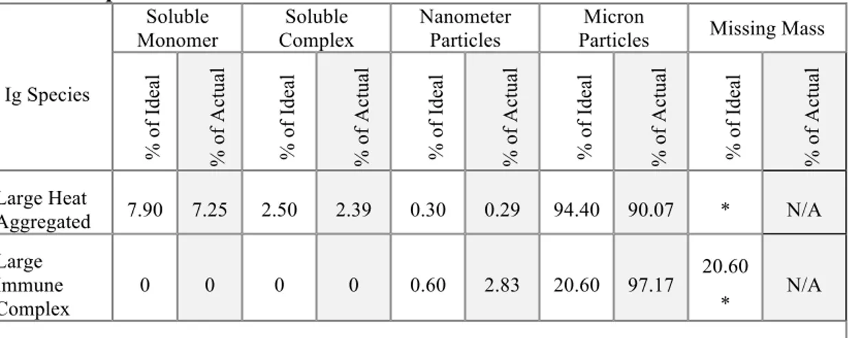
ix III.2.6 Summary of mass distribution of Ig species ... 54 III.2.7 Mass distribution of the alternative “large” heat aggregates and “large” immune complexes ... 54 III.2.8 Limulus amebocyte lysate assay does not rule out the possibility of LPS contamination ... 59 III.2.9 mAb 36-71 does not act as a non-specific adjuvant in an in vivo model ... 60 III.2.10 Heat aggregated mAb 36-71 is a more potent antigen for in vitro CA30 stimulation than monomeric Ig ... 61 III.3 Discussion ... 66 IV. IG AGGREGATION LEADS TO PRODUCTIVE, CD4-MEDIATED HUMORAL IMMUNITY WHILE MONOMERIC IG STIMULATES AN EXISTENT, BUT NON-PRODUCTIVE, CD4+ RESPONSE ... 71
IV.1 Introduction ... 71 IV.2 Results ... 73
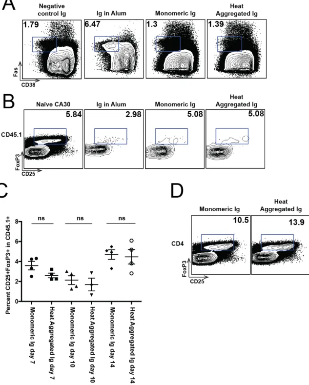
IV.2.1 Identification of CA30 T cells by using congenic markers in the B6AF1 system ... 73 IV.2.2 Monomeric Ig induces primary CA30 T cell proliferation, but heat aggregated Ig generates a weak humoral immune response ... 73 IV.2.3 Heat aggregated, but not monomeric, Ig primes for a memory humoral response ... 75 IV.2.4 Heat aggregated Ig drives CA30 T cells through more division cycles than monomeric Ig in the spleen ... 81 IV.2.5 Heat aggregates induce an increase in CA30 T follicular helper (Tfh) cells by day 14 ... 89
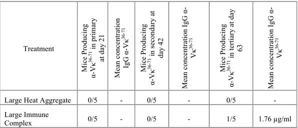
IV.2.6 Immune complexes generate a similar humoral immune response to heat aggregated Ig in the CA30 adoptive transfer model ... 96 IV.2.7 Alternative “large” heat aggregates and immune complexes induce divergent proliferative profiles at day 5 ... 104 IV.2.8 The lack of a functional Fc diminishes presentation of a peptide in the Fab region ... 107
x
IV.2.9 B cells are an important source of presentation for heat aggregated Ig antigen to CA30, but not critical for presentation
of immune complex or monomeric Ig ... 110
IV.3 Discussion ... 119
V. THE EFFECT OF IG FORM ON THE EXPANSION OF MEMORY CA30 T CELLS ... 125
V.1 Introduction ... 125
V.2 Results ... 127
V.2.1 CA30 T cells exposed to monomeric Ig can expand in a memory response, but this expansion is similar to that of CD4+ T cells exposed to a tolerogen ... 127
V.2.2 The primary expansion of residual CA30 T cells in negative control mice is larger than that of mice treated with various mAb 36-71 species ... 131
V.2.3 Monomeric Ig is a poor inducer of memory expansion of CA30 T cells ... 133
V.2.4 Memory expansion of CA30 T cells primed with aggregated Ig species does not appear to be dependent upon rechallenge with the same antigen ... 137
V.3 Discussion ... 141
VI. DISCUSSION ... 147
VI.1 Context and Discussion of the Findings ... 147
VI.1.1 Chapter III: Are particle mass and LPS contamination as important as immune complexes with regard to immunogenicity? ... 147
VI.1.2 Chapter IV: Monomeric Ig versus immune complexes: differential inducers of Th1 and Th2 development, or tolerogenic versus immunogenic stimuli? ... 152
VI.1.3 Chapter V: Evidence of Tfh memory or a critique of my experimental protocol? ... 162
VI.2 Limitations ... 164
xi VI.2.2 The use of a single isotype ... 166 VI.3 Recommendation for future work ... 168 VI.3.1 Building and transitioning to the B6.3kλ Tg mouse as a source of Ig ... 168 VI.3.2 The immunogenicity of immune complexes in
monoclonal antibody therapy ... 170 VI.3.3 The tendency of immune complexes to generate Th2 responses ... 171
REFERENCES………174 APPENDIX
A. PERCENTAGES OF CD45.1+ CELLS IN EACH CFSE DIVISION FOR PROLIFERATION EXPERIMENTS………...200
xii LIST OF TABLES
Table
2.1 Reagents used for flow cytometry. ... 39 3.1 Mass distribution of experimental Ig species compared to ideal and recovered mass ... 55 3.2 Mass distribution of large heat aggregate Ig and large immune complex species compared to ideal and recovered mass ... 56 4.1 Heat aggregated Ig generates a weak humoral response in some recipients of the CA30 T cell. ... 75 4.2 Heat aggregated Ig, but not monomeric Ig, primes for a memory humoral response ... 81 4.3 Immune complexes prime for a memory humoral response ... 99 4.4 Large heat aggregates and large immune complexes do not prime for a memory humor response ... 106 5.1 CA30 T cells exposed to monomeric Ig expand in a memory
response, but this expansion is similar to that of CD4+ T cells exposed to a tolerogen ... 131 5.2 The primary expansion of residual CA30 T cells in negative control mice is larger than that of mice treated with various mAb 36-71 species ... 136 5.3 Monomeric Ig is a poor inducer of memory expansion of CA30 T cells ... 140 5.4 Memory expansion of CA30 T cells primed with aggregated species does not appear to be dependent upon rechallenge with the same antigen ... 143 A1 Mean number of divisions: monomeric and heat aggregated Ig ... 200 A2 Mean number of divisions: monomeric and immune complex ... 200 A3 Mean number of divisions: large head aggregated Ig and large immune complex ... 200 A4 Mean number of divisions: F(ab')2, Fab, and
xiii A5 Mean number of divisions: Isotype control and α-CD20 mIg2A5D2 ... 201
xiv LIST OF FIGURES
Figure
3.1 Precipitation of mAb 36-71 with arsanilated mouse serum albumin ... 47 3.2 SEC chromatograms of experimental Ig species ... 48 3.3 Particle size distributions and integrated mass for nano-sized populations in experimental Ig species ... 50 3.4 Particle size distributions, integrated mass and representative images of micron-sized particles in experimental Ig species ... 52 3.5 SEC, PTA, and MFI analyses of the large heat aggregated Ig and large immune complex samples ... 57 3.6 mAb 36-71 does not act as a non-specific adjuvant in an in vivo model ... 62 3.7 Heat aggregated Ig is a more potent antigen than monomeric Ig for in vitro CA30 cell stimulation ... 65 4.1 Primary proliferative time course of the CA30 T cell in response to monomeric Ig and heat aggregated Ig ... 76 4.2 Heat aggregated Ig, but not monomeric Ig, primes for a memory humoral response ... 80 4.3 Evidence of humoral memory response to a unique heat aggregated Ig species ... 82 4.4 Heat aggregated Ig drives CA30 T cells through more division cycles than monomeric Ig in the spleen, but not lymph nodes ... 86 4.5 Heat aggregated Ig induces an increase in CA30 T follicular helper (Tfh) cells by day 14 ... 93
4.6 Immune complexes generate a similar primary expansion and humoral response to heat aggregates ... 100 4.7 Large heat aggregates and large immune complexes drive fewer divisions than expected ... 108 4.8 The lack of a functional Fc diminishes presentation of a peptide in the Fab region but an F(ab')2 complex can rescue presentation ... 111
xv 4.9 α-CD20 therapy diminishes proliferation of CA30 T cells in response to heat aggregated Ig ... 115 5.1 CA30 T cells exposed to monomeric Ig expand in a memory response, but this expansion is similar to that of CD4+ T cells exposed to a tolerogen ... 129 5.2 The primary expansion of residual CA30 T cells in negative control mice is larger than that of mice treated with various mAb 36-71 species ... 134 5.3 Monomeric Ig is a poor inducer of memory expansion of CA30 T cells ... 138 5.4 Memory expansion of CA30 T cells primed with aggregated species does not appear to be dependent upon rechallenge with the same antigen ... 142
1
CHAPTER I INTRODUCTION
I.1 Prologue
When Emil von Behring and Shibasaburo Kitasato first identified a component of serum that could transfer passive protection against tetanus or diphtheria toxin between immunized and naïve animals, they arrived at the simple, yet powerful, conclusion that this discovery would spark a revolution in therapeutics. To say that their hunch augured well vastly understates the pharmaceutical-industrial complex that has emerged around the production of these antibodies, the name coined by Paul Ehrlich, for the use as therapy in an increasing number of disease states. It has been projected that in 2014, the year that this thesis was written, monoclonal antibodies will account for 166 billion dollars in sales for pharmaceutical companies, which will be approximately one third of the total prescription drug market [1].
Towards the end of their seminal publication, Kitasato and Behring sought to relay an optimistic message of therapeutic potential by quoting von Goethe’s Faust Part I, “Blood is a very special juice.” In the spirit of Mephistopheles, the speaker goading Faust into signing a contract for his eternal soul, this benefit came with a price [2]. The early history of commercial antibody production, initiated by Behring himself, would also presage more complicated aspects of our current understanding of the
immunoglobulin he co-discovered. In a subsequent monograph, Behring stated a goal of mass production of antitoxin for the treatment of diphtheria in humans and set forth the experimental procedures by which he would ascertain an appropriate therapeutic protocol [3]. Starting in 1892, he worked with Farbwerke Hoechst to produce sheep
anti-2
diphtheria toxin to be used in humans. This antitoxin was a clinical success, but there was a proviso: it was evident from the earliest usages that patients often developed skin rashes in response to multiple injections and that some patients developed systemic symptoms (fever, arthralgias) that Clemens von Pirquet and Bela Shick later described as “serum sickness” [4, 5]. Behring agreed with the hypothesis of Pirquet and Shick: there was something foreign about transferring antitoxin from animals into humans that led to rejection. Behring spent years devising purification techniques to rid the antitoxin of this consequence.
While the modern techniques to produce therapeutic mAbs have sought to
eliminate any traces of non-human structure, there are nevertheless immune responses to virtually all of these agents in some fraction of patients. This is a therapeutic sequela of a paradox of Ig ontogeny that has long fascinated the immunological community: if the immune system recognizes non-self, amino acid sequence diversity in pathogens and antibodies generate amino acid sequence diversity to bind pathogens, how does the immune system avoid responding to the antibodies mediating its own action? To answer this question definitively in the context of every aspect of the immune system during a graduate thesis would border on megalomania. Instead, the aims of this thesis will focus on the capacity for CD4+ T cells to perceive and respond to an antigenic sequence within an Ig by initiating a productive humoral immune response. The ability, or lack thereof, for a CD4+ T cell to initiate this response has significant ramifications for the success of therapeutics in some fraction of the patient population for virtually every mAb, as well as implications for the more general biological question posed above: how do we maintain antibody diversity without rejecting antibody diversity?
3
Since the history of research surrounding Ig spans over one hundred years, it is unsurprising that there is a wealth of published experimental data that must be considered when addressing questions in this vein. To confound the analysis further, much of this research falls into one of two diametrically opposed categories: Ig as tolerogenic antigen or Ig as immunogenic antigen. For the purposes of the introduction to this thesis, I hope to present a survey of the literature detailing these competing views. This summary of an established division in the literature will be followed by a discussion of Ig
immunogenicity and CD4+ T cell response with a particular emphasis on the CA30 Tg T cell model system that was used in the experiments detailed in the later chapters. With this background established, I will present the hypotheses explored in the data chapters of the thesis and continue to the presentation of the body of the work.
I.2 Immunoglobulin as tolerogenic antigen
The 1940s to early 1960s was an era in which convergent lines of research laid a foundation for the study of immunological tolerance. In the 1930s, researchers had come to the conclusion that young animals made poor immune responses when challenged with commonly studied antigens, which was considered to be a function of some deficiency in Ig production [6-12]. In the 1940s, this work was extended through the study of serum gamma globulin to assay for changes in protein character during animal development; it became obvious that young animals tended to have a delay between birth and the capacity to produce specific, high titer Ig responses upon challenge [12-18]. Around the same time, Ray Owen identified fetal “graft” tolerance through his studies of the circulations of fraternal twin calves; twin calves that gestated with a common placental circulation, but had divergent blood types, could contain red blood cells that displayed red cell antigens
4
that came from either of the twins. Many of the twins assayed were adults, which implied a transfer of a regenerating progenitor cell that could continuously produce red blood cells in the recipient twin. Owen commented on the potential for immunological implications of this finding [19]. Frank Macfarlane Burnet and Frank Fenner cited both of these phenomena, the variation in Ig production during youth and the fetal tolerance in fraternal twin calves, in the second edition of “The Production of Antibodies” [12] Burnet would eventually win the Nobel Prize for his analysis of Owen’s experiment, hypothesizing that the transfer of foreign cells into an embryo could yield indefinite immunological tolerance to the cells, which was later borne out in the lab of co-Prize winner Peter Medawar. While it was not stated explicitly that these phenomena were the impetus for his conclusion, Burnet went on to state that it was likely that antibody
production was affected by both the age of the animal as well as “the nature and frequency of the antigenic stimulus.” Considering this conclusion with his hypothesis regarding fetal tolerance merely a chapter before, it is reasonable to consider Burnet to be an intellectual forefather of thought on how tolerance to serum proteins is achieved and maintained [12, 20, 21].
A year before the reissue of “The Production of Antibodies”, Lloyd D. Felton delivered the presidential address to the American Association of Immunologists and spoke about a harmful manifestation of tolerance, “immune paralysis.” By injecting 500 µg of pneumococcus polysaccharide into mice, his group was able to eliminate the
capacity for these mice to respond to an immunizing dose of polysaccharide [22]. During the 1950s, this “immune paralysis” intrigued a number of researchers who attempted to recapitulate these results by injecting large quantities of heterologous protein such as
5
albumin or gamma globulin, rather than polysaccharide, into rabbits or outbred mice. However, they could not repeat the paralysis until they followed the strategy implied by the work of Burnet and Medwar, by injecting the tolerizing dose of protein into neonatal animals. This protein-specific tolerization persisted for extended periods of time after its induction and could be maintained even longer by administering booster immunizations of the experimental antigen [20, 21, 23-42].
When David Dresser began his experiments injecting bovine gamma globulin (BGG) into CBA mice, he was extending knowledge of this tolerization phenomenon in an inbred strain of mouse, using a published method to detect antigen-elimination, and investigating kinetic variables. At first his results mirrored those of previous groups: the tolerization phenomenon occurred only in neonatal mice or, inconsistently, when BGG was injected intravenously. However, while studying the role of lipids as adjuvants, he made a notable and divergent finding: if he removed particulate matter by
ultracentrifugation, the BGG tolerized adult animals via any route of administration. This finding was subsequently recapitulated in guinea pigs. While previous tolerance
regimens had required large doses (hundreds of milligrams) of antigen and been inconsistent, ultracentrifuged BGG tolerized all adult mice at relatively low doses (hundreds of micrograms). Dresser had identified heterologous, deaggregated gamma globulin as a potent tolerogen that could be used in studies ranging from the testing of adjuvants to understanding the kinetic of immunological tolerance [40, 42-52].
William Weigle recognized the novelty of this tolerization by gamma globulin immediately and directed his research group to dissect the cellular mechanisms, a research question that he would follow throughout the rest of his career. Within two
6
years of Dresser’s discovery of the unique function of BGG, Weigle’s group
demonstrated that the same phenomenon occurred with human gamma globulin (HGG) and that the splenocytes of these mice remained tolerant even if adoptively transferred into an irradiated, secondary host. This “adoptive tolerance” intrigued the researchers, as it could be mediated by either neonatally or adult tolerized splenocytes, and indicated to them that the mechanism of tolerance might be similar in both groups. Continuing their studies with HGG, the group challenged the tolerization phenomenon with intricate time courses and injections of adjuvant in the context of different mouse strains, describing subtleties regarding the onset and abatement of tolerance that will be discussed in the data chapters of this thesis [53-66].
The original description of “adoptive tolerance” to HGG utilized splenocytes, a bulk population that contained both T and B cells. In 1966, Henry Claman and his colleagues described the necessity for synergism between thymic- and bone marrow-derived cells for Ig production [67]. Jacques Chiller, Gail Habicht, and Weigle made the logical leap and initiated experiments to test which cell population, the thymic or the bone marrow, was responsible for the tolerance to HGG. The simple, surprising answer: both populations of cells were tolerized by the deaggregated HGG. The kinetics of the phenomena were different, bone marrow cells were tolerized for about two months while the thymocytes were tolerized for over four months, but both populations of cells had to be functional to initiate an antibody response to immunogenic HGG and it was possible to show that tolerized cells from either location maintained tolerance in a transfer experiment [68-77]. This differentiation between tolerance in the B and T cell
7
was republished in the Journal of Immunology “Pillars of Immunology” series with a commentary about its historical significance to the field of self-tolerance [78]. From the perspective of Ig, it raised the questions: how does heterologous Ig tolerizing the two types of cells, and was there a reason that Ig would induce this response rather than another serum protein such as albumin [54]?
To continue this progression of thought, it is valuable to consider a vein of Ig research that occurred in the years after Chiller, Habicht, and Weigle, but that ultimately drifted from the mainstream of immunology research. Concurrent with the development of this suppression literature, other groups were building a compelling dataset to verify the Clonal Selection hypothesis, conceived in parts by Burnet, Talmage, and Jerne [79-82]. As an important component of clonal selection involves the unique specificity of Ig receptors on lymphocytes, immunologists became intrigued by extensive diversity evident within the lymphocyte repertoire: how many specificities could a mouse have? Could you transfer specificities from mouse to mouse? What was the cellular basis of Ig allotype suppression [83-85]? Researchers injected Ig from myelomas into mice and observed tolerance or, if aggregated, immunogenicity [86]. The immune system became an infinite hall of mirrors: if antibodies could elicit antibodies against themselves and those antibodies could have antibodies against themselves as well, was this suppression in immune response to something like HGG just an extension of battling antibody specificities? Although perhaps an inelegant description, this was the nexus of the “network theory of the immune system” of Jerne that became a significant topic of research from 1974 to roughly 1982 [84]. An important component of the theory as it developed was the existence of T cells that suppressed antibody responses due to
8
idiotypic-anti-idiotypic interactions, which was thought to explain the tolerized
thymocytes of Chiller et al; these T cells were known as “suppressors” and contained a gene that mapped a region of the Major Histocompatibility Complex called “I-J”. The existence of these suppressor T cells was debated within the literature with skeptics (including the chair of this thesis committee) and proponents based upon experimental data, until ultimately RNA hybridization revealed that the putative “I-J” gene did not exist at the site where others claimed to map it [83, 87-105]. To a great extent, network theory was relegated to the scrap heap of immunological thought. However, this does not mean that every experimental finding from the period is completely irrelevant. For example, regulatory T cells (Treg) function in some of the ways that the literature
predicted as mechanisms for the suppressor T cell. The capacity to suppress antibody responses with anti-idiotypic antibodies is still an intriguing finding and one must consider why this is, or is not, the norm in natural immune responses.
After the exodus from the realm of network theory, a few labs persisted in their efforts to define a mechanism for the tolerogenic effect of gamma globulin. The HGG tolerization was robust and highly reproducible. Many of the experimental observations were reminiscent of data that would be described in later, more controlled models testing central and peripheral tolerance, however, researchers struggled to find a definitive, unifying theories to explain the phenomenon. Ideas ranged from antibody-dependent cell cytoxicity to a lack of appropriate costimulation in both the B and T cell compartments, a suppressive effect mediated by macrophages to an interaction with B cells that
specifically targeted IL-4 secreting Th2 T cells [106-128]. The striking binary nature of
9
virtually no proliferation or cytokine production in response to a secondary challenge in vitro.
In recent years, focus on the tolerogenic effect of Ig has often shifted to a consideration of issues related to therapeutics. Multiple groups have identified the inclusion of peptides into the structure of IgG as a potential tolerogenic delivery
mechanism that can decrease experimental immune responses or even temper pathology in a mouse model of autoimmune disease [129-148]. Findings such as these, in
conjunction with computational methods of tolerogenic peptide analysis, have led to an intriguing new hypothesis: some IgG may contain naturally tolerogenic peptides that generate the production of Treg cells that can suppress ongoing response to other peptides
within the structure [149-157]. Other hypotheses surrounding IgG suppression of the immune response derive from knowledge about the function of inhibitory receptors for the constant region of IgG (Fcγ) or sugar moieties (sialic acid) that are attached to the IgG during trafficking in the endoplasmic reticulum. A notable example of the former is the work of Solveig Reitan and Kristian Hannestad, who examined the adjuvanticity of Ig containing an immunogenic idiotope but paired with differing isotypes. They found that IgG and dimeric IgA were tolerogenic, while IgE, native, pentameric IgM, and F(ab’)2
were immunogenic [158-160]. While they were unable to completely eliminate this dichotomy (tolerogenicity/immunogenicity) in various Fc receptor knockout mice, they concluded that it was likely that multiple Fc mediated mechanisms were involved in the process, including an inhibitory signal from the FcγRIIB on the surface of B cells and ADCC induced by stimulatory Fcγ on the surface of NK cells. In the case of sugar moieties, the laboratory of Jeffery Ravetch has identified a receptor for sialic acid
(SIGN-10 R1 in mice, DC-SIGN in humans) as the critical component for the suppression of
immune function by high doses of intravenous administration of Ig (IVIg) [161-164]. As opposed to much of the tolerogenic phenomenology discussed thus far, IVIg mediates a generalized down regulation of immune function rather than a targeted tolerization, and has been used to alleviate acute episodes of autoimmune disease. From their data, the group has surmised that SIGN-R1 is initiating this downregulation in function, however the direct mechanism of this systemic downmodulation is still in question. Ravetch and his colleagues propose that engagement of SIGN-R1 is causing the secretion of a soluble factor that leads to upregulation of the inhibitory FcγRIIB on effector macrophage populations that suppress ongoing inflammatory responses.
To summarize this historical progression of work in regards to the tolerogenicity of Ig: the administration of deaggregated gamma globulin, whether heterologous or isologous, does not yield a productive immune response as defined by the development of anti-gamma globulin antibody. Furthermore, animals treated this way are tolerized in so far as they do not make a secondary or primary response upon challenge with an
immunogenic form of the Ig and adoptive transfer of lymphocytes has demonstrated that they cannot participate in a productive response even when removed from the tolerizing environment. There is mixed evidence for this tolerance being active (i.e. regulatory cells) vs. deletion of potential effectors, and both mechanisms may be able to play some role within both the B and T cell compartment. More recent research has suggested roles for either cellular regulation or deletion, although much of the regulatory literature has either been predominantly putative or phenomenological at the level of suppression of autoimmune function. One deficit in a great deal of this literature has been its reliance on
11 assumed behavior of bulk populations rather than on cells with identifiable or defined antigenic specificity, and as such, it has been difficult to dissect the potential for regulation versus deletion. The physiological results obtained are of great interest, and their relevance should not be discounted, but one of the compelling reasons for the pursuit of this thesis is the capacity of our lab to track T cells of a defined specificity, as will be described in more detail.
I.3 Immunoglobulin as immunogenic antigen
In 1905, Pirquet and Schick described “Die Serumkrankheit” (The Serum Sickness), the systemic symptoms associated with an adaptive immune response to an administered heterologous horse serum in humans [5]. They recognized that the kinetics of the reaction, and the presence of a precipitin in some patients, indicated that the horse antibodies were acting as an antigen for ongoing production of an anti-horse Ig response. This recognition that a heterologous serum protein could induce a productive immune response was well established by the time that Dresser and others were injecting animals with various species of albumin or gamma globulin. In fact, it was the work of Dresser with BGG that was unusual, if adult animals were injected with purposefully aggregated or not ultracentrifuged Ig, they made a productive humoral immune response that was in contrast to the tolerance that he observed. These results complemented findings from groups investigating the capacity of antibody-antigen immune complexes to initiate cutaneous skin reactions, recapitulating the effect originally described by Nicolas Arthus in 1903 [165, 166]. In the case of the work of Dresser, a component of gamma globulin that could be eliminated through centrifugation (i.e. an aggregated species) was
12 its tolerizing counterpart; in the Arthus recapitulations, immune complexes formed in moderately high antigen excess along the precipitation curve, a situation where the majority of the Ig could be expected to be in a complexed form, were highly efficient at inducing cutaneous inflammation or systemic anaphylactic response [167-180]. The discovery of the existence and function of the Fc receptors would give these observations a plausible mechanism [181, 182]. Futhermore, while B cells could utilize antigen-specific, receptor-mediated intake to increase antigen presentation as a mechanism for increasing their capacity to stimulate T cells, Ig and Fc receptors presented an avenue through which other antigen presenting cells (APC) could bind antigen in a specific and receptor-mediated fashion (immune complexes) to augment presentation. While these observations did not speak specifically to the nature of the antigenicity of Ig per se, in retrospect, they did demonstrate that immune complexes have a natural adjuvanticity that can increase the presentation of antigenic peptides from within the complex to T cells. It is important to mention that FcγRIIB has been shown to deliver an inhibitory signal upon the binding of aggregated IgG, and that this inhibition is believed to regulate antibody production during an immune response [183]. While this caveat certainly must be taken into account when considering the totality of the effect of immune complexes on the progression of the immune response, the observations commented upon above suggests that this inhibition is not completely eliminating immune function when tested
experimentally. Furthermore, FcγRIIB deficient mice can be made tolerant in Ig tolerization experiments, and the receptor has always been cited as one component in a milieu of regulatory factors [120, 160].
13 An alternative method for evaluating the immunogenicity of Ig is to examine humoral responses against Ig-derived epitopes in either the constant or the variable region. Rheumatoid factor (RF) is the classical example of an Ig that targets the Fc of IgG in the system. It was discovered by two research groups independently in the years bookending World War II, the fog of war surrounding whether the original report was disseminated widely [184, 185]. In both cases, it was identified as a factor, found in the sera of some patients with rheumatoid arthritis (RA), which could agglutinate sheep red blood cells (SRBC) if an anti-SRBC antibody was also present in the reaction. RF was most often described as being an IgM anti-Fcγ, although it has been shown that RF can be any isotype, not only IgM. The validity of testing for RF as an assay for RA, and the role of RF in the pathophysiology of RA have both been questioned as knowledge about the disease has become more sophisticated [186]. However, for the purposes of
evaluating the capacity for the Fcγ to act as an immunogenic antigen for a productive immune response, the existence of RF is confirmation. Rheumatoid factor B cells are a common constituent of the natural repertoire and have been shown to expand during adaptive immune responses to unrelated protein antigens [187-194]. Recent work with transgenic mice expressing Ig chains from an RF has shown that RF B cells can exist quiescently within a normal mouse without becoming tolerized or deleted, but that these cells may secrete RF when bred to an autoimmune background [195, 196].
While RF targets the Fc component of the IgG, there is also ample experimental evidence to indicate that the variable regions of an Ig can also induce a productive
humoral response [197-204]. This is particularly interesting given the knowledge that the variable region contains somatic diversity from several mechanisms that occur both
14 during development of the B cell receptor and the affinity maturation that occurs in the germinal center during a humoral response. Isologous anti-idiotypic antibodies are a manifestation of the immunogenicity of this somatic antibody diversity. Again, the literature supporting the network theory relied upon the generation of these anti-idiotypic antibodies to create the eponymous network, and results of associated studies must be considered despite the consensus movement away from the theory since the early 1980s. More recently, the work of Reitan and Hannestad examining anti-idiotype generation demonstrated that isotypes that are typically found in polymeric form in the body (IgM, IgA) could elicit anti-idiotypic responses in the absence of adjuvant [158-160].
Amidst the years of controlled experimental investigation in animals, the clinical community has been carrying out a larger, less controlled, and higher stakes experiment into the immunogenicity of Ig: the usage of monoclonal antibodies in patients. Since the first published mAb treatment in a cancer patient in 1980, and particularly since the FDA approval of Muromanab-CD3 for allograft rejection in 1984, physicians, academic scientists, and pharmaceutical companies have sought ways to bring these mAb to the clinic [205, 206]. There are currently over 30 mAbs approved for therapy by the FDA and reports of over one hundred in ongoing clinical trials. In 2009, the FDA published a guide suggesting to pharmaceutical companies that assaying for immunogenicity should be an important component of drug development with mAbs and other biologics [207]. The scientific community had already sounded the alarm about immunogenicity of mAbs years earlier. As of the early 1990s, researchers had observed that chimeric mouse antibodies used in therapy regularly generated an anti-Ig response, which they dubbed a HAMA (human anti-mouse antibody) or HACA (human anti-chimera antibody) and are
15 still a concern to this day [208-224]. They concluded that it was likely an immune
response to murine components of the chimeric Ig, but some also conceded that it was likely that the humanization of therapeutic Igs, or even fully human Igs, would probably still lead to idiotypic immune response (later to be called a HAHA – human anti-human antibody) [207, 219, 221, 225]. While it was difficult to draw a direct correlation between the development or quantity of the HAMA and a specific clinical outcome, it was clear to these groups that this immune response had potential to end therapeutic efficacy and create pathology for patients. Pharmaceutical companies responded by attempting to develop reagents that incorporated increasing amounts of human protein structure or, in recent years, were derived completely from human Ig genes. Although it is rarely an emphasized point in papers about clinical trials, evidence of an immune response against a given mAb is generally a component of the adverse outcomes reported by researchers. Sometimes they will report an anti-Ig response; sometimes they report outcomes associated with anti-Ig antibodies like infusion anaphylaxis or delayed-type hypersensitivity reactions at the injection site. Most mAbs, if not all, generate evidence of an anti-Ig response whether they are chimeric or completely derived from human genes. The rates of reaction or measured anti-Ig development range based upon the specific Ig, the disease being treated, and the schedule of administration; the anti-CD20 antibody rituximab yielded an anti-Ig response in less than 10% of lymphoma patients, but in as high as 60% in one study of patients being treated for systemic lupus
erythematosus [217, 218]. This knowledge has led physicians to design treatments with the co-administration of a mAb and chemotherapeutic agents such as methotrexate or azathioprine in the hopes that these agents will ablate any immune response [226].
16 However, the correlation between anti-Ig development and the success of treatment is still murky, at best, for most agents and the evidence for this co-administration of chemotherapeutic is generally anecdotal in part due to an insufficient study population. One notable exception to this paucity in data is in the usage of anti-TNF-α mAbs, particularly for rheumatoid arthritis. In the last year, researchers have begun to make a quantitatively justified case that the development of anti-Ig responses has a significantly negative effect on hypersensitivity reactions, therapeutic response and soluble drug levels over time [227]. In concordance with the theoretical underpinnings, administration of a chemotherapeutic with the mAb could eliminate some of the negative outcomes
associated with developing an anti-Ig response. While more substantive data will be needed to make clinical judgments about treatment regimens, the fact remains that the therapeutic use of Ig has demonstrated that it can be immunogenic. The rates of anti-Ig antibody development may vary based upon a number of factors, but some percentage of patients is able to make a productive immune response against virtually every mAb and ongoing data collection is finally beginning to justify the concern that has existed for over two decades.
The release of the guide on immunogenicity testing of biologics by the FDA in 2009 hinted at another complicated aspect of this line of research: immunologists and clinicians are no longer the only scientists hoping to dissect the capacity for mAbs to induce or evade the immune response. Pharmaceutical scientists, those that examine the physicochemical attributes of manufactured pharmaceuticals, have begun to question what controllable aspects of the proteins they make can be modified to decrease rates of rejection. The variables they test can range from modifying the glycosylation patterns of
17 the Fc to optimizing amino acids in the variable domains to decrease the immunogenicity [228-230]. The influence of antigen size on Ig immunogenicity has been a subject of investigation for some time; the original tolerance protocol of Dresser relied upon the ultracentrifugation of BGG to remove polymeric complexes, and larger accumulations of antigen such as heat aggregates and immune complexes have been shown to induce humoral and DTH responses. Pharmacologists recognize that aggregated Ig could be immunogenic and have begun to study Ig species that have been manipulated
aggressively to simulate mishandling during production, storage, or administration [231-233]. While there are definitely cultural differences between the immunologists and the pharmacologists in terms of their techniques and the extent to which data between the two realms can be compared, it is valuable to consider this type of experimentation into aggregate size as a complementary element of immunogenicity research.
I.4 CD4+ T cells and the perception of immunoglobulin as antigen: model systems
While there is a substantial history of literature examining the extent to which Ig activates a productive immune response, the vast experimental data have a deficiency in regards to the role of individual CD4+ cells in tolerization or activation. Certainly, there have been observations about CD4+ cells and indications about how they might perceive immunoglobulin. A tolerized thymic compartment could not provide sufficient help to a naïve bone marrow compartment to yield a productive anti-HGG response [68, 69]. HGG treatment seemed to decrease both IL-2 and IL-4 production in T cell populations leading to the interpretation that the tolerogenic Ig could suppress both Th1 and Th2 CD4+ cells, with an increased emphasis on suppression of Th1 cells [116, 234].
18 Sequencing data have implied that the Fc may contain tolerogenic peptides that induce the proliferation of suppressive Treg cells [153-155, 157]. However, the production of
high affinity IgG anti-mAb responses in patients against therapeutic Ig implies that there must be CD4+ cells participate in productive immune responses to the mAb. Precursor percentages of some anti-mAb CD4+ cells have been defined in a naïve human repertoire [235, 236].
The biggest deficiency in all of this data is the reliance on experimental results that imply, rather than directly show, the effect of Ig on single T cells. Prior to the era of transgenic mice, it was difficult, if not impossible, to track a monoclonal population of T cells that were activated by an Ig-derived peptide. There are currently two mouse models with transgenic CD4+ T cells that recognize specific peptides generated via somatic mutation in isologous Ig V regions. Both have been used to study central CD4+ tolerance to Ig-derived peptides as well as the consequences of failures of tolerance. Both will be described, in particular the CA30 – Vκ36-71 model which is the basis for the work in this thesis.
The first model derived from the observation that injections of an isologous IgA antibody derived from a myeloma could induce anti-idiotypic antibodies [201]. This anti-idiotypic response was T cell-dependent, and the antigenic light chain epitope was determined via antigen presentation with T cell clones derived from sensitized mice [200, 237-245]. The researchers created two transgenic mice: one expressing the lambda light chain with the antigenic somatically mutated residues (λ2315) and the other expressing an
α/β T cell receptor which recognized the antigenic peptide in the context of the class II MHC I-Ed. If the two mouse strains were crossed, there was a profound deletion in
19 CD4+/CD8+ thymocytes indicating that central tolerance mechanisms were influenced by the presence of the λ2315 light chain in the mouse [246]. Interestingly, large injections of IgG bearing the λ2315 could also delete the TCR-Tg cells in the T cell-only transgenic animal. If a mother expressed the λ2315 and the TCR-Tg but the offspring expressed only the TCR-Tg, the transferred maternal IgG was insufficient to delete the TCR-Tg in the neonatal animal due to its low concentration. This finding indicated that high
concentrations of IgG could lead to Ig-derived antigen presentation to CD4+ cells, particularly in the thymus. Further work with these mice focused more on the
consequences of T cell/B cell cooperation and the illicit licensing of B cells expressing the λ2315 to secrete autoreactive antibodies due to CD4+ recognition of the light chain epitope [247-250].
The second model, used in this thesis, was developed in our lab to investigate the development of tolerance to Ig-diversity. The λ2315 system had begun as an investigation into the capacity for large amounts of isologous IgG to induce an anti-idiotypic response with the assumption that idiotypic antigen were probably immunogenic, just expressed at low enough levels within the system to evade tolerance induction. In 1995, our lab asked a more fundamental question: if T cells perceive peptides derived from Ig V regions can all Ig V regions induce CD4+ regulated humoral responses or is there a requirement for non-germline, somatic diversity in the Ig to direct a CD4+ mediated response [251]? To investigate this question, it was important to inject a panel of Ig that utilized the same germline genes, but some of which utilized somatically mutated receptors that would hypothetically contain an antigenic peptide. There was such a panel of Ig available that bound the hapten Ars (p-azophenylarsonate) and had been characterized extensively for
20 Ig genes and somatic mutation [252-254]. The experiment yielded T cell hybridomas that revealed the answer: animals were tolerant to isologous germline Ig sequences, but could generate a fuseable CD4+ response to Ig with somatically generated diversity. These results could be replicated by utilizing a different strain, C58 as opposed to A/J, that possessed the I-Ak MHC II, but did not possess the germline Ig V gene to which the A/J mouse had shown tolerance. By generating and challenging C58 hybridomas, the lab could demonstrate that A/J APC were able to present peptides from the germline Ig, but unable to make a CD4+ T cell response against it and thus tolerant. To study the mechanisms behind this tolerance, our lab generated two transgenic mice: the Vκ36-71 mouse, which expressed the somatically mutated κ light chain of the Ars-reactive mAb 36-71, and the CA30 Tg, which expressed an αβ T cell receptor specific for an epitope in the Vκ36-71 framework-1 region [255]. Crossbreeding between the two transgenic strains led to deletion of the CA30 Tg T cells during development, while transfer of large
numbers of CA30 Tg T cells into mice expressing only the Vκ36-71 caused development of a lupus-like autoimmune disease. Both of these experiments relied upon interactions between large numbers of transgenic T cells and B cells, and so the lab shifted to model more physiological circumstances to investigate the role of tolerance in the system. A series of bone marrow chimeras demonstrated multiple levels of CD4+ tolerance, both central and peripheral, that could serve as the regulatory mechanisms observed in the previous experiments [256]. Similarly, a dual transfer model in which Vκ36-71 B cells and CA30 T cells were transferred into a secondary animal to simulate the introduction of a novel B cell clone with a somatically mutated receptor containing antigenic peptides showed a mechanism of peripheral regulation, in which there was a disruption of memory
21 development by Tg B cells in an ongoing immune response and rapid differentiation of Tg B cells into short lived plasmablasts [257]. In both the bone marrow chimeras and dual transfer model, there were preliminary data indicating that the secretion of antigenic Ig may have played a role in some of the observed mechanisms of tolerance. In
particular, in the dual transfer model, there was early evidence that a transfer of sera from
a Vκ36-71 Tg animal could induce CA30 T cell proliferation when both were injected into
a κ-/- recipient. However, sera was used rather than a monoclonal reagent, so it was possible that this result was a function of T cell stimulation by any isotype. Regardless, this model presented an excellent opportunity for investigating the questions expressed over the course of this introduction, namely the extent to which Ig, and particularly IgG, could tolerize or activate CD4+ cells to make a productive humoral response to novel epitopes within the IgG itself.
I.5 Conclusion and the scope of the thesis
There has been a debate within the immunological literature since at least 1961 as to the nature of perception of isologous Ig by the immune system. Depending on
preconceived bias, one can point to a long progression of papers to support a hypothesis either that Ig is intrinsically tolerogenic or immunogenic. While the history of mAb therapeutics and the development of HAMA and HACA made an increasingly compelling argument for Ig being at least partially immunogenic, pharmaceutical scientists and their belief that aggregated, damaged species of Ig are responsible for inducing anti-Ig immune responses was sufficiently compelling to suspend disbelief at the outset of our work. Given that our lab has a unique and powerful system to transfer and track transgenic, Ig-specific CD4+ cells, we started the project with a goal: to
22 characterize the CD4+ response to an IgG1 containing an antigenic sequence within its
light chain. Our initial hypothesis was an extension of the landmark work by Chiller et al: we believed that the CA30 T cells would not make a primary CD4+ response to a monomeric, deaggregated species of IgG1 containing an antigenic sequence.
In Chapter III, I will describe the production and purification of antibody, initial in vitro testing of presentation of antibody species, and a panel of tests to characterize particle concentration within the samples that would be used in in vivo transfer
experiments. I characterized my monoclonal reagent as a predominantly monomeric protein with very small populations of potentially immunogenic particles and little evidence of adjuvant activity in vivo or in vitro. Conversely, I characterized heat aggregated Ig and immune complexes as reagents with large percentages of soluble and insoluble complexes that displayed early in vitro evidence of strong immunostimulatory capacity. These data describe the first particle concentration analysis of immune
complexes, which are naturally occurring structures.
In Chapter IV, I will describe a series of novel in vivo adoptive transfers that explored the primary response of a monoclonal CD4+ TCR Tg cell to species of
immunoglobulin without adjuvant. While others have reported the capacity for TCR Tg cells to proliferate after in vivo exposure to antigenic Ig, this is the first presentation of the full contraction and expansion of the primary response to monomeric Ig as well as two species hypothesized to be immunogenic: heat aggregated Ig and immune
complexes. I demonstrated that both heat aggregated Ig and immune complexes, but not monomeric Ig, can prime mice to make a consistent secondary humoral response, and that this difference is not due to stimulation of regulatory T cells by monomeric Ig, but
23 instead appears to be a function of T follicular helper differentiation in response to heat aggregated Ig and immune complexes, which has never been reported. Furthermore, I will describe a differential pattern of early proliferation in T cells exposed to monomeric Ig vs the immunogenic species that is a unique phenomenon in which T cells exposed to the immunogenic species have higher percentages of cells residing a later stages of division than those exposed to monomeric Ig, which has not been described without the use of adjuvant. I identified an important role for the Fc in the early presentation of Ig-derived peptides, in contrast to monomeric Ig, which was readily presented in vivo. Finally, I will present an early exploration of the role of B cells in the early presentation of Ig-derived peptides and will show evidence that exogenously administered heat aggregated Ig may be dependent on B cells for presentation in the spleen, whereas immune complexes are not, which has important ramifications for mAb therapy.
In Chapter V, I will describe experiments examining the dynamics of CA30 cells after a secondary challenge. I will present data that suggests that monomeric Ig
generates a poor memory population in comparison to peptide, and that this poor memory response is not augmented extensively by LPS or immune complexes. Furthermore, I will show experiments suggesting that even with a strong adjuvant, the memory response to Ig is weaker than peptide and that monomeric Ig is a poor stimulator of secondary response. This is the first report of the memory behavior of a TCR Tg cell exposed to Ig species.
24 CHAPTER II
MATERIALS AND METHODS
II.1 Production and Characterization of of Reagents II.1.1 T Cell medium (TCM)
For in vitro and adoptive transfer experiments, cells were washed and cultured in T cell medium (TCM): RPMI 1640 supplemented with 2-mercaptoethanol (50 µM), penicillin G (105 U/L), streptomycin sulfate (100 mg/L), sodium bicarbonate (2g/L) (Sigma Aldrich, St. Louis, MO), HEPES (7.5 mM) (Fisher Scientific, ), L-glutamine (2 mM), 1x MEM essential amino acids, 1x non-essential amino acids, 1x sodium pyruvate (Gibco-BRL, Grand Island, NY), and 10% fetal calf serum.
II.1.2 Hank’s Buffered Salt Solution (HBSS)
For cell harvesting for flow cytometry, tissues were processed in Hank’s Buffered Salt Solution (HBSS): 0.137 M NaCl, 0.25 mM Na2HPO4, 4.2 mM NaHCO3, 5.4 mM
KCl, 0.44 mM KH2PO4, 1.3 mM CaCl2, 1.0 mM MgSO4, and 0.1 g glucose.
(Sigma-Aldrich, St. Louis, MO).
II.1.3 Antibody production in mice
C;129S4-Rag2tm1.1Flv Il2rgtm1.1Flv/J (Rag2-/-cγ-/-) mice were injected i.p. with 0.2 ml incomplete Freund’s adjuvant (IFA) (Sigma Aldrich, St. Louis, MO) 5 days before the i.p. injection of 5 x 10⁶ hybridoma cells in phosphate buffered saline (PBS). Ascitic fluid began to accumulate 5 to 7 days later and was collected via peritoneal lavage after mice were sacrificed in accordance with guidelines of the National Jewish Health Institutional Animal Care and Use Committee (IACUC).
25 II.1.4 Handling and dialysis of ascitic fluid for anion exchange chromatography
Ascitic fluid was placed at 37°C for 20 minutes and then incubated on ice for 1 hour. After induction of clots, the fluid was centrifuged at 20,000 x g for 30 minutes at 4°C, removed from the clot and visible lipid fraction with a Pasteur pipette, and passed through a 0.22 µm filter (Millipore, Billerica, MA) under standard sterile cell culture precautions. Filtered ascites fluid was then immediately precipitated or stored at 4°C for less than 24 hours. Ascitic fluid was precipitated while stirring on ice; saturated
(NH4)2SO4 solution was added to the ascitic fluid at a rate of 1 ml/min until reaching 45%
(v/v). This solution was incubated on ice with stirring for 1 hour to maximize
precipitation. After precipitation, the solution was centrifuged for 1 hour at 20,000 x g at 4°C and the supernatant removed and tested for residual antibody. The precipitate was dissolved in a similar volume of phosphate buffered saline with 0.01% NaN3 (PBS-A) to
the original precipitated solution volume, as this minimized protein precipitation during dialysis, and stored at 4°C or immediately dialyzed for anion exchange chromatography. For dialysis, the dissolved precipitate was placed in dialysis tubing with a molecular weight cutoff of 35,000 kilodaltons (kD) and dialyzed against ≥20 volumes of 10mM NaPO4 pH 7.9. The protein was dialyzed over 48 hours which included 4 to 5 changes of
10mM NaPO4 buffer, the first two changes typically containing 0.01% NaN3 to prevent
bacterial growth and the final 2 to 3 changes eliminating its inclusion. After dialysis, protein was immediately subjected to anion exchange chromatography and never stored for >12 hours at 4°C.
26 II.1.5 Anion exchange chromatography
Diethylaminoethyl (DEAE) anion exchange cellulose DE52 (Whatman, GE-Healthcare, Pittsburgh, PA) was used in the purification of IgG1. DE52 beads were
swollen in distilled H2O for 1 hour. The beads were then incubated with 5 equilibrations
of 100 mM NaPO4 pH 7.9 buffer, followed by 10 equilibrations of 10 mM NaPO4 ph7.9.
After this equilibration, the beads were loaded into a 50 mL syringe with a small plug of glass wool in the base, with a target volume 0.15 ml packed resin for every 1 mg of protein to be loaded onto the column. After packing the column, the dialyzed protein in 10mM NaPO4 pH 7.9 was passed through the column and loaded onto the anion
exchange resin. The column was washed 3 times, each wash containing 1 bed volume of the column of NaPO4 pH 7.9. Protein was obtained by progressive column elutions in
which the column was subjected to 2 times the bed volume washes with 10 mM NaPO4
pH 7.9 with salt concentrations starting at 0.01 M NaCl and increasing 0.01 M with each subsequent wash. The fraction obtained during each wash cycle was concentrated in an Amicon Ultra spin filter (Millipore, Billerica, MA) with a molecular weight cutoff of 50 kD and then assayed for protein content using the absorbance at 280 nm measured by spectrophotometer. Once the elution point had been defined for an antibody, in the case of this work typically 0.03 M NaCl, this concentration was used for subsequent anion exchange purifications.
II.1.6 Low aggregation pharmaceutical buffer and Ig storage
Protein was buffer exchanged from 10mM NaPO4 ph7.9 into a low aggregation
pharmaceutical buffer that has been previously described (20 mM histidine, 222 mM trehalose dihydrate pH 5.5) using ≥4 spins in an Amicon Ultra spin filter, MW cutoff
27 50kD[233]. Ig concentration was brought to 4 mg/ml in the pharmaceutical buffer, and then the entire volume was passed through a 0.22 µm filter. Polysorbate 80 (PS80) (Sigma Aldrich, St Louis, MO) was added to 0.02% (v/v). Ig was then distributed into 2 mg aliquots in sterile 1.5 ml microcentrifuge tubes and stored at -20°C. Individual tubes of this Ig were subjected to a single freeze-thaw cycle prior to injection into animals. II.1.7 Arsanilation of mouse serum albumin
Mouse serum albumin was dissolved in a solution of boric acid and sodium chloride (0.16 M H3BO3, 0.16 M NaCl) to a concentration of 10 mg/ml. Two solutions
were prepared and incubated on ice: arsanilic acid was dissolved in 1 ml of 1 N HCl to create a 0.15 M arsanilic acid solution and NaNO2 was dissolved in cold water to create a
0.2 M solution. While stirring the arsanilic acid solution, the 0.2 M NaNO2 was titrated
into the arsanilic acid in 50 µL drops to create a diazonium salt. To assess this titration after the addition of each drop, 5 µl of the resultant solution was tested using starch iodide strips to assess for the presence of HNO2. When the strip showed a blue hue
immediately upon testing with the solution, the diazonium salt was prepared for coupling. This diazonium salt solution was titrated into the mouse serum albumin solution in 50 µL drops while stirring on ice. The pH of the resulting solution was maintained between at pH 9.5 by adjusting with 0.5 N NaOH. As the titration was occurring, the coupling of the hapten to the protein was assessed by diluting the solution 1/50 in 0.2 N NaOH and reading the absorbance at 477nm. Coupling could be monitored by using the extinction coefficient of 8.85 x 10³ to calculate the number of moles of Ars in the solution and then
dividing by the moles of MSA in the reaction. Once a coupling of 12 – 15 Ars per MSA
28 dialysis tubing with a MW cutoff of 35,000 kD, and against ≥20 volumes of PBS. The protein was dialyzed over 48 hours which included 4 to 5 changes of PBS, the first two changes typically containing 0.01% NaN3 to prevent bacterial growth and the final 2 to 3
changes eliminating its inclusion. After dialysis, the protein concentration was calculated based upon the end dialysis volume, the protein was passed through a 0.22 µm filter and divided in small volumes into 1.5 ml microcentrifuge tubes and stored at -20°C.
II.1.8 mAb 36-71 Fab and F(ab’)2 generation
As mAb 36-71 is an IgG1, the sulfhydryl protease ficin could be used to generate
both Fab and F(ab’)2 fragments. For both procedures, mAb 36-71 was buffer exchanged
from the low aggregation pharmaceutical buffer to a final concentration of 10mg/ml in a 0.1 M citrate buffer pH6.0 using an Amicon Ultra spin column with MW cutoff of 50kD. Immobilized ficin (Thermo Fisher Scientific, Waltham, MA) was equilibrated with 10 times resin slurry volume (2mL) of Fab digestion buffer (0.1M citrate, 5mM EDTA, 25mM cysteine) or F(ab’)2 digestion buffer (0.1M citrate, 5mM EDTA, 4mM cysteine).
Equilibrated ficin was incubated with 1 ml of mAb 36-71 in Fab or F(ab’)2 digestion
buffer for 40 hours (F(ab’)2) or 5 hours (Fab) while being rotated in a 37°C incubator.
The digested material was buffer exchanged into PBS-A and concentrated using an Amicon Ultra spin column with MW cutoff of 30kD. Both species were size excluded independently via fast protein liquid chromatography (kindly completed by Fran
Crawford in the Kappler/Marrack lab, NJH) and the fractions containing the two species pooled independently. Both the Fab and F(ab’)2 were buffer exchanged into the low
aggregation pharmaceutical buffer, passed through a 0.22 µm filter, and divided into sterile 1.5 ml microcentrifuge tubes and stored at -20°C.
29 II.1.9 Heat aggregation of Ig
mAb 36-71 was buffer exchanged from low aggregation pharmaceutical buffer into PBS at a concentration of 7 mg/ml using an Amicon Ultra spin column with MW cutoff of 50 kD. Individual 1 ml aliquots were incubated in a water bath at 63°C for 20 minutes and then immediately placed on ice for 1 hour, by which time large insoluble aggregates were visible within the solution. Insoluble aggregates were spun down by centrifuging at 1°,000 x g for 5 minutes, and the supernatant was measured for
absorbance at 280nm using a spectrophotometer, allowing for an estimation of the mass of the insoluble fraction. The soluble fraction was removed, pooled, and frozen at -20°C. The precipitated aggregates were washed 3 times with low aggregation pharmaceutical buffer and then divided into sterile 1.5 ml microcentrifuge tubes and stored at -20°C. II.1.10 Generation of Immune Complexes with Ars-MSA
Due to the specificity of mAb 36-71, it was possible to generate immune complexes with the Ars-MSA reagent. Due to some variability between batches of arsanilated albumin, it was necessary to determine an equivalence point for each batch prior to the generation of complexes for in vivo or in vitro experiments. 100 µg of mAb 36-71 were mixed with varying amounts of Ars-MSA and diluted to 100 µl with PBS. These mixtures were incubated at 37°C with rotation for 3 hours and then centrifuged at 13,000 x g for 5 minutes. After centrifugation, the supernatants were measured for absorbance at 280 nm using a Nanodrop 1000 spectrophotometer (Thermo Fisher Scientific, Waltham, MA) to establish a precipitation curve. Once an equivalence point had been established, experimental immune complexes were generated using similar conditions and either Ars-MSA at the equivalence point (referred to later as “large”
30 immune complexes due to the insoluble fraction) or at an amount of Ars-MSA 4 times larger than the mass at the equivalence point (referred to as immune complexes). Similar amounts of Ars-MSA created in antigen excess were also incubated with Fab and F(ab’)2
fragments, although the amounts of Fab (66 µg) and F(ab’)2 (73 µg) were varied to
normalize the amount of antigenic 36-71 epitope in the sample. Immune complexes were never frozen or stored prior to injection into animals or usage in in vitro culture and were used within 2 hours of generation.
II.1.11 Ultracentrifugation of Ig
To obtain monomeric Ig, multiples of 100 µg of mAb were diluted in sterile PBS to a concentration of 1 mg/ml and centrifuged at 165,000 x g in a fixed angle TLA-120.1 rotor (Beckman Coulter, Brea, CA) for 3 hours. The top 2/3 of the supernatant was removed for injection to avoid disturbing the pellet, and the sample was stored in a sterile 1.5 ml microcentrifuge tube until used for experimentation within less than 6 hours. II.1.12 Size-exclusion chromatography
Analytical size-exclusion chromatography was performed using an Agilent 1100 chromatography system (Agilent Technologies, Santa Clara, CA) as has been described previously[233]. Prepared stocks of ultracentrifuged Ig, heat aggregated Ig, immune complexes, or diluent PBS was spun at 13,000 x g and the supernatant removed carefully to eliminate large insoluble particles. Protein was loaded onto a Tosoh G3000 SWXL 7.8 x 30 cm column (Tosoh Bioscience, Tokyo, Japan) and eluted with a mobile phase of PBS pH 7.4 at a flow rate of 1 mL/min. The eluate was monitored at 280 nm and 215 nm to assess for protein recovery and percentages of monomer and dimer. Triplicate samples were analyzed for each Ig preparation. Monomer and dimer percentages were calculated