Alpha-actinin - an amazing
journey through time and species
Barbara Addario
Department of Chemistry Umeå university, Sweden Doctoral Thesis 2011
Copyright© Barbara Addario, 2011 ISBN: 978-91-7459-205-4
Cover: EM image of F-actin bundles induced by Entamoeba histolytica α-actinin2 Printed by: VMC KBC, Umeå University
To my family
“Considerate la vostra semenza:
fatti non foste a viver come bruti,
ma per seguir virtute e canoscenza"
(Dante Alighieri, Inferno XXVI, 118-120)
Table of Contents
Table of Contents 1 Abstract 2 List of papers 3 List of Abbreviations 4 Introduction 5 Cytoskeleton 5 Microtubules 5 Microfilaments 6 Intermediate filaments 7 Actin 7 Actin-binding proteins 9 α-actinin 11α-actinin in non-muscle cells 12
α-actinin in muscle cells 13
α-actinin domain structure 15
Actin-binding domain 16
Rod domain 18
Calcium-binding domain 19
α-actinins of Entamoeba histolytica 21
Experimental techniques 22
Circular dichroism spectroscopy 22
Actin co-sedimentation assay 23
X-ray crystallography 23
Gel filtration or size exclusion chromatography 24
Dynamic light scattering (DLS) 25
Aim of the study 26
Summary of the papers 27
Characterization of Entamoeba histolytica α-actinin2 (Paper I, II and III) 27 Characterization of another atypical α-actinin (Paper IV) 29
Concluding remarks 31
Appendix 32
Bioinformatics: terminology 32
Acknowledgements 34
Author
Barbara Addario
Title
Alpha-actinin - an amazing journey through time and species
Abstract
In eukaryotes, the actin cytoskeleton plays an important role in a large variety of cellular events. Its reorganization is regulated by a plethora of actin-modulating proteins, such as α-actinin.
α-actinin is a ubiquitous actin-binding protein that belongs to the spectrin superfamily. This family, besides α-actinin, includes spectrin, dystrophin and utrophin. Phylogenetic analyses have indicated that the family members arose after several intragene duplications and rearrangements of a common ancestral α-actinin isoform. Up to the invertebrate-vertebrate bifurcation, organisms seemed to have a single, calcium-dependent α-actinin. After the split, invertebrates have kept this single isoform, in contrast to vertebrates that acquired four distinct isoforms. Of the four vertebrate α-actinin isoforms, the two present in non-muscle cells are typically calcium sensitive while the two muscle isoforms are calcium insensitive.
α-actinin in higher organisms is characterized by the presence of three distinct structural domains: a highly conserved N-terminal actin-binding domain, a central rod domain with four spectrin repeats and a calcium-binding C terminus with EF-hand motifs. In some primitive organisms, such as protozoa and fungi, the rod domain of α-actinin contains only one or two spectrin repeats. With the completion of an ever increasing number of genomes, new and atypical α-actinin sequences had been available that have not been characterized yet. To obtain a firmer understanding of the evolutionary history of α-actinin, the main objective of this study was to identify, purify and biochemically characterize atypical α-actinin or α-actinin-like proteins of the parasite Entamoeba histolytica and of the fungus Schizosaccharomyces pombe. Our results show that both isoforms, despite the much shorter rod domain, are able to bind and cross-link actin filaments and therefore can be considered genuine α-actinins.
List of papers
This thesis is based on the work contained in the following papers, referred to by Roman numerals (I-IV) in the text.
I. Virel, A., B. Addario, and L. Backman
Characterization of Entamoeba histolytica alpha-actinin2. Mol Biochem Parasitol, 2007. 154(1): p. 82-9
II. Addario, B and L. Backman
The domain structure of Entamoeba histolytica alpha-actinin2. Cell Mol Biol Lett, 2010 15(4): p.665-78
III. Addario, B., H. Shenghua, U. Sauer, and L. Backman
Crystallization and preliminary X-ray analysis of the Entamoeba histolytica alpha-actinin2 rod domain. Submitted, 2011
IV. Addario, B and L. Backman
Alpha-actinin of Schizosaccharomyces pombe. Manuscript, 2011
Reprints have been included within this thesis, with permission from the publishers.
List of Abbreviations
Å Ångstrom (10-10 m)ABD Actin-binding domain ABP Actin-binding protein ABS Actin-binding site
ACTN α-actinin
ADF Actin depolymerization factor ADP Adenosine diphosphate Arp Actin-related protein Asp Aspartic acid
ATP Adenosine triphosphate CD Circular dichroism
CH Calponin homology domain CryoEM Cryoelectron microscopy DLS Dynamic light scattering ECM Extracellular matrix EM Electron microscopy F-actin Filamentous actin G-actin Globular actin
Gal/GalNAc Galactose and N-acetyl-D-galactosamine GFP Green fluorescent protein
Glu Glutamic acid Kd Dissociation constant
kDa Kilodalton
NCBI National Center for Biotechnology Information NMR Nuclear magnetic resonance
PDB Protein data bank
PIP2 Phosphatidylinositol 4,5-bisphosphate
Introduction
Cytoskeleton
The cytoskeleton is a dynamic structural “scaffold” present in the cytosol of eukaryotic (Frixione, 2000) and prokaryotic (Michie and Lowe, 2006) cells. Up to one third of the total protein content of animal cells is used for this purpose. This dynamic, three-dimensional structure plays a pivotal role in cell mechanics including mitosis, cell division, cytokinesis, intracellular transport, cell motility, establishment and regulation of cell polarity, and internal organization. In animal cells, the cytoskeleton maintains the shape of the cell, participates in the movements of organelles within the cytosol, and enables the cell as a whole to move, by using structures such as cilia and flagella. Unlike the human skeleton, the cytoskeleton is extremely dynamic. This dynamic nature is one of most important features of the cytoskeleton and is also necessary for cells to change shape, complete cell division, and migrate. In addition, all of these filament systems share a critical feature: they are composed of proteins that have the unique property of being able to self-assemble into a filamentous network. Each of the self-assembling proteins has a characteristic concentration, or "critical concentration", below which, the monomer state is favoured and above which, the polymer state is favoured. An increase in the subunit concentration favours filament building, while a decrease in concentration favours filament dissociation. This property allows the cell to rapidly control cytoskeleton structure. Three types of cytoskeletal filaments are present within the cytoplasm of most vertebrate cells: microtubules, actin filaments or microfilaments, and intermediate filaments.
Microtubules
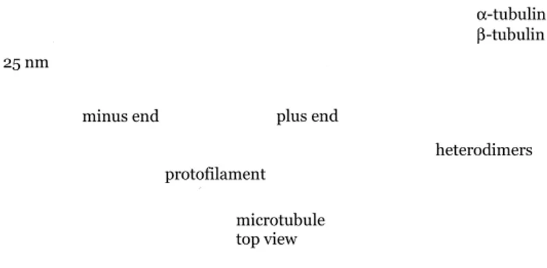
Microtubules are hollow cylinders approximately 25 nm in diameter, formed by α and β tubulin subunits. Microtubules are involved in maintenance of cell shape, cell motility, and chromosome movement (Figure 1). In animal cells, microtubules organization originates at the centrosome, while in plants it originates at many sites scattered throughout the cell. Microtubules serve as tracks for organelle movement within the cell. When the cell is about to divide, bundles of microtubules, known as kinetochore microtubules, come together and extend across the cell to assist in the movement of chromosomes during cell division. In animal cells the microtubules help to form the cleavage furrow, while in plant cells, they form the cell plate.
Figure 1. Microtubule structure. Microfilaments
Microfilaments, the thinnest elements of the cytoskeleton (≈ 7 nm in diameter), are formed by a two-start right-handed helix (Figure 2). As opposed to microtubules, actin microfilaments are not hollow. Actin monomers (G-actin) can bind ATP (adenosine triphosphate) which increases stability and causes the filaments to grow; when ATP is hydrolyzed to ADP (adenosine diphosphate), the filaments become unstable and shrink. Microfilaments are involved in most cellular processes including cell motility, intracellular trafficking, endo/exocytosis and the maintenance of cell shape and polarity (dos Remedios et al., 2003).
Figure 2. Helical structure of actin filaments.
Microtubules and microfilaments are linked to specific associated proteins that determine their function in place and time. Animal cells have actin filaments mostly concentrated in the cortex, beneath the plasma membrane, whereas the microtubules have an astral configuration. In plant cells have microtubules and actin filaments are co-aligned during most of their cell cycle. plus end minus end 7 nm Actin subunit microtubule top view plus end heterodimers protofilament 25 nm minus end α-tubulin β-tubulin
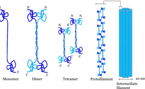
Intermediate filaments
Intermediate filaments (≈ 8-11 nm in diameter) are the most stable and heterogeneous constituents of the cytoskeleton. The first stage of filament assembly is the formation of dimers. Two dimers then associate in a staggered antiparallel fashion to form tetramers. Tetramers pack together end-to-end to form protofilaments. The intermediate filament itself is a bundle of eight protofilaments wound around each other to create a rope-like structure (Figure 3).
Figure 3. Assembly of intermediate filaments.
Intermediate filaments are found in almost all eukaryotic cells with the exception of fungi. Their function varies from forming the nuclear lamina (a net-like meshwork array that lines the inner nuclear membrane and governs the shape of the nucleus) (Coulombe and Wong, 2004), to anchoring the nucleus and other organelles as well as supporting the machinery responsible for muscle contraction. Unlike microfilaments and microtubules, intermediate filaments are non-polarized structure and are not involved in cell movement.
Actin
Actin, one of most abundant proteins in eukaryotic cells, is a ≈ 42 kDa globular protein with the ability to polymerize into long filaments under physiological salt concentration. Actin is also one of the most highly
10 nm Intermediate filament Tetramer
Monomer Dimer Protofilament
N N N C C C N N C C N N C C
conserved proteins. The only known eukaryotic cell not containing actin is the nematode sperm, which has developed another polymer-forming protein that appears to function akin to actin in several respects (Pring et al., 1992; Bullock et al., 1998). In prokaryotes, many proteins distantly related to actin have also been identified (Sanchez et al., 1994; van den Ent et al., 2001). The actin amino acid sequence is extremely conserved among different organisms, illustrated by the fact that non-muscle human β-actin is 89% identical to yeast actin (Gallwitz and Sures, 1980; Ng and Abelson, 1980). Six actin isoforms have been identified within Homo sapiens, four muscle specific and two non-muscle specific. The different isoforms are grouped into three classes: α-actin (present in muscle cells), β- and γ-actin (both found in non-muscle cells). Different attempts to solve the structure of F-actin have been made, but at present, are yet to be successful. The structure of monomeric actin has been determined in complexes with different actin-binding proteins: bovine pancreatic DNAse I (Kabsch et al., 1990), profilin (Schutt et al., 1993), and gelsolin (McLaughlin et al., 1993); all proteins that prevent actin polymerization.
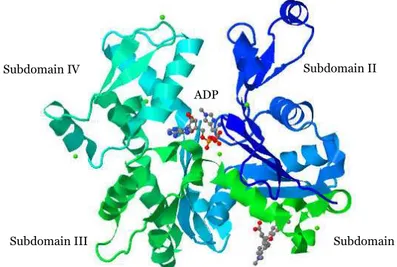
Actin consists of four subdomains, referred to as subdomains I, II, III, and IV (Figure 4). Subdomains I and II are separated from subdomains III and IV by a cleft, with a binding site for ATP and a divalent cations (Mg2+ or
Ca2+). Actin filament dynamics is regulated by a conformational change that
is coupled to ATP hydrolysis (Otterbein et al., 2001; Graceffa and Dominguez, 2003).
Figure 4. Ribbon representation of the structure of uncomplexed actin α−actin1 in the ADP state. PDB accession number: 1j6z.
Subdomain IV Subdomain II
Subdomain III Subdomain I
The actin molecule rapidly denatures in the absence of ATP and divalent cations (Valentin-Ranc and Carlier, 1991). Given the proper salt conditions (e.g. 1 mM MgCl2 and/or 50-100 mM KCl), monomeric actin polymerizes
into helical filaments (F-actin), while in low salt concentration, actin is monomeric.
All actin monomers self-assemble in a defined orientation such that the filaments have a defined directionality or polarity and their ends - labelled plus (barbed) and minus (pointed) ends – are distinguishable from one another. This polarity can be visualized by decorating actin filaments with myosin S1 fragments. Myosin fragments bind at a preferred angle creating the appearance of an arrowhead pattern, observed in electron micrographs (Huxley, 1963). Actin filaments can grow at either end, but the rate of growth is faster at the plus end than at the minus end. ATP-actin monomers are incorporated to the fast-growing plus end of the actin filaments; while the ADP-actin monomers are dissociated from the slow-growing minus end. This dynamic process is known as treadmilling (Pollard et al., 2000; Disanza et al., 2005).
Actin-binding proteins
In addition to actin’s self association as it assembles to form F-actin filaments, actin interacts with a multitude of proteins in the cell. Actin-binding proteins (ABP) are proteins that bind filamentous and/or monomericactin and regulate the architecture and motility of the cell (dos Remedios et al., 2003).
Actin filaments are nucleated by proteins, such as the Arp2/3 complex. According to the currently accepted model, the Arp 2/3 complex, consisting of two actin-related proteins (Arp2 and Arp3) and five smaller (Arc) proteins, polymerizes new actin filaments from the side of existing filaments, to form Y-branched networks (Winder and Ayscough, 2005). Contrary to the current dogma, a recent study by electron tomography (Urban et al., 2010) demonstrated that actin networks within protruding lamellipodia are formed by overlapping filaments. Although there is no proper evidence of branched arrays of actin filaments, the possibility that branching is required for lamellipodia formation can not be excluded.
Capping and severing proteins regulate the length of the actin filaments. Capping proteins such as gelsolin and tensin regulate the filament length by blocking the addition of new monomers to the plus end. In addition, gelsolin has the ability to sever actin filaments, thereby promoting their disassembly (Burtnick et al., 2004). Another important and well characterized protein
family that drives depolymerization is the actin depolymerization factor (ADF) and the cofilin family (Moon and Drubin, 1995). The ADF/cofilin plays a central role in actin turnover by depolymerizing filaments from their pointed ends (Carlier et al., 1997) . Like gelsolin, ADF/cofilin can also sever actin filaments. However unlike gelsolin, ADF/cofilin does not remain associated with the filaments after severing (McGough et al., 2003).
Actin filaments can be stabilized against spontaneous depolymerization by proteins like tropomyosins, which bind along the length of the filament (Winder and Ayscough, 2005).•
The rapid turnover of actin filaments, essential in motile cells, depends on the availability of actin monomers. Actin-monomer-sequestering proteins (i.e. proteins that bind monomeric actin and block filament formation) regulate the availability of monomeric actin for polymerization. By binding to actin monomers, profilins and β-thyomosins interfere with their incorporation into filaments (Perelroizen et al., 1996; Paavilainen et al., 2004).
Myosins are a large family of motor proteins that use actin filaments tracks. They are prime partners of actin in generating contractile structures in both non-muscle (Bresnick, 1999) and muscle cells (Huxley, 1998).
Proteins linking actin to other cytoskeletal elements and membrane complexes also belong to the actin–binding protein family. Some of these proteins, such as dystrophin, utrophin, talin and vinculin connect the actin cytoskeleton to membrane proteins, while others such as annexins can bind directly to membranes.
Actin-bundling and actin cross-linking proteins are responsible for the three-dimensional organization of actin filaments. In order to bind two different actin filaments, these proteins must either contain two actin-binding sites within their sequence or function in a multimeric form. Their different sizes and binding modes control the properties of the three-dimensional actin structures formed (Puius et al., 1998). Bundles and networks of actin serve as supporting mesh for the plasma membrane. In bundles the actin filaments are closely packed and aligned, whereas in networks they form angles of various degrees and are loosely packed. In spectrin, filamin and dystrophin, the two actin-binding domains are separated by a large flexible rod domain and therefore, they generally form actin networks. Bundles are primarily formed by small cross-linking proteins (such as fascin), or by proteins that have a shorter rod domain, like fimbrin and α-actinin.
α-actinin - the central focus of this thesis - plays a crucial role in three-dimensional arrangement of the actin filaments. The interaction between actin and α-actinin is rather complex. At high α-actinin concentrations actin bundles are formed, whereas at low concentration networks are formed. The binding affinity (Kd) of α-actinin for actin varies between species and ranges
from 0.6-4.7 μM (Wachsstock et al., 1993).
αααα
-actinin
α-actinin belongs to an ancient family of actin-binding proteins. Due to its ability to form antiparallel homodimers, α-actinin cross-links actin filaments into extended networks or bundles. α-actinin is conserved among organisms, but surprisingly, it is not present in neither plants nor baker’s yeast (Saccharomyces cerevisiae) (Virel and Backman, 2004).
α-actinin is the smallest member and the most probable ancestor of the spectrin superfamily. This family, besides α-actinin, includes spectrin, dystrophin and utrophin (Broderick and Winder, 2005). The hallmark of the spectrin superfamily is the presence of three structural domains: an N-terminal actin-binding domain (ABD) composed of two calponin homology (CH) domains, a central rod domain with a varying number of spectrin repeats (SR) and a C-terminal domain with EF-hand calcium binding motifs (Blanchard et al., 1989; Broderick and Winder, 2002; Otey and Carpen, 2004; Broderick and Winder, 2005). Phylogenetic analyses have indicated that the family members arose after several intragene duplications and rearrangements of a common ancestral α-actinin isoform (Thomas et al., 1997; Viel, 1999; Baines, 2003; Virel and Backman, 2004; Broderick and Winder, 2005). During the evolution of α-actinin, the rod domain was the domain that changed the most: as its length and composition varies considerably from lower to more complex organisms. The actin-binding domain and (in most cases) the C-terminal EF-hands have been conserved throughout evolution.
Although the sequence identity between repeats is low, the structure of the spectrin repeats is well conserved among the members of the spectrin superfamily (Djinovic-Carugo et al., 2002). As pairwise comparative analysis has shown, the differences between spectrin repeats within the same isoforms are larger than the differences between the same repeat in different organisms. Spectrin repeat 1 (SR1) and spectrin repeat 2 (SR2) of human α-actinin2, for instance, are only 13% identical. In contrast, SR1 of human α-actinin2 is 100% identical to SR1 of mouse α-actinin2 (Virel and Backman, 2007).
The number of spectrin repeats determines the length and the flexibility of the protein and therefore the nature of the three-dimensional organization of actin filaments. Vertebrate α-actinins contain four spectrin repeats, whereas dystrophin contains 24 repeats, β-spectrin 17 repeats and α-spectrin contains 20 (Broderick and Winder, 2005). In some lower organisms, like protozoa and fungi (Paper IV), the rod domain of α-actinin contains only one or two spectrin repeats (Virel and Backman, 2004). Up to the invertebrate-vertebrate bifurcation, organisms seemed to have a single, calcium-dependent α-actinin. After the split, invertebrates have kept this single isoform, in contrast to vertebrates that acquired four distinct isoforms.
Even though it was first purified from rabbit skeletal muscle, α-actinin is found in a large variety of tissues (Ebashi and Ebashi, 1965; Maruyama and
Ebashi, 1965). Depending on their tissue localization, expression patterns and sensitivity to calcium, α-actinin isoforms are classified into two distinct
groups: muscle (calcium insensitive) and non-muscle (calcium sensitive) isoforms (Burridge and Feramisco, 1981; Noegel et al., 1987; Blanchard et al., 1989; Otto, 1994). Non-muscle α-actinins are inhibited from binding to F-actin by micromolar concentration of Ca2+, while muscle α-actinins,
lacking the required residues for calcium coordination, are calcium insensitive (Burridge and Feramisco, 1981; Rosenberg et al., 1981).
αααα
-actinin in non-muscle cellsIn vertebrates four α-actinin genes (ACTN 1-4) have been identified. The non-muscle α-actinin isoforms, α-actinin1 and α-actinin4, both function as cytoskeletal proteins and are ubiquitously expressed. The calcium sensitivity of α-actinin1 and α-actinin4 regulates their ability to bind actin by decreasing binding affinity as calcium concentration increases (Noegel et al., 1987). α-actinin1 and α-actinin4 are both localized along actin stress fibers and focal-adhesion sites (Lazarides and Burridge, 1975; Youssoufian et al., 1990). α-actinin4, in contrast to α-actinin1, has also been shown to localize to the nucleus (Honda et al., 1998; Kumeta et al., 2010).
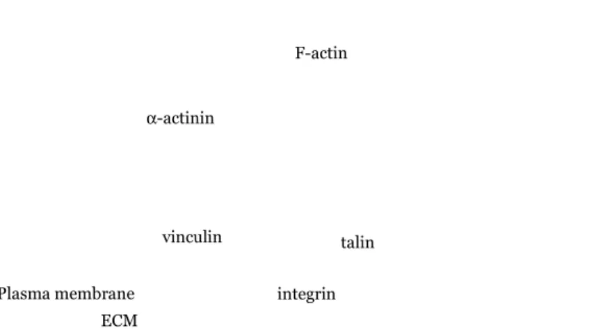
Focal adhesions are dynamic protein complexes through which the intracellular actin cytoskeleton connects to the extracellular matrix (ECM) outside the cell (Figure 5). At focal adhesion sites, non-muscle α-actinin links the cytoplasmic tails of β-integrin (Otey et al., 1990) to filamentous actin, but also interacts with other cytoskeletal proteins, such as vinculin (Belkin and Koteliansky, 1987), and talin (Gautel, 2010). The interactions between α−actinin and its molecular partners are generally mediated by the spectrin repeat region of α-actinin (Djinovic-Carugo et al., 2002).
Figure 5. Schematic representation of the focal adhesion site showing
α-actinin (green) and its interacting partners: vinculin (grey), talin (black) and β-integrin (light blue).
Stress fibers are highly ordered structures composed of bundles of aligned microfilaments and actin-associated proteins such as myosin, tropomyosin, titin and α-actinin, to name a few (Eilertsen et al., 1994; Katoh et al., 1998). α-actinin is localized at the dense bodies - the stress fiber analogues to the sarcomeric Z-disk structures of striated muscle – where it interacts with several dense region-associated molecules, including the PDZ and LIM domain-containing proteins (Vallenius et al., 2004; Klaavuniemi et al., 2009). Recent studies suggest that non-muscle α-actinins may be involved in the development and progression of different types of cancer, such as breast (Guvakova et al., 2002), colorectal (Honda et al., 2005), pancreatic (Kikuchi et al., 2008), and ovarian carcinomas (Yamamoto et al., 2009).
αααα
-actinin in muscle cellsThe striated (skeletal and cardiac) and smooth (visceral) muscle α-actinin isoforms, α-actinin2 and α-actinin3, are highly expressed in muscle, where they act as major structural components of the muscle contraction machinery at the Z-disk (Z-line) (Beggs et al., 1992). Layers of α-actinins cross-link the barbed ends of overlapping, antiparallel actin filaments. This results in the formation of a lattice-like structure that stabilizes the anchorage of the actin filaments at the Z-disk of striated muscle sarcomeres (Figure 6). ECM F-actin integrin α-actinin talin vinculin Plasma membrane
αααα
-actinin domain structure
Native α-actinin is composed of two monomers of approximately 100 kDa each, arranged in an antiparallel manner to form a rod-shaped molecule with an actin-binding domain at both ends (Endo and Masaki, 1982; Tang et al., 2001). The molecule has a high α-helical content and appears, in electron micrographs, to be a long rod 30-40 nm by 2-4.5 nm (Suzuki et al., 1976; Burridge and Feramisco, 1981). Each monomer contains an N-terminal ABD with two CH domains, a central rod domain with a varying number of spectrin repeats, and a C-terminal region containing EF-hand calcium binding motifs (Endo and Masaki, 1982; Blanchard et al., 1989). The number of spectrin repeats within the rod domain has changed during evolution. The rod domain of all known vertebrates consists of four spectrin repeats (SR1-SR4) (Figure 7). Primitive organisms such as the protozoa Trichomonas vaginalis and the yeast Schizosaccharomyces pombe have a rod domain with one or two spectrin repeats, respectively ((Wu et al., 2001; Virel and Backman, 2004, 2007) and Paper IV). The parasite Entamoeba histolytica expresses two different isoforms of α-actinin. Depending on the isoform the rod domain contains either one or two spectrin repeats (Virel and Backman, 2006) and Paper I, II and III).
Figure 7. Domain organization of vertebrate α-actinin homodimer. The actin-binding regions (ABD) are located at the N-termini of the antiparallel homodimer, followed by four spectrin repeats (SR) and the C-terminal calcium binding regions (EF).
The rod domain is not only important for the dimerization of the homodimers, but also has a role as a scaffold or docking site for many signalling proteins (Djinovic-Carugo et al., 2002).
At present, there is no atomic structure of the entire α-actinin molecule or of any other member of the spectrin superfamily. However, NMR and X-ray structures of its domain or those of homologues have been reported, allowing three-dimensional reconstructions, based on cryoEM, of the whole α-actinin molecule (Tang et al., 2001; Liu et al., 2004).
Actin-binding domain
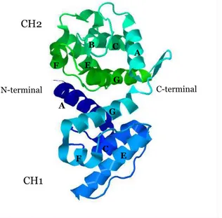
The actin-binding domain (ABD) of α-actinin isoforms consists of a type 1 and type 2 calponin homology domains (CH1 and CH2) in a tandem arrangement (Stradal et al., 1998; Gimona et al., 2002). The calponin homology domain gets its name from calponin - an actin-binding protein involved in the regulation of muscle contraction (Castresana and Saraste, 1995). Each CH domain is composed of six helical segments: two minor (B and F) and four major helices (A, C, E and G). The core of the domain is formed by the four principal helices (Franzot et al., 2005) (Figure 8).
Figure 8. Ribbon representation of the structure of human α-actinin3
actin-binding domain. PDB accession number: 1wku.
The two CH domains act cooperatively in conferring high-affinity binding to actin. CH1 has the intrinsic ability to bind F-actin albeit with lower affinity than the complete ABD. On the other hand, CH2 alone is not sufficient to bind filamentous actin (Way et al., 1992; Banuelos et al., 1998; Gimona and Mital, 1998). NMR, cross-linking, and mutational studies of various actin-binding domains from different proteins have led to the identification of three major actin-binding sites (ABS 1-3). ABS1 corresponds to the N-terminal A helix of CH1, ABS2 to the C-terminal G helix of CH1, and ABS3 corresponds to the interdomain link and the N-terminal A helix of CH2 (Bresnick et al., 1990; Bresnick et al., 1991; Hemmings et al., 1992; Levine et al., 1992). B G G E A C F E N-terminal A C F C-terminal CH2 CH1
Recently, the structures of ABD from human α-actinin isoform 1 (Borrego-Diaz et al., 2006), isoform 3 (Franzot et al., 2005) and isoform 4 (Lee et al., 2008) have been solved by X-ray crystallography. In all of these structures, CH1 and CH2 adopt a closed conformation with the C-terminal region of CH2 (with the helices F and G) packing against the helices A and G of CH1 (Sjöblom et al., 2008b; Sjöblom et al., 2008a). This compact conformation, characterized by extensive contacts between the two CH domains, is similar to the conformation observed in the crystal structure of the actin-binding domain of fimbrin (Goldsmith et al., 1997) and plectin (Garcia-Alvarez et al., 2003). By contrast, the structures of dystrophin (Keep et al., 1999) and utrophin (Norwood et al., 2000) show the ABD in an open conformation with the two CH domains separated by a long central helix.
In the 3D reconstructions of chicken smooth muscle α-actinin based on cryoEM, both open and closed conformations have been observed, suggesting two different actin-binding models - compact and extended (Liu et al., 2004). The compact model postulates that the ABD undergoes minor changes in order to interact with F-actin, whereas the extended model implies that the two CH domains become fully separated upon binding. In a recent study from 2010, Galkin et al. suggest not only that the two CH domains of α-actinin bind F-actin in an open conformation, but also that the opening of domains may be one of the regulatory mechanisms for proteins containing tandem CH domains (Galkin et al., 2010). This interpretation suggests that the opening of the CH1-CH2 interface is required to eliminate the steric clash caused by CH2, when binding to F-actin.
In muscle isoforms, the interaction between F-actin and α-actinin is regulated by the binding of phosphatidylinositol (4,5)-bisphosphate (PIP2) to the CH2 domain (Fukami et al., 1992; Fukami et al., 1996; Young and Gautel, 2000). In the closed conformation, the calcium-binding domain interacts with a region between the ABD and the SR1 of the opposite antiparallel monomer. The mechanism proposed by Young and Gautel (2000) implies that binding of PIP2 to ABD induces a conformational change that relieves this interaction and allows the binding of both F-actin (Fukami et al., 1992) and titin. The precise details of the mechanism still remain to be elucidated.
Rod domain
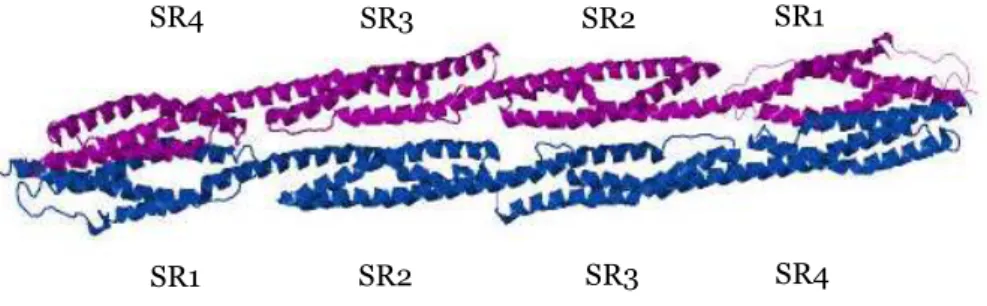
A flexible neck region connects the actin-binding domain to the central rod domain. The rod domain of α-actinin is generally composed of four spectrin repeats (SR) named for their similarity with the repetitive structures first identified in spectrin (Figure 9).
Figure 9. Ribbon representation of the structure of human α-actinin2 rod
domain. PDB accession number: 1HCI.
Spectrin repeats are left-handed antiparallel triple-helical coiled coil repeats (Pascual et al., 1997b) varying in size. They consist of 106 amino acid residues in spectrin (Speicher and Marchesi, 1984), 110 in dystrophin (Koenig et al., 1988) and 122 in α-actinin (Davison et al., 1989). The length of the rod domain determines the density and the flexibility of the cross-linked actin filaments – i.e. a short rod will create denser fibers.
Each individual spectrin repeat consists of three antiparallel α-helices (helices A, B and C) separated by two loops (loops AB and BC) (Pascual et al., 1996). Analysis of the primary sequence reveals a periodicity, a heptad pattern, of charged and hydrophobic amino acids. The residues forming the heptad pattern are named from a to g, with a and d representing the hydrophobic core of the repeat. The residues in position a and d, generally apolar, are packed along the axis of the repeat and are shielded from the aqueous environment. These hydrophobic residues are highly conserved (Pascual et al., 1997a; Djinovic-Carugo et al., 1999). Interhelix ionic interactions are stabilized by residues in position e and g, which are often charged (Parry et al., 1992; Grum et al., 1999). The heptad pattern has been suggested to promote stable folding of the single repeat (Parry et al., 1992).
The electrostatic potential of the dimer interface shows a charge gradient going from a slight positive N-terminal (SR1) to a more negative C-terminal
SR4 SR2 SR3 SR4 SR1 SR2 SR3 SR1
(SR4). Crystal structure of the complete rod domain in human skeletal muscle α-actinin has shown a left-handed twist of 90ο of the entire rod domain (Djinovic-Carugo et al., 1999; Ylänne et al., 2001a, b). As a consequence, the dimer interface is curved with an overall acidic concave face. The concave acidic surface may be involved in the association of the rod domain with other proteins and cytoplasmic structures (Ylänne et al., 2001a).
The structure of the spectrin repeats accounts for two vital characteristics of the α-actinin rod domain: structural rigidity and flexibility. α-actinin must be stable to withstand mechanical stress and be flexible in order to maintain binding in a dynamic environment. Hydrophobic interactions and salt-bridges within each repeat stabilize the secondary structure (Yan et al., 1993). Electrostatic interactions between the spectrin repeats and strong salt bridges between SR1 and SR4 on both monomers confer a highly stable dimer surface. The flexibility in the neck region coupled with the 90 degree twist along the axis of the dimer and the concave acidic surface may account for the α-actinin rod´s structural rigidity and bending flexibility (Sjöblom et al., 2008b; Golji et al., 2009).
Calcium-binding domain
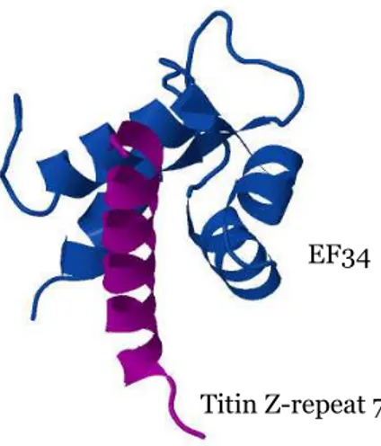
The C-terminal region of α-actinin contains two calcium-binding EF-hand motifs. The term “EF-hand” was coined in 1975 for the helix-loop-helix calcium-binding motif that was discovered in the crystal structure of parvalbumin (Moews and Kretsinger, 1975). Parvalbumin’s three calcium-binding domains were designated (from N-terminus to C-terminus) AB, CD and EF. The C-terminal (or EF domain) was the term first used to refer to the calmodulin (CaM) fold domain, hence the designation EF-hand or calmodulin-like domain. The EF-hand fold consists of two perpendicular α-helices and a calcium-binding loop, which usually contains 12 amino acid residues (Figure 10).
Calcium, when present, is coordinated by the side chains (mainly carboxylates or carbonyls) of six amino acid residues, whose positions are approximated by the vertices of an octahedron. The residues involved in Ca2+
coordination are labelled X, Y, Z, -X, -Y and -Z (Kretsinger, 1980; Nakayama and Kretsinger, 1994). Aspartic acid (Asp) is usually found in position X and glutamic acid (Glu) is often found in position –Z. Muscle α-actinin isoforms are calcium insensitive due to substitution in position Y (Kretsinger, 1980; Tang et al., 2001; Grabarek, 2006).
Figure 10. Ribbon diagram of the structure of EF34 from α-actinin2 (blue) in complex with titin Z-repeat 7 (purple). PDB accession number: 1H8B.
The binding of calcium induces a conformational change from an “closed” (the two helices are almost perpendicular) to a “open” (the two helices are almost antiparallel) conformation (Ikura, 1996). The reorientation of the helices leads to the exposure of a hydrophobic pocket on the surface of the protein, alowing the binding of different target partners.
Titin Z-repeat 7 EF34
αααα
-actinins of Entamoeba histolytica
The complete determination of the genomic sequence of several different organisms has led to the discovery of new α-actinin isoforms. The enteric protozoan parasite Entamoeba histolytica has two different α-actinin isoforms. α-actinin1 contains a single repeat (Virel and Backman, 2006) whereas α-actinin2 contains two putative repeats (Paper I, II and III). E. histolytica is the etiological agent of human amoebiasis - the third most common human parasitic disease after malaria and schistosomiasis. The World Health Organization estimates that E. histolytica causes 100,000 deaths per year, primarily in developing countries of Central and South America, Africa and Asia. To date, there is no approved vaccine against this pathogen (WHO/PAHO/UNESCO, 1997; Haque et al., 2003; Vanacova et al., 2003). Depending on the spread of the parasite, the clinical manifestation of parasite infection can vary from amoebic colitis to amoebic liver abscess and other extraintestinal lesions (Espinosa-Cantellano and Martinez-Palomo, 2000; Stanley, 2001, 2003). The sequencing of the entire E. histolytica genome has been one of the most important approaches to understand and characterize the mechanism and the proteins involved in the infection (Loftus et al., 2005). Microbial adhesion is often a first step during infection. In addition to extracellular cysteine proteases (Que and Reed, 2000), the attachment and penetration of E. histolytica into the host cells requires a Gal/GalNAc (galactose- and N-acetyl-D-galactosamine) lectin (Seigneur et al., 2005; Blazquez et al., 2007) as well as the reorganization of cytoskeletal and actin-binding proteins (Voigt and Guillen, 1999; Petri, 2002; Marion et al., 2004). Analysis of proteins associated with the Gal/GalNAc lectin has shown that α-actinin is one of the proteins attached to the lectin (Seigneur et al., 2005). In other protozoan such as Acanthamoeba (Mullins et al., 1998) or Trichomonas vaginalis (Addis et al., 1998) α-actinins are responsible for attaching the actin cytoskeleton to the plasma membrane and mediating morphological changes during infection. Therefore, it is tempting to suggest a similar role in E. histolytica.
Experimental techniques
Circular dichroism spectroscopy
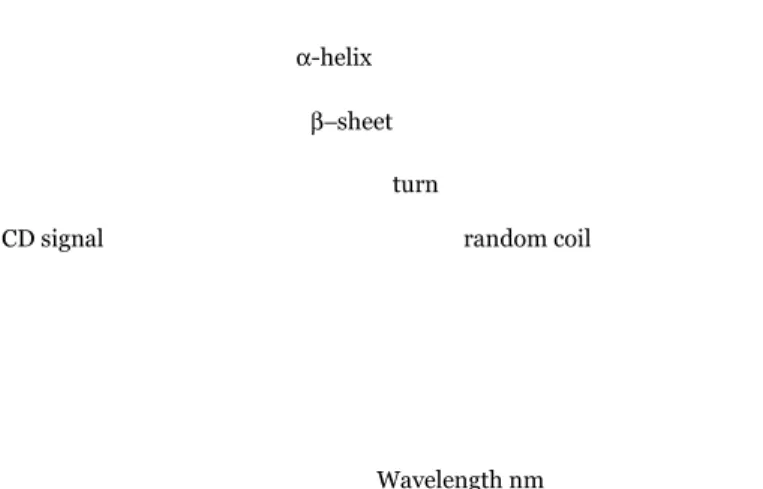
Circular dichroism (CD) spectroscopy measures the difference in absorbance of right- and left-circularly polarized light. The phenomenon of CD is sensitive to the secondary structure of polypeptides and proteins (Kelly et al., 2005; Whitmore and Wallace, 2008; Ranjbar and Gill, 2009). CD spectra, between 250 and approximately 190 nm, can be analyzed for the different secondary structural types: α-helix, parallel and antiparallel β-sheet, turn, and random coil (Figure 11).
Figure 11. Far-UV CD spectra characteristic of the four main protein
secondary structures.
In far-UV CD spectroscopy (190-250 nm), protein secondary structure and their amount can be determined. At these wavelengths the chromophore is the peptide bond, and the signal arises when it is located in a regular, folded environment. CD data in far-UV, or amide region, can be used to determine the relative amount of the secondary structure elements of a protein. In this region, a CD spectrum can be represented as a linear combination of CD spectra of each contributing secondary structure type (e.g. “pure” α-helix, “pure” β-strands etc.), weighted by abundance in the polypeptide conformation. In particular, α-helix displays a strong and characteristic CD spectrum in this region with prominent bands at 222 nm and 208 nm. The spectral contributions of the other elements of secondary structures are less well defined. CD signal Wavelength nm random coil α-helix β−sheet turn
Near-UV CD spectroscopy (250-350 nm) is dominated by contribution from aromatic residues and disulfides bonds in the polypeptide chain. The CD spectrum of a protein in this spectral region is usually sensitive to small changes in tertiary structure due, for instance to protein-protein interactions and/or changes in solvent conditions.
Since the CD signal is related to the structure, it is possible to determine whether a protein is folded or not, by monitoring the changes of the CD spectrum. One major advantage of using CD is given by the possibility to study conformational changes during, or because of, interaction with other entities, e.g. ligands, other proteins or nucleic acids. In addition, CD is a valuable technique for studying the conformational stability of a protein under stress (for example thermal stability, pH stability, and stability to denaturants), or how this stability is altered by buffer composition.
Actin co-sedimentation assay
The actin co-sedimentation assay is an in vitro assay used to analyze the actin-binding activity of proteins or protein domains. A low speed centrifugation (13,000 rpm, 15 min) is sufficient to pellet cross-linked actin filaments or actin bundles whereas high speed centrifugation (90,000 rpm, 60 min) is required to pellet actin filaments. The basic principle of the assay involves the incubation of the full length (or domain/s) of the protein of interest with F-actin, an ultracentrifugation step to pellet F-actin and the analysis of the protein co-sedimenting with F-actin. It is important to note that this is not really a quantitative assay for bundling, as it requires that large bundles of actin filaments to be formed and smaller bundles cannot be sedimented. However, it is still a useful means for estimation of extensive bundling.
X-ray crystallography
X-ray crystallography and nuclear magnetic resonance (NMR) are two of the most important techniques for elucidating the conformation of a protein and its function. Both techniques allow a resolution at the level of distinguishing individual atoms and have different advantages and disadvantages in terms of sample preparation and data collection and analysis. NMR experiments are limited to small molecules or molecular complexes. NMR is able to resolve the atomic structure of proteins with an upper molecular weight limit of approximately 25 kDa, whereas X-ray crystallography is more suitable for determining the structure of larger proteins.
X-ray crystallography can reveal the three-dimensional position of most atoms in a protein molecule; however it requires that all molecules are precisely oriented. The rate-limiting obstacle in x-ray crystallography is to obtain diffracting crystals of high quality. Some proteins crystallize readily, whereas others require specific conditions to be crystallized. Approximately two-third of all the proteins that enter crystallization trials will fail to generate protein crystals. In addition, only half of those that actually form crystals can be optimized to form suitable crystals that will allow structure determination. Thus, the estimated final success rate from a pure protein sample to its determined molecular structure is a mere 15% (Dong et al., 2007). Due to inter-domain flexibility, modular proteins like α-actinin are more problematic to crystallize. Being regarded as “evolutionary units”, the study of protein domains is often interesting by itself. Usually individual domains of a protein will crystallize better and produce better diffracting crystals relative to the corresponding full-length protein.
The success of crystallization depends on both the chemical purity of the protein and its homogeneity (uniformity of particle sizes). The purity of the protein sample is routinely assessed by SDS-PAGE. Several methods are available to assess protein homogeneity, such as gel filtration and dynamic light scattering (DLS).
Gel filtration or size exclusion chromatography
Gel filtration chromatography is a widely used technique, which separates molecules on the basis of both mass and shape. The principle of the technique is based on the fact that the space inside the gel particle (matrix) is available to smaller molecules but unavailable to larger molecules, which are therefore excluded. When a solution is applied to a properly packed gel column, only the external space between gel particles (void volume) is available to the excluded molecules. Therefore, larger molecules will have a smaller elution volume and elute first from the column. Molecules with partial access to the pores of the matrix will elute in order of decreasing size from the column.
Gel filtration is one of the most powerful and yet simplest methods to estimate the molecular weight of proteins. This method relies on the comparison of the elution profile (chromatogram) of the molecule of interest with the values obtained for several calibration protein standards of known molecular weight (Andrews, 1965). The greatest source of error in gel filtration comes from the requirement that the unknown protein should be similar in size and shape to the protein standards. Since the protein standards are usually globular proteins, the estimated molecular weight (i.e.
size) of rod shape proteins can be quite inaccurate due to their anomalous behavior (in the column). Another problem can be represented by proteins that are in complex with other molecules (e.g. detergents). Since it is the size (or rather Stokes radius) and not the molecular weight that is determined by this technique, the estimated molecular weight of detergent-solubilized proteins may be unreliable.
Dynamic light scattering (DLS)
Dynamic light scattering measures fluctuations in the intensity of light scattered by particles in solution as a function of time. If the molecule is stationary, the amount of light scattered would be constant. However, since all molecules in solution diffuse with Brownian motion, there are fluctuations in the scattered light intensity. The slower the particles diffuse, the slower the intensity will change and vice versa. Thus, the speed of these changes is directly related to the motion of the molecule. The intensity of the scattered light fluctuates at a rate that is dependent upon the size of the particles; the bigger the molecules, the slower they move. By measuring the time scale of light intensity fluctuations, DLS can provide information regarding the average size, size distribution, and oligomeric state and homogeneity (monomer, dimer, oligomer, or a mixture of these) of molecules and particles in solution. Investigating the homogeneity of a protein sample is important in determining optimal crystallization conditions ahead of X-ray crystallography studies of a protein (Baldwin et al., 1986; Price et al., 2009). Both conformational homogeneity and protein purity increase the chance of successful crystallization.
Aim of the study
From an evolutionary point of view, α-actinin is an extremely interesting protein family. This protein is considered to be the most probable ancestor of the spectrin superfamily, which dates back at least 500 million years (Muse et al., 1997). Gene duplication and alternative splicing are two of the most common mechanisms involved in the generation of protein functional diversity. Both mechanisms have played major roles in the evolution of the α-actinins, making this protein a valuable tool in learning about the processes that guide evolution.
Considerable knowledge about fruit fly (Drosophila melanogaster), slime mold (Dictyostelium discoideum), chicken (Gallus gallus) and human (Homo sapiens) α-actinins has accumulated over the years. And yet, very little is known about atypical α-actinins which have been found in earlier organisms, such as protozoa and fungi. To obtain a firmer understanding of the evolutionary history of α-actinin, the main objective of this study was to identify, purify and biochemically characterize atypical α-actinin or α-actinin-like proteins of the parasite E. histolytica and in the fungus S. pombe. Special attention was paid to E. histolytica α-actinin2; a protein that has been suggested to be involved in the parasite´s mechanism of infection in humans (Seigneur et al., 2005).
Summary of the papers
Characterization of Entamoeba histolytica αααα-actinin2 (Paper I, II
and III)
E. histolytica is considered to be one of the earliest diverged eukaryotes (Roger and Silberman, 2002). Within the E. histolytica genome two α-actinin genes have been identified (Loftus et al., 2005; Clark et al., 2007). These two genes code for α-actinin1 and α-actinin2 with molecular weights of 63 and 72 kDa, respectively.
The gene coding for E. histolytica α-actinin2, was cloned from a genomic library and then inserted into an expression vector (pET-TEV). This plasmid is well suited for the high-level protein expression that is required for further characterization of the protein. The subsequent expression and purification procedure that was undertaken gave a reasonably (>95%) pure preparation of α–actinin2.
Studies of the amino acid sequence by protein structure prediction and modelling revealed that the essential domains of a typical α-actinin are conserved in E. histolytica α-actinin2. However, the predicted intervening rod domain appears to be shorter compared to that the rod domain of vertebrate α-actinins. Instead of the typical four spectrin repeats, it contains only two. Phylogenetic analyses have shown that the rod domain of α-actinin of yeast and other fungi also contain only two spectrin repeats (Virel and Backman, 2007).
One of the key features of the α-actinin homodimer is its ability to cross-link actin filaments. Gel filtration was used to show that E. histolytica α-actinin2 is capable of forming dimers and therefore should be able to cross-link or bundle actin filaments.
The ability to bind and bundle actin in a calcium-sensitive manner was determinate by an actin co-sedimentation assay. E. histolytica α-actinin2 was pelleted with actin at low speed, implying bundle and/or network formation. The results also showed that calcium reduced the amount of co-pelleted E. histolytica α-actinin2, indicating a calcium-dependent interaction. The bundling and the calcium sensitivity data were also corroborated by negative stain electron microscopy (Figure 12).
Figure 12. F-actin bundles induced by Entamoeba histolytica α-actinin2. Actin was incubated alone (A) or with E. histolytica α-actinin2 in the absence of calcium (B) or presence of 1 mM calcium (C).
To gain insight into the secondary structure of E. histolytica α-actinin2, circular dichroism (CD) was used. The CD data indicated that the α-helical content of α-actinin2 is ~40%, which is in agreement with the estimated value obtained from the sequence. The results from these experiments showed that despite its shorter overall length, E. histolytica α-actinin2 displays all the characteristics of a typical α-actinin. This includes the ability to cross-link actin filaments in a calcium sensitive manner (Paper I).
It has been suggested that an α-actinin-like protein may be involved inthe morphological changes that occur during amoeboid movement and in phagocytosis (Seigneur et al., 2005; Blazquez et al., 2007). Both processes are critical for the invasive behaviour and virulence of the parasite (Guillen, 1996; Meza et al., 2006). In order to gain a more detailed insight into the structure and function of E. histolytica α-actinin2, we cloned and characterized the structural domains both individually and in combination with each other (Paper II). Gel filtration results showed that all constructs, with the exception of the actin-binding domain, formed dimers. Since the rod domain was the smallest of the constructs able to dimerize, it appeared likely that the E. histolytica α-actinin2 rod domain is responsible for formation of antiparallel dimers.
Previous studies have suggested that there is direct binding between E. histolytica α-actinin2 and the cytoplasmic domain of a lectin. This hypothesis was tested using a GFP (green fluorescent protein) fragment reassembly screen (details of this procedure can be found in Paper II). Although it is not possible to exclude the possibility of an indirect binding via another protein/s, our results suggested that there is no direct interaction between E. histolytica α-actinin2 and the lectin.
A key feature of α-actinin as well as of other members of the spectrin superfamily is the presence of spectrin repeats. The rod domain of this atypical α-actinin, characterized by the presence of only two spectrin repeats, was crystallized (Figure 13) and preliminary diffraction data collected (Paper III).
Figure 13. Crystal of the rod domain of Entamoeba histolytica α-actinin2. Attempts to use molecular replacement for structure determination were not successful due to the low sequence identity (<25%) of the two E. histolytica α-actinin2 spectrin repeats to that of solved structures of related proteins.
Characterization of another atypical
αααα
-actinin (Paper IV)In contrast to Saccharomyces cerevisiae (baker’s yeast), the genome of Schizosaccharomyces pombe (fission yeast) contains a gene coding for α-actinin. This isoform appears to be similar to E. histolytica α-actinin2 yet atypical to conventional α-actinin, as sequence analysis indicates that the rod domain is only long enough to encompass two spectrin repeats (Virel and Backman, 2004, 2007). In order to characterise this isoform, the DNA coding for S. pombe α-actinin was isolated from a genomic DNA library. The isolated gene contained two introns. These non-coding sequences (51 and 140 bp long respectively) were then removed by site-directed mutagenesis. DNA sequencing of the recombinant plasmid proved to be quite a difficult challenge. The results of the DNA sequencing contained DNA traces with mixed signals, noisy data with weak signals and multiple overlying peaks. We tried to identify the underlying cause of such poor DNA sequencing results by verifying that only one DNA template was present within the sequencing reaction. Additionally, we checked the template for possible multiple priming sites and reviewed the PCR conditions to be certain that only one single product would be amplified and with no mispriming products. When we finally succeeded to sequence the full-length gene, we discovered that there were seven point mutations and one deletion in the
nucleotide sequence. The sequence was then corrected by using site-directed mutagenesis. Thereafter, S. pombe α-actinin’s individual structural domains were cloned, purified and characterized.
Using gel filtration, DLS and transmission electron microscopy we have characterized S. pombe α-actinin. Similar to E. histolytica α-actinin2, gel filtration showed that S. pombe α-actinin forms dimers and that dimer formation requires the rod domain. These results were supported by transmission electron microscopy.
Concluding remarks
This thesis provides new insights into the evolutionary history of α-actinin ancestral isoforms. We have successfully identified, purified and biochemically characterized two atypical α-actinins from two simple organisms: the parasite E. histolytica and the fungus S. pombe. Even though these α-actinin isoforms have a shorter rod domain, our study showed that they are indeed genuine α-actinins.
Many large proteins, especially if they (like α-actinin) contain flexible parts have proven extremely difficult to crystallize at full length. No atomic structure of the entire α-actinin molecule or of any other member of the spectrin superfamily is so far available. We tried to crystallize each of the structural domains of both α-actinin isoforms and managed to crystallize the rod domain of E. histolytica α-actinin2. However, due to the low sequence identity of the two spectrin repeats to related proteins, our attempts to use molecular replacement for structure determination of the rod domain were not successful. Nonetheless, it would be extremely interesting to see if these repeats fold in the same way as typical spectrin repeats. We have therefore used seleno-methionine to label the E. histolytica α-actinin rod domain with the expectation to determine the structure by anomalous diffraction methods. The elucidation of the three-dimensional structure of this atypical α-actinin would greatly contribute to shed considerable light on the role it plays within the life cycle of this parasite.
With the completion of an ever increasing number of genomes, new α-actinin sequences have been available. Further studies on the characterization of α-actinin from lower organisms, such as protozoa, would not only extend the understanding of the evolution of the spectrin superfamily but also could help establish the root of the eukaryotic tree.
Appendix
Bioinformatics: terminology
"Understanding nature's mute but elegant language of living cells is the quest of modern molecular biology” (NCBI)
First there was in vivo biology then in vitro biology and now the discipline is moving towards in silico biology. Bioinformatics is a new discipline that has evolved in the past ten years. According to the National Center for Biotechnology Information (NCBI), bioinformatics is a very interdisciplinary, fast-growing scientific field in which biology, computer science, and information technology merge together into a single discipline. Bioinformatics deals with the analysis and interpretation of various types of data, including nucleotide and amino acid sequences, protein domains, and protein structures. In other words, bioinformatics is going 3D.
Some of the common terms used in the field of bioinformatics are listed below.
Alignment
The process of lining up and comparing two or more DNA or protein sequences in order to determine their degree of similarity and the possibility of homology between two or more genes or gene products.
BLAST
A computer program that searches for sequence similarities and homologies in different organisms. The acronym stands for Basic Local Alignment Search Tool.
Domain
A protein domain is an independently folding structural unit. Each domain has a distinctive function, secondary structure content and a hydrophobic core. Homologous domains with common functions usually show sequence similarities.
Homology
The significant degree of similarity in sequence of two or more gene or protein sequences that share a common evolutionary ancestor.
Motif
Sequence motifs are short conserved regions of a protein sequence alignment that usually correlate with a particular function. The motif can either be represented as a pattern or a profile.
Similarity
How a nucleic acid or protein is related to another. The extent of similarity between two sequences is based on the percentage of sequence identity and/or conservation.
Similarity (homology) search
Homology methods are used to identify genes or gene products that share a significant similarity and hence might give information on the ancestry and possible function of the query. The method is based on the detection of significant extended sequence similarity to a protein of know structure, or of a sequence pattern characteristic of a protein family. When the similarity is high and extended in length, the transfer of structure/function information to a potentially homologous protein is straightforward. When the similarity is weak or restricted to a short sequence the assessment of structural significance can be difficult.
Multiple sequence alignment
A representation of a set of sequences (protein, DNA or RNA) where equivalent residues are aligned. Multiple sequence alignment can provide evolutionary information about protein families by displaying which positions are crucial for maintaining the fold and function of the proteins of the investigated family.
Pattern
A fairly small set of residues that is unique to a family of proteins. Conserved sequence patterns in proteins usually have important biological functions and may be indicative of e.g., a protein structural domain, enzyme active site, or a binding site for another protein or metal ion.
Acknowledgements
“No man is an island, entire of itself;
every man is a piece of the continent, a part of the main”
(John Donne)Even though it was written almost four hundred years ago, the above statement is still true for this thesis. A large number of people helped me thrive during these years in Umeå. I would like to take this opportunity to thank all those people who stood by, helped and guided me in this incredible journey.
First and foremost I would like to show my sincerest gratitude to my supervisor, Lars Backman, who has supported me throughout the years with his patience, experience and knowledge whilst allowing me the room to work in my own way. Tack för den hjälp och stöd du gett mig genom åren. Tack också till Anna, jag har verkligen uppskattat er gästfrihet och all god mat!!! Grazie di cuore.
Special appreciation goes to Gerhard Gröbner, Uwe Sauer and Magnus
Wolf-Watz for constructive criticisms, valuable comments, suggestions and
fruitful scientific discussions.
All the co-authors for good collaboration and pleasant team-work.
Annelie, Jonas, Tina, Malin, Anna, Elin, Lisa tack så mycket för att ni
tog hand om mig när jag precis hade kommit till Sverige och det enda svenska ord jag kunde var “hej”!!!
All past and present PhD students and post-docs for making my life easier, both in the scientific field and outside. I could not have wished for better company at lunch breaks, late evenings at the department, after works, fika, conferences, dinners and weekends at work…
They say life is like a crossword puzzle. It is up to us to fill it in and once that page is done we're off to a new page in our life. Thank you for helping me to fill some of my pages…and now…let’s have a little bit of fun!!!
Across
1: the best personal trainer ever!!! 3: knight in shining armour 6: gorgeous bread baker 8: smiling cultural ambassador 10: administrative angel 11: checkmate!!!
12: origami expert 13: my Spanish idol 15: my Swedish lab teacher 17: the biker
18: sakura and koi fish 20: exotic Mate drinker 25: the ultimate gentleman 27: pratar du spanska? 28: yummy cheesecake 29: cioccolatino 30: el biologo 32: Totoro
33: gorgeous explosion of energy
Down
2: my rock climbing heroine 4: Clark Kent
5: Casanova 7: violin virtuoso
9: buy a train ticket…find love 14: HV71 number one fan 16: technology expert 19: the dancing queen 21: innebandy master 22: my partner in crime 23: snygg-Tobbe 24: radio lab
26: human encyclopaedia on plants 28: latte detergente
Harald one of my favourite gerrrrrman...äh Ösi ☺…for fixing my bathroom
(at least I had a hammer!!!)…for baking my birthday cakes…Vielen Dank.
Helder my big baby Helderino! Você é uma pessoa fantástica e merece tudo
de melhor que o futuro pode te oferecer.
Marcus…Marcus…Marcus…thank you…you made me want to become a
better person…just because you are who you are…
Ximena with a smile that can illuminate an entire room. Gracias por todos
los momentos que hemos compartido y sobre todo, amistad.
Miriam my lunch-gossip buddy with a passion for fashion and a heart of
gold.
Tania, who makes me feel at home, helps me with my error-prone Italian ☺
and always makes it fun to go to work. Non ti preoccupare… arriverà quello giusto…ne sono sicura!!!
Liudmila for your friendship, for the time in the “garden”, for taking care of
me…We will always have Paris…oops…Stockholm !!! I mean…Congo!
Peter for being you…for all the fun we had together…grazie.
Nat who always manages to put a smile on my face, even when I feel low.
Non so come avrei fatto senza di te in queste ultime settimane ☺
Gautam it’s been great to have such a good friend to share a passion for
books, music, movies, art, travel and food!!! I truly admire your neverending enthusiasm for science…don’t ever change.
Ana, Franziska and Kristina… the sisters I never had…thanks for always
being there for me…for listening…for caring…for all the crazy memories…for our trips…for the fun...for sharing my joy in the “ups” and giving me plenty of energy when I felt deflated in the “downs”… for being my family …vi voglio bene.
Thanks to my family for always being there when I need you. My brother,
Antonio, a great role model of strength and character. Thank you for giving
me the wings and teaching me how to fly. Non ti potro’ mai ringraziare abbastanza. Ti voglio bene fratellino.
I miei genitori, Rossella e Raffaele, per essere una presenza costante nella mia vita, per accettare i miei sogni e per far di tutto per aiutarmi a realizzarli. Per avermi insegnato il significato del duro lavoro, del rispetto, della perseveranza, di come essere indipendente. Non ci sono parole per esprimere tutta la mia gratitudine per tutto quello che avete fatto e continuate a fare per me. Vi voglio bene.