Faculty of Veterinary Medicine and Animal Science
Long term outcome and quality of life in
cats and dogs suffering from pelvic
fractures
Elin Orrenius
Uppsala 2019
Long term outcome and quality of life in
cats and dogs suffering from pelvic
fractures
Elin Orrenius
Supervisor: Annika Bergström, Department of Clinical Sciences Assistant Supervisor: Maria Dimopoulou, UDS
Examiner: Jens Häggström, Department of Clinical Sciences
Degree Project in Veterinary Medicine
Credits: 30
Level: Second cycle, A2E Course code: EX0869 Place of publication: Uppsala Year of publication: 2019
Online publication: https://stud.epsilon.slu.se
Key words: pelvic fracture, quality of life, conservative, surgical treatment, prognosis, questionnaire Nyckelord: bäckenfraktur, livskvalitet, konservativ, kirurgisk behandling, prognos, frågeformulär
Sveriges lantbruksuniversitet
Swedish University of Agricultural Sciences Faculty of Veterinary Medicine and Animal Science Department of Clinical Sciences
SUMMARY
Pelvic fractures are a common injury in cats and dogs, mostly due to hit by car or falling from heights. There are several components that determine what treatment is the best in each case. Literature and leading surgeons suggest fractures of the weight bearing axis (iliosacral joint (SI joint), ilium body and acetabulum) should be treated surgically, but there are few studies comparing surgical and conservative treatment. Fractures of the pelvic floor (os pubis, pelvic symphysis and os ischium) and fractures of the ilium wing are rarely treated surgically. This study aims to describe what fractures were the most common, what treatment was chosen and to evaluate long term prognosis and quality of life in dogs and cats after suffering from a pelvic fracture.
The study consists of review of patient records, owner-based questionnaires and a clinical part with long term follow up of clinical outcome. A total of 196 cats and dogs suffering from pelvic fractures during the years 2007 to 2017 were treated at the University animal hospital of the Swedish University of Agricultural Sciences. Questionnaires used were the Feline Musculo-skeletal and Pain Index (FMPI) and the ACVS Canine Orthopedic Index (COI), the result of each questionnaire was calculated into a percentage that was comparable between the questionnaires. Twenty-one cat owners and 16 dog owners answered the questionnaire. Thirteen cats and 11 dogs participated in the clinical study and were subjected to a thorough orthopedic and neurological examination.
Review of patient records showed that the most common fractures in cats were fractures of the SI joint and amongst dogs the most common fractures were fractures of the pelvic floor. Multiple fractures occurred more often than fractures in one or two sites in both cats and dogs. Treatment of pelvic fractures differed between cats and dogs, cats were euthanized to a greater extent than dogs due to their pelvic fracture. Dogs were more commonly treated surgically compared to cats. In cats 46,4% were treated conservatively, 9,3% were treated surgically and 44,4% were euthanized. In dogs 41,3% were treated conservatively, 39,1% were treated surgically and 19,6% were euthanized.
The questionnaire showed with statistical significance, and 95% certainty, that cats recover better and have a better quality of life than dogs after suffering from a pelvic fracture. 57,1% of the cats recovered completely and 6,9% of the dogs recovered completely, according to the questionnaires. The clinical examination showed that the most common complication to pelvic fracture was decreased range of motion in the hip joint. Lameness in one or both of the hind limbs occurred in 25% of the cats and dogs. None of those who had neurological deficits reported on initial clinical presentation had remaining neurological deficits at follow up examination, although, 16,7% of the dogs had neurological deficits at follow up examination. Unfortunately, the population was too small and heterogenous to make comparison and draw conclusions about whether surgical or conservative treatment is the ultimate treatment for the different types of pelvic fractures. Further studies are needed in the subject of treatment and long-term prognosis of pelvic fractures.
CONTENT
Introduction ... 1
Literature review ... 1
Healing process in skeleton and tendons ... 1
Basics in orthopedic implants... 1
Screws ... 1
Plates ... 2
Initial clinical presentation ... 2
Diagnostics ... 3 Initial treatment ... 3 Choice of treatment ... 3 Methods of treatment ... 4 Conservative treatment ... 4 Surgical treatment ... 5 Postoperative care ... 7
Complications and prognosis ... 7
Fractures of the SI joint and the ilium ... 8
Fractures of the acetabulum ... 8
Fractures of the os pubis and os ischium ... 8
Difference in recovery between surgical and conservative treatment ... 8
Material and methods ... 9
Review of patient records ... 9
Questionnaires ... 10 Questionnaire feline ... 10 Questionnaire canine ... 10 Clinical study ... 10 IT-security ... 11 Results ... 12
Review of patient records ... 12
Causes to pelvic fractures ... 12
Age, sex and breed related to pelvic fractures... 12
Clinical presentation ... 13
Type of fractures ... 14
Treatment of pelvic fractures ... 15
Questionnaires ... 20
ACVS Canine Orthopedic Index (COI) ... 21
Comparison FMPI and COI... 23
Clinical study ... 23
Comparison treatment method vs FMPI and COI vs clinical exam ... 24
Discussion ... 26
Etiology to pelvic fractures and initial clinical presentation ... 26
Treatment of pelvic fractures in cats and dogs ... 27
Quality of life and long-term prognosis ... 28
Sources of error ... 29 Future studies ... 30 Conclusions ... 31 Populärvetenskaplig sammanfattning ... 32 References ... 35 Appendix 1 ... 37 Appendix 2 ... 39 Appendix 3 ... 8
1 INTRODUCTION
Pelvic fractures are common injuries amongst dogs and cats, most often caused by hit by car accident, or falling from high heights. There are several ways to treat pelvic fractures described in veterinary literature, but there are few studies that describe long term follow up of patients after treatment.
The aim of this study was to estimate the quality of life, evaluate long term complications, describe the most common pelvic fracture types and how they were treated amongst cats and dogs. Moreover, the study compared treatment results between surgical- and conservative-treatment and compared the long term outcome between cats and dogs.
To achieve these goals the thesis consists of three parts - a review of patient records, an owner-based questionnaire and a clinical study. To better understand the treatment options described the thesis begins with a literature review on basic anatomy, healing processes, the typical patient and available treatment methods of pelvic fractures.
LITERATURE REVIEW
Healing process in skeleton and tendons
Fracture healing consists of three main phases: inflammation phase, regeneration phase and remodulation phase. Instantly after the injury a hematoma forms in the area of the fracture. The hematoma brings growth-factors and inflammatory cells. After 24 to 48 hours the regeneration phase starts with mesenchymal cells which later creates connective tissue, cartilage and, finally, bone. During this phase there is also a vascular growth, neovascularisation. After about 36 hours there is formation of woven bone, but there is no stability and it takes four to six weeks until callus forms and creates stability. The process of callus transforming into laminar bone takes several months to years (Zachary, 2012).
Tendons also heal in three phases: inflammation, cell- and matrix proliferation and remodeling with maturation. To avoid adhesions to surrounding tissue there should be some movement during the healing process. Movement during the healing process also increases the strength in the tendon. It takes a long time for a tendon to heal, and the phase of remodeling and maturation begins in approximately six to eight weeks past the injury (Zachary, 2012).
Basics in orthopedic implants
Screws and plates are often used in orthopedic surgery, including pelvic fracture surgery. To understand the different surgical methods used as treatment of pelvic fractures there is a need to understand the basic principles of screws and plates.
Screws
There are several types of screws depending on where they are used and what the purpose of the fixation is. There are screws used for cortical bone or cancellous bone. The screws used for cancellous bone have wider pitch, i.e. they are wider between the threads, than the screws used for cortical bone. Lag screws are used to create a pressure between two fracture fragments.
2
Different types of screws can work as a lag screw if inserted correctly. Some screws are self-tapping and some need manual self-tapping before inserting the screw. When the space is limited in the wound and there is a need to be certain that the screw is inserted correctly there is a screw with a hollow shaft within, where it is possible to use guiding instruments to insert the screw. These screws are called cannulated screws. A position screw is threaded all the way and prevents fragments from dislocating without creating pressure between the fragments. Position screws are often used in intraarticular fractures (Tobias & Johnston, 2012).
Screws can be used as the only fixation or together with a plate. When used together with a plate the screw can either create compression between the plate and bone due to lag, or lock onto the plate (Tobias & Johnston, 2012).
Depending on where the screw is placed and what type of movement it is supposed to prevent there is a large number of sizes of the screw, both in diameters and in length. Both screws and plates are made of 316L stainless steel or titanium (Tobias & Johnston, 2012).
Plates
There are many shapes and sizes of plates, depending on the purpose with the plate and where it is used. The size of the plate is based on the screw used and the total amount of holes. The most common plate is a dynamic compression plate (DCP), which creates a pressure between the plate and the bone. There is a similar plate that is called limited contact dynamic compression plate which spreads the pressure all over the plate and prevents the pressure to center around the screws. There are cuttable plates that can be altered in length to optimize the fit, although these plates are weaker than non-cuttable plates. There are also plates that are softer than regular plates and can be shaped during surgery to fit the specific needs. These are called reconstructive plates and are preferred on bones like the pelvis, where perfect fit is difficult if the shape cannot be altered to match the contour of the bones. Locking compression plates (LCP) have a different configuration of the holes which makes it possible for the screws to lock into the plate. This makes the fixation more stable. One disadvantage of the LCP is that the screw has to be in 90 degrees angle to the long axis of the plate to lock to the plate, which can be difficult to achieve in the pelvic area due to the irregular shape of the bones (Tobias & Johnston, 2012). String of pearls (SOP) is another plate that is suitable for fixating pelvic fractures. SOPs is a locking plate that can be rotated and bent to create the best fit (Orthomed, 2017).
Initial clinical presentation
The typical canine patient, with a pelvic fracture, shown in a previous study (Butterworth et al., 1994) is male and younger than four years old. Another study (Denny, 1978) showed that 53% of the dogs injured were females and 60% of the dogs were younger than two years old, but the peak incidence was two to three years of age. The most common breeds injured were Jack Russel Terriers and crossbreeds. The typical feline patient is 2 years old, male and domestic shorthaired (Langley-Hobbs et al., 2009).
3
According to literature research the most common cause of all pelvic fractures is being hit by car or falling from high heights (Côté, 2015), which is confirmed in most of the clinical studies of pelvic fractures.
When arriving to the clinic the patient is usually presented with lameness or paralysis in one, or both, hind limbs (Côté, 2015). In some cases, the patient can support the hind limbs and it might be difficult to palpate an instability in the pelvis when the animal is awake. After sedation it might be easier to palpate an instability (Fossum Welch, 2013). Palpation of the pelvis can result in crepitation and pain. Manipulation of the hind limbs is often painful as well. Rectal exam can reveal a malalignment in the pelvic canal (Côté, 2015).
Most patients, as high as 76%, with a pelvic fracture suffer from multiple pelvic fractures, which means more than 3 fracture locations in the pelvis (Messmer & Montavon, 2004). In a study from 1978 (Denny, 1978) only 6% of the injured dogs suffered from soft tissue injuries related to their pelvic fractures previous to treatment, for example, sciatic nerve paralysis, ruptured urethra or urinary bladder, and abdominal hernia.
Diagnostics
Diagnosis is determined by history, clinical exam and radiography or computed tomography (CT). Differential diagnoses are skeletal injuries in the spine and tail, injuries to the spinal cord, fractures in the hind legs or coxofemoral luxation (Côté, 2015).
A study performed 2015 in the United States (Stieger-Vanegas et al., 2015) compared the accuracy of using radiography versus CT when evaluating pelvic fractures. The result showed that if the person interpreting the images was educated in CT evaluation the accuracy was 100%. If the person interpreting the CT images was not educated in CT evaluation it takes longer time to evaluate the fractures. There was better accuracy in evaluating fractures in the os pubis and os ischium in CT than in radiography. In other sites of the pelvis the accuracy was equal between CT and radiography.
Initial treatment
Before beginning the treatment of the pelvic fracture, it is necessary to perform a neurological exam, and an evaluation of the urinary tract. Damage to the urinary bladder and to the urethra is associated with trauma to the pelvis (Fossum Welch, 2013).
Initial pain management is important. Avoid Non-Steroidal Anti-Inflammatory Drugs (NSAID) until the urinary tract and the status of the cardiovascular system are evaluated since NSAIDs are contraindicated when hypovolemia is present (Harasen, 2007).
Choice of treatment
Many components matter in the choice of treatment. The whole situation must be taken into consideration. Even if the fracture is treatable there might be other injuries that the animal might not recover from. If everything is considered and the fracture is treatable the choice is between conservative or surgical treatment. Which treatment method is most suitable from a medical
4
view will be discussed below, but there are other components besides from the injuries that are important as well in the choice of treatment.
Factors that matter in treatment of pelvic fractures are the age of the animal, weight of the animal, area of use of the animal and future demands on function (Innes & Butterworth, 1996). If it is an elderly animal there is a risk there are elderly changes in, for example, the liver and kidneys that increase the risk of anesthesia. In the case where anesthesia is not recommended conservative treatment might be a better option. Obesity might result in increased loading of the limbs, and therefore the fracture, during a conservative treatment.
The time since the injury also matters in deciding what treatment to choose. Surgical treatment is recommended within seven to ten days since the injury (Innes & Butterworth, 1996) but the optimal time range for surgical treatment is within 48 to 72 hours (Harasen, 2007). If the fracture is intraarticular the surgical treatment should be completed as soon as the animal is stabilized to avoid complications (Tobias & Johnston, 2012). The animal owner’s private economy is also a matter to be considered (Innes & Butterworth, 1996).
Methods of treatment
Conservative treatment
The pelvis is surrounded by large muscles that can stabilize eventual fractures which provides the option to treat these fractures conservatively (Harasen, 2007). Conservative treatment can be an option for cats or small dogs. Conservative treatment is not recommended for all pelvic fractures. Fractures that can successfully be treated conservatively are fractures in the os ischium, os pubis and os ilium cranially to the SI joint (Côté, 2015). The fracture must not be severely dislocated if conservative treatment is considered (Fossum Welch, 2013). A recent study (Meeson & Geddes, 2017) showed that only 26% of the cats with pelvic fractures were treated conservatively.
Initially conservative treatment consists of strict rest (cage rest) for three to four weeks. The
initial strict rest is followed by controlled movement until four to eight weeks has passed since the injury (Côté, 2015; Fossum Welch, 2013). A resting period of four weeks was evaluated in a previous study (Denny, 1978) where they also compared fractures of the ilium, SI luxation fractures and fractures of the acetabulum, which were treated conservatively to the same fractures treated surgically. Function of the hind limbs were evaluated by the animal owner through a questionnaire. Of the patients with ilium fractures treated conservatively 14 of 17 regained full function of the hind limbs according to the owner. Of the patients with SI luxation fractures five of six regained full function of the hind limbs with conservative treatment. Of the patients with acetabular fractures 10 of 17 regained full function of the hind limbs according to the owner with conservative treatment. Although, a surgical treatment of the fracture in acetabulum shortens the period of convalescence and decreases the risk of chronic osteo-arthritis.
In the literature, fractures of the acetabulum require surgical treatment to heal properly. Nevertheless - a review (Butterworth et al., 1994) of 34 patients with acetabular fractures
5
evaluated conservative treatment and 38% of these cases were treated conservatively. The result was evaluated by the owners in a questionnaire. All cases treated conservatively recovered successfully and 76% of those treated surgically recovered successfully. Successful treatment allowed mild intermittent lameness. There was a lack of information about recovery in some cases, which makes these figures uncertain.
Surgical treatment
Surgical treatment is often necessary when fractures cause a dislocation in the acetabulum, os ilium, SI joint or sacrum (Côté, 2015). These structures are involved in the weight bearing axis. A dislocation of the pelvis cannot occur without bilateral SI joint luxation and/or fractures in at least three different sites of the pelvis, where SI joint luxation is counted as a fracture. Surgical treatment often results in shorter period of convalescence (Fossum Welch, 2013). When SI joint luxation occurs the most common direction of displacement of the ilium is craniodorsally (DeCamp et al., 2016). In a previous study (Meeson & Geddes, 2017) 60% of the SI joint fractures, 82% of the ilial fractures, 58% of the acetabular fractures and 3% of the pubic fractures were treated surgically in cats.
Fractures of the SI joint
There are two open reduction approaches to the SI joint: dorsolateral approach or ventral approach. The dorsal approach can be used if there is a fracture of the acetabulum on the same side that also needs surgical fixation. The ventral approach can be used if there is a fracture in the ilium on the same side that needs surgical fixation. Fixation of the SI-joint is achieved by one or two lag screws through the body of ilium into the sacrum (DeCamp et al., 2016). In a review of six dogs that were treated surgically (Denny, 1978) the fracture was stabilized using lag screws and in two cases lag screws combined with pins. These patients regained full function in the fractured hind limb.
There is also a study reporting that closed reduction using lag screws is a considerable treatment option (Tomlinson et al., 1999). The surgical technique was based on IM pins, Kirschner wires and intraoperative fluoroscopy to identify the correct insertion site for the lag screw and to temporary stabilize the fracture during lag screw insertion. A single lag screw was used and were anchored in the sacrum. The technique gives less soft tissue trauma than an open approach. Only one dog of 13 examined suffered from complications such as screw loosening and persistent ischiatic nerve damage, but, this dog did not follow the post-operative regime. This approach allows earlier use of the limb than an open approach. A more recent study evaluating closed reduction approach (Tonks et al., 2008) shows the same result, but only radiographic follow-up was performed. Three of 24 dogs suffered from screw loosening, mostly as the result of osteomyelitis and were treated with antibiotics. All the 24 dogs had healed properly on follow up radiographs.
Fractures of the ilium
The open approach to the ilial body is the same as the ventrolateral approach to the SI joint. Fixation of the fracture can be achieved in several ways. The most common fixation is by plate.
6
What type of plate depends on the space available. Preferable is a six-holes straight plate, where one or several screws can be inserted into the sacrum to increase the strength of the fixation. If there is not enough space for two screws in the caudal segment a T-shaped, a L-shaped or a reconstruction plate might be used instead. To prevent narrowing of the pelvic canal due to the ilial fracture, the plate must be bent more concave than the usual shape of the pelvis (DeCamp et al., 2016).
Locking T-plates were evaluated in cats and small dogs (Scrimgeour et al., 2017) with the result of no case with screw loosening, compared to standard compression plate where 50% of the cases reported screw loosening.
DeCamp et al. (2016) describes two other techniques also used for ilial body fractures. The fracture can be fixated using lag screws. If the animal is too small and lag screws cannot be used, they can be substituted with pins and compression wire.
A study of cats with ilial fractures (Langley-Hobbs et al., 2009) also described a dorsal approach to the ilium. In both the lateral approach and the dorsal approach, a plate was used for fixation. These two approaches were compared in the study. The authors concluded that dorsal plating might result in lower the risk of narrowing of the pelvic canal. Dorsal plating also resulted in fewer implant-associated complications.
Fractures of the acetabulum
Open reduction and fixation are achieved by a dorsolateral approach to the hip joint. It might be necessary to perform an osteotomy of the greater trochanter to provide a proper view of the joint. Bone plates and screws tend to have the best result. Type of plate depends on the fracture, commonly used are straight-, reconstruction-, cuttable-, acetabular- and small fragment plates. Lag screws may also be used, depending on the type of fracture. If the animal is too small for a plate or lag screws, tension band wire in combination with pins and Kirschner wire can be used instead, but this is not as stable as lag screws or plates (DeCamp et al., 2016).
In a review of 14 dogs with fractures of the acetabulum (Denny, 1978) the fracture was reduced with two lag screws or a plate, depending on what part of the acetabulum that was injured. Lag screws were used if the fracture originated from the ilium and continued through the acetabulum.
If the acetabulum fracture cannot be reduced or fixated in a proper way and there is a high risk of osteoarthritis development, a femoral head and neck ostectomy can be considered. This is considered an acceptable treatment method for small dogs and cats. The surgery is performed through a craniolateral approach to the hip joint. Luxation of the hip joint is necessary to be able to proceed with the ostectomy of the femoral head and neck. After an ostectomy physical therapy should be started instantly to provide the best use of the limb and minimize post-surgical muscular atrophy. Known complications are persistent lameness, limb shortening, muscle atrophy, decreased range of motion and patellar luxation (Tobias & Johnston, 2012).
7
Other fractures of the pelvis
Fractures of the os ischium and os pubis are often stable without fixation if there are no other fractures, or if the other fracture involving the weight bearing axis is fixated and stable. These kinds of fractures rarely need surgical treatment (DeCamp et al., 2016).
A study of 10 cats (Kipfer & Montavon, 2011) showed successful outcome in surgical reduction of pelvic floor fractures (fracture of pelvic symphysis, os pubis or os ischium). The study suggests that in cases where a SI joint luxation is difficult to reduce it might help to stabilize the pelvic floor before reducing the SI joint luxation. Fixation of these fractures also reduce pain during recovery. Fixation can also be used to prevent narrowing of the pelvic canal.
Postoperative care
If the patient is non-ambulatory in the hind limbs post-surgery a schedule for turning the patient is necessary to prevent decubital ulcers (DeCamp et al., 2016).
After surgery the animal should be supported by abdominal support when moving and have restricted and controlled movement for one to two months (Côté, 2015; Fossum Welch, 2013), but other sources claim that the patient should have controlled movement which increases slowly during a period of three weeks (Fossum Welch, 2013). Early movement is better for healing, but to allow this the fracture must be completely stable and the exercise controlled. If the owner cannot follow the directions it is better to prevent the animal to use the injured side for one to two weeks initially, this can be achieved through using an Ehmer sling for example. If the patient has problem with adduction a sling to prevent abduction might be necessary for the first week (DeCamp et al., 2016).
During this period pain management using NSAID can be used if indicated (Côté, 2015) (Fossum Welch, 2013). During this period, it is also important to monitor defecation and treat with laxative if the patient have problems with defecation (Fossum Welch, 2013).
Complications and prognosis
There are complications associated with pelvic fractures overall, but there are also complications that are specific for where the fracture is located – these are described below. Possible complications to a pelvic fracture can be an uneven healing of the fracture if the fracture fragments are not well aligned and have not been fixated correctly. A malaligned healing could result in narrowing of the pelvic canal which can cause obstipation or dystocia. There might also be damage to the urethra, therefore it is very important to monitor the patients urinating behavior (Côté, 2015). Problems with defecation could also result from narrowing of the pelvic canal, soft tissue swelling or pain.
A gait analysis using a pressure-sensing walkway in dogs with pelvic fractures treated conservatively was performed after a minimum of four months after injury (Vassalo et al., 2015). The study showed that 73,3% of the dogs had a visual lameness and 66,7% of the dogs had an abnormal weight distribution between the limbs or showed kinetic changes. Thirty-three
8
percent of the dogs showed signs of pain or restricted movement when passively extending the hip joint.
Fractures of the SI joint and the ilium
The femoral nerve and the sciatic nerve are located near the SI joint and the nerves might be affected by a fracture (Fossum Welch, 2013). One study of peripheral nerve injuries in patients with pelvic fractures (Jacobson & Schrader, 1987) showed that the most common fractures associated with peripheral nerve injuries were ilial fractures with displacement of the fragments and luxation of the SI joint. Persistent neurological deficit in the caudal part of the body is a known complication, likewise, is persistent lameness in the hind limbs (Côté, 2015). Neuro-logical deficits might occur when a fracture in the sacrum crosses the canalis vertebralis or crosses the sacral foramina (Fossum Welch, 2013).
In a review of 11 dogs with ilium fractures treated surgically (Denny, 1978), 10 dogs regained full function of the hind limbs. The 11th dog suffered from persistent peroneal nerve paralysis.
In a study of 10 cats with ilial fractures (Langley-Hobbs et al., 2009) only one cat had persistent neurologic deficits. In a more recent study (Meeson & Geddes, 2017) 23% of the cats had neurological deficits at initial presentation, but 79% of these had regained full neurological function after six months.
Fractures of the acetabulum
If the acetabulum is fractured there is a known risk of development of osteoarthritis due to incongruence in the joint. In a study from 2015 (Vassalo et al., 2015) all dogs with intraarticular fractures had radiological signs of osteoarthritis.
In a review of 14 dogs with acetabular fractures treated surgically (Denny, 1978), nine regained full function of the hind limbs. Those who did not regain full function suffered from intermittent lameness and persistent sciatic nerve paralysis.
Fractures of the os pubis and os ischium
If os pubis is fractured there is a risk of abdominal hernia if the fragments are dislocated or due to an avulsion in the tendons attaching to os pubis (Côté, 2015).
Difference in recovery between surgical and conservative treatment
In an old review by Denny (1978), 123 dogs with pelvic fractures were evaluated. There was a small difference in recovery in patients treated surgically compared to those treated conservatively. Seventy-eight percent of the patients treated surgically had a full recovery, versus 75% of the patients treated conservatively had a full recovery.
9 MATERIAL AND METHODS
The thesis is based on three different parts: a review of patient records, owner questionnaires about pain and life quality after pelvic fracture and a clinical study. These three parts are described in detail below.
Review of patient records
Review of patient records consists of a retrospective study of patients treated for pelvic fractures at the University animal hospital, Uppsala, between the years 2007 to 2017. Data was collected containing information on what type of fracture, the cause of the fractures, to what extent animals are euthanized because of the fractures, other injuries associated with pelvic fractures and the treatment method. Based on the journal studies patients were selected to participate in the questionnaires and the clinical study.
When collecting information about the type of fractures that occur the fractures were classified depending on the area of the pelvis that were fractured, but also if it was a unilateral fracture or a bilateral fracture. The areas were: ilium wing, ilium body, SI joint, acetabulum, os pubis, pelvic symphysis and os ischium. Fractures of the sacrum were not included in the study, but all fractures and soft tissue injuries in the patient presented with the pelvic fracture were reported as well.
Inclusion criterium to participate in the questionnaire and the clinical study:
- The patient is a dog or a cat.
- The patient suffered from pelvic fracture in the time between the years 2007 to 2017 and was treated at the University animal hospital in Uppsala.
- Diagnostic imaging of the pelvis was performed at the University animal hospital in Uppsala on the initial visit.
- The patient has not had any leg amputated and has not had a femoral head and neck ostectomy. These procedures change the normal pattern of movements, and the gait can therefore not be analyzed to determine if there is a change in movement because of the pelvic fracture.
- Cats younger than 15 years, small dogs younger than 13 years and large dogs younger than 10 years were included in the owner questionnaires and a clinical follow up study. - Contact details to the owner of the animal are available and the owner does not have
protected contact details.
Twenty-three dogs and 59 cats fulfilled the inclusion criteria. The owners were sent a letter including information about the study and were asked to answer the questionnaire and participate in the clinical study. The letters are found in Appendix 1. Out of 23 dogs 12 owners decided to participate in both the questionnaire and the clinical study, and four decided to only participate in the questionnaire. Out of 60 cats, 13 owners decided to participate in both the questionnaire and the clinical study. Eight cat owners only participated in the questionnaire and declined the invitation to the clinical study. Eight cat owners reported their cat dead or missing and could not answer the FMPI. These eight cat owners evaluated the result of the treatment outside the FMPI.
10 Questionnaires
Part two of the thesis consists of owner-based questionnaires. To participate in this part of the study the patients needed to fulfill the inclusion criteria’s stated above. The questionnaires provided information about the animal’s quality of life after treatment of a pelvic fracture, based on the owner’s experiences. The questionnaires also provided information about complications and how common these are in the long term. The questionnaires used are found in Appendix 2. Dogs and cats are different in their behavior, in their movement, and how they live. Therefore, there are two different questionnaires, one directed to dog owners and one directed to cat owners. Both the questionnaires are validated. The result of the questionnaires was translated into values which are comparable between the cats and dogs.
The American College of Veterinary Surgeons Canine Orthopedic Index has been validated by Brown as described, translated and validated in Swedish by Andersson and Bergström (2019). The questionnaire used for the cats is called Feline Musculoskeletal Pain Index (FMPI) and is created by Dr D. Lascelles as described and translated by Sarah Stadig in her doctoral thesis “Evaluation of physical dysfunction in cats with naturally occurring osteoarthritis” (2017).
Questionnaire feline
Maximum score was 85, and the higher the score the poorer quality of life after the pelvic fracture. Total score of 17 means the patient recovered completely from the pelvic fracture and had the best quality of life possible. Quality of life (QOL) was scored outside the total score. Zero equals good quality of life. Maximum score in QOL is 3 and equals poor quality of life. If a question was not answered the total score was reduced with four points per question not answered. A relative score was calculated in percent, the lower score the better result. In the FMPI the relative score was the result of the total score and the QOL score combined. Relative score 20,0% equals normal life, these figures are comparable between the FMPI and the canine orthopedic index (COI) questionnaire. A score lower than 20,0% equals better than normal in some questions.
Questionnaire canine
Maximum score was 80 and 16 was the lowest score possible if all questions were answered. If a question was not answered the total score was reduced with four points per question not answered. Low scores equal a life with low levels of stiffness, pain, problems with movement and in function, and a good quality of life. The lowest relative score was 20% and indicates that there was no stiffness, decreased function, impairment in movement and equals good quality of life.
Clinical study
To proceed to clinical study the participant needed to fulfill the inclusion criteria’s and answer the questionnaire. The clinical study consisted of a general health exam, orthopedic exam and neurological examination of the hind limbs. The examination sought to determine if the patient had regained full function in the hind limbs after treatment, or if the patient suffered from
11
orthopedic or neurological disabilities due to the injury. Description of the clinical examination is found in Appendix 3.
The patient received a grade from 0 to 3, where 0 equals normal, 1 equals mildly abnormal, 2 equals moderately abnormal and 3 equals severely abnormal in the areas examined (these areas are found in Appendix 3). The total score estimated how well the animal recovered after the pelvic fracture. The same grading system was used on both cats and dogs. If the patient suffered from other injuries or diseases, for example osteoarthritis, it was considered when interpreting the result.
IT-security
According to GDPR the participants need to be informed about IT-security. In the survey the owners could choose to not share their phone number or email address, and they were informed about what the information was needed for. The information was collected to be able to contact the owners for a follow up examination. Before the follow up examination the owners received information about IT-security as follows in Swedish:
“Persondatan som samlats in syftar till att kunna länka patienten i fråga till rätt journal och för att kunna kalla patienten till ett återbesök. När studien är klar raderas all information berörande personuppgifter. Inga uppgifter kommer lämnas ut i den slutgiltiga rapporten som kan länka patienten till dess ägare”.
12 RESULTS
Review of patient records
There were 196 cases diagnosed with pelvic fractures at the University animal hospital at the Swedish University of Agriculture Sciences between the years 2007 to 2017. Out of these 196 cases 150 were cats and 46 were dogs.
- 58 cats matched the inclusion criteria’s for participating in the questionnaires.
- 92 cats did not match all the inclusion criteria’s and were not offered to participate in the questionnaires and the clinical study.
- 23 dogs matched the inclusion criteria’s to be participating in the questionnaires. - 23 dogs did not match all the inclusion criteria’s and were not offered to participate in
the questionnaires and the clinical study.
Causes to pelvic fractures
The most common cause of pelvic fractures was the patient being hit by a motor vehicle, for example a car, a motorcycle or a train, and the cause in 97 patients out of 196. There was lack of information in many cases, for example cats disappeared and came back injured a couple of days later. The owners often thought the cause of the injury was hit by car on a nearby road. Other common reasons to pelvic fractures were falling from heights or injured by another animal (for example dog, horse or wild boar). See detailed proportions in Table 1.
Table 1. Cause of pelvic fractures in cats and dogs
Cause of pelvic fracture Cats (%) Dogs (%) Total (%)
Unknown 74 (49,3) 2 (4,3) 76 (38,8)
Hit by motor vehicle 63 (42,0) 34 (73,4) 97 (49,5)
Falling from height 9 (6,0) 2 (4,3) 11 (5,6)
Injured by animal 0 5 (10,9) 5 (2,6)
Pathologic fracture 3 (2,0) 0 3 (1,5)
Jumped over fence 0 1 (2,2) 1 (0,5)
Item falling on the animal 1 (0,7) 1 (2,2) 2 (1,0)
Traffic road accident 0 1 (2,2) 1 (0,5)
Total 150 (100) 46 (100) 196 (100)
Age, sex and breed related to pelvic fractures
Median age amongst the cats was two years of age and 51,3% of the cats injured were zero to two years of age. The most common age was one-year old cats. 32% of the cats were between three to six years old and only 6,7% were older than 10 years of age. The oldest cat presented with a pelvic fracture was 17 years of age.
Median age amongst the dogs was two years old and 60,9% of the dogs injured were zero to two years of age. 30,4% of the dogs were between three to six years old and only 4,3% were older than 10 years of age. The oldest dog presented with a pelvic fracture was 13 years of age. Detailed distribution is shown in Figure 1.
13
Figure 1. Ages amongst cats and dogs presented with a pelvic fracture.
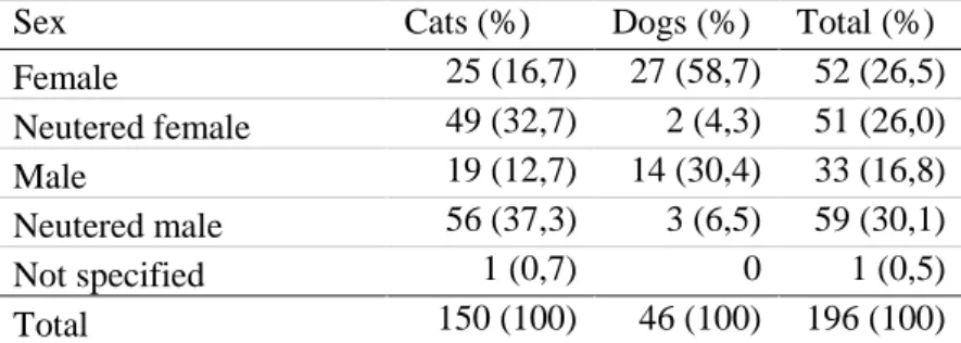
Table 2 describes the distribution between the sexes.
Table 2. Sex of the cats and dogs that suffered from pelvic fractures
Sex Cats (%) Dogs (%) Total (%)
Female 25 (16,7) 27 (58,7) 52 (26,5) Neutered female 49 (32,7) 2 (4,3) 51 (26,0) Male 19 (12,7) 14 (30,4) 33 (16,8) Neutered male 56 (37,3) 3 (6,5) 59 (30,1) Not specified 1 (0,7) 0 1 (0,5) Total 150 (100) 46 (100) 196 (100)
The most common cat breed with a pelvic fracture was the domestic shorthaired or longhaired cat (84,7%). Only 15,3% of the cats were pure bred and the most common breeds were Norwegian forest cat, Abessiner and Cornish Rex. Other injured breeds were Birma cat, Siamese, Maine Coon, Ocicat, Siberian cat, Sphynx, Bengal and Ragdoll.
The most common dog breed with pelvic fractures was the mixed breed (26,1%). Jack Russel Terrier was presented with 13,0%, followed by Dachshund (8,7%) and German shepherd (6,5%). Other injured breeds were Basset fauve de Bretagne, Golden retriever, Miniature poodle, Poodle, Cairn terrier, Norwegian moose dog, Danish-Swedish yard dog, Mittelspitz, Collie, Bichon frisé, Swedish white moose dog, Border collie, Australian kelpie, Vorsteh, Papillon, Basenji and Chihuahua.
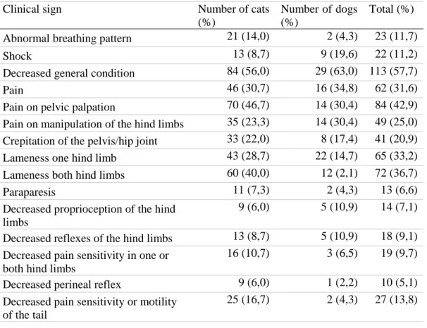
Clinical presentation
On arrival to the clinic a clinical examination was performed. Reported clinical signs were noted in the patient’s record. Table 3 shows the clinical signs associated with initial arrival to the clinic of cats and dogs with pelvic fractures. The most commonly occurring clinical signs were decreased general condition and pain on palpation of the pelvis. Lameness in both hind limbs were slightly more common than lameness in one hind limb, although almost a quarter (23,5%) of the cats and dogs that did not show any signs of lameness.
0 5 10 15 20 25 30 <1 1 2 3 4 5 6 7 8 9 >10 Dog Cat Number of patients Years of age
14
Table 3. Clinical signs of cats and dogs presented with pelvic fractures
Clinical sign Number of cats
(%)
Number of dogs (%)
Total (%) Abnormal breathing pattern 21 (14,0) 2 (4,3) 23 (11,7)
Shock 13 (8,7) 9 (19,6) 22 (11,2)
Decreased general condition 84 (56,0) 29 (63,0) 113 (57,7)
Pain 46 (30,7) 16 (34,8) 62 (31,6)
Pain on pelvic palpation 70 (46,7) 14 (30,4) 84 (42,9) Pain on manipulation of the hind limbs 35 (23,3) 14 (30,4) 49 (25,0) Crepitation of the pelvis/hip joint 33 (22,0) 8 (17,4) 41 (20,9)
Lameness one hind limb 43 (28,7) 22 (14,7) 65 (33,2)
Lameness both hind limbs 60 (40,0) 12 (2,1) 72 (36,7)
Paraparesis 11 (7,3) 2 (4,3) 13 (6,6)
Decreased proprioception of the hind limbs
9 (6,0) 5 (10,9) 14 (7,1) Decreased reflexes of the hind limbs 13 (8,7) 5 (10,9) 18 (9,1) Decreased pain sensitivity in one or
both hind limbs
16 (10,7) 3 (6,5) 19 (9,7)
Decreased perineal reflex 9 (6,0) 1 (2,2) 10 (5,1)
Decreased pain sensitivity or motility of the tail
25 (16,7) 2 (4,3) 27 (13,8)
Other orthopedic injuries presented at the same time as the pelvic fractures amongst cats were 26 cases of fractures of the hind limbs, 18 cases of fractures of the sacrum and 11 cases of fractured tails. Other less common orthopedic injuries presented at the same time as the pelvic fracture in cats were fractured ribs, fractures of the spine, ruptured cruciate ligament and fractured front limbs.
In dogs, common orthopedic injuries presented at the same time as the pelvic fracture were fractures of the spine, sacrum and hind limbs. Other concurrent injuries were fractures of the ribs and rupture of cruciate ligament or collateral ligaments.
Soft tissue injuries presented at the same time as the pelvic fracture were almost the same in cats and dogs. The most common soft tissue injuries were skin wounds, trauma to the thorax (i.e. pneumothorax or bleeding in the lung) and bleedings, both internal and external. Only three cats and two dogs were presented with abdominal wall hernia and one cat presented with a diaphragmatic hernia. Seven cats had either urinary incontinence or urinary bladder atony, but only two of these had suspicion of a ruptured urinary bladder. One cat and one dog also had nerve damage in one of the front limbs.
Type of fractures
The most commonly occurring fractures amongst cats were SI joint luxation followed by fractures of the pelvic floor. Amongst dogs the most commonly occurring fracture were fractures of os pubis followed by fractures of the ischium. Amongst fractures involving
weight-15
bearing axis fractures of the ilium body was the most common type in dogs. In dogs 44,8% of the fractures were left sided and 31,2% were right sided. Cats had almost the same amounts of fractures in either side of the pelvis, 38,2% right sided and 32,9% left sided. Bilateral fractures were less common than unilateral fractures, 16,2% in cats and 19,2% in dogs. Two cats were diagnosed with pelvic fracture on clinical examination and then euthanized, therefore, no further information on fracture type was available. Detailed distribution is showed in Table 4.
Table 4. Type of pelvic fractures in cats and dogs
Fractured area Unilateral Sinister Dexter Bilateral Total (%) Cat Dog Cat Dog Cat Dog Cat Dog Cat Dog Os pubis 76 29 32 17 36 12 24 10 100 (67,1) 39 (84,8) Os ischium 90 27 42 17 40 10 12 9 102 (68,5) 36 (78,3) Pelvic symphysis 28 4 - - - 28 (18,8) 4 (8,7) Ilium wing 7 3 1 1 6 2 0 1 7 (4,7) 4 (8,7) Ilium body 43 17 22 12 22 4 2 0 46 (30,9) 17 (37,0) SI joint 75 9 32 3 40 5 30 4 107 (71,8) 13 (28,3) Acetabulum 27 12 9 6 16 6 2 0 29 (19,5) 12 (26,1)
One cat was counted twice due to suffering from pelvic fractures twice and the two cats with no radiological diagnosis were excluded (149 cats in total).
In cats 75,8% had three or more fractures of the pelvis and in dogs 69,6% had three or more fractures of the pelvis.
SI joint luxation was recorded in 71,8% of the cats and 28,3% of the dogs (Table 5). The most commonly occurring direction of the luxation in cat was cranial luxation (81,6%). Other directions reported in cats were craniolateral (5,7%), craniomedial (2,3%), cranioventral (2,3%), caudal (2,3%), caudodorsal (1,1%), dorsal (1,1%), ventrolateral (1,1%), caudolateral (1,1%) and craniodorsal (1,1%). In dogs the most common direction of the luxation was, like in cats, cranial luxation (81,8%). Other directions of SI luxation in dogs were craniodorsal luxation (9,1%) and caudal luxation (9,1%).
Treatment of pelvic fractures
Between 2007 and 2017, 150 cats were presented with pelvic fractures, one of these cats suffered from pelvic fractures twice and therefore the following numbers count 151 cats. Of these 151 cats 70 were treated conservatively and 14 were treated surgically. Some of the cats treated conservatively had surgery to the tail or limbs due to other concurrent injuries. Sixty-seven cats died or were euthanized due to their pelvic fracture. One cat had no medical record else than the radiological evaluation and therefore there was no information about treatment. In the same period 46 dogs were presented with pelvic fractures. Of these 46 dogs 19 dogs were treated conservatively and 18 dogs were treated surgically. Nine dogs were euthanized due to their pelvic fracture.
16
Euthanasia related to pelvic fractures
Seventy-six animals were euthanized or died due to their pelvic fractures. In 15 of the cases studied the reason for euthanasia was not stated in the patient record. The most common reason for euthanasia in both cats and dogs was related to poor prognosis for recovery. Figure 2 below describes the causes of euthanasia in patients with pelvic fractures.
Sixty-five cats of total 151 cats suffering from pelvic fractures were euthanized. The most common reason for euthanasia was poor prognosis (40,0%) and the second most commonly known reason for euthanasia due to pelvic fractures in cats was due to the owner’s economy (15,4%). There were four reasons for euthanizing cats with a pelvic fracture that did not occur amongst the dog population: the owner declined surgery or amputation, due to factors concerning the owner’s current living situation, due to animal welfare or due to concern that the animal was too old to undergo treatment. Two additional cats died during treatment and could not be saved.
In total there were nine dogs that were euthanized out of total 46 dogs suffering from pelvic fractures. The most common reasons for dogs to be euthanized were poor prognosis (33,3%) and that the dog could not go through rehabilitation and get back to its normal activities, such as hunting (22,2%). Only one dog was euthanized because of the owner’s economy (11,1%). No dog died during treatment and none were euthanized because of animal welfare considerations.
Figure 2. Reasons for euthanasia due to pelvic fractures in cats and dogs.
Conservative treatment
During the stated period of time 70 cats out of 151 were treated conservatively. Figure 3 combined with Table 5 describes the fracture combinations that were treated conservatively. In cats 100% of the ilial wing fractures were treated conservatively. Ilium body fractures were treated conservatively in 85,7% of the cases. SI joint luxation were treated conservatively in 93,2% of the cases. Acetabulum fractures were treated conservatively in 25,0% of the cases. Fractures of os pubis, the symphysis and os ischium were treated conservatively in 100% of the cases. 20% 38% 14% 4% 2% 3% 4% 8% 4% 3% Unknown Poor prognosis Economy Concurrent diseases Age
Died during treatment Animal welfare
Rehabilitation impossible/can not work as before Owner declined surgery
17
Figure 3. Diagnosed fracture types treated conservatively in cats.
During the same period of time 19 dogs out of 46 were treated conservatively. Figure 4 combined with Table 6 describes the fracture combinations that were treated conservatively. Fractures of the ilial wing were treated conservatively in 100% of the cases. Fractures of the ilium body were treated conservatively in 46,2% of the cases. Fractures of the SI joint were treated conservatively in 60,0% of the cases. Fractures of the acetabulum were treated conser-vatively in 20,0% of the cases. Fractures of the os pubis, symphysis and os ischium were treated conservatively in 100% of the cases.
Figure 4. Diagnosed fracture types treated conservatively in dogs.
Surgical method used in the cases treated surgically
During the stated period of time 14 of 151 (9,3%) cats had pelvic fractures that were treated surgically. Table 5 describes the fractures in cats and the surgical method used. During the same period of time 18 of 46 (39,1%) dogs had pelvic fractures that were treated surgically. Table 6 describes the fractures in dogs and what surgical method that was used. Fractures of the ilium wing, os pubis, pelvic symphysis and os ischium were not treated surgically in any case during this period. 0 5 10 15 20
Ilium wing Ilium body SI joint Acetabulum Os pubis Pelvic symphysis
Os ischium Treated conservatively
Treated conservatively but had other fractures treated surgically Fractures treated surgically
Number of patients 0 10 20 30 40 50 60
Ilium wing Ilium body SI joint Acetabulum Os pubis Pelvic symphysis
Os ischium Treated conservatively
Treated conservatively but had other fracture treated surgically Fractures treated surgically
18
Fractures of the ilium body were surgically treated in three cats and seven dogs, 14,3% of the ilium body fractures in cats and 53,8% of the ilium body fractures in dogs. Fractures of the ilium were surgically fixated with a plate in 100% of the cases in cats. In dogs three ilium fractures were fixated with a DCP, one fracture was fixated with a SOP, one fracture was fixated with a LCP, one fracture was fixated with a reconstruction plate and one fracture was fixated with a screw.
Fractures of the SI joint were surgically fixated in four cats and four dogs, 6,8% of the SI joint fractures in cats and 40,0% of the SI joint fractures in dogs. In cats two SI joint fractures were fixated with cortical screws, one fracture was fixated with a pin and a nylon line and one fracture was fixated with a lag screw. In dogs one fracture was fixated with a lag screw, one was fixated with a cancellous screw, one fracture was fixated with a cortical screw and one was fixated with a lag screw combined with a trans-ilial pin. Only in one case a bilateral fracture was surgically treated bilaterally.
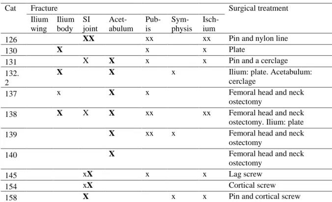
Fractures of the acetabulum were surgically treated in nine cats and nine dogs, 75,0% of the fractures in cats and 90,0% of the fractures in dogs. In cats seven acetabular fractures were treated with femoral head and neck ostectomy. One was treated with a pin and cerclage, and one fracture was treated with only cerclage. In dogs three fractures were treated with a SOP. Three fractures were treated with a femoral head and neck ostectomy. One acetabular fracture was treated with a C plate and one fracture were treated with a LCP. One fracture was treated with a femoral head and neck ostectomy in combination with a cerclage.
Table 5. Fracture type and surgical treatment of cats. “x” represents one sided fracture and. “xx”
bilateral fracture. “BOLD” shows which fractures were treated surgically. Total C shows the total number of fractures treated conservatively and total S shows the total number of fractures treated surgically
Cat Fracture Surgical treatment
Ilium wing Ilium body SI joint Acet-abulum Pub-is Sym-physis Isch-ium
126 XX xx xx Pin and nylon line
130 X x x Plate
131 X X x x Pin and a cerclage
132. 2
X X x Ilium: plate. Acetabulum:
cerclage
137 x X x Femoral head and neck
ostectomy
138 X X X xx xx Femoral head and neck
ostectomy. Ilium: plate
139 X xx x Femoral head and neck
ostectomy
140 X Femoral head and neck
ostectomy
145 xX x x Lag screw
154 xX Cortical screw
19
168 x X X x Femoral head and neck
ostectomy
169 x X X x Femoral head and neck
ostectomy
170 X X x x Femoral head and neck
ostectomy Total C 0 3 5 0 9 3 8 Total S 0 3 4 9 0 0 0
Table 6. Fracture type and surgical treatment of dogs. “x” represents one sided fracture and “xx”
bilateral fracture. “BOLD” shows which fractures were treated surgically. Total C shows the total number of fractures treated conservatively and total S shows the total number of fractures treated surgically
Dog Fracture Surgical treatment
Ilium wing Ilium body SI joint Aceta-bulum Pubis Symp-hysis Ischi-um
202 X Xx xx xx Ilium: SOP. SI joint: pin
and lag screw
205 X xx x SOP
206 X x x xx DCP
207 X X LCP
212 X xx xx Pin and cancellous screw
214 X X x x SI joint: pin and cortical
screw. Ilium: reconstruction plate
215 X x Plate
225 XX x x Lag screw and pin
226 X x x Femoral head and neck
ostectomy
227 x X x x Femoral head and neck
ostectomy
228 X SOP. Femoral head and
neck ostectomy later
229 X xx Reoperation of old screw
fixation. The screw was removed
230 x X x x Femoral head and neck
ostectomy and cerclage
231 X x x SOP
232 X x x DCP
233 X xx xx DCP
234 X Femoral head and neck
ostectomy Total C 0 1 2 0 14 0 12 Total S 0 7 4 9 0 0 0
20 Questionnaires
Feline Pain and Musculoskeletal Index (FMPI)
Twenty-one cat owners answered the FMPI questionnaire. Eight cat owners described the time after the injury, outside the FMPI, because their cat was dead or missing and could therefore not answer the complete FMPI. Follow up time from injury varied between six months to 11 years, with a mean follow up time of 4,3 years, and the owners were asked questions about the past month. Table 7 shows the scores of the cats participating in the study.
Of all 21 cats, 12 cats had normal, or better than normal, activity and therefore, no indication of pain in their normal life. Nine cats had a higher relative score than 20,0%, which indicates some level of pain in their normal life. Only four owners thought their cat was in pain in their normal life, but none thought that this impacted on their cat’s quality of life. Those who scored their cats with pain in their normal life had relative scores of 30,6%, 33,8%, 22,4% and 27,5%. These were not the highest relative scores, the highest relative scores were 33,8%, 30,6%, 28,2% and 27,5%. Median relative score was 20,0% which equals normal life with no pain. Mean relative score was 23,0%.
The most common questions to grade as “not normal” were the questions about normal gait, 28,6% did not have a normal gait, and being carried or petted, 28,6% could not be carried or petted normally. No cat had problems with urination or defecation. Jumping up and down was a problem for 23,8% of the cats. Walking downstairs was easier than walking upstairs, 85,7% of the cats walked upstairs normally and 90,5% of the cats walked downstairs normally. The owners could leave a comment to the questionnaire. Two owners commented that their cat was lame, two owners commented that their cat was stiff in its hind limbs or back, one owner told their cat was asymmetric in its pelvis and one owner commented that their cat walked with stomping sounds with its hind limbs. Two owners also expressed that their cat did not like being petted or carried before the injury and did not like it after the injury either. Owner of cat 162 reported that the first years after the injury the cat did not show any pain, but when time had passed, they could see that their cat had changed its movement in a way that the owner perceived as a pain induced change.
Eight owners described their cats’ time after the injury without answering the FMPI questionnaire due to death of their cat or that the cat was missing. Six of these cats recovered completely according to the owner and behaved the same way they did before the injury. One cat did not recover and the owner regrets that they did not euthanize the cat at the time of the injury. Another owner that participated in the clinical study expressed the same thing, although this cat did recover from the injury. They thought the treatment and the period after the injury were too tough. They expressed that they did not fully understand what the treatment meant and that if they were with the same choice again, they would not go through with treatment. The last cat almost recovered, it had a disturbance in movement, but it did not affect the cat in its normal life. The cat behaved the same as before the injury.
21
Table 7. FMPI scores of cats that suffered from pelvic fractures. Conservative treatment = C, surgical
treatment = S. Quality of life (QOL) is shown separately to better show the owners opinion on QOL. Relative score of ≤20,0% indicates normal function. Higher relative score indicates poorer function
Cat Treatment Total score Relative score (%) QOL score
101 C 16/80 20,0 0 105 C 16/85 18,8 0 106 C 17/85 20,0 0 111 C 17/85 20,0 0 114 C 22/80 27,5 0 116 C 17/85 20,0 0 117 C 17/85 20,0 0 118 C 16/80 20,0 0 119 C 24/85 28,2 0 122 C 17/85 20,0 0 123 C 17/85 20,0 0 129 C 26/85 30,6 2 130 S 21/85 24,7 0 131 S 18/85 21,2 0 132 S 27/80 33,8 2 133 C 19/85 22,4 2 135 C 17/85 20,0 0 150 C 17/85 20,0 0 153 C 22/80 27,5 0 158 S 17/85 20,0 0 162 C 22/80 27,5 2
ACVS Canine Orthopedic Index (COI)
Sixteen dog owners answered the COI questionnaire. Follow up time from injury varied between six months to 11 years, with a mean follow up time of 4,3 years, and the owners were asked about the past month. The result of the questionnaire is seen below in Table 8.
One dog had a relative score of 20%. Three dogs had a stiffness score of five, which indicates no stiffness in normal life. Seven dogs had a function score that indicates full function in their normal life. Four dogs had a gait score that indicates no lameness in their normal life. Six dogs had best quality of life possible, or the owners were not worried that the injury would affect the animal. Median relative score was 28,8% and mean relative score was 37,7%.
One female dog had problems with urinating and defecation. The dog had sometimes trouble getting in position and ended up urinating standing up. Twenty-five percent of the dogs never showed any kind of disturbance in movement due to the injury. The questionnaire asked directly about lameness in the movement part of the questionnaire, 43,9% of the dogs had varying degrees of lameness during light activity and 37,5% had varying degrees of lameness during moderate activity. The day after moderate activity 43,7% of the dogs had varying degrees of lameness.
22
The part of the questionnaire about stiffness showed that 31,3% of the dogs never showed any stiffness. 68,7% of the dogs had stiffness in the morning and 56,2% of the dogs had stiffness after 15 minutes rest, while 43,7% of the dogs had problems rise after 15 minutes rest. Although, it is important to note that 68,7% of the dogs had problems with their joints in general, not only the joints of the pelvis, according to their owner.
The part of the questionnaire about function showed that the most difficult movement was to jump up on something, 43,8% of the dogs had a normal jump. One dog could not jump up at all, this dog could not jump down either. Jumping down was easier, 62,5% of the dogs jumped down normal. Climbing was easier than jumping, 60% of the dogs could climb up and down. The owners could comment the questionnaire and two owners thought that their dogs’ stiffness and lameness were not correlated to the pelvic fracture, one of the dogs had been lame previous to the accident and both dogs showed lameness in their front limbs. One dog showed lameness when it had not had proper training, usually they exercise previous to the hunting season and the dog was fit, and then the dog did not show any lameness. Another dog showed lameness after a tough whole days hunting, but when it hunted only half days the dog was not lame. One owner could not tell if the dog did not want to walk or if it could not walk due to pain, because when they turned to go home the dog walked normal again. Several owners pointed out that when the questionnaire was answered the outside temperature was much higher than normal which impacted on the dogs’ quality of life at the moment.
Table 8. COI scores in dogs that suffered from pelvic fractures. Conservative treatment = C, surgical
treatment = S. Relative score of ≤20,0% indicates normal function. Higher relative score indicates poorer function
Dog Treatment Total score Relative score (%) Stiffness score Function score Gait score QOL score 201 C 38/80 47,5 12/25 8/20 7/20 11/15 202 S 45/80 56,3 15/25 7/20 20/20 3/15 203 C 47/80 58,8 15/25 10/20 16/20 6/15 204 C 17/75 22,7 6/25 4/20 5/20 2/10 206 S 18/70 25,7 6/25 2/10 7/20 3/15 207 S 16/80 20,0 5/25 4/20 4/20 3/15 213 C 53/80 66,3 19/25 10/20 16/20 8/15 215 S 17/80 21,3 5/25 4/20 4/20 4/15 216 C 23/80 28,8 9/25 5/20 6/20 3/15 217 C 46/80 57,5 11/25 14/20 10/20 11/15 218 C 47/80 58,8 18/25 9/20 15/20 5/15 221 C 21/80 26,3 10/25 4/20 4/20 3/15 222 C 21/80 26,3 8/25 4/20 5/20 4/15 223 C 28/80 35,0 10/25 5/20 8/20 5/15 224 C 23/80 28,8 8/25 6/20 4/20 5/15 225 S 18/80 22,5 5/25 4/20 6/20 3/15
23
Comparison FMPI and COI
The relative scores in the FMPI and the COI were comparable, and a Mann-Whitney test was performed to test if the difference in the relative scores in the FMPI and the COI were statistically significant. Median value of the relative score of cats was 20,0% and median value of the relative score of dogs was 28,8%. The Mann-Whitney test showed that with 95% certainty cats had less pain and a better quality of life than dogs after a pelvic fracture. Figure 5 shows the distribution of relative scores between cats treated conservatively, cats treated surgically, dogs treated conservatively, and dogs treated surgically.
Figure 5. Distribution of relative scores in cats and dogs. C = conservative treatment, S = surgical
treatment.
Clinical study
The different parts examined and how they were graded is seen in Appendix 3. In Table 9 and 10 the result of the clinical examination is described. Thirteen cats and 11 dogs participated in the clinical study. Follow up time from injury varied between six months to 11 years, with a mean follow up time of 4,3 years.
Table 9. Clinical signs on physical exam in cats that suffered from pelvic fractures Cat Treat-ment Hind limb muscle Lame-ness Pain on palpa-tion Pelvic symmetry ROM hind limbs Hind limb proprioception Spinal reflex hind limbs Function of the tail Total score 105 C 0 0 0 0 1 0 0 0 1 111 C 0 0 0 0 0 0 0 0 0 116 C 0 0 0 0 0 0 0 0 0 117 C 0 0 0 0 0 0 0 0 0 119 C 0 0 2 2 0 0 0 0 4 123 C 0 0 0 0 0 0 0 0 0 129 C 1 1 0 3 1 0 0 0 6 130 S 2 1 2 1 0 0 0 0 6 131 S 1 0 0 0 0 0 0 0 1 132 S 0 0 0 1 1 0 0 0 2 0 2 4 6 8 10 12 20 21,2 21,3 22,4 22,5 22,7 24,7 25,7 26,3 27,5 28,2 28,8 30,6 33,8 35 47,5 56,3 57,5 58,8 66,3 Cats C Cats S Dogs C Dogs S
Number of patients
Relative scores
24 133 C 0 0 1 0 0 0 0 0 1 150 C 0 0 0 2 0 0 0 0 2 162 C 0 0 0 0 0 0 0 0 0 Tot-al (%) 13 3 (23,1) 2 (15,4) 3 (23,1) 5 (38,5) 3 (23,1) 0 0 0
0 = normal, 1 = mild changes, 2 = moderate changes, 3 = severe changes. C = conservative treatment, S = surgical treatment.
Table 10. Clinical signs on physical exam in dogs that suffered from pelvic fractures Dog Treat-ment Hind limb muscle Lame-ness Pain on palpa-tion Pelvic symme try ROM hind limbs Hind limb propriocep tion Spinal reflex hind limbs Function of the tail Total score 201 C 1 0 2 1 1 2 1 0 8 202 S 1 2 1 1 0 0 0 0 5 204 C 0 1 0 0 1 0 0 0 2 213 C 1 1 0 0 1 2 0 0 5 215 S 0 0 0 0 2 0 0 0 2 216 C 0 0 0 0 0 0 0 0 0 218 C 1 0 0 1 3 0 1 0 6 221 C 0 0 0 0 1 0 0 0 1 223 C 0 0 0 0 1 1 0 0 2 224 C 0 0 0 0 0 0 0 0 0 225 S 1 1 1 2 1 0 0 0 6 Total (%) 11 5 (45,5) 4 (36,4) 3 (27,3) 4 (36,4) 8 (72,7) 3 (27,3) 2 (18,2) 0
0 = normal, 1 = mild changes, 2 = moderate changes, 3 = severe changes. C = conservative treatment, S = surgical treatment.
According to Tables 9 and 10 twice as many dogs had some degree of lameness compared to cats. One owner described that their dog started to ambler after the injury, the dog had normal gait before the injury. Five cats (38,5%) and four dogs (36,4%) had an asymmetry in their pelvis. Five of these were treated conservatively and two were treated surgically. Patient number 215 had puppies, normal delivery. No cat had neurological deficits, while four dogs had some degree of neurological deficit. According to patient records none of these dogs had known neurological deficits on initial presentation. Patient number 117, 129, 130, 202 and 225 had neurological deficits on initial clinical examination but did not show this on the follow up examination.
Comparison treatment method vs FMPI and COI vs clinical exam
Table 11 describes the correlation between type of fracture, treatment method, the relative score of the questionnaire and the score of the clinical examination.