This is an author produced version of a paper published in International
Journal of Oral and Maxillofacial Surgery. This paper has been peer-reviewed
but does not include the final publisher proof-corrections or journal pagination.
Citation for the published paper:
Chrcanovic, Bruno; Santiago Gomez, Ricardo. (2019). Melanotic
neuroectodermal tumour of infancy of the jaws : an analysis of diagnostic
features and treatment. International Journal of Oral and Maxillofacial
Surgery, vol. 48, issue 1, p. null
URL: https://doi.org/10.1016/j.ijom.2018.08.006
Publisher: Elsevier
This document has been downloaded from MUEP (https://muep.mah.se) /
DIVA (https://mau.diva-portal.org).
IntroductionMelanotic neuroectodermal tumour of infancy (MNTI) is a rare benign neoplasm of infancy with a rapid growth pattern. It was first described by Krombecher in 1918 as congenital melanocarcinoma1. Difficulty in determining the cellular origin of this tumour has led to numerous names. Borello and Gorlin were the first to observe an association between elevated levels of urinary vanillylmandelic acid (VMA) and MNTI, and they suggested that the lesion was of neural crest origin2. Later, Hoshino et al. demonstrated increased urinary levels of catecholamines in MNTI, supporting the neural crest origin of the tumour3. The usual treatment for MNTI is surgical removal, although a considerable number of cases develop recurrence. The aim of this study was to integrate the available published data on MNTI into an updated comprehensive comparative analysis of their clinical and radiological features, with emphasis on the predictive factors associated with recurrence.
Materials and methods
This study followed the guidelines of the PRISMA Statement4.Search strategies
An electronic search without time restriction was undertaken in March 2018, in the PubMed/MEDLINE, Web of Science, Science Direct, J-Stage, and LILACS databases. The following terms were used in the search strategies: ((melanotic neuroectodermal tumor of infancy) OR (neuroectodermal tumor of infancy) OR (melanotic progonoma) OR (pigmented melanoameloblastoma) OR (retinal anlage tumor) OR (congenital melanocarcinoma) OR (melanotic Systematic Review Clinical PathologyMelanotic neuroectodermal tumour of infancy of the jaws: an analysis of diagnostic features and treatment
B.R. Chrcanovic1, ⁎ bruno.chrcanovic@mau.se, brunochrcanovic@hotmail.com R.S. Gomez2 1Department of Prosthodontics, Faculty of Odontology, Malmö University, Malmö, Sweden 2Department of Oral Surgery and Pathology, School of Dentistry, Universidade Federal de Minas Gerais, Belo Horizonte, Brazil ⁎Address: Bruno Ramos Chrcanovic, Department of Prosthodontics, Faculty of Odontology, Malmö University, Carl Gustafs väg 34, SE-214 21 Malmö, Sweden. Tel: +46 725 541 545; Fax: +46 40 6658503. AbstractThe purpose of this study was to integrate the available published data on melanotic neuroectodermal tumour of infancy (MNTI) of the jaws into a comprehensive analysis of its clinical/radiological features, with emphasis on the predictive factors associated with recurrence. Eligibility criteria included publications with sufficient clinical/radiological/histological information to confirm the diagnosis. A total of 288 publications reporting 429 MNTI cases were included. MNTIs were slightly more prevalent in males and markedly more prevalent in the maxilla. Most of the lesions were asymptomatic, presenting cortical bone perforation and tooth displacement. Nine lesions were malignant, with metastasis in five cases. Enucleation was the predominant treatment (67.2%), followed by marginal (18.4%) and segmental resection (6.1%). Eighty-one of 356 lesions (22.8%) recurred. Recurrence rates were 61.5% for curettage, 25.3% for enucleation alone, 16.2% for enucleation + curettage, 20.0% for enucleation + peripheral osteotomy, 11.3% for marginal resection, 10.0% for segmental resection, 30.0% for chemotherapy, and 33.3% for radiotherapy. Enucleation and resection presented significantly lower recurrence rates in comparison to curettage. Curettage appears not to be the best form of treatment, due to its high recurrence rate. As resection (either marginal or segmental) is associated with higher morbidity, enucleation with or without complementary treatment (curettage or peripheral osteotomy) would appear to be the most indicated therapy.
adamantinoma) OR (melanotic ameloblastoma) OR (progonoma) OR (pigmented epulis of infancy) OR (pigmented congenital epulis) OR (melano-ameloblastomata) OR (melanoameloblastoma)) AND (maxilla OR mandible OR jaw OR oral OR maxillofacial OR head). Google Scholar was also checked.
A manual search of related journals was performed, including Acta Odontologica Scandinavica, Acta Oto-Laryngologica, Annals of Otology, Rhinology and Laryngology, British Journal of Oral and Maxillofacial Surgery, Cancer, Head and Neck, Head and Neck Pathology, International Journal of Oral and Maxillofacial Surgery, International Journal of Paediatric Dentistry, Japanese Journal of Oral and Maxillofacial Surgery, Journal of Dental Research, Journal of Craniofacial Surgery, Journal of Cranio-Maxillofacial Surgery, Journal of Japanese Society of Oral Oncology, Journal of the Japanese Stomatological Society, Journal of Laryngology and Otology, Journal of Maxillofacial and Oral Surgery, Journal of Nihon University School of Dentistry,
Journal of Oral and Maxillofacial Surgery, Journal of Oral Pathology and Medicine, Journal of the Stomatological Society, Laryngoscope, Oral Diseases, Oral Oncology, Oral Surgery, Oral Medicine, Oral Pathology, Oral Radiology, Otolaryngology—Head and Neck Surgery, Paediatric Dentistry, and Quintessence International. The reference lists of the studies identified and reviews relevant to the subject were also checked for possible additional studies. Publications with lesions identified by other authors as being MNTI, even in the absence of the term ‘melanotic neuroectodermal tumour of infancy’ in the title of the article, were also re-evaluated by one of the present review authors.
Inclusion and exclusion criteria
Eligibility criteria included publications reporting cases of MNTI. The studies needed to contain sufficient clinical, radiological, and histological information to confirm the diagnosis. The definitions and criteria of the WHO Classification of Head and Neck Tumours were used to diagnose a lesion as MNTI5. Randomized and controlled clinical trials, cohort studies, case–control studies, cross-sectional studies, case-series, and case reports were included. Exclusion criteria were immunohistochemical studies, histomorphometric studies, radiological studies, genetic expression studies, histopathological studies, cytological studies, cell proliferation/apoptosis studies, in vitro studies, and review papers, unless any of these publication categories reported any cases with sufficient clinical, radiological, and histological information. Although the tumour generally presents two cell types (proliferation of small rounded neuroblast-like cells and large epithelioid cells with eosinophilic cytoplasm containing variable amounts of melanin), the predominance or presence of only one type did not lead to the exclusion of cases.
Study selection
The titles and abstracts of all reports identified through the electronic searches were read independently by the review authors. For studies appearing to meet the inclusion criteria, or for which the data in the title and abstract were insufficient to make a clear decision, the full report was obtained. Disagreements were resolved by discussion between the authors. The clinical and radiological aspects, as well as the histological description of the lesions reported in the publications, were thoroughly assessed by one of the authors (R.S.G.) in order to confirm the diagnosis of MNTI.Data extraction
The review authors independently extracted data using a specially designed data extraction form. Any disagreements were resolved by discussion. For each of the studies identified and included, the following data were then extracted, when available: year of publication, number of patients, patient sex and age (in months), duration of the lesion prior to treatment, location of the lesion (maxilla or mandible; locations were indicated as predominantly on the right or left side, or in the anterior region of the jaws), lesion size, perforation of the cortical bone, locularity appearance on radiological examination (unilocular/multilocular), presence of radiopacities on radiological examination, association of the lesion with a tooth (the tooth could either be erupted with the entire root(s) encompassed by the lesion or unerupted encompassing the entire tooth), tooth displacement due to lesion growth, expansion of the osseous region adjacent to the tumour, presence of clinical symptoms, whether the lesion was benign or malignant, whether there was a single lesion or multiple lesions, whether the lesion was present or not at birth, presence of elevated levels of urinary VMA or serum alpha-fetoprotein (AFP), percentage of Ki-67, details of the treatment performed (curettage, enucleation, partial resection, resection with continuity, other), recurrence, recurrence period, metastasis, death, and the follow-up period. The lesion size was determined according to the largest diameter reported in the publications. Authors were contacted for possible missing data.Analyses
A descriptive analysis was performed based on mean, standard deviation (SD), and percentage values. The Kolmogorov–Smirnov test was used to test the normality of the distribution of variables, and Levene’s test was used to evaluate homoscedasticity. The Student t-test or Mann–Whitney test was performed to compare two independent groups, depending on the normality of the data. The Pearson χ2 test or Fisher’s exact test was used for categorical
variables, depending on the expected count of events in a 2 × 2 contingency table. Whenever possible, the probability of recurrence was calculated using the odds ratio (OR) and 95% confidence interval (95% CI). The association between Ki-67 and several factors was tested using Pearson correlation. The level of statistical significance was set at P < 0.05. All data were analyzed using IBM SPSS Statistics for Windows, version 25.0 (IBM Corp., Armonk, NY, USA).
Literature search
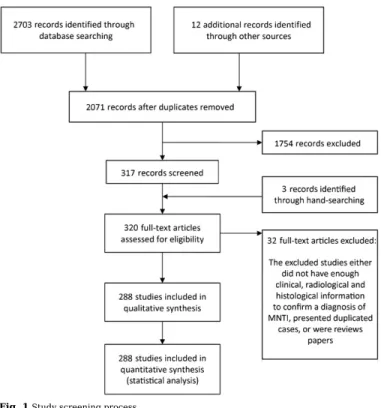
The study selection process is summarized in Fig. 1. The search strategy in the databases resulted in 2703 papers. The search in Google Scholar resulted in 12 eligible papers not found in the five main databases. There were 644 duplicate articles that were cited in more than one database. The reviewers independently screened the abstracts for those articles related to the aim of the review. Of the resulting 2071 studies, 1754 were excluded for not being related to the topic or not presenting clinical cases. An additional hand-search of relevant journals and of the reference lists of selected studies yielded three additional papers. Assessment of the full texts of the remaining 320 articles led to the exclusion of 32 because they did not meet the inclusion criteria. The excluded studies did not have sufficient clinical, radiological, and histological information to confirm the diagnosis of MNTI. Thus, a total of 288 publications were included in the review (see Supplementary Material, Appendix).
Description of the studies and analyses
A total of 288 publications reporting 429 cases of MNTI were included in this review. Table 1 presents the demographic and clinical features of all cases. The lesion was slightly more prevalent in males than females, with a ratio of 1.28:1. The mean age of the patients was 5.4 ± 10.7 months (range 0–144 months). Fig.ure 2 shows the distribution of the lesions according to age. The highest prevalence was seen in those between the ages of 3 months and 6 months. Twenty-eight lesions (9.7%) had been noticed at birth. The lesions were more prevalent in the maxilla than in the mandible (Fig. 3), and three lesions were reported in the cheek.
Table 1 Demographic characteristics and clinical features of melanotic neuroectodermal tumours of infancy in the jaws described in the literature. alt-text: Table 1 Variables Resulta Number 429 Age (months) 5.4 ± 10.7 (0–144) (n = 427) Fig. 1 Study screening process. alt-text: Fig. 1
Male 5.9 ± 13.3 (0–144) (n = 220) Female 5.1 ± 7.5 (0–96) (n = 173) P-valueb 0.936 Duration (months) 2.7 ± 10.3 (0–136) (n = 256) Lesion noticed at birth Yes 28 (9.7%) No 261 (90.3%) Unknown 140 Ki-67 (%) 16.6 ± 17.1 (0–65) (n = 36) Recurrent lesions 23.1 ± 24.5 (0–65) (n = 7) Non-recurrent lesions 15.0 ± 14.9 (1–60) (n = 29) P-valuec 0.610 Elevated levels of urinary VMA Yes 24 (34.8%) No 45 (65.2%) Unknown 360 Elevated levels of serum AFP Yes 4 (80.0%) No 1 (20.0%) Unknown 424 Sex Male 221 (56.1%) Female 173 (43.9%) Unknown 35 Jaw Maxilla 384 (89.9%) Mandible 40 (9.4%) Cheek 3 (0.7%) Unknown 2 Symptomatic Yes 8 (3.8%) No 204 (96.2%) Unknown 217 Cortical bone perforation
Yes 117 (86.0%) No 19 (14.0%) Unknown 293 Locularity Unilocular 147 (94.8%) Multilocular 8 (5.2%) Unknown 274 Tooth displacement/unerupted Yes 219 (99.5%) No 1 (0.5%) Unknown 209 Treatment None 1 (0.3%) Curettage 15 (3.8%) Enucleation 266 (67.2%) Enucleation alone 184 Enucleation + curettage 72 Enucleation + peripheral osteotomy 10 Marginal resection 73 (18.4%) Segmental resectiond 24 (6.1%) Debulking 1 (0.3%) Chemotherapy 10 (2.5%) Radiotherapy 6 (1.5%) Unknown 33 Recurrence Yes 81 (22.8%) No 275 (77.2%) Unknown 73 Time to recurrence (months) 3.5 ± 5.2 (0.3–24) (n = 67) Behaviour Benign 420 (97.9%) Malignant 9 (2.1%) Unknown 0 Metastasis
Yes 5 (1.4%) No 353 (98.6%) Unknown 71 Died Yes 9 (2.5%) No 349 (97.5%) Unknown 71 Follow-up time (months) 49.8 ± 65.1 (0.5–408) (n = 341) Lesion size (cm) 3.3 ± 1.9 (0.4–18.0) (n = 288) AFP, alpha-fetoprotein; VMA, vanillylmandelic acid. aResults are presented as the mean ± standard deviation (range), or as the number (%). bComparison of the mean age between male and female subjects (Mann–Whitney test). cComparison of the mean percentage Ki-67 between recurrent and non-recurrent lesions (Mann–Whitney test). dResection with continuity defect. Fig. 2 Distribution of melanotic neuroectodermal tumours of infancy according to age, up to 18 months. Cases for which patient age was available are included (n = 427). Six patients were older than 18 months of age (24, 34, 36, 96, 132, and 144 months old). alt-text: Fig. 2 Fig. 3 Topographical distribution of the known precise locations of melanotic neuroectodermal tumours of infancy (n = 368). Locations are indicated as predominantly either on the right or left side, or in the anterior region of the jaws. For the rest of the lesions (n = 61), the location was the ‘maxilla’ (n = 48), ‘mandible’ (n = 8), ‘cheek’ (n = 3), and not available (n = 2). alt-text: Fig. 3
Measurements of the levels of urinary VMA were reported for 69 cases, and elevated levels were seen in 24 (34.8%). Measurements of the levels of serum AFP were reported for only five cases, with elevated levels reported for four of them. There was information about the percentage of Ki-67 in 36 cases; the mean percentage was 16.6 ± 17.1% (range 0–65%). In general, the lesions were unilocular, asymptomatic, with cortical bone perforation, and tooth displacement. There was no statistical correlation between Ki-67 and several factors (recurrence, cortical bone perforation, age, lesion size, duration of the lesion, or VMA) and no statistically significant difference in mean percentage Ki-67 values between lesions that recurred and lesions that did not recur. Several lesions were reported to have shown a rapid increase in size between the incisional biopsy, initial consultation, or hospitalization and the excisional surgery. The mean lesion size was 3.3 ± 1.9 cm (range 0.4–18.0 cm; n = 288).
Concerning the presence of a biphasic cell population, the vast majority of the cases had both components, but it was not possible to assess their proportions because of the limitations of a retrospective study. In some cases, the authors reported that there were two components, but the histopathological images did not show that. In other cases, the authors reported that the lesions did not present both components, but the histopathological images indicated otherwise. Nine lesions were malignant, with metastasis in five cases. The four cases without metastases were diagnosed as malignant MNTIs due to the high number of mitoses. Nine patients died, including four malignant cases (three with metastases). The cause of death was unknown in two patients. One patient died of cardiac arrest and another died on the sixth postoperative day, probably due to post-surgery complications. In the other patient, the tumour presented a rapid increase in size after biopsy, and this tumour continued to grow to monstrous dimensions; it was considered that operative surgery could not be performed in this case. The patient died 1 month after the biopsy. Most of the lesions were treated by enucleation (67.2%), followed by marginal (18.4%) and segmental resection (6.1%). Teeth involved in the lesion were preserved in only 0.8% of the cases treated by enucleation. Eighty-one of 356 lesions (22.8%) recurred, with a mean time to recurrence after treatment of 3.5 ± 5.2 months (range 0.3–24 months). Table 2 shows the recurrence rate according to the treatment performed. With the exception of debulking, which was performed in only one case, curettage was the treatment with the highest rate of recurrence (8/13, 61.5%). Enucleation with complementary treatment (curettage or peripheral osteotomy) showed lower recurrence rates when compared to enucleation alone, although the difference was not statistically significant. Marginal and segmental resection showed the lowest recurrence rates among the surgical therapies. The duration of follow-up was reported for 341 lesions, with a mean of 49.8 ± 65.1 months (range 0.5–408 months).
Table 2 Treatment recurrence for the lesions with available information about treatment and recurrence. alt-text: Table 2 Treatment Recurrence/total (% recurrence) Curettage 8/13 (61.5) Enucleation 52/232 (22.4) Enucleation alone 39/154 (25.3) Enucleation + curettage 11/68 (16.2) Enucleation + peripheral osteotomy 2/10 (20.0) Marginal resection 7/62 (11.3) Segmental resectiona 2/20 (10.0) Chemotherapy 3/10 (30.0) Radiotherapy 2/6 (33.3) Debulking 1/1 (100) Total 75/344 (21.8) aResection with continuity defect.
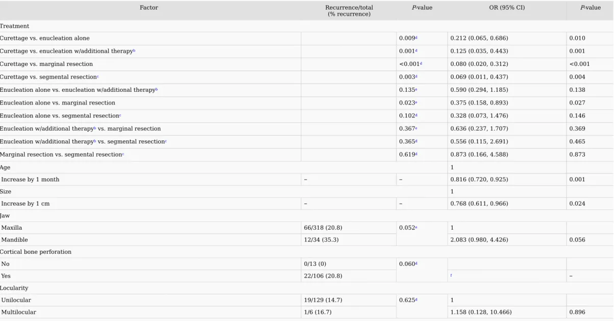
significantly with increasing patient age and with increasing size of the lesion. However, in relation to this, older patients and larger lesions were more often submitted to more radical surgery (Table 4).
Table 3 Recurrence rate for melanotic neuroectodermal tumours of infancy in the jaws according to different factorsa. alt-text: Table 3
Factor Recurrence/total
(% recurrence) P-value OR (95% CI) P-value
Treatment Curettage vs. enucleation alone 0.009d 0.212 (0.065, 0.686) 0.010 Curettage vs. enucleation w/additional therapyb 0.001d 0.125 (0.035, 0.443) 0.001 Curettage vs. marginal resection <0.001d 0.080 (0.020, 0.312) <0.001 Curettage vs. segmental resectionc 0.003d 0.069 (0.011, 0.437) 0.004 Enucleation alone vs. enucleation w/additional therapyb 0.135e 0.590 (0.294, 1.185) 0.138 Enucleation alone vs. marginal resection 0.023e 0.375 (0.158, 0.893) 0.027 Enucleation alone vs. segmental resectionc 0.102d 0.328 (0.073, 1.476) 0.146 Enucleation w/additional therapyb vs. marginal resection 0.367e 0.636 (0.237, 1.707) 0.369 Enucleation w/additional therapyb vs. segmental resectionc 0.365d 0.556 (0.115, 2.691) 0.465 Marginal resection vs. segmental resectionc 0.619d 0.873 (0.166, 4.588) 0.873 Age 1 Increase by 1 month
–
–
0.816 (0.720, 0.925) 0.001 Size 1 Increase by 1 cm–
–
0.768 (0.611, 0.966) 0.024 Jaw Maxilla 66/318 (20.8) 0.052e 1 Mandible 12/34 (35.3) 2.083 (0.980, 4.426) 0.056 Cortical bone perforation No 0/13 (0) 0.060d Yes 22/106 (20.8) f–
Locularity Unilocular 19/129 (14.7) 0.625d 1 Multilocular 1/6 (16.7) 1.158 (0.128, 10.466) 0.896 CI, confidence interval; OR, odds ratio. aRecurrence rate for the lesions with information available about recurrence and the factors included here. bAdditional therapy could be curettage or peripheral osteotomy. cResection with continuity defect.dFisher’s exact test. ePearson χ2 test. fIn at least one case, the value of the weight variable was zero. Such cases are invisible to statistical procedures and graphs, which need positively weighted cases. Table 4 Comparison of patient age and lesion size in relation to the surgical treatment performed. alt-text: Table 4 Treatment Age (months) mean ± SD (range) P-value a Size (cm) mean ± SD (range) P-value a
Curettage 3.6 ± 2.3 (1.5–10) (n = 15) C vs. E: 0.188 2.8 ± 0.9 (2.0–4.0) (n = 9) C vs. E: 0.879
Enucleation 4.2 ± 2.3 (0–15) (n = 265) C vs. MR: 0.009 2.9 ± 1.3 (1.0–7.5) (n = 186) C vs. MR: 0.266
Marginal resection 5.6 ± 4.8 (1.5–36) (n = 73) C vs. SR: 0.007 3.9 ± 2.8 (0.4–18.0) (n = 53) C vs. SR: 0.001
Segmental resection 18.5 ± 38.3 (1–144) (n = 23) E vs. MR: 0.010 6.1 ± 2.9 (2.0–12.0) (n = 14) E vs. MR: 0.006
E vs. SR: 0.003 E vs. SR: <0.001 MR vs. SR: 0.123 MR vs. SR: 0.002 aComparisons between the different surgical treatments (Mann–Whitney test): C, curettage; E, enucleation; MR, marginal resection; SR, segmental resection.
Discussion
The aim of this study was to integrate the available published data on MNTI into an updated comprehensive comparative analysis of their clinical and radiological features, as well as the frequency of recurrence. A review of pathological lesions is important because it provides information that can improve diagnostic accuracy, allowing pathologists and surgeons to make informed decisions and refine treatment plans to optimize clinical outcomes6–10. This review revealed that MNTIs were slightly more prevalent in males and were markedly more prevalent in the maxilla, and most of the lesions were asymptomatic, presenting cortical bone perforation and tooth displacement. The fact that about 10% of the lesions were observed at birth supports the hypothesis of an embryonic origin of the tumour.Some authors have suggested additional examinations to help in the diagnosis or to assist in the determination of the most suitable therapy for each case. High levels of urinary excretion of VMA and serum AFP are characteristic of this tumour. A return of urinary VMA levels to normal after surgery was observed in some of the studies (e.g., the cases reported by Johnson et al.11 and Howell and Cohen12, among others). In some cases, this examination was found to be useful to monitor the patient when the level was high at the initial examination. However, it was found that in most patients, the presurgical values were normal. Although this test is indicated as a complement to the diagnosis, only a third of the patients showed high levels of urinary excretion of VMA. Unfortunately, serum AFP levels were reported for only five patients, making it impossible to draw reliable conclusions.
No association between Ki-67 and clinical or radiographic parameters could be identified, and the mean percentage Ki-67 values did not differ between recurrent and non-recurrent lesions. At present, no reliable histological or immunophenotypical markers predict clinical aggressiveness or the need for complete excision13. Moreover, although some recurrent tumours are aneuploid, recurrent diploid tumours have also been reported14. Therefore, ploidy analysis is not a good predictor for tumour recurrence.
Concerning the treatment of MNTIs, some authors have suggested that wide surgical margins may not be necessary because tumour islets may regress spontaneously after incomplete excision. Numerous clinicians have advocated allowing residual portions of the tumour to remain when radical excision would be mutilating15–18. Shafer and Frissell reported that at the time of the operation it was impossible to tell whether the tumour had been completely excised, and their case was followed up for 66 months with no recurrence19. Van Middlesworth et al. stated that at the completion of the surgical excision, it was evident that there was some inaccessible tumour remaining, and still no recurrence was observed at 1 year postoperative20. Judd et al. did not remove the whole lesion in two enucleations in order to avoid mutilation of other structures, and no recurrence was observed after 30 months17. On the other hand, other authors have suggested tumour resection with wide surgical excision of at least 5 mm21,22, stating that recurrence was due to incomplete removal of the lesion22. In addition to incomplete excision, recurrence could also be a consequence of dissemination during surgery or multicentric tumours, factors that have been suggested in some case reports23,24.
recurrence rates. Although resection results in higher morbidity, the vast majority (99.2%) of the cases treated by enucleation resulted in concomitant removal of the involved teeth. As both marginal and segmental resection are related to higher morbidity, enucleation with or without complementary treatment (curettage or peripheral osteotomy) would appear to be the most indicated therapy. Some authors used additional therapeutic methods to avoid mutilating surgeries. Maroun et al. administered neoadjuvant chemotherapy prior to surgery, thereby obtaining adequate shrinkage of the tumour to allow better resectability and easier surgical access, which minimized the need for wider margins25. In the case reported by Enriquez and Carnate, microscopic sections of the resected mass showed post-chemotherapy-related changes consisting of predominantly melanin-containing epithelioid cells and reduced or absent neuroblast-like cells26. The final microscopic features of the case reported by Davis et al. revealed that chemotherapy essentially obliterated the neuroblastic cell nests, yet was minimally effective in reducing the epithelioid cell island population27. Further case reports are necessary to confirm these findings.
There is an interesting point to make concerning metastases. In the case reported by Navas Palacios, there were many metastatic foci in several submaxillary and parotid lymph nodes28. The cytological appearance of the metastases was that of the solid areas of the tumour, which closely resembled neuroblastoma. It was suggested that the malignant behaviour of this case was probably due to the neuroblastoma-like transformation of the neuroblastic component of the tumour. Similar changes were observed by Dehner et al.29. The metastases showed exclusively solid areas containing neuroblast cells14.
Previous reviews have found a correlation between patient age ≤4.5 months and a significantly higher recurrence rate30,31. The results of the present study appeared to show a decrease in recurrence rate with increasing patient age and with increasing size of the lesion. However, older patients and larger lesions were more often submitted to more radical surgery, and more radical surgery was found to be associated with a lower recurrence rate.
The results of this study should be interpreted with caution because of its limitations. First, all included studies were retrospective reports, which inherently result in errors, with incomplete records. Second, many of the published cases had a short follow-up, which could have led to an underestimation of the actual recurrence rate. However, it is difficult to define what would be considered a short follow-up period for the evaluation of recurrence in such lesions. Third, many of the cases described were published as isolated case reports or small case-series. To conclude, curettage appears not to be the best form of treatment, due to its high recurrence rate. As resection (either marginal or segmental) is associated with higher morbidity, enucleation with or without complementary treatment (curettage or peripheral osteotomy) would appear to be the most indicated therapy.
Acknowledgements
The authors would like to thank Mrs Sabrina Avendaño and Mrs Claudia Rossi, Librarians of the Asociación Odontológica Argentina, who provided us with some of the articles from the Revista de la Asociación Odontológica Argentina, and Dr Anne-Flore Derache for providing us with her thesis. The authors would also like to thank Dr Audrey Moreau and Dr Natacha Kadlub for providing missing information for their studies. Last but not least, we would like to thank the librarians of Malmö University (with a special thanks to Ms Anneli Svensson), who helped us to obtain some articles. RSG is a research fellow at CAPES, Brazil (Proc. 88881.119257/2016-0).Funding
None.Competing interests
None.Ethical approval
Not applicable.Patient consent
Not required.Appendix A. Supplementary data
Supplementary data associated with this article can be found, in the online version, at https://doi.org/10.1016/j.ijom.2018.08.006.References
1. E. Krombecher, Zur Histogenese und Morphologie der Adamantinome und sonstiger Kiefergeschwulste, Beitr Pathol Anat Allg Pathol 64, 1918, 165–197.
2. E.D. Borello and R.J. Gorlin, Melanotic neuroectodermal tumor of infancy—a neoplasm of neural crest origin: Report of a case associated with high urinary excretion of vanilmandelic acid, Cancer 19, 1966, 196–206. 3. S. Hoshino, H. Takahashi, T. Shimura, S. Nakazawa, Z. Naito and G. Asano, Melanotic neuroectodermal tumor of infancy in the skull associated with high serum levels of catecholamineCase report. Case report, J Neurosurg
80, 1994, 919–924.
4. D. Moher, A. Liberati, J. Tetzlaff, D.G. Altman and PRISMA Group, Preferred Reporting Items for Systematic Reviews and Meta-Analyses: the PRISMA Sreporting items for systematic reviews and meta-analyses: the PRISMA statement, Ann Intern Med 151, 2009, 264–269, W64. 5. World Health Organization, WHO classification of head and neck tumours, Fourth edition, 2017, IARC Press; Lyon. 6. B.R. Chrcanovic and R.S. Gomez, Peripheral calcifying cystic odontogenic tumour and peripheral dentinogenic ghost cell tumour: an updated systematic review of 117 cases reported in the literature, Acta Odontol Scand 74, 2016, 591–597. 7. B.R. Chrcanovic and R.S. Gomez, Calcifying epithelial odontogenic tumor: an updated analysis of 339 cases reported in the literature, J Craniomaxillofac Surg 45, 2017, 1117–1123. 8. B.R. Chrcanovic and R.S. Gomez, Cementoblastoma: an updated analysis of 258 cases reported in the literature, J Craniomaxillofac Surg 45, 2017, 1759–1766. 9. B.R. Chrcanovic and R.S. Gomez, Glandular odontogenic cyst: an updated analysis of 169 cases reported in the literature, Oral Dis 24, 2018, 717–724. 10. B.R. Chrcanovic and R.S. Gomez, Squamous odontogenic tumor and squamous odontogenic tumor-like proliferations in odontogenic cysts: an updated analysis of 170 cases reported in the literature, J Craniomaxillofac Surg 46, 2018, 504–510. 11. R.E. Johnson, B.W. Scheithauer and D.C. Dahlin, Melanotic neuroectodermal tumor of infancy. A review of seven cases, Cancer 52, 1983, 661–666. 12. R.E. Howell and M.M. Cohen, Jr., Pathological case of the month. Melanotic neuroectodermal tumor of infancy, Arch Pediatr Adolesc Med 150, 1996, 1103–1104. 13. D.J. Fowler, J. Chisholm, D. Roebuck, L. Newman, M. Malone and N.J. Sebire, Melanotic neuroectodermal tumor of infancy: clinical, radiological, and pathological features, Fetal Pediatr Pathol 25, 2006, 59–72. 14. G. Pettinato, J.C. Manivel, E.S. d’Amore, W. Jaszcz and R.J. Gorlin, Melanotic neuroectodermal tumor of infancy. A reexamination of a histogenetic problem based on immunohistochemical, flow cytometric, and ultrastructural study of 10 cases, Am J Surg Pathol 15, 1991, 233–245. 15. D.M. Crockett, T.J. McGill, G.B. Healy and E.M. Friedman, Melanotic neuroectodermal tumor of infancy, Otolaryngol Head Neck Surg 96, 1987, 194–197. 16. J.R. Hupp, R.G. Topazian and D.J. Krutchkoff, The melanotic neuroectodermal tumor of infancy. Report of two cases and review of the literature, Int J Oral Surg 10, 1981, 432–446. 17. P.L. Judd, K. Harrop and J. Becker, Melanotic neuroectodermal tumor of infancy, Oral Surg Oral Med Oral Pathol 69, 1990, 723–726. 18. G.W. Thoma, Ameloblastic tumors, Cancer Bulletin 24, 1972, 98–99. 19. W.G. Shafer and C.T. Frissell, The melanoameloblastoma and retinal anlage tumors, Cancer 6, 1953, 360–364. 20. D.G. Van Middlesworth, R.C. Fox and M.J. Freeman, Melanotic neurectodermal tumor of infancy: a review of histogenesis and report of a case, ASDC J Dent Child 44, 1977, 137–139. 21. S. Hamilton, D. Macrae, S. Agrawal and D. Matic, Melanotic neuroectodermal tumour of infancy, Can J Plast Surg 16, 2008, 41–44. 22. Y. Hoshina, Y. Hamamoto, I. Suzuki, T. Nakajima, H. Ida-Yonemochi and T. Saku, Melanotic neuroectodermal tumor of infancy in the mandible: report of a case, Oral Surg Oral Med Oral Pathol Oral Radiol Endod 89, 2000, 594–599. 23. B. Steinberg, C. Shuler and S. Wilson, Melanotic neuroectodermal tumor of infancy: evidence for multicentricity, Oral Surg Oral Med Oral Pathol 66, 1988, 666–669.
Queries and Answers
Query: The author names have been tagged as given names and surnames (surnames are highlighted in teal color). Please confirm if they have been identified correctly. Answer: Yes Query: Please confirm that the provided email brunochrcanovic@hotmail.com is the correct address for official communication, else provide an alternate e-mail address to replace the existing one, because private e-mail addresses should not be used in articles as the address for communication Answer: It is correct. Query: The text has undergone minor rephrasing throughout. Please check sentences marked with this query carefully and advise of any further changes required. Answer: It is OK. Query: “Your article is registered as a regular item and is being processed for inclusion in a regular issue of the journal. If this is NOT correct and your article belongs to a Special Issue/Collection please contact j.faur@elsevier.com immediately prior to returning your corrections.” Answer: It is a regular item. Query: “recurrence period” – should this be “time to recurrence”? Answer: Yes, it should be "time to recurrence". Query: The text has undergone minor rephrasing throughout. Please check sentences marked with this query carefully and advise of any further changes required. Answer: It is OK. Query: The text has undergone minor rephrasing throughout. Please check sentences marked with this query carefully and advise of any further changes required. Answer: It is OK. 24. S. Young and F. Gonzalez-Crussi, Melanocytic neuroectodermal tumor of the foot. Report of a case with multicentric origin, Am J Clin Pathol 84, 1985, 371–378. 25. C. Maroun, I. Khalifeh, E. Alam, P.A. Akl, R. Saab and R.V. Moukarbel, Mandibular melanotic neuroectodermal tumor of infancy: a role for neoadjuvant chemotherapy, Eur Arch Otorhinolaryngol 273, 2016, 4629–4635. 26. A.M. Enriquez and J.M. Carnate, Melanotic neuroectodermal tumor of infancy, Philippine Journal of Otolaryngology Head and Neck Surgery J Otolaryngol Head Neck Surg 26, 2011, 51–54.27. J.M. Davis, M. DeBenedictis, D.K. Frank and M.E. Lessin, Melanotic neuroectodermal tumor of infancy: a wolf in sheep’s clothing, Ann Otol Rhinol Laryngol 124, 2015, 97–101. 28. J.J. Navas Palacios, Malignant melanotic neuroectodermal tumor: light and electron microscopic study, Cancer 46, 1980, 529–536. 29. L.P. Dehner, R.K. Sibley, J.J. Sauk, Jr, R.A. Vickers, M.E. Nesbit, A.S. Leonard, D.E. Waite, J.E. Neeley and J. Ophoven, Malignant melanotic neuroectodermal tumor of infancy: a clinical, pathologic, ultrastructural and tissue culture study, Cancer 43, 1979, 1389–1410. 30. B. Kruse-Lösler, C. Gaertner, H. Bürger, L. Seper, U. Joos and J. Kleinheinz, Melanotic neuroectodermal tumor of infancy: systematic review of the literature and presentation of a case, Oral Surg Oral Med Oral Pathol Oral Radiol Endod 102, 2006, 204–216. 31. S. Rachidi, A.J. Sood, K.G. Patel, S.A. Nguyen, H. Hamilton, B.W. Neville and T.A. Day, Melanotic neuroectodermal tumor of infancy: a systematic review, J Oral Maxillofac Surg 73, 2015, 1946–1956.
Appendix A. Supplementary data
The following is Supplementary data to this article: Multimedia Component 1Query: This paragraph has been rephrased. Please advise of any changes required. Answer: It is OK. Query: The text has undergone minor rephrasing throughout. Please check sentences marked with this query carefully and advise of any further changes required. Answer: It is OK. Query: The text has undergone minor rephrasing throughout. Please check sentences marked with this query carefully and advise of any further changes required. Answer: It is OK. Query: The text has undergone minor rephrasing throughout. Please check sentences marked with this query carefully and advise of any further changes required. Answer: It is OK. Query: The text has undergone minor rephrasing throughout. Please check sentences marked with this query carefully and advise of any further changes required. Answer: It is OK. Query: Please check the abbreviated journal titles in reference [26]. Answer: It is OK. Query: The text has undergone minor rephrasing throughout. Please check sentences marked with this query carefully and advise of any further changes required. Answer: It is OK. Query: Please check the layout of all the Tables, and correct if necessary. Answer: It is OK. Query: Table 4: details in the P-value columns and footnote ‘a’ have been changed. Are these changes correct? Answer: Yes, they are.