An epidemiological study of Swedish Campylobacter jejuni isolates from
humans and broilers using multilocus sequence typing
Tora Lövström
Master program in Biomedical Science 120 hp
University of Kalmar, Pure and Applied Natural Sciences Examination project work 45 hp
Supervisors:
Jonas Waldenström, Assoc prof Department of Pure and
Petra Griekspoor, M Sci Applied Natural Sciences
University of Kalmar
391 82 KALMAR
Examiner:
Michael Lindberg, Prof Department of Pure and
Applied Natural Sciences
University of Kalmar
391 82 KALMAR
Abstract
Campylobacter jejuni is the main cause of bacterial diarrhoeal illness in developed countries, with ~7000
cases being reported each year in Sweden. C. jejuni has received growing attention since it’s recognition as a human pathogen in the 1970s, but its epidemiology is complex and much still remains unknown. There are several potential reservoirs for C. jejuni, including environmental sources as water and soil, wild and domesticated animals, particularly poultry, but also other livestock and pets.
In this study 348 Swedish C. jejuni isolates from the year 2000 from humans (n = 164) and broilers (n = 184) were characterized with multilocus sequence typing (MLST) with the aim of comparing the
population structures and diversity of C. jejuni between isolates from the two hosts. MLST is a method for characterization of bacterial isolates that indexes the variation in DNA sequence of multiple protein encoding housekeeping genes. A secondary aim in this study was to compare populations of C. jejuni from 11 subgroups of isolates based on location of the sampling.The overlap between the populations was analyzed numerically based on genotypes detected and with analysis of phylogeny, gene flow and molecular variation.
It was shown that the population structure of C. jejuni isolates from broilers and humans show a high degree of similarity, supporting broilers as an important source of human infection. However, even though the population structure of human and broiler C. jejuni were almost genetically indistinguishable other sources of C. jejuni infections in humans cannot be ruled out since the same genotypes can be found in other sources as well. Analysis of the 11 subgroups suggested that there may be a difference in populations infecting humans in different Swedish regions, and between populations of C. jejuni in broilers from different slaughterhouses. But this could be a result of chance since most of the subgroups were small. Future studies to improve the understanding of C. jejuni epidemiology, for which MLST has proven itself as a valid method, is important to develop control strategies to prevent infection with this common cause of diarrhoeal illness.
2
POPULÄRVETENSKAPLIG SAMMANFATTNING
Campylobacter är den vanligaste orsaken till bakteriell matförgiftning. Bara i Sverige rapporteras ca ~7000 fall varje år där de flesta har smittats utomlands. En infektion med Campylobacter är en så kallad zoonos, dvs en sjukdom som kan smitta från djur till människa. Det är mycket sällsynt att bakterien smittar från människa till människa utan vi är oftast en återvändsgränd för bakterien. När en människa blir smittad uppstår ofta akut diarré som kan vara blodig, magsmärtor, illamående och feber. I de flesta fall går infektionen över av sig själv men i sällsynta fall kan komplikationer uppstå i form av t.ex. ledbesvär eller Guillain-Barré som är en sjukdom med tillfällig förlamning. Det finns olika arter av Campylobacter där Campylobacter jejuni (förkortas C. jejuni) är den vanligast förekommande i fall där människor blir infekterade. C. jejuni förekommer hos många djur, både vilda och tama, och i naturen i t.ex vatten eller sand. Det är i de flesta fall inte klart varifrån smittan kommer när en person insjuknar, men en av de viktigaste källorna antas vara kyckling. C. jejuni finns i mycket av kycklingköttet som konsumeras här i Sverige.
För att skapa en bild av hur viktiga just kycklingar är för infektioner med C. jejuni i Sverige undersöktes i den här studien hur populationerna av C. jejuni från människor och från
kycklingar i livsmedelsindustrin såg ut. Bakterier som tillhör arten C. jejuni kan ha skillnader från varandra i sin arvsmassa som uppstår bland annat genom mutationer. Genom att titta på DNA-sekvensen i sju olika avsnitt från varje bakteries arvsmassa kunde en så kallad
sekvenstyp bestämmas för varje bakterie. Bakterier som har exakt likadan DNA-sekvens i de sju avsnittet tillhör samma sekvenstyp, men förekommer skillnader tillhör de olika
sekvenstyper. DNA-sekvensen kan även ge ledtrådar om hur nära besläktade två bakterier är även om de inte skulle tillhöra exakt samma sekvenstyp. Denna metod för karaktärisering av bakterier kallas ”multilocus sequence typing” (förkortas MLST) och används flitigt i olika delar av världen. Genom en stor internationell databas kan resultat från studier med MLST delas med andra.
När alla bakterier från människor och kycklingar i den här studien hade karaktäriserats med MLST kunde resultaten jämföras mellan de två populationerna. Inom varje population fanns mycket variation och många olika sekvenstyper hittates både från människor och kycklingar. Men det visade sig att populationerna hade väldigt stora likheter, dvs att i stort sett samma sekvenstyper påträffades hos bakterier från människa som från kycklingar. Detta tyder på att de flesta fallen då en människa smittats med C. jejuni i Sverige kan ha orsakats av bakterier från kycklingar. Med hjälp av den internationella databasen kunde man dock se att många av dessa sekvenstyper hade hittats även i andra källor som boskap eller vilda fåglar. Det går därför inte att utesluta att även andra källor än kyckling kan vara viktiga smittkällor för människor. Genom att fortsätta studera hur olika populationer av C. jejuni ser ut och jämföra dem med de som smittat människor kan vi öka förståelsen för hur vi blir smittade och
3
CONTENTS
POPULÄRVETENSKAPLIG SAMMANFATTNING ... 2 CONTENTS ... 3 INTRODUCTION ... 4 Campylobacter jejuni ... 4 Typing of microorganisms ... 5Multilocus sequence typing (MLST) ... 6
MATERIALS AND METHODS ... 8
Isolates ... 8 Preparation of DNA ... 8 Multiplex-PCR ... 8 MLST ... 9 Analysis of sequences ... 10 RESULTS ... 11
Sequence typing of C. jejuni isolates ... 11
Clonal complexes of C. jejuni isolates ... 11
Allelic diversity ... 13
Association between clonal complex and the source of isolation ... 13
New STs and alleles ... 16
Phylogenetic analysis of isolates ... 16
Diversity between groups of isolates ... 18
DISCUSSION ... 19
Conclusion ... 23
ACKNOWLEDGEMENTS ... 23
4
INTRODUCTION
Campylobacter jejuni
The gastrointestinal bacterium C. jejuni is the most frequently reported zoonotic pathogen in humans (2, 3) and the main cause of bacterial diarrhoeal illness in developed countries. It is also an important pathogen in developing countries, with millions being infected each year worldwide (1-4). In Sweden, around 7000 cases of campylobacteriosis, i.e. infections caused by Campylobacter spp., are reported each year and the corresponding number for Europe is ~200,000 cases (2, 3). However, the Swedish institute of food and agricultural economics estimates that the actual number of annual cases of campylobacteriosis in Sweden is ~80,000, resulting in major expenses for society in form of health care and production loss (42). Campylobacteriosis in humans is caused by thermophilic campylobacters, i.e. those species that grow in temperatures from 30°C to 46°C, and most commonly C. jejuni followed by C. coli and C. lari (1-3). The genus Campylobacter belongs to the epsilon class of proteobacteria in the order Campylobacterales, the family of Campylobacteriaceae, and contains 18 species and 6 subspecies (1, 5). All Campylobacter species are small, spiral shaped, gram-negative and microaerobic bacteria (1, 4).
C. jejuni was not recognized as a human pathogen until the 1970s when it was for the first time successfully isolated from stool samples (31). The bacterium has since then received growing attention but C. jejuni epidemiology is complex and much still remains unknown. There are several potential reservoirs for C. jejuni, including environmental sources as water and soil, wild and domesticated animals, particularly poultry, but also other livestock and pets (1, 2, 4,). Consumption of poultry meat, contact with live poultry or pets, drinking raw milk or untreated or contaminated water have been identified as the main causes of human cases of campylobacteriosis (1, 2, 4). Poultry meat has the highest proportion of samples positive with C. jejuni, with an average of 26% of samples of fresh poultry meat in Europe being positive (1, 2). Although the number of positive samples decreases later in the food chain, the levels still remain high at the consumer level (2). C. jejuni are detected in pig and bovine meat as well, but less frequently compared to poultry (2).
Poultry meat can be contaminated with C. jejuni during slaughter if the live chickens carry the bacterium in their intestines. The transmission routes of C. jejuni into broiler flocks is not completely elucidated, but the presence of hygiene barriers is an important factor for lowering the risk of flock colonization (27). C. jejuni spreads rapidly within a flock of broilers, and can reach a prevalence of 100% within a flock before slaughter (28). Furthermore, studies have shown that C. jejuni can infect and multiply within amoebae, and that intra-amoeba C. jejuni are able to colonize broilers (13, 33). Protozoa and C. jejuni have been detected in poultry drinking water and this environment can facilitate biological interaction between these organisms. Amoebae may serve as a reservoir of C. jejuni and provide protection from a hostile environmental thus complicating the control of the bacteria (13, 32-34).
5
Most cases of campylobacteriosis in humans are sporadic, however C. jejuni have occasionally been associated with large food or water borne outbreaks (3, 4). C. jejuni epidemiology has a marked seasonality, with peaks during summer months in temperate climates (1-3). The infectious dose of C. jejuni is low, with 500-800 bacteria being enough to cause disease in humans (1, 2, 5). The majority of cases with campylobacteriosis in Sweden are imported, with only about 30% or less being domestically acquired infections (2,3). Age of the host is also a factor in C. jejuni epidemiology, with children under the age of five having the highest notification rate for campylobacteriosis, with an average of 120 cases per 100,000 people (2). Corresponding numbers for other age groups are less than half of that number (2).
Gastroenteritis is the clinical syndrome most common in infections with C. jejuni in humans, where acute, often bloody diarrhoea develops after an incubation period of 24-74 hours. The diarrhoea is frequently accompanied by abdominal pain, fever, nausea and headache (1-4). In the majority of cases, campylobacteriosis is self-limiting with the resolution of symptoms within a week (1, 2, 4). Treatment with antibiotics is only recommended for severe cases and erythromycin is then often the drug of choice (1, 3, 7). The unfortunate fact that resistance to antimicrobial drugs is increasing in C. jejuni complicates the treatment of infections (1, 11, 12). In some cases late complications can arise, including intestinal haemorrhage, reactive arthritis or Guillain-Barré syndrome (GBS), the most common form of acute neuromuscular paralysis (1, 4). Infection with C. jejuni is the most commonly identified predisposing factor with about 1 in 1000 infections leading to GBS (1).
In humans C. jejuni bypass the mechanical and immunological barriers of the gastrointestinal tracts and interact with epithelial cells (5, 8). It is also known to invade these cells and to pass through to the subepithelium causing a strong inflammatory response. Although C. jejuni causes severe disease and inflammation in humans, the bacterium can be carried in very high numbers in the intestinal tracts of many domestic and wild animals without causing disease (1, 2, 4, 5). It is not well understood why C. jejuni give rise to such different outcomes in humans compared to most animals (5). Despite its importance as a human pathogen much is still unknown about the pathogenesis of C. jejuni and the host response in humans (5). One important limitation is the lack of a good animal model that mimics the disease caused in humans (5, 9).
Typing of microorganisms
When working with microorganisms, whether in medical practice or in research, there is a need for a common nomenclature, a system for subtyping or characterization. Typing schemes are crucial for understanding the epidemiology of the microorganisms and must provide enough information to distinguish different types from each other without making every isolate unique, and define clusters of closely related isolates. To be able to provide large collections of information, the typing scheme must gain wide acceptance and therefore be relatively cheap, easy, reproducible and comparable between laboratories (14, 22). The earlier methods for typing include culturing, phenotyping and serological characterization (14). Along with the progress of molecular techniques and the availability of complete genome sequences several new characterization methods have been developed and some have gained wide acceptance (1, 14).
6
Genetic studies are an important tool in the exploration of the epidemiology and population structure of pathogens. The population structure of a microorganism is affected by point mutations, recombination, positive and negative selection and population growth or reduction (4). C. jejuni is naturally competent and can take up DNA from the environment, enabling recombination between strains (5). It is known that both plasmid and chromosomal DNA is transferred horizontally (5). When choosing a typing method it is important to consider the resolution requested. Genes with high mutation frequency or even subjected to positive selectivity may be a good choice for separation of closely related isolates. If relationships are to be found in wide populations housekeeping genes, i.e. genes involved in basic functions needed for the maintenance of the cell, may be preferable because of their relatively
conserved DNA sequence (14, 15). With the latter method, high discrimination can instead be accomplished by using multiple genes (15). Since many bacteria have a complex population structure and lack a tree-like phylogeny there can be an advantage in using multiple loci in the characterization (14).
Multilocus sequence typing (MLST)
MLST is a recently developed method for characterization of bacterial (or fungi) isolates that indexes the variation in DNA sequence of multiple protein encoding housekeeping genes (14, 15, 17). MLST has several advantages compared to the older methods. The method can easily be reproduced in different laboratories, the results are unambiguous and the allelic profiles can easily be stored and compared through an international database. MLST therefore
provides a portable typing method that enables global epidemiology (14-17). There are today databases and schemes for the characterization of many bacterial species with MLST, and the method can be applied to all or almost all bacterial species (15, 17). Neisseria meningitidis was the pathogen for which MLST was first developed and is still the bacteria most
comprehensively studied with this approach (14).
Several MLST schemes have some loci in common but it has not been possible to identify a complete set of housekeeping genes that can be applied to all bacterial pathogens (14). Loci used in MLST encode metabolic proteins which remain conserved due to stabilizing selection to preserve the function (4, 14). All loci used in a scheme should ideally have a similar level of variation (14). The C. jejuni MLST scheme was developed in 2001, by Dingle and co-workers, using C. jejuni isolates from a variety of environmental, animal and human sources (18). The characterization of C. jejuni is based on the nucleotide sequence of seven
housekeeping genes (Figure 1) (18, 20). A MLST scheme using the same loci has been developed for C. coli and MLST can therefore be used for direct comparison of the two closely related human pathogens (21).
7
Figure 1: Chromosomal locations of the seven loci used in C. jejuni MLST (18).
For each locus, all unique alleles are assigned arbitrary allele numbers in the order that they are identified resulting in seven numbers for each isolate. The seven numbers constitute an allelic profile, or sequence type (ST). STs are in turn assigned arbitrary numbers in the order of description (18, 20). STs with close relation have been grouped into clonal complexes (CCs) where each complex is thought to be descended from the central genotype, which also provides the name for the CC (4). The C. jejuni database is the third largest MLST database, with 3953 sequence types and 7747 isolates as of April 2009 (17). Its applications to date include population structure analysis and epidemiological surveillance of food borne infections (17). Studies using MLST have indicated that C. jejuni has a weakly clonal population structure, i.e. lacking the consistent tree-like phylogeny present in asexual
populations (19). Furthermore, studies have shown that CCs persist during time and space and that they in part can be coupled to different hosts, environments and to human disease (4, 19, 23-26, 43-46).
C. jejuni is a major public health problem, with high incidence, risks of severe complications, complex epidemiology and many reservoirs (4). It is of great importance to extend the
knowledge about its epidemiology, population structure and transmission routes to develop strategies to prevent infection in humans. Molecular epidemiology can give information about the relationship between genotypes of C. jejuni and different hosts and disease in humans (4). In this study 348 Swedish C. jejuni isolates, from humans (n = 164) and broilers (n = 184) were characterized with MLST with the aim of comparing the population structures and diversity of C. jejuni between the two hosts. How well the C. jejuni types from the two hosts overlap can give indications on the importance of broilers as a source of C. jejuni infection in humans. A secondary aim in this study was to compare C. jejuni types from subgroups of isolates from the two hosts.
8
MATERIALS AND METHODS
Isolates
The isolates used in this study were provided by the Swedish National Veterinary Institute (SVA). A total of 348 Swedish C. jejuni isolates from the year 2000 were included, of which 164 were isolated from humans and 184 from broilers. The human isolates were stool samples collected at four Swedish hospitals in different parts of Sweden, including Uppsala (n= 36), Halmstad (n=36), Sundsvall (n=24) and Kalmar (n=68). Only isolates from patients with domestic infections were included in this study. The broiler isolates came from caecal samples taken at slaughter from broilers (27, 28). Broiler isolates were divided in seven groups based on the slaughterhouse in which the samples were taken; Chicken 1 (Skåne, 29 isolates), Chicken 3 (Västergötland, 21 isolates), Chicken 4 (Södermanland, 29 isolates), Chicken 5 (Öland, 31 isolates), Chicken 6 (Blekinge, 33 isolates), Chicken 7 (Södermanland, 25 isolates) and Chicken 8 (Halland, 16 isolates). Isolates were handled at SVA’s Community Reference Laboratory (CRL) for Campylobacter according to routine procedures and sent to us on swabs with Amie’s transport medium with charcoal.
Preparation of DNA
C. jejuni isolates from Amie-swabs were immediately grown on blood agar plates (Columbia agar II containing 8% [vol/vol] whole horse blood) in a microaerobic atmosphere, using CampyGen™ (CN0025A; Oxoid, Ltd., Basingstoke, United Kingdom) and the BBL GasPak system (BD, Franklin Lakes, NJ), at 42°C for 48 hours. The CampyGen™ sachet produces an environment with 5% O2, 10% CO2 and 85% N2 (29). A loop of bacterial cells were
transferred to tubes with MQ-H2O and lysed through boiling to release DNA. For difficult
isolates, DNA were extracted with MagAttract DNA mini M48 kit (Qiagen, Hilden, Germany) according to manufacturers’ instruction in a BioRobot M48 (Qiagen, Hilden, Germany). DNA extracts were stored at -20°C prior to PCR. Live bacterial cells were stored in freeze medium at -80°C.
Multiplex-PCR
For discrimination of C. jejuni and C. coli when uncertainty arose, a multiplex PCR was used (36, 37). Two μl of each isolate was mixed with 1.25 units Taq DNA polymerase (Qiagen, Hilden, Germany), 3μl 25mM MgCl (Qiagen, Hilden, Germany), 5μl 10X PCR Buffer (Qiagen, Hilden, Germany), 10μl 5X Q-solution (Qiagen, Hilden, Germany), 2μl 10mM dNTP (Invitrogen, Carlsbad, USA) and 2μl of four 20μM primers to a reaction volume of 50μl. One of the primer pair amplifies C. coli at a size of ~350bp (Col1: 5'-AG GCA AGG GAG CCT TTA ATC-3', Col2: 5'-TAT CCC TAT CTA CAA ATT CGC-3') and the other primer pair amplifies C. jejuni at a size of ~760bp (Jun3: 5'-CA TCT TCC CTA GTC AAG CCT-3', Jun4: 5'-AAG ATA TGG CTC TAG CAA GAC-3'). The reactions were performed using a touchdown protocol with denaturation at 94ºC, primer annealing at temperatures ranging from 64ºC to 54ºC, and extension at 72ºC for a total of 40 cycles. Amplification products were separated with 1.5 % agarose gel electrophoresis and stained with ethidium bromide.
9
MLST
The seven loci amplified and sequenced in C. jejuni MLST include aspA (protein product aspartase A), glnA (glutamine synthetase), gltA (citrate synthase), glyA (serine
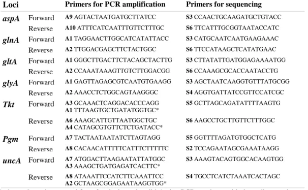
hydroxymethyltransferase), pgm (phosphoglucomutase), tkt (transketolase), and uncA (ATP synthase α subunit) (18, 20). PCR was performed according to C. jejuni MLST homepage (20). The primer pairs (Table 1) for each locus provide amplification products of about 1000bp. For the uncA and tkt loci, alternative primers were used for some isolates to get PCR-products. In a 50μl reaction volume the following reagents were mixed: 1.25 units Taq DNA polymerase (Qiagen, Hilden, Germany), 3μl 25mM MgCl (Qiagen, Hilden, Germany), 5μl 10X PCR Buffer (Qiagen, Hilden, Germany), 10μl 5X Q-solution (Qiagen, Hilden, Germany), 1μl 10mM dNTP (Invitrogen, Carlsbad, USA), 4 or 5μl 10μM forward and reverse primer, water and 2μl template DNA. Conditions for the PCR reaction were denaturation at 94ºC for 2 minutes, primer annealing at 50ºC for 1 minute and extension at 72º C for 1 minute for 35 cycles.
Table 1: Primers used for PCR amplification (left column) and sequencing (right column).
Loci Primers for PCR amplification Primers for sequencing
aspA Forward A9 AGTACTAATGATGCTTATCC S3 CCAACTGCAAGATGCTGTACC
Reverse A10 ATTTCATCAATTTGTTCTTTGC S6 TTCATTTGCGGTAATACCATC
glnA Forward A1 TAGGAACTTGGCATCATATTACC S3 CATGCAATCAATGAAGAAAC
Reverse A2 TTGGACGAGCTTCTACTGGC S6 TTCCATAAGCTCATATGAAC
gltA Forward A1 GGGCTTGACTTCTACAGCTACTTG S3 CTTATATTGATGGAGAAAATGG
Reverse A2 CCAAATAAAGTTGTCTTGGACGG S6 CCAAAGCGCACCAATACCTG
glyA Forward A1 GAGTTAGAGCGTCAATGTGAAGG S3 AGCTAATCAAGGTGTTTATGCGG
Reverse A2 AAACCTCTGGCAGTAAGGGC S4 AGGTGATTATCCGTTCCATCGC Tkt Forward A3 GCAAACTCAGGACACCCAGG A1 TTTAAGTGCTGATATGGTGC* S5 GCTTAGCAGATATTTTAAGTG Reverse A6 AAAGCATTGTTAATGGCTGC A4 CATAGCGTGTTCTCTGATACC* S6 AAGCCTGCTTGTTCTTTGGC Pgm Forward A7 TACTAATAATATCTTAGTAGG S5 GGTTTTAGATGTGGCTCATG
Reverse A8 CACAACATTTTTCATTTCTTTTTC S2 TCCAGAATAGCGAAATAAGG
uncA Forward A7 ATGGACTTAAGAATATTATGGC
A3 AAAGCTGATGAGATCACTTC*
S3 AAAGTACAGTGGCACAAGTGG
Reverse A8 ATAAATTCCATCTTCAAATTCC A2 GCTAAGCGGAGAATAAGGTGG*
S4 TGCCTCATCTAAATCACTAGC
* alternative primers used for a few isolates that didn't give PCR-products with the ordinary primers.
For isolates that didn't give an amplification product with the conditions described above, the following reagents were mixed in a 25μl reaction volume: 0.5 units Platinum Taq DNA polymerase (Invitrogen, Carlsbad, USA), 0.75μl 50mM MgCl (Invitrogen, Carlsbad, USA), 2.5μl 10X PCR Buffer (Invitrogen, Carlsbad, USA), 0.5μl 10mM dNTP (Invitrogen,
Carlsbad, USA), 2μl 10μM forward and reverse primer, water and 2μl template DNA. PCR conditions were adjusted to denaturation at 95ºC for 30 seconds, primer annealing at 50ºC for 30 seconds and extension at 72º C for 1 minute for 40 cycles. PCR products were visualized by 1.5 % agarose gel electrophoresis.
10
PCR-products were precipitated with Fast-PEG solution (10% poly ethylene glycol, 1M NaCl, 30% isopropanol). After centrifugation at 4000 rpm for 45 minutes the pellet was washed with 70% ethanol, air dried and dissolved in 5mM Tris-HCl. The purified PCR-products were sent away for sequencing by Eurofins MWG Operon (Germany). Cycle sequencing technology with heat resistant DNA polymerase, 4 dNTPs, 4 ddNTPs (dideoxy terminator nucleotides) fluorescently labelled with four different dyes were used on ABI 3730XL sequencing machines (30). Primers for sequencing (Table 1) are nested inside the PCR products of the initial PCR to give sequence data of approximately 600 bp. Every locus was sequenced at least once with forward or reverse primer. The sequences were compared with existing alleles in the MLST C. jejuni database to enable determination of allele
numbers, STs and clonal complexes (20). To assign an isolate with an allele number a 100% correct DNA sequence was demanded. All new alleles were sequenced with both reverse and forward primers. New alleles and STs were reported to the database and assigned with new numbers. The MLST database was searched for previous isolation sources and number of STs and isolates in clonal complexes.
Analysis of sequences
Concatenated sequences, i.e. all seven loci sequences assembled in order as one sequence of 3309bp, for each of the isolates were obtained from the MLST-database (20). BioEdit Sequence Alignment Editor (35) was used for assembly, editing and alignment of the sequences. MEGA (Molecular Evolutionary Genetics Analysis) 4 (38) were used for phylogenetic analyses, where neighbour-joining trees were constructed with nucleotide sequences and bootstrap of 500 replications. DNAsp (DNA sequence polymorphism) 5.0 (39) was used to estimate the gene flow between isolates from the two hosts or the 11 subgroups. Gene flow is measured by a FST value which falls between 1 and 0, where a value of 1
indicates that the two populations share no gene flow and a value of 0 indicates that the two populations are panmicitic. In some analyses a population of 97 C. jejuni isolates from Black-headed gulls (Larus ridibundus) from a different study (47) was included to put the results in perspective. FST values obtained in DNAsp calculations were used to construct phylogenetic
trees as a visualization of the variation between the groups. Arlequin ver 2.000, software for population genetics data analysis (40, 41) was used to calculate the sources of variation in the population of C. jejuni isolates in this study, whether among the two large groups of human and broiler isolates, among the 11 subgroups or within each subgroup.
11
RESULTS
Sequence typing of C. jejuni isolates
In this study MLST was used to characterize 348 C. jejuni isolates from human stool samples and caecal samples from broilers. C. jejuni isolates from human and broiler samples were analysed either as two large groups depending only on host, or as 11 separate subgroups based on in which Swedish hospital or slaughterhouse they were isolated. The four groups of human samples are referred to as: “Human Halmstad”, “Human Kalmar”, “Human Sundsvall” and “Human Uppsala”. The seven groups of broiler isolates are referred to as: “Chicken 1”, “Chicken 3”, “Chicken 4”, “Chicken 5”, “Chicken 6”, “Chicken 7” and “Chicken 8” (see materials and methods for details about each group).
Of the total of 348 C. jejuni isolates, 97% was successfully characterized with MLST and assigned to a ST. Six isolates were excluded from the results either because they were contaminated with another sequence type of C. jejuni (n=2, “Chicken 5” and “Chicken 6”), had less than five sequenced loci (n=1, “Chicken 1”), or that species identity was C. coli instead of C. jejuni (n=3, “Chicken 1”, “Chicken 5” and “Human Kalmar”). An additional 4 isolates gave results for only five or six of the seven loci and could therefore not be assigned with ST and were left out of some parts of the results.
In this study a total of 78 STs were obtained from 338 isolates (Table 2), with 50 different STs among human isolates and 48 among broiler isolates. The five most common STs (ST-45 [n=40], ST-48 [n=39], ST-21 [n=27], ST-677 [n=19] and ST-148 [n=17]) accounted for 42% of all isolates. As many as 41 STs were detected only once among the isolates. Fifty-seven STs, containing ~92% of all isolates, were assigned to 21 previously described CCs, i.e. larger groups of STs thought to be descended from the same central genotype. The CCs of ST-21 and ST-45 had the highest number of different STs (n=13 and n=11 respectively) assigned to them. None of the other CCs were assigned with more than three different STs detected in this study. Twenty-one STs, containing ~8% of all isolates, could not be assigned to any
previously described CC and are referred to as unassigned STs. Table 4 shows all STs detected in this study and the CCs they were assigned to.
Clonal complexes of C. jejuni isolates
The percentages of broiler and human isolates in each CC are shown in Figure 2. Of the 21 noted CCs, 11 were found among both human and broiler isolates, containing 89% and 79% of isolates respectively. Five CCs (ST-1332, ST-1034, ST-702, ST-692 and ST-1275) were only represented by broiler isolates and another five CCs (ST-353, ST-362, ST-574, ST-607 and ST-952) only had isolates from human stool samples. There were isolates from both humans and broiler that were unassigned to any CC. The allelic profile of the central genotype of each CC is shown in Table 2. Many of the CCs have alleles in common with other CCs. For instance, ST-21 has 4 alleles in common with the ST-48 and ST-206 complexes.
12
Figure 2: Clonal complexes (CC) of human and broiler C. jejuni. (ua=unassigned)
Table 2: Allelic profiles of the central genotype of each of the 21 CCs detected in this study. CC Allelic profile of central genotype of CC
aspA glnA gltA glyA pgm tkt uncA
21 2 1 1 3 2 1 5 22 1 3 6 4 3 3 3 45 4 7 10 4 1 7 1 48 2 4 1 2 7 1 5 52 9 25 2 10 22 3 6 61 1 4 2 2 6 3 17 206 2 21 5 37 2 1 5 257 9 2 4 62 4 5 6 283 2 7 40 4 42 51 1 353 7 17 5 2 10 3 6 362 1 2 49 4 11 66 8 574 7 53 2 10 11 3 3 607 8 2 5 53 11 3 1 658 2 4 2 4 19 3 6 677 10 81 50 99 120 76 52 692 37 52 57 26 127 29 23 702 2 52 29 48 127 99 23 952 18 22 77 98 122 86 16 1034 2 61 4 64 74 25 23 1275 27 33 22 49 43 82 31 1332 2 1 29 28 58 25 58 21 45 48 677 283 1332 61 52 658 206 257 22 353 1034 362 702 574 607 692 952 1275 ua 0% 5% 10% 15% 20% 25% 30%
35% Clonal complexes of human and broiler C. jejuni isolates
Human Broiler CC is ol at es in CC ( %)
13
Allelic diversity
The genetic diversity differed considerably between the seven loci used for typing of C. jejuni (Table 3). Generally, the proportion of alleles found from each locus in this study resembled the reported data in the MLST-database (20). Among the 342 isolates of C. jejuni almost twice as many different alleles were found for the loci glyA and pgm (n=33) as for aspA (n=18). As of May 2009, the pgm locus has the highest number (n=456) of reported alleles in the C. jejuni MLST database (20), and aspA the lowest number (n=247). Among the alleles found in this study, aspA had the lowest number of variable sites, and uncA the highest number. Phylogenetic relationship between C. jejuni isolates differed a considerable amount between neighbour-joining trees constructed with sequences from each of the seven alleles alone (not shown).
Table 3: Diversity at the seven loci of C. jejuni MLST. Locus Fragment
size (bp)
Number of variable sites *
Number of different alleles among 342 isolates in this study
Number of different alleles in MLST- database aspA 477 17 18 247 glnA 477 25 22 331 gltA 402 28 27 268 glyA 507 51 33 362 pgm 498 62 33 456 tkt 459 58 30 376 uncA 489 81 22 271
*calculated with all STs found in this study
Association between clonal complex and the source of isolation
The ST-21, ST-45 and ST-48 complexes were the three most common among both human and broiler isolates, representing 63% (n=102) and 60% (n=105) of all isolates, respectively. The ST-677 complex was the fourth most common among human isolates, containing 12% (n=20). It however only contained 1% (n=2) of broiler isolates. The ST-1332 complex was in turn the fourth most common among broilers containing 7% (n=12) of the isolates, but no human isolates were assigned to it.
In the MLST database the ST-21, ST-45 or ST-48 complexes are the most frequently detected in human C. jejuni samples (20). Among chicken C. jejuni samples reported to the database the ST-21 and ST-45 complexes are the most frequently reported, while the ST-48 CC is the sixth most common. The ST-45 complex is furthermore one of the three most common among samples from wild birds reported to the database. The three most common sources of each of the CCs detected in this study are listed in Table 4. The percentage of isolates from each of the eleven subgroups assigned to each CC is also shown in Table 4. All 11 subgroups had one of the ST-21, ST-45 or ST-48 complexes as the most common or second most common CC. In this study the ST-21 complex was the only one to which isolates were assigned from all 11 groups. The ST-45 complex had isolates assigned to it from all subgroups except “Chicken 4” and “Chicken 6” and the ST-48 complex had isolates assigned to it from all human groups but only three subgroups of isolates from broilers.
14
Table 4: Distribution of CCs and STs among 338 C. jejuni isolates for each of the eleven groups in this study.
Previous sources together with the number of STs and isolates belonging to each of the encountered CCs in the
C. jejuni and C. coli MLST-database are given as of May 2009 (20).
CC STs among isolates
Isolates from each group assigned to CC (%): MLST-database HH HK HS HU C1 C3 C4 C5 C6 C7 C8 Number of STs / isolates Sources (%) * 21 19, 21, 50, 53, 104, 141, 148, 201, 262, 842, 883, 3937, 3944 36 39 8 17 37 45 32 3 16 16 13 392 / 1176 H (50), Ca (12), Ch (9) 22 22, 1966 1 10 4 4 43 / 108 H (69), Ch (8), Ca (8) 45 11, 45, 137, 230, 233, 334, 583, 1326, 3727, 3934, 3940 28 13 38 17 15 10 72 44 33 215 / 526 H (46), Ch (20), Wb (6) 48 48, 475, 918 14 6 4 31 4 59 8 132 / 323 H (62), Ch (9), Ca (9) 52 52 3 3 4 5 7 62 / 85 H (67), Ch (13) 61 61,3936 3 7 3 4 114 / 245 Ca (39), H (38), Sh (13) 206 46, 290, 572 1 4 4 3 13 81 / 179 H (59), Ch (13), Sh (13) 257 257, 584, 3949 3 10 11 109 / 243 H (69), Ch (12) 283 267, 564 3 4 8 3 15 10 13 32 / 55 H (55), Ch (24) 353 5, 353, 524 6 1 145 / 245 H (69), Ch (18) 362 362 3 8 / 26 H (84) 574 305 1 46 / 116 H (53), Ch (8) 607 3939 4 35 / 60 H (63), Ch (22) 658 658 8 13 31 / 61 H (80) 677 677, 794, 3933 3 9 33 14 8 12 / 20 Wb (30) 692 3948 4 24 / 44 Wb (54), H (14), Ch (14) 702 702 7 8 / 29 Wb (90) 952 2311 3 19 / 20 H (25) 1034 1709, 3946 4 13 44 / 70 Wb (56), H (26), Ch (10) 1275 637 7 41 / 51 Wb (41), Ew (35) 1332 696 10 36 13 / 23 Wb (61) ua 586, 995, 997, 1367, 1371, 1673, 1911, 1938, 2349, 2407, 2491, 3322, 3935, 3938, 3941, 3942, 3943, 3945, 3947, 3950, 3951 8 9 3 11 10 7 14 6 16 7 1214 / 1574 H (30), Wb (30), Ch (14)
ua=unassigned, CC=clonal complex, HH=Human Halmstad (n=36), HK=Human Kalmar (n=67), HS=Human Sundsvall
(n=24), HU=Human Uppsala (n=36), C1=Chicken 1 (n=27), C3=Chicken 3 (n=20), C4=Chicken 4 (n=28), C5=Chicken 5 (n=29), C6=Chicken 6 (n=32), C7=Chicken 7 (n=25) and C8=Chicken 8 (n=15).
* The three most common sources for each CC are given if the source contains > 5 isolates and 5% of isolates. H=Human,
15
The 36 isolates from the subgroup “Human Halmstad” were assigned to seven different CCs where the two most common were ST-21 (36%) and ST-45 (28%). From the 67 isolates from “Human Kalmar” 12 different CCs were detected, where the two most common were ST-21 (39%) and ST-45 (13%). The 36 isolates from “Human Uppsala” were assigned to nine different CCs where the most common was ST-48 (31%). From the 24 isolates from “Human Sundsvall” seven different CCs were detected, where the two most common were ST-45 (38%) and ST-677 (33%). The 27 isolates from the source “Chicken 1” were assigned to eight different CCs and the 20 isolates from “Chicken 3” were assigned to six different CCs, where the most common for both was ST-21 (37% and 45% respectively). The 28 isolates from “Chicken 4” were assigned to six different CCs where the two most common were ST-21 (32%) and ST-1332 (36%). From the 29 isolates from “Chicken 5” only three different CCs were detected, where the most common was ST-45 (72%). The 32 isolates from “Chicken 6” were assigned to five different CCs where the most common was ST-48 (59%). The isolates from the sources “Chicken 7” (n=25) and “Chicken 8” (n=15) were assigned to six different CCs each, where the most common for both was ST-45 (44% and 33% respectively). All sources, except “Human Sundsvall”, had isolates that were unassigned to any existing CC.
Table 5: 19 new STs found in this study. Allelic numbers in bold are new alleles.
ST Allelic profile of ST Source
aspA glnA gltA glyA pgm tkt uncA
3933 10 81 268 87 120 76 52 Human Uppsala 3934 4 7 10 360 1 7 1 Human Uppsala 3935 64 22 22 97 134 301 16 Human Halmstad 3936 1 4 267 2 6 3 17 Human Halmstad 3937 2 1 1 359 2 1 5 Human Halmstad 3938 64 22 68 97 134 301 16 Human Halmstad 3939 8 2 5 53 456 3 1 Human Sundsvall 3940 4 7 10 4 125 7 1 Human Kalmar 3941 18 70 77 97 205 86 47 Chicken 1 3942 64 22 164 230 123 101 60 Chicken 3
3943 18 89 72 109 122 94 16 Chicken 5, two isolates
3944 2 4 42 3 2 1 5 Chicken 6
3945 18 33 22 142 134 51 243 Chicken 7
3946 22 61 4 64 57 25 23 Chicken 8, two isolates
3947 2 7 4 48 131 24 57 Chicken 1
3948 37 52 72 26 127 29 23 Chicken 1
3949 9 1 4 62 4 2 6 Chicken 4
3950 64 89 77 357 94 160 16 Chicken 5, two isolates
16
New STs and alleles
Of the 78 different allelic profiles detected in this study, 19 had not been detected previously in the MLST-database and were assigned to new STs (Table 5). Three of the new STs were detected in two separate isolates, but from the same subgroup. At least one new ST was found among isolates in each of the 11 subgroups and in the subgroup “Human Halmstad” four new STs were found. Seven of the new STs were due to one or two new alleles and 12 were due to new combinations of previously found alleles. New alleles were found for the loci gltA (n=2), glyA (n=4), pgm (n=1) and uncA (n=1). All new alleles differed with only one nucleotide from previously reported alleles.
Phylogenetic analysis of isolates
Phylogenetic trees constructed with the 3309bp long concatenated nucleotide sequences of all seven loci (bootstrap with 500 replications) are shown in Figure 3 and 4. In Figure 3A, the central genotype sequence for each CC detected in this study was used to construct a
neighbour-joining tree showing the genetic relationships between all CCs. Interestingly, even though ST-21 share 4 alleles with ST-48 and ST-206 they do not show the closest
phylogenetic relationship. Notable is that the ST-61 complex formed an outlier cluster in the phylogenetic analysis. In figure 3B one sequence of the species C. coli (the central genotype of the ST-828 complex) was included, showing that the ST-61 complex overlay C. jejuni and C. coli genetically, but that it is closer to C. jejuni. Because of the genetic difference of ST-61 from the rest of the C. jejuni isolates a multiplex PCR was run with all isolates in the ST-61 complex, and confirmed its species identity as C. jejuni.
Figure 3: (A) A Neighbor-joining tree showing the genetic relationship between all CCs found among isolates in
this study. (B) A Neighbor-joining tree showing the genetic relationship between all CCs and the ST-828 complex of the species C. coli. ST-61 is marked with a black dot.
B A
17
In Figure 4A one sequence per ST was included and in Figure 4B and C all 342 isolate sequences were used to construct the tree. The topology of human and broiler isolates in the phylogenetic trees showed high similarity. There was no apparent difference in the
distribution in the phylogenetic tree of the STs found among broiler and human isolates (Figure 4A). STs only found among human isolates seemed to be closely related to isolates found among broiler isolates, and vice versa. When isolates from either human or broilers were marked in the neighbour-joining tree where all 342 isolate sequences were used, the isolates were spread in a similar way across the tree (Figure 4B-C).
Figure 4: (A) A Neighbor-joining tree constructed with concatenated sequences from all STs found in this study.
STs without any marking were found among both human and broiler isolates. STs marked with a circle were only present in human isolates, while those marked with a triangle only were found among broiler isolates. (B and C) Neighbor-joining tree constructed with concatenated sequences from all isolates of C.
jejuni in this study (n=342). In 4B all human isolates is marked with a black dot, and in 4C all broiler
isolates is marked in the same way.
A
B
18
Diversity between groups of isolates
Gene flow analyses showed that human and broiler isolates were almost genetically
indistinguishable. An analysis of genetic subdivision depending on host gave a FST value of
0.01, i.e. close to panmictic. To put this figure in perspective, the data was reanalyzed
together with a population of 97 C. jejuni isolates from Black-headed gulls (Larus ridibundus) from a different study (47). The FST values between human and gull isolates and between
broiler and gull isolates were much higher (0.26 and 0.23, respectively) than between broilers and humans. Another gene flow analysis of the isolates divided in the 11 subgroups gave FST
values that ranged from 0.002 to 0.6. When the population of C. jejuni isolates from Black-headed gulls were included as a 12th group the gene flow analysis no longer showed the gull isolates as a distinct outgroup, as the FST values between gull isolates and each of the 11
subgroups ranged from 0.2 to 0.5. Neighbour-joining trees constructed with FST values are
shown in Figure 5. Analysis of molecular variance (AMOVA) was used to calculate the source of variation among isolates and showed that there was no significant variation among isolates depending on host. The genetic variation among isolates originated from within each of the 11 subgroups (87%), and among the 11 subgroups (13%).
.
Figure 5: A visualization of the gene flow analysis: Neigbour-joining trees constructed with FST values for C.
jejuni isolates from humans, broilers and gulls. (A) The relationship between three groups of isolates depending
on host. (B) The relationship between all 11 subgroups and gull isolates
B A
19
DISCUSSION
There is no doubt that C. jejuni is an important pathogen that lead to suffering and risks for individuals infected and also represents a significant economic burden (1). To prevent
infection in humans it is of great importance to extend the knowledge about the epidemiology of this important pathogen. In this study MLST was used to characterize C. jejuni isolates from two different host, humans and broilers, with the aim of comparing their population structure. Since the human and broiler isolates originated from four different Swedish
hospitals and seven broiler slaughterhouses the results were broken down and reanalyzed with the secondary aim of comparing the population structures of all 11 subgroups. The overlap between the populations was analyzed on different levels; numerically with the STs detected and with analysis of phylogeny, gene flow and molecular variation.
The population of C. jejuni characterized in this study showed a high degree of diversity, even within the subgroups. From the 338 isolates of human and broiler C. jejuni that was
successfully characterized with MLST 78 different STs were found, among which 19 STs were completely new to the MLST-database. In the population as a whole the population structure was not strongly clonal, and it was clear that recombination had a role in forming the population. However, the majority of the STs could be grouped into 21 larger complexes of genetically related STs. Within the clonal complexes the phylogenetic relationship had a more clonal structure. The structure that allows grouping into CCs in this diverse and recombining population can be imposed by different evolutionary forces like niche adaptation and
geographic isolation.
Although some STs were found only among human or broiler isolates they were closely related to STs detected among isolates from the other host. Eleven of the detected CCs, containing 89% of all human and 79% of all broiler isolates, were detected among both
human and broiler C. jejuni. There was isolates from both groups that could not be assigned to any of the previously described CCs (6% of human and 10% of broiler isolates). Even though the large CCs were present among isolates from both humans and broilers there were some notable differences in the fourth most common CC from each of the hosts. The ST-677 contained 12% of human isolates, but only 1% of broiler isolates. The ST-1332 complex however, contained 7% of the broiler isolates, but no human isolates. In the MLST-database both of these CCs have few previous records (n=20 and 23 respectively) and has wild birds as the most frequent source.
The fact that some CCs were found only among human and not in broiler isolates and vice versa could implicate that some human infections have sources other than broilers and that some broiler isolates are unable to infect humans. But it could on the other hand be a result of mere chance since this only was the case for CCs with fewer isolates. However, if there in fact exist C. jejuni genotypes that aren’t pathogenic to humans they would be interesting to
20
The genetic diversity was similar in the two populations, with the same number of different CCs detected in both groups. Phylogenetic analyses showed similar topology for isolates from the two hosts. Gene flow analysis gave a FST value of 0.01, i.e. close to panmictic, indicating
that the two populations are almost genetically indistinguishable. This FST value was put in
perspective by including a third population of C. jejuni from black headed gulls (from a different study) in the analysis. The human and broiler populations were then shown to be separated from the gull population. The similarity between isolates from the two hosts was further supported by the analysis of molecular variance (AMOVA) which showed that no significant variation among the isolates could be explained by host origin.
When the isolates were divided in the 11 subgroups depending on source a higher degree of difference could be detected. The ST-21 complex was the only CC detected among isolates in all 11 groups. Within these smaller groups more uncommon CCs could contain a large
proportion of the isolates. For example, the ST-677 complex accounted for one third of the isolates from “Human Sundsvall”, and the ST-1332 complex accounted for 36% of “Chicken 4”. Even though the subgroups were small it could be seen that most of them were quite genetically diverse and that the genetic diversity within these small subgroups generally reflected the diversity seen in the population as a whole. The degree of genetic diversity was however dissimilar among the subgroups. In “Chicken 8” six different CCs (and one
unassigned ST) were detected among 15 isolates. “Chicken 5” on the other hand had 72% of the 29 isolates in ST-45 complex and the rest of the isolates divided between two CCs (or were unassigned). But new STs were detected in all subgroups.
Gene flow analysis calculated that some of the populations had a high degree of similarity but that some groups were quite distant from each other genetically. When isolates of C. jejuni from black headed gulls were included it was calculated that some groups (“Human
Sundsvall”, “Chicken 5”, “Chicken 7” and “Chicken 8”) were genetically closer to the gull population than to the other subgroups. Common to those four groups of isolates was that they were the only ones with more than 30% of isolates in CC ST-45, a CC commonly detected in wild birds as well as humans and broilers according to the MLST-database (20). In addition “Human Sundsvall” had 33% in CC ST-677, “Chicken 8” also was the only source with isolates from CC ST-1275, and had 13% in CC ST-1034. All of these CCs have wild birds as the most common source. AMOVA showed that 13% of the variation of the entire population originated from among the 11 subgroups. However, 87% of the variation could not be
explained by either host origin or the 11 subgroups but existed within the subgroups.
The ST-677 complex was detected in all human subgroups, containing as many as one third of isolates in “Human Sundsvall”, but was detected in only one chicken subgroup. It could be an indication of another source than broilers being responsible for the infection in those cases. The ST-677 only has few previous records in the MLST database, yet was the fourth most common among human isolates in this study. It could implicate that this is a C. jejuni type more common in Sweden than other places where C. jejuni have been typed with MLST before. Some CCs like ST-1332 and ST-1034 complex, mainly found among wild birds previously, were detected among broiler isolates. This may indicate that there could exist genetic exchange between the populations of C. jejuni in broilers and wild birds.
21
When human and broiler isolates are considered as two populations the number of isolates in each population is high and the reliability of the results is therefore high. When attention is shifted to the 11 subgroups of isolates the reliability of the results decrease. The size of the subgroups ranged from 67 isolates in “Human Kalmar” to 15 isolates in “Chicken 8”. When the sample size is as small as the smallest of the 11 groups the results can be significantly affected by chance events. The trends and differences detected among the 11 groups can therefore not be ruled out to be a result of too small sample sizes, especially as the populations showed out to be so genetically diverse. If any real conclusion is to be drawn from the isolates from subgroups more isolates should be included. It is still somewhat interesting to compare these subgroups to detect trends and to generate hypotheses to be tested in larger populations. It is important to remember that the samples in this study
(whether considering the two large populations or the 11 smaller subgroups) do not constitute all C. jejuni isolated from humans and broiler during that year but a randomly picked sample. In addition, considering that the reported cases of campylobacteriosis is believed to be only a small proportion of the actual number of infections, the isolates of C. jejuni taken at hospitals may not be representative of the entire population of C. jejuni that infect humans. Also, this study included only domestic infections.
The epidemiology of C. jejuni is complex and much is still unknown about the routes of transmission important in human infections. There are many reservoirs of C. jejuni, including all animals used in food production, domestic pets, wild animals and environmental sources, but it is not yet completely elucidated how significant these reservoirs are as sources of human infection (1). Several studies have improved the understanding of C. jejuni
epidemiology by studying C. jejuni populations from these reservoirs. In a study by Manning et al (2003) MLST was applied to isolates of C. jejuni from human campylobacteriosis as well as cattle, sheep, poultry, pigs, pets and environment (45). The study showed that the
populations of veterinarian and human isolates overlap more or less, implicating that most veterinarian sources are possible reservoirs of C. jejuni pathogenic for human if given the opportunity. Wilson et al (2008) genotyped 1,231 isolates of C. jejuni from patients in
England and concluded that chicken and cattle were the principal sources in those cases (44). In a recently published study by Rodin et al (2008) Swedish C. jejuni isolates from humans and broilers were analyzed with MLST, PFGE and microarray-based comparative genomic hybridizations (43). Only a smaller number of isolates were characterized with these three methods, but from those no inherit difference could be identified between isolates from humans and broilers. Hänninen et al (2000) characterized samples of C. jejuni from human infections and broilers in the Helsinki area with PFGE (pulsed-field gel electrophoresis). They showed that most of the predominant genotypes found among human isolates were also found among chicken isolates, but some genotypes were only found in one of the hosts (46). The results from that study, performed in another country using another method, were in line with the results obtained in the present study. Even if the populations of C. jejuni from humans and broilers in this study overlap it cannot be concluded that broilers is the source of all the
human infections since many of the STs detected in these populations are found among isolates from other sources as well (20). Previous studies have shown, and it can be seen in the MLST database, that some genotypes seem to be partly host specific. The fact that some CCs are overrepresented in particular isolation sources supports the view that CCs may be maintained by niche adaptation (19, 20). C. jejuni from many CCs however, seem to be able to colonize a variety of hosts and infect humans (45, 20).
22
This and many previous studies support broilers as an important source of human
campylobacteriosis. Why broilers are such an important reservoir is not completely known, but may result from the observations that birds seem to have a higher rate of C. jejuni carriage than other animals together with the fact that broiler meat is consumed and handled in large quantities (1, 4). New knowledge about the epidemiology of C. jejuni can be used to develop control strategies to prevent human infection. Direct measures can be taken to prevent infection with or spread of C. jejuni in broiler flocks. By using the knowledge obtained from other studies implicating amoebas as a reservoir of C. jejuni in water systems measures can for example be aimed at the amoebas.
MLST enabled the typing of a large number of isolates in this study and the comparison of the results with previously characterized C. jejuni from all over the world and from different sources through the C. jejuni MLST-database. The method was quite easy to learn and more importantly leave no room for subjective interpretations in obtaining the sequence types. It can therefore easily be performed by different persons without affecting the results. This method is relatively expensive since seven separate loci must be amplified and sequenced for each isolate, but the lack of a treelike phylogeny justifies the use of multiple loci. One
drawback of the method was that a large number of isolates were included in the large CCs (the ST-21 complex contained 87 isolates and the ST-45 complex contained 77 isolates). For these isolates a method that gives a higher resolution may be a good complement. Rodin et al (2008) showed that MLST gave lower resolution than PFGE and microarray-based
comparative genomic hybridizations (43). All methods however gave results that were in line with each other. Despite a lower resolution than some other methods MLST has proven itself as a useful tool in the investigation of C. jejuni epidemiology. As the C. jejuni
MLST-database expands an even larger body of isolates, from different geographical locations and different sources, is provided to enable epidemiological studies that wouldn’t be possible without a reproducible and electronically portable typing method.
One direct continuation of the present study would be to compare the isolates from the year 2000 to isolates from the same sources but another year. It would of course also be interesting to collect samples from additional sources in Sweden like cattle, wild birds or pigs or to include isolates obtained in other countries. In this study only domestic infections were included, but it could be appealing to include infections acquired abroad as well to see if the genotypes responsible for infection in those cases are different. If it were possible to use more detailed data on the patients, for example if any late complications like GBS developed, it could be used to define new subgroups of isolates depending on disease pattern. There have already been studies linking some genotypes of C. jejuni with GBS (19). Extended knowledge about which genotypes that may cause severe disease can give new information about C. jejuni pathogenicity. Despite C. jejuni´s importance as a pathogen less is known about its pathogenicity and biology than other less prevalent pathogens (5). Some newly developed small animal models shows promise but the future holds what they can bring to the research on C. jejuni (9, 10). Along with further studies of the population structure and epidemiology of C. jejuni there should ideally be studies to extend the knowledge about its pathogenicity and biology.
23
Conclusion
A better understanding of the epidemiology of C. jejuni is important to develop control
strategies to prevent infection with this common cause of diarrhoeal illness. MLST has proven itself as a valid method for investigating the epidemiology of C. jejuni. However, additional methods can sometimes be necessary depending on the resolution requested. In this study it was shown that C. jejuni populations from broilers in Swedish food production and from patients with campylobacteriosis show a high degree of similarity, supporting broilers as an important source of human infection. Even though the population structure of human and broiler C. jejuni are almost genetically indistinguishable other sources of C. jejuni infections in humans cannot be ruled out since the same STs are found in other sources as well. The results from this study also suggest that there may be a difference in populations infecting humans in different Swedish regions and between populations of C. jejuni in broilers from different slaughterhouses. But the differences were not clear and most of the samples small. Further studies with larger samples are therefore needed to draw conclusions about
differences between the subgroups.
ACKNOWLEDGEMENTS
I would like to express my gratitude to professor Björn Olsen for letting me do my
examination project work in his group for Zoonotic ecology and epidemiology. I couldn’t have ended up in a more amazing group than this.
Petra Griekspoor for letting me in on a part of her project and teaching me the method of MLST as well as helping me out with other lab issues.
Jonas Waldenström for making me feel like a part of the group for the last 30 weeks, and being a fantastic supervisor. You will be a true role model for me in my future career.
REFERENCES
1. Humphrey T, O´Brien S, Madsen M. (2007) Campylobacters as zoonotic pathogens: A food production perspective. International journal of food microbiology 117: 237-257
2. European food safety authority (2009) The Community Summary Report on Trends and Sources of Zoonoses and Zoonotic Agents in the European Union in 2007, The EFSA Journal Jan 223
3. Swedish institute of infectious disease control.
http://www.smittskyddsinstitutet.se/sjukdomar/campylobacterinfektion/
4. Ketley J M, Konkel M E (2005). Campylobacter - molecular and cellular biology. Horizon Bioscience, Norfolk
5. Young K T, Davis L M, DiRita V J. (2007). Nature reviews – Microbiology. 5: 665-677 6. Broman T, Waldenström J, Dahlgren D, Carlsson I, Eliasson I, Olsen B (2004). Diversities
and similarities in PFGE profiles of Campylobacter jejuni isolates from migrating birds and humans. Journal of applied microbiology 96: 834-843.
24
7. Läkemedelsboken (2007/2008) Apoteket AB. Stockholm
8. van Alphen L B, Bleumink-Pluym N M C, Rochat K D, van Balkom B W M, Wösten M M S M, van Putten J P M (2008). Active migration into the subcellular space precedes
Campylobacter jejuni invasion of epithelial cells. Cellular microbiology 10(1):53-66
9. Newell D G (2001) Animal models of Campylobacter jejuni colonization and disease and the lessons to be learned from similar Helicobacter pylori models. Journal of applied
microbiology 90:57s-67s
10. Mansfield L S, Bell J A, Wilson D L, Murphy A J, Elsheikha H M et al (2007) C57BL/6 and congenic interleukin-10-deficient mice can serve as models of campylobacter jejuni
colonization and enteritis. Infection and immunity. Mar. 1099-1115
11. Alfredson D A, Korolik V (2007). Antibiotic resistance and resistance mechanisms in Campylobacter jejuni and Campylobacter coli. FEMS Microbiology letters 277:123-132 12. Waldenström J, Mevius D, Veldman K, Broman T, Hasselquist D, Olsen B (2005).
Antimicrobial resistance profiles of Campylobacter jejuni isolates from wild birds in Sweden. Applied and environmental microbiology. May: 2438-2441
13. Axelsson-Olsson D, Waldenström J, Broman T, Olsen B, Holmberg M (2005). Protozoan Acanthamoeba polyphaga as a potential reservoir for Campylobacter jejuni. Applied and environmental microbiology. Feb:987-992
14. Maiden M C (2006). Multilocus sequence typing of bacteria. Annual reviews microbiology 60:561-88
15. Maiden M C et al (1998) Multilocus sequence typing: a portable approach to the identification of clones within populations of pathogenic microorganisms. Proceedings of the National Academy of Sciences USA 95: 3140-3145.
16. Maiden M C, Urwin R (2003) Multi-locus sequence typing: a tool for global epidemiology. Trends Microbiology. 11: 479-487
17. PubMLST: publicly accessible Multilocus Sequence Typing (MLST) databases and software. http://pubmlst.org/
18. Dingle, K.E., Colles, F.M., Wareing, D.R.A., Ure, R., Fox, A.J., Bolton, F.J., Bootsma, R.J.L., Willems, R., Urwin, R., Maiden, M.C.J (2001). Multilocus Sequence Typing System for Campylobacter jejuni. Journal of Clinical Microbiology 39 (1), 14-23.
19. Dingle K E, Maiden C J et al (2002). Molecular characterization of campylobacter jejuni clones: a basis for epidemiologic investigation. Emerging infectious disease 8(9):949-955 20. Campylobacter jejuni and Campylobacter coli MLST Home Page. Developed by Keith Jolley
and Man-Suen Chan and sited at the University of Oxford.
http://pubmlst.org/campylobacter/
21. Dingle K E, Maiden M C (2005). Sequence typing and comparison of population biology of Campylobacter coli and Campylobacter jejuni. Journal of clinical microbiology. Jan 43(1):340-347
22. Turner K M E, Feil E J (2007). The secret life of multilucus sequence type. International journal of antimicrobial agents 29:129-135
23. Kwan P S L, Barrigas M, Bolton F J et al (2008). Molecular epidemiology of campylobacter jejuni populations in dairy cattle, wildlife, and environment in a farmland area. Applied and environmental microbiology. Aug 74(16): 5130-5138
24. Lévesque S, Frost E, Arbeit R D, Michaud S (2008). Multilocus sequence typing of campylobacter jejuni isolates from humans chickens, raw milk and environmental water in Quebec, Canada. Journal of clinical Microbiology. Oct 46(10): 3404-3411
25. Litrup E, Torpdahl M, Nielsen E M (2007). Multilocus sequence typing on Campylobacter coli isolates from humans, broilers pigs and cattle originating in Denmark. Journal of applied microbiology 103:210-218
25
26. Mickan L, Doyle R, Valcanis M, Dingle K E, Unicomb L, Lanser J (2007). Multilocus sequence typing of Campylobacter jejuni isolates from New South Wales, Australia. Journal of applied microbiology 102:144-152
27. Hansson I, Forshell L, Gustafsson P, Boqvist S, Lindblad J, Olsson Engvall E, Andersson Y, Vågsholm I (2007). Summary of the Swedish Campylobacter Program in broilers 2001 through 2005. Journal of food protection 70(9):2008-2014
28. Hansson I, Olsson Engvall E, Lindblad J, Gunnarsson A, Vågsholm I (2004). Surveillance programme for Campylobacter species in Swedish broilers, July 2001 to June 2002. Veterinary record 155:193-196
29. Hardy diagnostics:
https://www.hardydiagnostics.com/catalog2/hugo/CampyGenAnaeroGenSys.htm 30. Eurofins MWG Operon: www.eurofinsdna.com
31. Butzler J P (2004) Campylobacter, from obscurity to celebrity. Clinical Microbiology infections. Oct: 10(10):868-876
32. Snelling WJ, McKenna JP, Lecky DM, Dooley JS (2005) Survival of Campylobacter jejuni in waterborne protozoa. Applied environmental microbiology. Sep: 71(9): 5560-5571
33. Snelling WJ, Stern NJ, Lowery CJ, Moore JE, Gibbons E, Baker C, Dooley JS (2008) Colonization of broilers by Campylobacter jejuni internalized within acanthamoeba castellanii. Archives of microbiology feb:189(2):175-179
34. Snelling WJ, McKenna JP, Hack CJ, Moore JE, Dooley JS (2006) An examination of the diversity of a novel Campylobacter reservoir. Archives of microbiology Jul:186(1):31-40 35. Bioedit sequence alignment editor: http://www.mbio.ncsu.edu/BioEdit/BioEdit.html
36. van Doorn LJ, Verschuuren-van Haperen A, Burnens A, Huysmans M, Vandamme P, Giesendorf BA, Blaser MJ, Quint WG (1999). Rapid identification of thermotolerant Campylobacter jejuni, Campylobacter coli, Campylobacter lari, and Campylobacter
upsaliensis from various geographic locations by a GTPase-based PCR-reverse hybridization assay. Journal of clinical microbiology June 37(6):1790-1796
37. Vandamme P, van Doorn L J, Al Rashid S T, Quint W G V, van Der Plas J, Chan V L, On S L W (1997). Campylobacter hyoilei Alderton et al. 1995 and Campylobacter coli Veron and Chatelain 1973 are subjective synonyms. International journal of systematic bacteriology Oct 47(4):1055-1060
38. MEGA (Molecular Evolutionary Genetics Analysis) 4: http://www.megasoftware.net/
39. DNA Sequence Polymorphism: http://www.ub.edu/dnasp/ 40. Arlequin ver 2.000: lgb.unige.ch/arlequin
41. Weir B S, Cockerham C C (1984) Estimating F-statistics for the analysis of population structure. Evolution 38(6):1358-1370
42. Sundström K (2007). Campylobacterios och salmonellos i Sverige - en beräkning av direkta och indirekta kostnader. Livsmedelsekonomiska institutet. Rapport 2007:1
43. Rodin S, Andersson A F, Wirta V, Eriksson L, Ljungström M, Björkholm B, Lindmark H, Engstrand L (2008). Performance of a 70-mer oligonucleotide microarray for genotyping of Campylobacter jejuni. BMC Microbiology 8(73)
44. Wilson D J, Gabriel E, Leatherbarrow A, Cheesbrough J, Gee S, Bolton E, Fox A, Fearnhead P, Hart C A, Diggle P J (2008). Tracing the source of campylobacteriosis. PLoS Genetics 4:9 45. Manning G, Dowson C G, Bagnall M C, Ahmed I H, West M, Newell D G (2003). Multilocus
sequence typing for comparison of veterinary and human isolates of Campylobacter jejuni. Applied and environmental microbiology Nov 69(11)
46. Hänninen M, Perko-Mäkelä P, Pitkälä A, Rautelin H (2000). A three-year study of Campylobacter jejuni genotypes in humans with domestically acquired infections and in chicken samples from the Helsinki area. Journal of clinical microbiology May 38(5)
47. Waldenström J (2005). Epidemiology and population structure of Campylobacter jejuni and related organisms in wild birds. Doctoral thesis. Lund University