Polypeptide-Based Nanoscale Materials
Daniel Aili
Division of Sensor Science and Molecular Physics Department of Physics, Chemistry and Biology
Linköping University, Sweden
Cover, front: (left) electron micrograph showing fibres composed of hetero-associated polypeptide fibre-forming units, (middle) a folded four-helix bundle (PDB entry 1u7j), and (right) polypeptide-decorated gold nanoparticles. Back: (left) ring structure composed of peptide fibres, (middle) polypeptide-decorated gold nanoparticles, and (right) polypeptide fibre.
During the course of the research underlying this thesis, Daniel Aili was enrolled in Forum Scientium, a multidisciplinary doctoral programme at Linköping University, Sweden.
© Copyright 2008 Daniel Aili, unless otherwise noted
Aili, Daniel
Polypeptide-Based Nanoscale Materials ISBN: 978-91-7393-818-1
ISSN: 0345-7524
Linköping Studies in Science and Technology. Dissertation No. 1207 Electronic publication: http://www.ep.liu.se
Abstract
Self-assembly has emerged as a promising technique for fabrication of novel hybrid materials and nanostructures. The work presented in this thesis has been focused on developing nanoscale materials based on synthetic de novo designed polypeptides. The polypeptides have been utilized for the assembly of gold nanoparticles, fibrous nanostructures, and for sensing applications.
The 42-residue polypeptides are designed to fold into helix-loop-helix motifs and dimerize to form four-helix bundles. Folding is primarily driven by the formation of a hydrophobic core made up by the hydrophobic faces of the amphiphilic helices. The peptides have either a negative or positive net charge at neutral pH, depending on the relative abundance of Glu and Lys. Charge repulsion thus prevents homodimerization at pH 7 while promoting heterodimerization through the formation of stabilising salt bridges. A Cys incorporated in position 22, located in the loop region, allowed for directed, thiol-dependent, immobilization on planar gold surfaces and gold nanoparticles. The negatively charged (Glu-rich) peptide formed homodimers and folded in solution at pH < 6 or in the presence of certain metal ions, such as Zn2+. The folding properties of this peptide were retained when immobilized directly on gold, which enabled reversible assembly of gold nanoparticles resulting in aggregates with well-defined interparticle separations. Particle aggregation was found to induce folding of the immobilized peptides but folding could also be utilized to induce aggregation of the particles by exploiting the highly specific interactions involved in both homodimerization and hetero-association. The possibility to control the assembly of polypeptide-functionalized gold nanoparticles was utilized in a colorimetric protein assay. Analyte binding to immobilized ligands prevented the formation of dense particle aggregates when subjecting the particles to conditions normally causing extensive aggregation. Analyte binding could hence easily be distinguished by the naked eye. Moreover, the peptides were utilized to assemble gold nanoparticles on planar gold and silica substrates.
Fibrous nanostructures were realized by linking monomers through a disulphide-bridge. The disulphide-linked peptides were found to spontaneously assemble into long and extremely thin peptide fibres as a result of a propagating association mediated by folding into four-helix bundles.
Populärvetenskaplig sammanfattning
Ingenjörer och vetenskapsmän har ofta inspirerats av naturen i sökandet efter lösningar på tekniska problem. Allt ifrån byggnadskonstruktioner, flygplans-vingar, kompositmaterial till kardborrebandet har skapats med utgångspunkt från förebilder i naturen. Många av de material och konstruktioner som återfinns i naturen har åtråvärda egenskaper som är svåra att erhålla i syntetiska matrial med traditionell teknik. Även om vi i flera fall kan härma sammansättningen och formen blir resultatet inte nödvändigtvis det samma. Den största skillnaden mellan syntetiska material och material producerade av levande organismer är hur deras komponenter sinsemellan är organiserade och sammansatta. I syntetiska material är komponenterna ofta inbördes mer eller mindre slumpvis ordnade medan de i biologiska material är organiserade med en oerhörd precision som sträcker sig ända ned på molekyl- och atomnivå. Naturens byggstenar har genom evolutionens gång förfinats för att spontant kunna organisera sig och bilda komplexa material och strukturer. Denna process, som styrs genom att många svaga krafter inom och mellan byggstenarna samverkar, kallas ofta för självorganisering och är en förutsättning för allt liv. Självorganisering har också blivit en allt viktigare metod inom nanotekniken för att konstruera material och strukturer med nanometerprecision.
I den här avhandlingen beskrivs en typ av självorganiserande material där byggstenarna utgörs av nanometerstora guldpartiklar och syntetiska proteiner. De syntetiska proteinerna är designade för att efterlikna naturliga biomolekyler och antar en välbestämd tredimensionell struktur när två av dem interagerar med varandra. Denna interaktion är mycket specifik men kan styras genom att variera kemiska parametrar som surhet och jonstyrka vilket ger en möjlighet att påverka och kontrollera proteinernas struktur. Proteinerna har vidare modifierats för att spontant organisera sig till fibrer som är flera mikrometer långa men endast några nanometer tjocka. Proteinfibrer utgör en mycket viktig typ av strukturer i biologiska system och finns i alltifrån spindelväv till muskler. Syntetiska proteinfibrer är därför både ett intressant modellsystem och ett material med många potentiellt intressanta användningsområden.
Genom att fästa de syntetiska proteinerna på ytan av guldnanopartiklar går interaktionerna mellan partiklarna att kontrollera på samma sätt som inter-aktionerna mellan proteinerna. Krafterna mellan proteinerna och interinter-aktionerna
involverade i proteinernas veckning har använts för att reversibelt aggregera och organisera nanopartiklarna. Ett antal olika byggstenar har studerats och utvecklats till något som liknar ett mycket enkelt nano-Lego, som på en given signal spontant bygger ihop sig eller trillar isär.
Guldnanopartiklar är intressanta eftersom de är stabila och lätta att modifiera kemiskt men också på grund av deras optiska egenskaper som ger dem en ovanligt vacker vinröd färg. Färgen uppstår på grund av partiklarnas ringa storlek och varierar naturligt med egenskaperna hos den omgivande miljön. Detta gör det enkelt att studera hur partiklarna interagerar eftersom de byter färg när de närmar sig varandra, men gör dem också intressanta för sensortillämpningar. En enkel och robust sensor beskrivs i avhandlingen där syntetiska proteiner, speciellt utformade för att upptäcka och binda andra molekyler, har fästs på nanopartiklarna. Med partiklarnas hjälp går det att med blotta ögat detektera ett mänskligt protein i koncentrationer under ett tusendels gram per liter. En tidig diagnos av sjukdomstillstånd kan i de flesta fall avsevärt underlätta behandlingen och behovet av enkla sensorer för att bestämma närvaro och koncentration av medicinskt intressanta molekyler är därför mycket stort.
List of publications
This thesis is based on the following papers, which are referred to in the text by their Roman numerals (I-VII).
I K. Enander, D. Aili, L. Baltzer, I. Lundström, B. Liedberg, “Alpha-Helix-Inducing Dimerization of Synthetic Polypeptide Scaffolds on Gold”,
Langmuir, 2005, 21, 2480-2487.
II D. Aili, K. Enander, J. Rydberg, I. Lundström, L. Baltzer, B. Liedberg, "Aggregation-Induced Folding of a De Novo Designed Polypeptide Immobilized on Gold Nanoparticles", J. Am. Chem. Soc., 2006, 128, 2194-2195.
III D. Aili, K. Enander, I. Nesterenko, F. Björefors, L. Baltzer, B. Liedberg, "Folding Induced Assembly of Polypeptide Decorated Gold Nanoparticles", J. Am. Chem. Soc., 2008, 130, 5780-5788.
IV D. Aili, H. S. Han, K. Enander, L. Baltzer, B. Liedberg, ”Controlled Assembly of Gold Nanoparticles using De Novo Designed Polypeptide Scaffolds”, Proc. SPIE, 2008, 6885, 688506-1-688506-8.
V D. Aili, F.-I. Tai, K. Enander, L. Baltzer, B. Liedberg, “Self-assembling Fibers and Nano-Rings from Disulphide-Linked Helix-Loop-Helix Polypeptides”, Angew. Chem., Int. Ed., 2008, 47, 5554-5556.
VI D. Aili, K. Enander, L. Baltzer, B. Liedberg, “Assembly of Polypeptide Decorated Gold Nanoparticles through a Hetero-Association and Folding Dependent Bridging”, Nano Lett., 2008, 8, 2473-2478.
VII D. Aili, R. Selegård, L. Baltzer, K. Enander, B. Liedberg, “Colorimetric Protein Sensing by Controlled Assembly of Gold Nanoparticles Functionalized with Synthetic Receptors”, In manuscript.
Contribution report
Paper I: D.A. and K.E. worked together with planning, performing and evaluating CD and SPR experiments. K.E. did all peptide synthesis. D.A. was responsible for all IR and ellipsometry measurements. K.E. did a major part of the writing.
Paper II: D.A. was responsible for planning, performance, and evaluation of experiments; except for peptide synthesis which was conducted by K.E. D.A. was responsible for a major part of the writing.
Paper III: D.A. was responsible for planning, and evaluation of experiments. Evaluation of impedance data was performed in collaboration with F.B. D.A. did most of the experimental work and a major part of the writing. Initial experiments on cation-induced folding were performed by J.R. Experiments on aggregation reversibility were performed by I.N.
Paper IV: D.A. and S.H. worked together with performing the experiments. D.A. did most of the planning, evaluation and most of the writing.
Paper V: D.A. was responsible for planning, performance, and evaluation of experiments. AFM was performed by F.-I.T. D.A. did most of the writing.
Paper VI: D.A. was responsible for planning, performance, and evaluation of experiments. D.A. did most of the writing.
Paper VII: D.A. was responsible for planning, and evaluation of the experiments. R.S. did the measurements on C-pTMVP, and K.E. expressed and purified the antibody fragment. D.A. did the experiments on HCAII and most of the writing.
The peptides utilized throughout this thesis were originally designed by Johan Rydberg, Sarojini Vijayalekshmi, and Karin Enander and are the result of many years of research in the group of Professor Lars Baltzer.
Papers not included in the thesis
VIII D. Aili, K. Enander, L. Baltzer, B. Liedberg, “Synthetic de novo designed polypeptides for control of nanoparticle assembly and biosensing”,
Biochem. Soc. Trans., 2007, 35, 532-534.
IX F. Nayeri , J. Xu , A. Abdiu , T. Nayeri , D. Aili , B. Liedberg , U. Carlsson, “Autocrine production of biologically active hepatocyte growth factor (HGF) by injured human skin”, J. Derm. Sci., 2006, 43, 49-56.
X C. Andrésen, S. Jalal, D. Aili, Y. Wang, S. Islam, A. Jarl, B. Liedberg, L.-G. Mårtensson, B. Wretlind, M. Sunnerhagen, ”Biochemical and biophysical analysis of multidrug resistant mutations in the Pseudomonas
aeruginosa efflux gene regulator MexR suggest that the intrinsic dynamics
of MexR subdomain interactions is critical for specific gene regulation”,
Submitted to Biochemistry.
XI F. Nayeri, D. Aili, T. Nayeri, J. Xu, S. Almer, I. Lundström, B. Åkerlind, B. Liedberg, ”Hepatocyte growth factor (HGF) in fecal samples: rapid detection by surface plasmon resonance”, BMC Gastroenterology, 2005, 5, 13.
XII F. Nayeri, T. Nayeri, D. Aili,L. Brudin, B. Liedberg, ”The clinical impact of real-time evaluation of biological activity and degradation of hepatocyte growth factor”, Growth Factors, 2008, 26, 163-171.
XIII J. Wetterö, T. Hellerstedt, P. Nygren, K. Broo, D. Aili, B. Liedberg, K.-E. Magnusson, ”Immobilized chemoattractant peptides mediate adhesion and
distinct calcium-dependent cell signaling in human neutrophils”,
Langmuir, 2008, 24, 6803-6811.
Other contributions
D. Aili, A. Larsson, B. Liedberg, “Attana Technical note - Sensor Surface for Immobilization of Biotinylated Ligands”, www.attana.com, 2004.
Conference contributions
D. Aili, K. Enander, J. Rydberg, I. Lundström, L. Baltzer, B. Liedberg, POSTER: “Immobilization and heterodimerization of helix-loop-helix polypeptides on gold surfaces – a model system for peptide-surface interactions” First World Congress
on Synthetic Receptors, 2003, Lisbon, Portugal.
D. Aili, K. Enander, J. Rydberg, I. Lundström, L. Baltzer, B. Liedberg, POSTER: ”Alpha helix-inducing dimerization of synthetic polypeptide scaffolds on gold - a model system for receptor mimicking and biosensing” The Eight World Congress
on Biosensors, 2004, Granada, Spain.
D. Aili, K. Enander, J. Rydberg, I. Lundström, L. Baltzer, B. Liedberg, POSTER: “Folding-induced aggregation of polypeptide-decorated gold nanoparticles – a nano-scale Lego for the construction of complex hybrid materials” The 5th
International Conference on Biological Physics – ICBP, 2004, Gothenburg,
Sweden.
D. Aili, K. Enander, I. Lundström, L. Baltzer, B. Liedberg, POSTER: “Towards novel functional materials and sensors using de novo designed polypeptides on gold nanoparticles” Europt(r)ode VIII, 2006, Tübingen, Germany.
D. Aili, K. Enander, L. Baltzer, B. Liedberg, ORAL COMMUNICATION: “Synthetic de novo designed polypeptides for control of nanoparticle assembly and biosensing” Bionanotechnology: from self-assembly to cell biology, 2007, Cambridge, UK.
D. Aili, K. Enander, L. Baltzer, B. Liedberg, POSTER: ”Complex gold nanoparticle- and polypeptide based supramolecular hybrid materials”,
Supramolecules & Assemblies, Chemistry of Functional Materials Through Bottom-Up Self-Assembly, 2007, Lucca, Italy.
Abbreviations
AFM Atomic Force Microscopy
CA Carbonic Anhydrase
CD Circular Dichroism
EDC EthylDimethylaminopropylCarbodiimide
Fcc face centered cubic
Fmoc Flourenylmethyloxycarbonyl
FT-IR Fourier Transform Infrared Spectroscopy HCAII Human Carbonic Anhydrase II
HPLC High Performance Liquid Chromatography LSPR Localized Surface Plasmon Resonance MPC Monolayer Protected Cluster
MALDI-TOF Matrix Assisted Laser Desorption Ionization Time of Flight
mrw mean residue weight
NHS N-HydroxySuccinimide
NMR Nuclear Magnetic Resonance
IRAS Infrared Reflection Absorption Spectroscopy PDEA 2-(2-PyridinylDithio)-EthylAmine
SAM Self-Assembled Monolayer
SEM Scanning Electron Microscopy SPR Surface Plasmon Resonance
STEM Scanning Transmission Electron Microscopy TEM Transmission Electron Microscopy UV-vis Ultra Violet - visible
Amino Acids
The three- and one-letter code of the twenty common amino acids.
Amino Acid Three-letter abbreviation One-letter abbreviation Alanine Ala A Arginine Arg R Asparagine Asn N
Aspartic acid Asp D
Cysteine Cys C
Glutamine Gln Q
Glutamic acid Glu E
Glycine Gly G Histidine His H Isoleucine Ile I Leucine Leu L Lysine Lys K Methionine Met M Phenylalanine Phe F Proline Pro P Serine Ser S Threonine Thr T Tryptophan Trp W Tyrosine Tyr Y Valine Val V
Contents
Preface 1
1 Introduction 3
2 Self-Assembly and Self-Organization 7
2.1 Supramolecular chemistry ……… 8
2.2 Bionanotechnology ………... 9
2.3 Self-assembly of hybrid materials and nanocomposites …………... 11
3 Peptides, Proteins and Protein Structures 13 3.1 Structure and folding of proteins ………... 14
3.2 The alpha-helix ………... 15
3.3 The four-helix bundle ……….. 16
3.4 Protein adsorption on surfaces ………...……… 17
3.5 Determination of protein structure ……… 17
3.5.1 Infrared spectroscopy ……….. 18
3.5.2 Circular dichroism spectroscopy ………. 20
4 Polypeptide Design and Synthesis 23 4.1 The JR-polypeptides ………. 24
4.2 The KE-polypeptides ………. 27
4.3 Peptide synthesis ……….. 28
5 Colloidal Gold 29 5.1 Synthesis of gold nanoparticles ………. 31
5.2 Optical properties of gold nanoparticles ………... 32
6 Nanoparticle Stability 37 6.1 van der Waals forces ………. 37
6.2 Electrostatic double-layer forces ………... 38
6.3 Hydration forces and the hydrophobic effect ..……….. 39
6.4 Steric- and bridging forces ……… 39
6.5 DLVO-theory ………. 40
6.6 Controlling nanoparticle stability using the JR-polypeptides ……… 41
7 Surface Functionalization of Gold 43 7.1 Self-assembled monolayers ……….. 43
7.3 Immobilization of the JR- and KE-polypeptides ………... 47 7.3.1 Immobilization on planar gold ………... 47 7.3.2 Immobilization on gold nanoparticles ……… 49
8 Gold Nanoparticles in Biosensors 51
8.1 Colorimetric biosensing using KE-polypeptides ………... 53
9 Summary of the papers 55
10 Future perspectives 61
Preface
The world is so much larger than we can comprehend, especially without a good microscope! Beyond the limits of our perception, there exists a micro-cosmos so different from our macroscopic world, yet so integrated into our reality. In the nano-regime, the true wonders of life take place and it is here where atoms assemble into molecules and molecules into living organisms. This thesis, on polypeptide-based nanoscale materials, intends to give a tiny glimpse into this fascinating world, and is an attempt to understand and to explain some of the underlying mechanisms and phenomena taking place there.
The work presented in this thesis has been carried out during approximately five years of studies at the Department of Physics, Chemistry and Biology (IFM) at Linköping University, from August 2003 to October 2008, and would not have been possible if it was not for the creative and friendly environment at IFM and at the division of Sensor Science and Molecular Physics. I am thus indebted to a great number of people for a great number of reasons: I would like to acknowledge and extend my heartfelt gratitude to my supervisors Professor Bo Liedberg and Dr Karin Enander who have kept me on track throughout the whole of this journey. Although they have given me the freedom to sometimes find my own way and get lost in the landscape of Science they have always been around for guidance and for sharing their great knowledge and brilliant ideas. I would also like to acknowledge Professor Lars Baltzer and Dr Johan Rydberg for giving me the opportunity to work with the JR-polypeptides, to Hsu Shu Han for being
PREFACE
patient enough to let me supervise her during her diploma work, Irina Nesterenko for all the hard work on peptide structures and gold nanoparticles, Robert Selegård for helping me with Paper VII, Feng-I Tai for the great work on peptide fibres, Daniel Kanmert for all the fruitful discussions on peptides and peptide fibres, Per Persson for helping me with the STEM, Per Björk, Jens Wigenius, and Maihar Hamedi for the interesting work on conjugated polymers. I am also very grateful for the collaborations with Fariba and Tayeb Nayeri, Maria Sunnerhagen and Cecilia Andrésen, and Henrik Andersson, which have given me the opportunity to widen my perspectives in many ways.
Special thanks to Stefan Klintström for managing Forum Scientium in such a great way and for all the advices and support, Ingmar Lundström, Kajsa Uvdal, Thomas Ederth who all have inspired me with their knowledge and enthusiasm which they have been very willing to share. I am also grateful to Annica M, Christian U, Jenny C, Andréas L, Goran K, Mattias Ö, Ramũnas V, Sophia F, Erik M, Luminita S, Richard B, Ye Z, Gunnar B, Chun-Xia D, Annica B, Cecilia V, Rodrigo P, Patrik N, Maria A, Timmy F, Tomas R, Alexander O, Magnus F, Linnéa S, Hung-Hsun L, Emma E, Lan B, and all the people in the group that has come and gone throughout the years, which have made my time inside as well as outside the lab very pleasurable and instructive. Without the invaluable help from Agneta Askendal, Bo Tunér, Jörgen Bengtsson, Pia Blomstedt, Anna Maria Uhlin, and Susann Årnfelt, non of this work would have been possible. Olle Andersson, Tobias Ekblad, Fredrik Björefors, Anders Lundgren and Lars Faxälv, thanks for friendship, for company on all the adventures all over the world, and for all the discussions concerning scientific matters as well as everything else between heaven and earth.
Finally, I would like to thank my parents, my sister and brother for supporting, encouraging and believing in me, my grandfather for sharing his wisdom and reminding me about the privilege of receiving a good education and the importance of the progress of science (and for all the good fishing tips), and Siri for all the patience and love.
Linköping, August 2008
“There’s plenty of room at the bottom” Feynman, 1959.[1]
1. Introduction
“There’s plenty of room at the bottom” was the title of a seminar talk given by Richard Feynman in 1959 where he first proposed the general ideas of nanotechnology – to manipulate individual atoms and molecules in order to construct materials and devices with nanoscale dimensions.[1] In this after dinner talk, Feynman proposed work in a new field of science “in which little has been done but an enormous amount can be done in principle”.[1] Several new avenues for research was pointed out that later came to define nanotechnology, such as making computers smaller and therefore faster and making “mechanical surgeons” that could travel to trouble spots in the body. Although the concepts of modern nanoscience was first described by Feynman, the term nanotechnology was not introduced until 15 years later, when Norio Taniguchi of the Tokyo University of Science suggested it to describe a technology that strives for precision at the level of about one nanometre.[2] About one decade later, in 1986, the implications and ideas of nanotechnology was brought to the general public by Eric Drexler in his controversial novel “Engines of Creation”.[3] Nanotechnology is nowadays an established field of science that is truly interdisciplinary and encompasses areas within chemistry, medicine, physics, biology, electronics and material science.
By definition, nanotechnology refers to the rational design of materials and devices with dimensions at the nano-scale. One nanometre is a millionth of a millimetre, which corresponds to the distance a standard beard in a standard face grows in 0.1 seconds.[4] By the time the razor has passed over the cheek, the
INTRODUCTION
beard has in other words already grown ten nanometres. On this unimaginably small length scale the properties of materials are very different from what is seen in the macroscopic world. Nanomaterials can be stronger, lighter, conduct electricity and heat in a different way, and possess very different optical properties as compared to the corresponding bulk materials. Nanomaterials can also be assembled in completely new ways, exploiting the same principles for fabrication as is utilized by living organisms. These assembly strategies allow ordering of building blocks into complex functional architectures and devices with sub-nanometre precision. As the most complex nanostructures can be found in Nature, it is not strange that life itself, and all the amazing processes going on at the molecular and cellular levels of living organisms, has been a source of inspiration for many scientists working within this field.
The work presented in this thesis has been focused on self-assembly processes of nanoscale objects exploiting specific biomolecular interactions, such as polypeptide folding. The constituents are however entirely synthetic although partly inspired by naturally occurring nanostructures. The aim has been to develop a kit of functional building blocks that allow flexible, controlled, and reversible assembly of nanoscale architectures, and to study their properties in order to elucidate more about the design rules for self-assembling systems. The term
functional building blocks, refers to their ability to respond in a predictable way to
changes in the physiochemical environment which can be utilized both as a trigger for assembly and disassembly as well as for sensor applications.
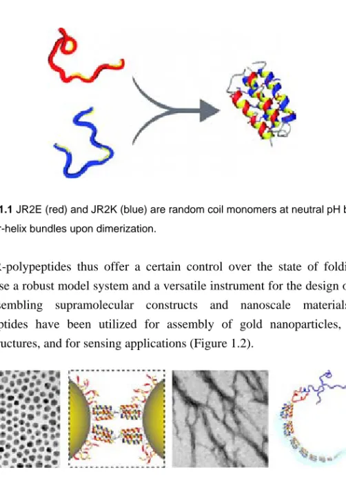
The common denominator of the included papers is a set of amphiphilic de novo designed synthetic helix-loop-helix polypeptides. The so called JR-polypeptides are 42-mer helix-loop-helix polypeptides that were designed to fold into four-helix bundles upon dimerization.[5] The JR-polypeptides are rich in either Glu (JR2E) or Lys (JR2K), which render them a high negative or positive net charge, respectively. Charge repulsion prevents homodimerization at neutral pH while promoting heterodimerization through the formation of stabilizing salt bridges. As monomers the polypeptides are random coil but they fold into four-helix bundles upon dimerization (Figure 1.1). Homodimerization can be induced at low and high pH, respectively, as well as at high salt concentrations or in the presence of certain metal ions.
Figure 1.1 JR2E (red) and JR2K (blue) are random coil monomers at neutral pH but fold
into four-helix bundles upon dimerization.
The JR-polypeptides thus offer a certain control over the state of folding and comprise a robust model system and a versatile instrument for the design of novel self-assembling supramolecular constructs and nanoscale materials. The polypeptides have been utilized for assembly of gold nanoparticles, fibrous nanostructures, and for sensing applications (Figure 1.2).
Figure 1.2 The polypeptides have been utilized for controlled and reversible assembly of
gold nanoparticles and fibrous nanostructures.
The thesis is divided into two parts, where the aim of the first part is to give an introduction and a brief overview of the subjects relating to the thesis work. The second part of the thesis is devoted to the obtained results, which are presented and discussed in seven papers. The papers are briefly summarized in chapter 9.
Molecular self-assembly is very likely to become an important methodology for nanofabrication. Building with molecules, however, requires a deep understanding of their individual structure, assembly properties and dynamic behaviour; questions that are addressed in this thesis.
“The objectives of self-assembly are to make structures that cannot be made by other means and to understand one aspect of life.” Whitesides, 2002.[6]
2. Self-Assembly and Self-Organization
The terminology of “Self-Assembly” and “Self-Organization” are not really clear-cut and the two words are often used interchangeably and sometimes in association. Whitesides defines self-assembly as “the autonomous organization of components into patterns or structures without human intervention”.[6] He further divides the process of assembly into two groups; static and dynamic self-assembly. Static self-assembly occurs as a result of free energy minimization in a closed system and leads to an equilibrium state, whereas dynamic self-assembly occurs far from equilibrium and as a result of energy dissipation. This definition embraces a wide variety of processes covering the whole length scale from nano to parsec but is limited to such interactions that involve pre-existing components (separate or distinct parts of a disordered structure), are reversible, and can be controlled by proper design of the components.[6, 7] Lehn, on the other hand, regards self-assembly “as simple collection and aggregation of components into a confined entity”[8] and distinguish it from self-organization which he proposes is “the spontaneous but information-directed generation of organized functional structures in equilibrium conditions”[8]. The information that directs the molecular species into ordered structures is encoded in the covalent framework of the involved molecules and the organization occurs as a result of specific supramolecular interactions. Kirschner et al. consider self-organization from a biological perspective, as an extension of self-assembly[9] where self-organization in contrast to self-assembly “gives structures under a wider set of conditions; the
SELF-ASSEMBLY AND SELF-ORGANIZATION
rules tend to be more general and the structures more variable”.[10] Kirschner et al. further concludes that self-organizing systems are characterized by reaching a steady state, where there is continuous energy consumption and gain and loss of material.[10] This view on self-organization is similar to Whitesides definition of dynamic self-assembly and both definitions also allude to the processes responsible for the assembly, and survival, of all living organisms.
Despite some discrepancies in definitions and terminology, there is a consensus in that self-assembly and self-organization are the perhaps only, or at least the most practical, strategies for making ensembles of nanostructures.[11, 12]
2.1 Supramolecular chemistry
Assembly of organic nanostructures and hybrid materials most often rely on non-covalent molecular interactions. This area of chemistry, focused on the association of molecular species that are held together solely by intermolecular forces, is called supramolecular chemistry and is often referred to as “the chemistry beyond the molecule”.[8] Whereas traditional chemistry focuses on the covalent bond, supramolecular chemistry exploits the weaker and reversible interactions between molecules such as:
- hydrogen bonding - metal coordination - hydrophobic effects - van der Waals forces - π-π interactions
- electrostatic interactions
These forces are utilized in order to form organized, complex entities from the association of two or more species. The assembly can, as in host-guest chemistry, be driven by association of smaller molecules and ions with larger molecules and complexes, or involve macromolecules of the same size range that interact through multiple interactions in a concerted fashion.[13] Examples of host-guest chemistry include binding of a substrate by an enzyme or the coordination of metal ions by a chelating ligand.
Molecular recognition (selectivity and specificity) is crucial in order to obtain predictable structures and is the basis for programmable assembly. Another key component in supramolecular chemistry is cooperativity. As the individual interactions in a supramolecule may be weak, the association of the components requires multiple interactions that act in concert. In this way the resulting binding strength can become synergistically very much stronger than the mere sum of the parts. It is also very common within self-assembling systems to have more than one type of interaction present. In such cases, the system acts to optimize the product by forming those structures that have the lowest overall free energy. The assembly then tends to occur stepwise where the most stabilizing interactions are formed first and then proceed in a descending hierarchical order.[13]
Supramolecular chemistry has provided new insights in how to design synthetic molecular compounds that self-assemble in a programmable fashion but has also put new light on the chemistry of life, which heavily relies on non-covalent interactions.
2.2 Bionanotechnology
Bionanotechnology is the area of science where biology and nanotechnology meet and are combined in order to create synthetic self-assembling nanostructures. Nature gives the most striking examples on the strength of self-assembly when it comes to producing complex functional nano- and micro-architectures.[12] And based on the definitions mentioned above, one can claim that the creation of all living organisms, from bacteria to humans, is a result of self-assembly.
Feynman considered biology as a source of inspiration for the new generation of physicists in their pioneering efforts within the then newly invented field of nanotechnology: “A biological system can be exceedingly small. Many of the cells are very tiny, but they are very active; they manufacture various substances; they walk around; they wiggle; and they do all kinds of marvellous things - all on a very small scale. Also, they store information. Consider the possibility that we too can make a thing very small which does what we want - that we can manufacture an object that manoeuvres at that level!”.[1]
SELF-ASSEMBLY AND SELF-ORGANIZATION
Nature is truly full of amazing examples of supramolecular constructs that spontaneously self-assemble into functional nanostructures far more complex than any man-made nanodevice. Just to mention a few; the ribosome which carry out DNA translation and synthesis of proteins, ATP-synthase which is a proton driven molecular machine that produces the intracellular energy carrier ATP, and not to forget the protein assemblies constituting the photosystems in photosynthetic cells that collects photons and converts their energy into chemical energy.[14]
It is therefore not surprising that much of the work on nanosized self-assembling systems has been based on biologically derived or biomimetic molecules and biomolecular interactions. DNA has for example been widely studied and employed as a building block for nanostructures as it combines self-assembly with programmability and a plethora of chemical techniques for its manipulation.[15] This has resulted in the development of DNA based molecular motors,[16, 17] and computational devices,[18] as well as astonishing two- and three-dimensional constructs of DNA and the development of the field called “DNA origami”.[19, 20] Micrometre long DNA chains can also be stretched on surfaces and used as wire templates for bioorganic electronic applications.[21, 22] In addition, DNA has also been demonstrated as an effective and specific means to control the assembly of nanoparticles.[23, 24]
Despite the many interesting features of DNA, it is chemically and structurally inferior to proteins. Proteins are far more complex and versatile when considering their three dimensional structure as well as their chemical and biological functionality. The complexity stems from the huge number of degrees of freedom owing to their chemical composition. Proteins have thus become flexible and powerful molecular tools for various technological applications. Of particular interest is the ability of some proteins to specifically recognize other molecular species, which has been extensively employed in bioassays. Proteins have also been extensively employed for creating supramolecular nano-structures, such as synthetic spider silk,[25] molecular wires,[26] and for assembly of nanoparticles.[27,
28]
Most proteins are, however, very sensitive to changes in their physiochemical environment and can easily and irreversible lose their native conformation. In this perspective, designed polypeptides, that spontaneously adopts an ordered secondary and tertiary structure has emerged as an interesting alternative for
proteins. Polypeptides do have the same chemical diversity as proteins but are typically more robust and can also be manufactured at a large scale to a relatively low cost. As the design rules for obtaining functional and folded polypeptides are gradually being elucidated, novel polypeptides with new shapes and new functions are being realized.[29, 30] Polypeptides have in recent years also been exploited in the design of self-assembling nanostructures,[31] and hybrid materials.[32, 33]
2.3 Self-Assembly of hybrid materials and nanocomposites
Numerous well-established man-made materials, known as composites, are made as mixtures between organic and inorganic compounds which are combined in such a way that the resulting materials attain properties that the individual components by them self cannot attain.[34] The inorganic building blocks in such materials are often of macroscopic dimensions and dispersed in an organic matrix, e.g. inorganic fibre-reinforced plastics. A large number of composite materials can also be found in nature, such as bone and teeth, which consists of hard inorganic crystals of hydroxyapatite, that are reinforced with collagen fibres.[35] Many natural composites have outstanding mechanical properties, far beyond those that can be achieved using similar synthetic materials. They typically consist of both inorganic and organic materials that are hierarchically and spatially organized at the nano-, micro-, and meso-levels. In addition, the dimensions of the inorganic components in natural composites are of approximately the same size as the organic species (< 50 nm), and consequently the properties of the materials will be very different showing characteristics in between the pure compounds or even display entirely new properties.[36, 37] Large efforts have therefore been made in order to develop composite materials with spatial and chemical control on the nanoscale which not only can provide improved mechanical properties but also afford so called functional or smart materials, that are switchable and able to react to changes in their physiochemical environment.[38]
These types of materials are usually divided into two subgroups, hybrid materials and nanocomposites, based on the size of their constituents.[38] Hybrid materials are typically considered as homogenous materials that includes two moieties, one inorganic and one organic, blended at the molecular level. The term nano-composite is used if one of the structural units, either the inorganic or organic, is
SELF-ASSEMBLY AND SELF-ORGANIZATION
in a defined size of 1-100 nm. There is however no clear boundary between the two types of materials and the terms are often used interchangeably.[38]
Apart from conventional soft chemistry routes, self-assembly has emerged as a natural method for fabrication of both hybrid materials and nanocomposites. The large variation in possible building blocks, both when considering the inorganic part as well as the organic components, allows fabrication of an amazing amount of architectures using very different assembling strategies. Nanocomposites containing for example noble metal particles with plasmonic properties have in recent years found many interesting applications and are a subject of intense research. A large part of the work in this thesis has been devoted to the study and development of such self-assembling materials.
“After taking into consideration the surrounding water, we actually have no idea of the magnitude of the interactions within folded proteins, of the real entropy balance, and there is no hope of evaluating them solely by the logical analysis of folding an abstract polypeptide chain into an abstract ordered conformation” Privalov, 1992.[49]
3. Peptides, proteins and protein structures
A peptide is a linear chain of covalently linked amino acids with a defined length and sequence. The amino acids are joined end-to-end through an amide bond that is formed when the carboxyl group of one amino acid condenses with the amino group of another (Figure 3.1). When the number of amino acids in the peptide exceeds about 20, it is usually referred to as a polypeptide. The number of amino acids in naturally occurring polypeptides, or proteins, range from approximately 50 up to as many as 27.000, and they generally have a defined three-dimensional structure under physiological conditions.[39, 40]
Figure 3.1 A peptide is formed when two or more amino acids are joined covalently through an amide bond (shaded area).
The peptide backbone has a distinct double bond character due to the electron delocalization over the π-orbital system involving the carbonyl oxygen, carbonyl carbon and the amide nitrogen. The atoms involved in the peptide bond and the connected α-carbons are positioned in a common plane. The only degrees of
PEPTIDES,PROTEINS AND PROTEIN STRUCTURES
freedom are thus rotations around the α-carbon, carboxyl carbon and amide nitrogen atoms.[41] The relative rotational positions of the backbone are defined by its dihedral angles.
There are 20 different naturally occurring amino acids, which are distinguished by their side-chain groups (R1 and R2 in Figure 3.1).[42] The different nature of the
side chains give the amino acids a variety of chemical properties that roughly can be divided into three groups; nonpolar, polar, and charged. When connected into a single molecule, the sequence and number of amino acids can be varied in an infinite number of ways, giving rise to polypeptides and proteins with chemical properties far more complex than just the sum of its constituents.
3.1 Structure and folding of proteins
Proteins assemble into their native fold, or three-dimensional structure, as a result of a large number of concerted inter- and intra-molecular interactions. The outcome is heavily dependent on the sequence of the amino acids. Many proteins fold spontaneously after biosynthesis whilst others require some assistance from e.g. chaperones or enzymes to adopt their native conformation.[43, 44] Because of the very large number of degrees of freedom in an unfolded polypeptide chain, the molecule has an astronomical number of potential conformations. Hence, the time for a protein to find its native fold would be enormous if the protein was to attain the correct fold by sequentially sampling through all possible conformations.[45, 46] This is obviously not the case as proteins typically fold on a timescale of seconds or less. Proteins rather seem to fold as a result of an energy gradient or rugged “funnel” that guides the random tangle to its native conformation.[47]
The interactions involved in protein folding are largely the same as those mentioned in chapter 2.1 on supramolecular chemistry (vide supra). In particular, the hydrophobic effect, which can be regarded as the tendency for nonpolar substances to aggregate in aqueous media, is considered to play a central role.[48] This is manifested by the burying and clustering of hydrophobic side chains in order to minimize their contact with water as the hydrophobic residues induce an ordering of the water molecules.[41, 48] The folding of a polypeptide chain thus involves a gain in entropy due to the burial of nonpolar residues. This entropy gain is however reduced by the loss in configurational entropy of the polypeptide,
which means that the overall entropy change of protein folding and unfolding is close to zero at physiological temperatures.[49] The net stabilization of proteins is therefore mainly a result of the cooperative action of a large number of intramolecular interactions, such as salt-bridge formation and hydrogen bonds. The stability of a protein hardly ever exceeds 50 kJ mol-1 in free energy, which corresponds to the energy of about three hydrogen bonds.[50]
The peptide backbone is very hydrophilic, with one hydrogen bond donor, NH, and one acceptor, C=O, in each peptide unit. As the interior of proteins typically is very hydrophobic the free energy is minimized through formation of hydrogen bonds between the polar atoms of the backbone. These hydrogen bonds result in a regular secondary structure, which is usually one of two types: alpha-helix or
beta-sheet.[51] The secondary structure elements then pack into often rather dense structures. The interior residues of proteins are actually packed as tightly as crystalline amino acids.[41]
Lack of ordered secondary structure is referred to as random coil, while a protein that is not fully folded neither fully unfolded is in a molten globule state.[40] Although many proteins adopt a regular and close-packed three dimensional structure they are not static, but constitute flexible molecules that can go through significant structural fluctuations.[41, 52]
3.2 The alpha-helix
When a stretch of consecutive amino acid residues all have the dihedral angles for rotation around the α-carbon (Φ, Ψ) close to -60o
and -50o, the polypeptide chain will adopt an α-helical conformation. Each completed turn incorporates 3.6 amino acid residues and raises 5.6 Å along the helix axis direction.[14] This conformation places the hydrogen bonding atoms in near perfect alignment. In addition, the radius of the helix allows for favourable van der Waals interactions across the helix axis and the side chains are well staggered which minimize steric interferences. Hydrogen bonds occur in the ordinary α-helix between the carbonyl carbon of residue n and the amide nitrogen of residue n+4. Since all the dipole moments of each peptide unit are directed in the same direction along the helix axis, the α-helix has a significant net dipole moment that gives rise to a partial
PEPTIDES,PROTEINS AND PROTEIN STRUCTURES
positive charge at the amino terminal and a partial negative charge at the carboxy-terminal. [51]
3.3 The four-helix bundle
A simple combination of a few secondary structures in a specific geometric arrangement is often referred to as supersecondary structure, or motif.[42] The simplest supersecondary structure with a specific function is the helix-loop-helix motif consisting of two α-helices joined by a loop region (Figure 3.2 a). An isolated α-helix is only marginally stable in solution due to the lack of stabilization of adjacent hydrophobic side chains. Stabilization can be achieved by the formation of domains. A domain is a combination of secondary structure elements and motifs, also commonly referred to as tertiary structure. A common naturally occurring domain is the four-helix bundle where four amphiphilic α-helices are arranged in a bundle with the helical axis almost parallel to each other (Figure 3.2 b). In this way the hydrophobic side chains are effectively buried between the helices and the hydrophilic side chains are exposed to the solvent.
a) b)
Figure 3.2a) A helix-loop-helix motif (PDB entry 2gp8). b) A four-helix bundle formed by the association of two helix-loop-helix motifs (PDB entry 1u7j). The structures were obtained in the Protein Data Bank (PDB)[53] and rendered using the Polyview-3D visualization server.[54]
3.4 Protein adsorption on surfaces
Proteins have a tendency to accumulate on surfaces as a result of the high standard free energy at the interface. When proteins adsorb on a surface, the free energy of the system decreases and the surface is thermodynamically stabilized.[55] The mechanisms of protein adsorption are very complex and there seems to be a large number of dynamic constraints affecting the process. The adsorption is most often irreversible, but exchange reaction may take place in the presence of other proteins. The adsorption can also result in two-dimensional phase transitions and crystalline packing of the proteins on the surface.[56] The amount of protein adsorbed is determined by several factors including properties of the proteins, the type of surface and the solvent. The properties of the proteins affecting adsorption are such as the charge, size, stability of the protein, amino acid composition, and conformation.[55] Proteins with low internal stability (e.g human serum albumin and immunoglobulin G) tend to adsorb on all types of surfaces irrespective of electrostatic interactions whereas proteins with high internal stability (e.g. ribonucleases and lysozymes), generally adsorb to a very low extent on hydrophilic surfaces, unless there is an electrostatic attraction.[55] Upon adsorption bonds are formed between the protein and the solid surface and the protein may unfold during the process of optimizing the contact with the surface. The unfolding is in general not complete and the degree of remaining secondary structure is often significant. Protein structure is, however, not necessarily disrupted upon adsorption and induction of secondary structure of proteins upon adsorption has also been observed.[57, 58] Possibilities to induce folding of synthetic de novo designed polypeptides by adsorption to monolayer protected gold clusters,[59] and silica nanoparticles[60, 61] have recently been demonstrated.
3.5 Determination of protein structure
To fully characterize the three-dimensional structure of proteins and polypeptides is tedious and time consuming work. Such an undertaking involves either X-ray crystallography or Nuclear Magnetic Resonance (NMR) spectroscopy. Fourier transform infrared (FT-IR) spectroscopy and circular dichroism (CD) spectroscopy are two frequently used techniques for structural characterization of proteins and polypeptides that are less laborious, but that still provide both qualitative and quantitative information.
PEPTIDES,PROTEINS AND PROTEIN STRUCTURES
3.5.1 Infrared spectroscopy of polypeptides
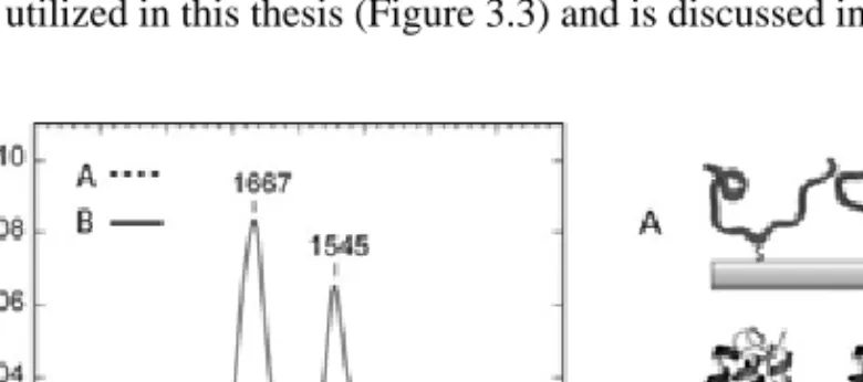
The infrared (IR) spectra of polypeptides show nine separate bands that originate from vibrational modes of the polypeptide backbone; amide A, amide B and amide I-VII. For structural characterization of polypeptides most information can be obtained from the amide I band. This vibrational mode is due to the C=O stretching vibration of the amide group coupled to the in plane bending of the N-H and stretching of the C-N bonds.[62] Amide II is also often studied when evaluating secondary structure. This vibrational mode is more complex and originates from an out of phase combination of NH in plane bend and CN stretch with minor contributions from CO in plane bending, CC stretch and NC stretch.[63] Unfortunately, many of the side chains of polypeptides have characteristic absorption bands in the same frequency range as the amide I and II bands and may account for as much as 15-20% of the total integrated intensity in this region.[62]
The amide bands, amide I in particular, are very sensitive to hydrogen bonding patterns, dipole-dipole interactions and the geometry of the polypeptide backbone. Different secondary structures therefore give rise to characteristic absorption frequencies. The formation of hydrogen bonds between the amide C=O and N-H groups in polypeptides leads to a redistribution of electrons among the atoms involved in the peptide bond. The original loss of negative charge at the donor atom due to the formation of the hydrogen bond tends to be overcompensated by the molecule, which actually leads to an increased electron density at the donor atom. The acceptor atom passes negative charge to the rest of the molecule to compensate for the loss of fractional positive charge. This results in weaker NH and CO bonds and stronger CN bonds.[64] The weakening of the CO bond makes the amide I band appear at lower absorption frequencies while the stronger CN bond will make the amide II band appear at higher frequencies. The stronger the hydrogen bonds the lower the wavenumber of the amide I band. The strongest hydrogen bonds are formed in extended and aggregated polypeptide chains due to the possibility for the chains to come in close proximity to each other and form very strong intermolecular hydrogen bonds. The amide I band for proteins known to be predominantly α-helical is commonly positioned between 1648 and 1660 cm-1 and for denaturated and aggregated proteins between 1610 and 1628 cm-1,[65] which is in good agreement with calculations made by for example Krimm and Bandekar.[66] Typical amide I frequencies for a number of common protein structures are presented in table 3.1. The amide II peak is not as sensitive to
differences in secondary structure as the amide I but is commonly seen in the spectral range 1545 to 1551 cm-1 for α-helical proteins.[65] In contrast to the amide I, the amide II mode is rather sensitive to dehydration and typically shifts by 5-8 cm-1 when dried as a result of the reduction of hydrogen bond interactions with water.[67]
Table 3.1 Common protein structures and corresponding amide I frequencies [65]
Structure Amide I frequency (cm-1)
antiparallel β-sheet/aggregated strands 1675-1695
310-helix 1660-1670
α-helix 1648-1660
random coil 1640-1648
β-sheet 1625-1640
aggregated strands 1610-1628
Data from studies of globular proteins known to be predominantly helical, such as albumin, also suggest that there exists a correlation between the intensity ratio of the amide II band to the amide I band and the α-helix content.[68] Typically the amide I to amide II intensity ratio adopts values of 1.5-2 in isotropic (non-polarized) transmission-absorption spectra of native proteins.[67] For unfolded proteins this ratio may decrease to about 1.[68] This has also been observed for polypeptides utilized in this thesis (Figure 3.3) and is discussed in Paper I.
Figure 3.3 Infrared reflection absorption spectra (IRAS) of a polypeptide (JR2EC)
immobilized on gold surfaces, before (- - -) and after (——) heterodimerization with the complementary polypeptide JR2K. Heterodimerization induces folding into four-helix bundles.
PEPTIDES,PROTEINS AND PROTEIN STRUCTURES
3.5.2 Circular dichroism spectroscopy
Circular dichroism (CD) spectroscopy is a widespread technique for studies of protein secondary structure.[69-71] Different secondary structure elements give rise to characteristic spectra that enables characterization and quantification of secondary structure elements. CD is observed for molecules that are optically active, or chiral, which means that they absorb left and right circularly polarized light to a different extent. For proteins the CD arises in the far-UV region (λ < 240 nm) as a result of the chiral properties of the polypeptide backbone. The difference in extinction coefficients (Δε) for right-handed (R) and left-handed (L) circularly polarized light results in a difference in absorption (ΔA) of the two
components according to the Lambert-Beer relation (eq 3.1).
eq. 3.1 ε Δ ⋅ ⋅ = ΔA l c
where l is the optical path length and c the sample concentration. CD is usually expressed in terms of the ellipticity Θ since the difference in amplitude between the two polarization components caused by the difference in absorption results in an elliptical polarization of the transmitted light. The ellipticity is defined by the angle between the major and minor axis of the ellipse described by the sum vector of the left and right polarized components and is measured as a function of wavelength and related to ΔA by the expression (eq. 3.2):
eq. 3.2 A A Δ ⋅ ≈ Δ ⋅ ⋅ = Θ 33 4 180 10 ln π
For proteins the result is commonly presented as the mean residue ellipticity [Θ] which is related to the ellipticity (Θ) by the following expression (eq. 3.3).
[ ]
c l mrw ⋅ ⋅ ⋅ Θ = Θ 10 eq. 3.3where mrw is the mean residue weight.
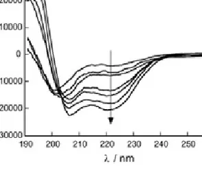
The CD spectra of helical proteins typically display pronounced minima at 208 and 222 nm and a maximum around 190 nm (Figure 3.4). The mean residue ellipticity at 222 nm is often used as a measure of the helical content of a protein.
Figure 3.4 CD-spectra in the far UV-region of a polypeptide (JR2E) that goes from
random coil to a helical conformation. The helicity increases in the direction of the arrow.
To obtain quantitative data from CD spectra, access to spectra of proteins with known structure is necessary. Given a data set of reference spectra, various algorithms implemented in software packages such as the CONTIN/LL,[72] SELCON3,[73] and CDSSTR,[74] can be employed to extract the amount of the various secondary structure elements that are present in the peptide or protein.
”Where Nature finishes producing its own species, man begins, using natural things and with the help of this nature, to create an infinity of species” Leonardo da Vinci (1452-1519).
4. Polypeptide design and synthesis
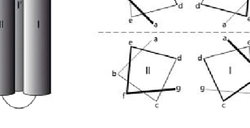
An increasing knowledge on how the sequence of amino acids determines the three-dimensional structure of a polypeptide chain has made it possible to design polypeptides that adopt a regular and ordered conformation. De novo design of proteins not only extend our knowledge about the rules governing protein folding but enables realization of new molecular entities similar to native proteins but with entirely new properties and functions. One of the most common design motifs is the four-helix-bundle formed from amphiphilic helices (Figure 4.1 a).[29] Typically, shape complementarities and hydrophobic interactions drive the formation of the supersecondary structure. Since one turn of an α-helix involves about four residues, the introduction of hydrophobic amino acids in approximately every fourth position results in an helix with a hydrophobic face. This approach can be described in terms of a heptad repeat (abcdefg)n pattern (Figure 4.1 b) were
the positions a and d usually contains hydrophobic residues that form the hydrophobic core upon folding. The residues in g and c positions are part of the solvent exposed face and the residues in b and e positions are at the dimer interface and control dimerization.[51] Two designed helix-loop-helix motifs can under the right conditions dimerize to form a four-helix bundle. The dimerization process can lead to a parallel, an anti-parallel [75] or a bisecting U motif [76].
The design of the polypeptides used throughout this thesis is based on the SA-42 polypeptide motif.[77, 78] SA-42 is a helix-loop-helix polypeptide that was de novo
DESIGN AND SYNTHESIS OF POLYPEPTIDES
a) b)
Figure 4.1a) The polypeptides utilized throughout this thesis dimerize in an antiparallel fashion to form four-helix bundles.[77, 78] b) The heptad repeat pattern is utilized to illustrate the relative position of the amino acids in the antiparallel helix-loop-helix dimers. The dashed line represents the dimerization interface. Figures redrawn from [79].
designed to dimerize and fold into a four-helix bundle. The polypeptide was also designed to catalyze ester hydrolysis, but its reactivity was found to be very low. SA-42 is largely helical with a mean residue ellipticity at 222 nm of -25 000 deg cm2 dmol-1, and from NMR-spectroscopy data it was concluded that it predominantly forms a hairpin helix-loop-helix that dimerize in an antiparallel mode. Dimerization was further confirmed using analytical ultra-centrifugation.[77]
4.1 The JR-polypeptides
The JR-polypeptides, JR2E and JR2K, were designed by Johan Rydberg and Sarojini Vijayalekshmi to form heterodimeric four-helix bundles.[80] The design of the 42-residue helix-loop-helix polypeptides was based on SA-42, but they were modified with a large abundance of either Glu (JR2E) or Lys (JR2K), giving them controllable dimerization properties. The amino acid sequences are presented in Figure 4.2. The net charge of JR2E and JR2K at pH 7 is -5 and +11, respectively. The charged residues are mainly incorporated in the b and e positions in the heptad repeat pattern, which are located on the faces of the helices that control dimerization.
JR2E 1 19 H3N+-N-A-A-D-L-E-K-A-I-E-A-L-E-K-H-L-E-A-K 20 23 -G-P-V-D 42 24 -OOC-G-A-R-E-F-A-E-F-A-Q-E-L-Q-K-E-L-Q-A-A JR2K 1 19 H3N+-N-A-A-D-L-K-K-A-I-K-A-L-K-K-H-L-K-A-K 20 23 -G-P-V-D 42 24 -OOC-G-A-R-K-F-A-K-F-A-Q-K-L-Q-K-K-L-Q-A-A
Figure 4.2 The amino acid sequences of JR2E and JR2K.
Due to the charge repulsion, the peptides exists a random coil monomers at neutral pH and at moderate salt concentrations (< 0.5 mM). When the two peptides are mixed at equimolar concentrations they heterodimerize and fold, resulting in a large increase in helicity (Figure 4.3 a). The driving force for folding is the formation of the hydrophobic core made up by hydrophobic residues in a and d position. Dimerization has been confirmed using analytical ultracentrifugation.[80]
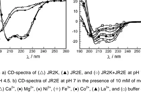
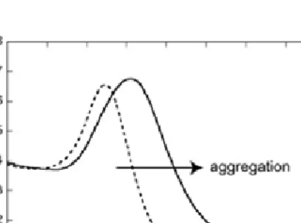
Homodimerization is induced when lowering the charge repulsion between the monomers by protonating or deprotonating acidic and basic residues, respectively. For JR2K this occurs at pH > 10,[80] whereas JR2E homodimerizes below pH 6 (Figure 4.3 a). The pH dependent homodimerization of JR2E is described in Paper I and Paper II. As is reported in Paper III, homodimerization of JR2E could also be induced at neutral pH in the presence of a number of metal ions, such as Ni2+, La3+, Co2+ and Zn2+ (Figure 4.3 b). The largest folding tendency was seen for Zn2+.
DESIGN AND SYNTHESIS OF POLYPEPTIDES
b) a)
Figure 4.3a) CD-spectra of (U) JR2K, (▲) JR2E, and (○) JR2K+JR2E at pH 7 and (●)
JR2E at pH 4.5. b) CD-spectra of JR2E at pH 7 in the presence of 10 mM of metal salts: (○) Zn2+ , (U) Ca2+ , (♦) Mg2+ , (×) Ni2+, (¨) Fe3+ , (●) Co2+ , (▲) La3+
, and (□) buffer only.
In order to allow for directed, thiol-dependent immobilization of the JR-polypeptides on gold substrates, Val22 was exchanged for Cys, affording the peptides JR2EC and JR2KC. Position 22 is located in the loop region and was chosen in order to minimize the influence of immobilization on dimerization. No effects of the Cys on the secondary structure were observed for either the homodimer or the heterodimer when in solution, irrespectively if homodimerization was induced by Zn2+. The Cys residue was also utilized to covalently join two peptide monomers via a disulphide bridge. The disulphide-linked peptides were found to spontaneously and rapidly assemble into micrometre long fibres as a result of a propagating folding mediated association (Paper V). Both hetero- and homo-associating fibres were obtained.
An additional peptide that was unable to dimerize and fold was designed and utilized as a reference polypeptide when investigating the influence of dimerization and folding on the assembly of peptide-modified gold nanoparticles and peptide fibres. This peptide, JR2ECref, has the same amino acid sequence as JR2EC with the exception that all L-Ala were replaced by D-Ala, while the other residues were kept in the L-state. Peptides with mixed D- and L-amino acids cannot generally adopt an ordered secondary structure.
4.2 The KE-polypeptides
The 42-residue helix-loop-helix polypeptide KE2 was designed by Karin Enander as a scaffold for biosensor applications.[81, 82] The design was based on LA-42b, which is closely related to SA-42, but modified in order to catalyze site-selective self-functionalization.[83] KE2 was modified with an affinity ligand and fluorescent probes to enable specific binding of target proteins and reporting on this event combined in the same molecular entity. A number of different derivatives of benzenesulphonamide were investigated as ligands and were attached to the polypeptide scaffold using an orthogonal protection group strategy. Benzenesulphonamide (H2NO2SC6H5) is an inhibitor of the 29 kDa, enzyme
human carbonic anhydrase II (HCAII) and binds with a Kd of 1.5 μM.[84] In order
to detect binding, a number of different environmentally sensitive fluorescent probes were incorporated in various positions. In addition, the affinity for HCAII could be tailored by varying the length and hydrophobicity of the spacer between the benzenesulphonamide moiety and the polypeptide, which was utilized in order to create an affinity array.[85]
In this thesis, KE2 was modified to allow for thiol dependent immobilization on gold surfaces by replacing Val in position 22 by Cys, affording the polypeptide KE2C (Figure 4.4). KE2C was further functionalized with a benzene-sulphonamide derivate with an aliphatic spacer having five methylene units in order to obtain the sensor peptide KE2C-C6. The corresponding fluorophore modified sensor peptide KE2-D(15)-5 binds HCAII with a Kd of 0.02 μM.[82]
KE2C 1 19 Ac-N-A-A-D-L-E-A-A-I-R-H-L-A-E-K-L-A-A-R 20 23 -G-P-C-D 42 24 -OOC-G-A-R-A-F-A-E-F-K-K-A-L-Q-E-A-L-Q-A-A
DESIGN AND SYNTHESIS OF POLYPEPTIDES
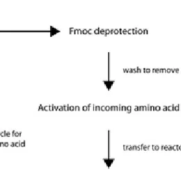
4.3 Peptide Synthesis
The peptides utilized throughout this thesis were prepared using an automated continuous flow solid-phase peptide synthesizer. The procedure is schematically summarized in Figure 4.5, below. The insoluble solid support allows soluble by-products and excess reagents to be conveniently removed. The C-terminal amino acid was covalently bound to the solid support (typically polyethylene glycol grafted polystyrene) and the following amino acids were added sequentially one at the time to the growing chain. The α-amino group of the amino acids was protected with a fluorenylmethyloxycarbonyl (Fmoc) group that was removed by treatment with a mild base before addition of the following activated amino acid. Amino acids with reactive side chains were protected with groups that could be removed under the conditions used when cleaving the peptide from the solid support.
After synthesis and cleavage from the support material the peptides were purified by reversed phase high-performance liquid chromatography (HPLC) and identified from their matrix-assisted laser desorption ionization time of flight (MALDI-TOF) spectra.
“The state of division of these particles must be extreme; they have not as yet been
seen by any power of the microscope.” Faraday, 1857.[92]
5. Colloidal Gold
In the bulk form, gold is a soft, yellow metal, with the face centred cubic crystal structure, a melting point of 1064o C and excellent electrical conductivity. Interestingly, not one of these properties necessary applies to gold colloids on the nanometre scale,[86] and the smaller they are the more do their properties deviate from the bulk material.[87, 88]
The many interesting properties of colloidal gold have fascinated humanity since the 5th or 4th century B.C. The technique of preparing “soluble gold” was first discovered and used in Egypt and China and adopted by the Romans who utilized it for staining of glass.[89] The Lycurgus Cup, is the perhaps most famous preserved piece from that time and is one of a class of Roman Vessels known as
cage cups or diatreta.[90] The Lycurgus Cup has a very remarkable feature; it appears ruby red in transmitted light while it has an opaque greenish-yellow tone in reflected light. The origin of this dichroic property has been confirmed to the presence of gold and silver particles, 50-100 nm in diameter, embedded in the glass.[91] The Roman glass makers, however, probably had a very vague idea about the cause of the beautiful colours and it was not until the mid 19th century that the properties of colloidal gold was examined and described in more detail by Michael Faraday. Faraday presented his results on the interaction of light with metal particles that were “very minute in their dimensions” in a ground breaking lecture to the Royal Society of London in 1857.[92] Although the term “colloid”
COLLOIDAL GOLD
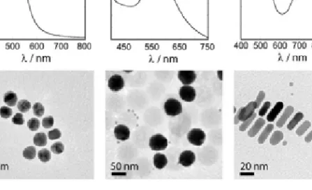
Figure 5.1(Left) Electron micrograph of a 13 nm gold nanoparticle (scale bar 5 nm), that are the cause of the beautiful colours of the dispersions in the photo in the middle. (Right) UV-visible spectrum of a dispersion of gold nanoparticles showing the characteristic extinction peak that gives rise to these colours. (Photo courtesy of Prof. M. Cortie, Institute for Nanoscale Technology, Sydney, Australia.)
was not coined until a few years later by Graham[93] from the Greek word for glue, Faradays work laid the foundation to modern colloid science.[94]
In addition to the interesting optical properties of colloidal gold that gives rise to their beautiful colours (Figure 5.1), monodisperse particles in the size range from ~2 to 100 nm can straightforwardly be prepared using standard protocols. Moreover, they do not easily oxidize and can be functionalized with a wide variety of different molecules, which are properties that make gold nanoparticles very attractive as components in various technological applications.
Except as a colourant in stained glass, the first practical use of gold nanoparticles was as immunohistochemical contrast agent for electron microscopy of tissue samples, a method still widely used.[95] When used for staining, antibodies raised against an antigen of interest are adsorbed on the surface of gold nanoparticles which are subsequently exposed to the sample. If the antigen is present the antibody coated particles might bind, which can easily be visualized using Transmission Electron Microscopy (TEM). Gold nanoparticles functionalized with a range of other biomacromolecules, such as lectins and polysaccharides, have also been employed for cytochemical applications.[96, 97] In addition, gold nanoparticles have been extensively used as a label in immunoassays,[98, 99] and in a number of commercially available products for self-testing, such as the home pregnancy test “First Response”,[100] and more recently as a component in a large number of nanocomposite materials.