Folding and interaction studies of
subunits in protein complexes
Ximena Aguilar Iturri
Umeå University
Department of Chemistry 901 87 Umeå, Sweden
This work is protected by the Swedish Copyright Legislation (Act 1960:729) ISBN: 978-91-7459-795-0
Cover: Protein Human Med25-ACID PDB-code 2XNF Electronic version available at: http://umu.diva-portal.org/ Printed by: VMC-KBC UMeå
Dedicado a toda mi familia en Bolivia, en especial a Mami Olga, mejor amiga Mary, mi ‘gemela’ Claudia y los mellizos Olga Jr y Rodrigo.
Suma yatekanani
“地図がなくても迷うことはないが、目的地がなければ迷ってしまう。” Takemoto Yuta, Honey & Clover
Table of Contents
Table of Contents i Abstract iii List of papers v Abbreviations vii 1. Introduction 1 1.1 Protein folding 1 1.1.1 Macromolecular crowding 3 1.2 Protein-protein interactions 4 1.2.1 Homo-oligomer: cpn10 5 1.2.2 Hetero-protein complex: Mediator 61.3 Mediator and transcription regulation 8
1.4 Molecular recognition - Coupled folding and binding 10
1.4.1 Med25 11
1.4.2 The plant-specific transcription factor Dreb2a 12 1.4.3 The human herpes simplex virus 1 VP16 protein 13
2. Aims 15
3. Methods 17
3.1 Protein stability, folding and binding 17
3.2 CD spectroscopy 19
3.3 Fluorescence spectroscopy 19
3.4 Nuclear Magnetic Resonance 20
3.4.1 Chemical shift perturbation 21
3.5 Surface plasmon resonance 22
3.6 Isothermal titration calorimetry 22
3.7 Cross-linking 23
3.8 GST pull-downs 23
4. Results 24
4.1 Summary of papers 24
4.1.1 Macromolecular crowding stabilizes cpn10 monomers 24 4.1.2 Conformational changes upon interaction between Med25 and Dreb2a 27 4.1.3 Similar binding affinities but different binding energetics 29
5. Discussion 33
5.1 Protein folding studies in cell-like environment 33
5.2 Interaction between transcriptional regulatory proteins 34
5.2.1 Small contacts might end up in great connections 34
6. Conclusions 37
7. Acknowledgements 38
Abstract
Proteins function as worker molecules in the cell and their natural environment is crowded. How they fold in a cell-like environment and how they recognize their interacting partners in such conditions, are questions that underlie the work of this thesis.
Two distinct subjects were investigated using a combination of biochemical- and biophysical methods. First, the unfolding/dissociation of a heptameric protein (cpn10) in the presence of the crowding agent Ficoll 70. Ficoll 70 was used to mimic the crowded environment in the cell and it has been used previously to study macromolecular crowding effects, or excluded volume effects, in protein folding studies. Second, the conformational changes upon interaction between the Mediator subunit Med25 and the transcription factor Dreb2a from Arabidopsis thaliana. Mediator is a transcriptional co-regulator complex which is conserved from yeast to humans. The molecular mechanisms of its action are however not entirely understood. It has been proposed that the Mediator complex conveys regulatory signals from promoter-bound transcription factors (activators/repressors) to the RNA polymerase II machinery through conformational rearrangements.
The results from the folding study showed that cpn10 was stabilized in the presence of Ficoll 70 during thermal- and chemical induced unfolding (GuHCl). The thermal transition midpoint increased by 4°C, and the chemical midpoint by 0.5 M GuHCl as compared to buffer conditions. Also the heptamer-monomer dissociation was affected in the presence of Ficoll 70, the transition midpoint was lower in Ficoll 70 (3.1 µM) compared to in buffer (8.1 µM) thus indicating tighter binding in crowded conditions. The coupled unfolding/dissociation free energy for the heptamer increased by about 36 kJ/mol in Ficoll. Altogether, the results revealed that the stability effect on cpn10 due to macromolecular crowding was larger in the individual monomers (33%) than at the monomer-monomer interfaces (8%).
The results from the interaction study indicated conformational changes upon interaction between the A. thaliana Med25 ACtivator Interaction Domain (ACID) and Dreb2a. Structural changes were probed to originate from unstructured Dreb2a and not from the ACID. Human Med25-ACID was also found to interact with the plant-specific Dreb2a, even though the ACIDs from human and A. thaliana share low sequence homology. Moreover, the human Med25-interacting transcription factor VP16 was found to interact with A. thaliana Med25. Finally, NMR, ITC and pull-down experiments showed that the unrelated transcription factors Dreb2a and
VP16 interact with overlapping regions in the ACIDs of A. thaliana and human Med25.
The results presented in this thesis contribute to previous reports in two different aspects. Firstly, they lend support to the findings that the intracellular environment affects the biophysical properties of proteins. It will therefore be important to continue comparing results between in vitro and cell-like conditions to measure the magnitude of such effects and to improve the understanding of protein folding and thereby misfolding of proteins in cells. Better knowledge of protein misfolding mechanisms is critical since they are associated to several neurodegenerative diseases such as Alzheimer’s and Parkinson's. Secondly, our results substantiate the notion that transcription factors are able to bind multiple targets and that they gain structure upon binding. They also show that subunits of the conserved Mediator complex, despite low sequence homologies, retain a conserved structure and function when comparing evolutionary diverged species.
Keywords: Macromolecular crowding, cpn10, Mediator, Med25, Dreb2a,
List of papers
I) Ximena Aguilar, Christoph F. Weise, Tobias Sparrman,
Magnus Wolf-Watz, and Pernilla Wittung-Stafshede. Macromolecular Crowding Extended to a Heptameric System: The Co-chaperonin Protein 10. Biochemistry 2011; 50 (14), 3034-3044.
Reprinted with permission from Biochemistry, 2011, 50 (14), 3034-3044. Copyright 2011 American Chemical Society.
II) Jeanette Blomberg, Ximena Aguilar, Kristoffer Brännström, Linn Rautio, Anders Olofsson, Pernilla Wittung-Stafshede, and Stefan Björklund. Interactions between DNA, transcriptional regulator Dreb2a and the Med25 mediator subunit from Arabidopsis thaliana involve conformational changes. Nucleic Acids Research 2012; 40: 5938 - 5950.
Reprinted with permission from Nucleic Acids Research, 2012, 40 (13), 5938 - 5950. Copyright 2012 Oxford University Press.
III) Ximena Aguilar, Jeanette Blomberg, Kristoffer Brännström,
Anders Olofsson, Jürgen Schleucher, and Stefan Björklund. Interaction studies of the Human and Arabidopsis thaliana Med25-ACID proteins with the Herpes Simplex virus VP16- and plant-specific Dreb2a transcription factors.
Manuscript.
Abbreviations
ACID ACtivator Interaction Domain
aMed25 Med25 from Arabidopsis thaliana
A. thaliana Arabidopsis thaliana
CD Circular Dichroism
Cpn10 Co-chaperonin protein 10
CSP Chemical Shift Perturbation
DBD DNA Binding Domain
Dreb2a Dehydration responsive element binding
protein
Dreb2af.l. Dreb2a full-length protein E. coli Escherichia coli
F Folded
GST Glutathione S-transferase
GuHCl Guanidine Hydrochloride
hMed25 Med25 from human
HSQC Heteronuclear Single Quantum Coherence
ITC Isothermal Titration Calorimetry
KD Dissociation constant
Med25 Mediator subunit 25
NMR Nuclear Magnetic Resonance
NRD Negative Regulatory Domain
PDB Protein Data Bank
PIC Pre-Initiation Complex
Pol II RNA polymerase II
SDS-PAGE Sodium Dodecyl Sulfate-Polyacrylamide Gel Electrophoresis
SEC Size Exclusion Chromatography
SPR Surface Plasmon Resonance
TAD Transcriptional Activation Domain
TF Transcription Factor
U Unfolded
UV Ultraviolet
VP16 Virion protein 16
1. Introduction
1.1 Protein folding
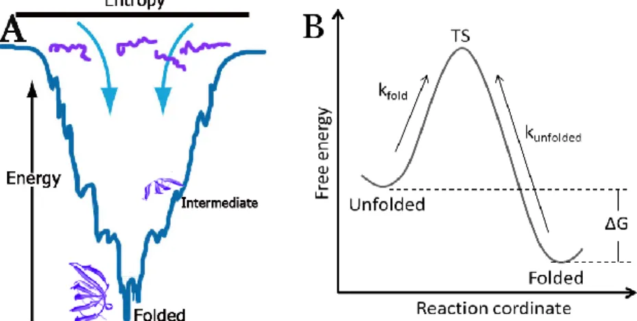
Protein folding is the final step in the central biological process where the genetic information stored in DNA is converted into functional proteins. Newly translated proteins need to acquire a unique structure in order to be functional. How this process works and the mechanisms behind it have been extensively investigated for more than fifty years. The importance to unravel the molecular mechanisms of protein folding relies on the impact proteins have for the proper function of the cell. Proteins are actually the hard worker molecules in living organisms and are involved in all cellular functions. Even though there is a rigid cellular assistance and control for folding - e.g. chaperones which are molecular helpers that prevent misfolding1 - still, polypeptides might escape cellular control to end up as misfolded proteins which have the potential to form aggregates or amyloid fibrils associated with neurodegenerative diseases such as Alzheimer’s and Parkinson’s2,3. A pioneering scientist in this field was Christian Anfinsen who already in the 1960’s, together with co-workers, showed that proteins can fold spontaneously and also refold in vitro. Anfinsen’s work suggested that a protein three-dimensional structure was encoded in its amino acid sequence4,5. However, this statement gave rise to a paradox noted by Cyrus Levinthal, who remarked that a polypeptide chain has a large degree of freedom - depending on the number of amino acids and peptide bonds - which means that if the process of folding is random, then the numbers of possible conformations are massive and it would take a long time for a protein to fold into the correct conformation6. This was called the Levinthal’s paradox since it was already known at that time that proteins fold in the milliseconds timescale. Levinthal therefore proposed that folding occurs through a series of partially structured intermediate states and other researchers reported on the existence of transient folding intermediates7-9. What emerged from these studies was a new way to look at proteins, they are not static and the transition from the unfolded to the folded state occurs in a cooperative manner. In the 1990’s, a theoretical concept was introduced to understand the mechanism of folding; the energy landscape theory10. This concept explains folding as a result of the nature of a molecule that strives for low free energy in order to obtain stability11. The energy landscape theory provides an understanding for the presence of intermediate states. In a simple way, the landscape might be viewed like a funnel-shaped topology, where polypeptides strive to reach a low energy state in a downhill manner
whereby it folds by adopting different intermediate states until reaching the minimal energy level, the folded state12,13 (Figure 1A).
Currently, protein folding is studied by applying thermodynamic laws which state that a system strives to reach a state of minimum energy. One important driving force for the folding process is the hydrophobic effect that excludes water molecules from contact with hydrophobic amino acids, and exposes the hydrophilic amino acids to the aqueous solvent. The energy diagram in Figure 1B illustrates a folding reaction where the principles of Gibbs free energy can be applied (the equations are included in section 3.1). The reaction involves a two-state process where the protein is in equilibrium between the folded and unfolded state, and there is an energy barrier or transition state (TS) to pass from one state to another14. This model can only be applied to proteins that show the two-state transition and as discussed above, most proteins fold in sequential steps by forming intermediates. Then, multiple energy barriers are needed to be passed in order to reach the folded state. That is the reason why the folding process of only certain proteins that “behave well” has been studied experimentally so far. However, new methods and approaches are continuously being developed in order to understand the chemistry and biology of protein folding15-20.
Figure 1. A) Illustration of the free energy landscape for protein folding. Proteins have a high energy level and a large degree of conformational freedom in the unfolded state. The folding process includes the formation of intermediates and the folded state is reached when the degree of conformational freedom and the energy levels are low. B) Energy diagram of a folding reaction for a two-state process. Proteins have to pass an energy barrier represented by the transition state (TS) to go from one state to another.
1.1.1 Macromolecular crowding
Almost all our knowledge about protein folding comes from experiments done in in vitro conditions. The methods that are currently available to study proteins are insufficient to monitor folding directly inside cells. A convenient way to study protein folding so far has been to use isolated, purified stable proteins (model proteins) in dilute buffers in vitro; an environment which is completely different from the crowded intracellular medium encountered within cells (Figure 2). Macromolecules (proteins, nucleic acids, carbohydrates, lipids) occupy a significant fraction of the total volume (10 – 40 %) in the cell and their concentration in the cytoplasm has been estimated to range from 80-400 mg/ml21-24. This crowded environment restricts the available space inside the cell and gives rise to an excluded volume effect termed as ‘macromolecular crowding’25-28. The excluded volume effect is a consequence of the steric repulsion (a molecule occupies a space not accessible to another) of macromolecules and has the potential to influence kinetics and equilibria of cellular reactions29. For individual proteins, the predicted effect is a stabilization of the folded state since compact conformations will be indirectly favored due to unfavorable effects on the extended unfolded states30-32. On the other hand, an increase in volume occupancy may also enhance unwanted association processes leading to aggregate formation33,34.
Figure 2. A) Illustration of a eukaryotic cell depicting subcellular organelles such as the nucleus, mitochondria, endoplasmic reticulum and the Golgi apparatus (asterisk). B) A 3D reconstruction from a high resolution electron tomography of the Golgi apparatus in a mammalian cell. The picture illustrates the high macromolecular content in the intracellular environment. Reprinted from (Marsh BJ 2005)35 Copyright 2005, Elsevier with permission of the Publisher.
Studies of macromolecular crowding effects on protein folding, conformational changes, thermodynamics, kinetics and dynamics is a research field that is currently getting increased attention. To address these questions, one has to perform studies in vivo or in cell-like conditions in vitro. A majority of the experimental crowding studies in vitro mimic the crowded environment by using inert, synthetic, sugar-based polymers termed crowding agents (Ficoll 70 and Dextran)36-40. In such studies, effects of macromolecular crowding on protein stability with respect to unfolding37,40-42, on association equilibria and rates36,43,44, and on enzyme activity45,46 have been reported. Today it is known that the interior cell milieu affects the biophysical properties of proteins and new approaches to investigate how they fold and function in their true environment are continuously being developed.
Part of this thesis (paper I) covers the impact of macromolecular crowding on the folding, assembly and overall stability of the model homo-oligomer protein, co-chaperonin protein 10 (cpn10), which is introduced in section 1.2.1.
1.2 Protein-protein interactions
The fate of a protein can take several directions. Some proteins accomplish their function as monomers, other proteins assemble into complexes, while the fold and function of other proteins depend on the interaction with their biological targets47. Interactions between proteins are essential for several aspects of cellular function, e.g. antigen-antibody binding, signal transduction, biosynthetic and degradation pathways, and regulation of gene expression. Studies have shown that the majority of proteins are involved in complex formation and that a protein on average has about eight interacting partners48-51. The way in which proteins associate ranges from simple oligomers to gigantic multi-protein complexes. Oligomers are present as homo-oligomers composed of several identical polypeptide chains or as hetero-oligomers which contain non-identical polypeptide chains52. These oligomeric states are predominant in the cell and studies have showed that a majority of them are present in cells as homo-oligomers52. Proteins that associate into large complexes function as protein machines, e.g. RNA polymerase II (Pol II), the proteasome and Mediator. The capacity of proteins to form multimeric complexes and their ability to network with other proteins has most likely been a requirement for the development of complex cellular processes53.
How oligomers and protein-complexes self-assemble and then physically interact with several other proteins is not entirely understood. Extensive
research has elucidated some aspects that play important roles for the specific molecular recognition events that are required for interaction between proteins54-58. Just in terms of chemical properties which are restricted to the inter- and intra-protein interfaces, the recognition between the correct partners is a result of several complementary attributes ranging from the amino acid composition, hydrophobicity, electrostatic interactions, hydrogen bonding and hydration. All these chemical attributes contribute to protein-protein recognition and determine the energetics buried at the protein interfaces, which will be distinct for every individual case. Still, in common for all kind of interactions is to keep the free energy of binding in balance in order to lend stability to the complex59-62.
In the following section, two protein complexes that have been part of my thesis project are introduced: 1) Proteins that associate into a homo-heptamer (cpn10) and 2) proteins that assembles into a large hetero-protein complex (Mediator).
1.2.1 Homo-oligomer: cpn10
Cpn10, also known as heat shock protein 10, is an essential protein found in the mitochondrial matrix. Cpn10 homologs are present in most organisms 63-65 and its function is to interact with another oligomer, the cpn60. The cpn60 oligomer forms a chamber, and the cpn10 oligomer operates as the lid of the chamber in an ATP-dependent chaperonin complex-system that assist in the folding process of certain proteins65. The cpn60-cpn10 proteins show conserved structure and function in all organisms and the most widely studied pair is the bacterial GroEL-GroES of E. coli65,66. In addition to the chaperonin function, new functions for human cpn10 have been reported, but they are still poorly understood. For example, cpn10 has been found to be identical to an immunosuppressive early pregnancy factor67. It has also been found in red blood cells68, as a protein which is over-expressed in carcinogenesis69, and it has been formulated as a therapeutic drug for treatment of rheumatoid arthritis70.
The structure of cpn10 is ring-shaped, and the complex is composed of seven identical subunits or monomers. Each monomer comprises 101 residues and each subunit is approximately 10 kDa71. The monomers assemble to form a 70 kDa homo-heptameric protein complex through anti-parallel β-strand pairing between the N-terminal of one monomer and the C-terminal of the neighboring monomer (Figure 3). Each monomer unit adopts an irregular β-barrel structure forming two flexible protruding loops72. Studies in vitro have shown that the monomers are able to fold but their stability in the monomeric state is low73,74. The overall cpn10 heptamer stability has been
shown to be dominated by inter-subunit interactions where the hydrophobic subunit-interfaces interact non-covalently75. The cpn10 oligomer unfolds reversibly in vitro and unfolding/refolding pathways for the cpn10 homologous from E. coli and Aquifex aeolicus have been characterized75-82.
Figure 3. Structure of the human cpn10 heptameric protein which is composed of seven identical monomers. One monomer is highlighted in pink. Subunit-subunit interfaces between monomers, formed between anti-parallel β-strands, are depicted in red and blue71.
Studies of oligomer folding are complicated since they involve both, intramolecular interactions (monomer folding) and intermolecular interactions (subunit assembly)83,84. For cpn10 folding studies, an apparent two-state process for unfolding coupled to dissociation can be applied. Therefore, the cpn10 protein represents an excellent model system for fundamental folding/assembly studies in cell-like conditions and it was thus used as model protein in our studies described in paper I.
1.2.2 Hetero-protein complex: Mediator
In addition to homo-oligomers such as the cpn10 described above, many proteins associate into hetero-protein complexes (e.g. ribosome, proteasome, Mediator). The Pol II transcription machinery in eukaryotes requires a Mediator complex for proper transcription regulation (Figure 4, 5). Mediator is a giant evolutionarily conserved multi-protein complex (~1.2 MDa) and it was originally identified in Saccharomyces cerevisiae in the 1990’s85,86. It is organized into four modules (head, middle, tail and kinase) containing all together between 25 (yeast) and 29 subunits (humans) which share a unified subunit nomenclature87 (Figure 5). Cross-species comparisons have shown that almost all yeast Mediator proteins have homologs in the human complex88. However, it has been found that the Med23, Med25, Med26, Med28 and Med30 (Figure 5) subunits are unique to higher eukaryotes such
as humans and plants89,90. This means that Mediator has diverged over time and an explanation for the existence of additional subunits in higher eukaryotes most probably originates from the requirement for the more complex transcription regulation that is found in multicellular organisms53,89. During the last decades, several studies have described the function and location of individual Mediator subunits. It was found that different subunits are involved in regulating distinct sets of genes by interacting with diverse transcription factors (TFs). It has also been reported that some subunits are essential for survival, e.g. Med17 and Med21 which are required for transcription of all protein-coding genes in yeast91,92. Non-essential subunits have more specialized roles in the regulation of selective genes. It has also been suggested that subunit composition depends of the cell-stage; proliferating cells might have the complete Mediator with all the subunits whereas differentiated cells probably have the Mediator with a subset of subunits93,94. Other studies have shown that subunits function individually, e.g. Med12 that might function independently of the rest of Mediator as a regulator of TGFB signaling in the cytoplasm95.
As exemplified above, the regulatory properties of Mediator are broad and include participation in transcription initiation and elongation96-99. In addition, Mediator has been reported to be involved in mRNA processing and chromatin architecture modification100-103. However, this thesis emphasizes on the role of Mediator when interacting with transcriptional regulatory proteins; an essential function by which Mediator transmit the regulatory signals to Pol II machinery. The interaction of a specific Mediator subunit protein (Med25) and specific TFs (Dehydration responsive element binding protein (Dreb2a) and VP16 were investigated in papers II and III.
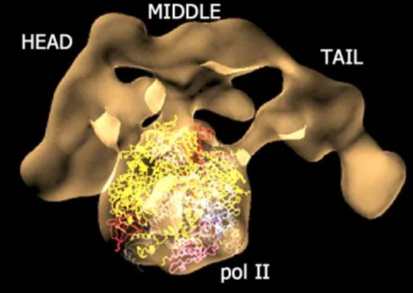
Figure 4. The Mediator complex interacting with Pol II. Cryo-EM structure of Mediator-pol II complex where pol II structure was docked in the central part. Reprinted from Roger D. Kornberg 2007104,105 Copyright 2007, with permission from the National Academy of Sciences U.S.A. and Nature Publishing Group.
1.3 Mediator and transcription regulation
Pol II transcribes all protein-coding genes in eukaryotes and this function is controlled by a range of transcriptional regulatory proteins. There is a group of general transcription factors (TFIIA, TFIIB, TFIID, TFIIE, TFIIF, TFIIH) that assembles and form a pre-initiation complex (PIC) which together with Pol II and Mediator constitute the transcription machinery (~4 MDa) (Figure 5) 105-107. Another important group of regulatory proteins are TFs that activate or repress expression of target genes in response to an environmental or cellular trigger. A feature of TFs is that they contain one or more DNA-binding domains (DBD) which bind to specific DNA sequences or in the promoters of genes. In bacteria, TFs interact directly with RNA polymerase and a Mediator complex is not present. In eukaryotes, TFs do not bind directly to Pol II; they control Pol II activity by interacting with Mediator and other co-factors through their transcriptional activation- or negative regulatory domains (TAD and NRD)108. The question is why the transcription machinery needs the large multi-protein complex Mediator and how exactly Mediator works. To answer these questions many efforts have been done in several groups to provide a deeper insight into the molecular mechanism of Mediator function. Mediator appears to function as a bridge between promoter-bound transcriptional regulators (activators/repressors) and the general Pol II transcription machinery (Figure 5).
The head module of Mediator interacts directly with Pol II in both human and yeast cells (Figure 4), and this interaction generates large structural changes109-111. Mediator binding to Pol II is the most functionally significant similarity among Mediators from different organisms. How can then Mediator transmit the regulatory signals from activators and repressors to Pol II? It has been reported that Mediator structure is dynamic and that it has a high degree of conformational flexibility112. This might be due to the high content of intrinsically disordered regions within the Mediator subunits, regions which have similar alignment among homologs113, and are prone to fold and undergo conformational changes upon interaction with activators, repressors and with other Mediator subunits. Several studies have shown that the Mediator complex undergoes global structural changes upon binding to different TFs, and the changes in Mediator were shown to be different depending on the bound TF112,114,115. The conformational changes in Mediator triggered by interaction with TFs might expose distinct protein surfaces or binding modes, which might induce unique protein-protein interactions between Mediator and the Pol II transcription machinery. Through this mechanism, Mediator would adopt an ‘active’ structural state when interacting with transcriptional activators that positively affects the
activity of the Pol II machinery. Conversely, interaction with transcriptional repressor proteins would elicit different conformational changes in Mediator that would have a negative effect on the Pol II machinery. The rearrangement of Mediator has also been described by a multiple allosteric model where a structural shift at one site propagates throughout a protein-protein network; this process implies coordination among the subunits and might require dissociation at one site and re-association at another site116. Consequently, protein-protein interactions underlie the mechanism of action of Mediator by forming a network that offers communication between transcriptional regulatory proteins and the Pol II machinery. It is therefore important to continue studying the interactions among Mediator proteins and TFs in order to reveal how Mediator works and how its subunits can function to integrate signals from different regulatory pathways.
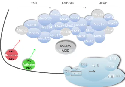
Figure 5. Schematic illustration of the transcription pre-initiation complex in eukaryotes where Mediator integrates signals from promoter-bound transcriptional regulators (repressors and activators). Mediator kinase module is not shown. Specific subunits present in higher Eukaryotes are colored grey. The location of Mediator subunit 25 (Med25: enlarged grey subunit) is undefined. Adapted from Ansari and Morse117
1.4 Molecular recognition - Coupled folding and binding
The enormous size of Mediator, its subunit complexity and its dynamic nature makes it a very challenging subject for applying biophysical studies. The complete Mediator has been studied by cryo-electron microscopy118,119 but a high-resolution structure is still not available. Only 13 subunits have been structurally characterized at atomic resolution by X-ray crystallography or solution NMR120,121. This means that there is still an immense lack of knowledge about the structure of Mediator and hence about key aspects of the mechanisms that regulate gene expression. Most knowledge about Mediator has so far been achieved by studies of interactions between specific Mediator subunits and individual TFs. The specific interactions occur between TADs of the TFs and the ACtivator Interaction Domains (ACIDs) in Mediator subunits. When studied in vitro, these interactions are generally found to be weak (in the micro-molar range), most likely since they are meant to be transient in the cells55,122,123. However, several weak interactions might cooperate to trigger structural shifts in other Mediator subunits in order to propagate the regulatory signals, similar to a Domino effect.Several studies have shown that a specific TAD can target several ACIDs in Mediator. For example Gcn4 targets Med2, Med3, Med15 and Med16124-128. On the other hand, different TFs interact with the same Mediator protein, such is the case for VP16129, IE62130, HNF4α131, SOX9132, ATF6α133 and ERM134 that all interact with the human Med25. The question is how this is possible and what the mechanisms for binding and recognition are. There is not one straight answer for those questions, since the process of protein binding is the result of several attributes at the proteinprotein interfaces -e.g. amino acid composition, hydrophobicity, electrostatic interactions, hydrogen bonding and energetics of binding55,56,135. It is known that TADs are highly diverse when comparing their amino acid sequences. However, some studies have revealed potential motifs of short amino acid sequences that are present in several TFs, e.g. p53, which mediate transient interaction with different targets123,136-140. Another aspect is that TADs are typically unfolded in their free state but can adopt structured conformations upon binding to their targets139,141-144. These structural transitions are dynamic and might explain the ability of TADs to adopt multiple conformations such that different TADs can share a common target surface on Mediator (fuzzy complex)124. Conversely, this conformational flexibility can also provide an explanation for how one specific TAD can target multiple Mediator subunits. Moreover, several TADs have acidic properties which makes it possible to interact with target proteins by complementary electrostatic interactions 57. Electrostatic interactions can promote a rapid initial contact and might dominate during the initial binding phase to increase the rate of complex
formation. Formation of the final complex might depend on hydrophobic interactions in a subsequent entropy driven phase. Finally, the binding energetics involve a balance between entropy and enthalpy changes, compensations and commitments of favorable or unfavorable effects that have to be kept in balance to lend stability to the complex54,59-61,145-148. The binding of a single TAD, for example the VP16 TAD which is composed of 40 residues, to its target might trigger small and large conformational changes, both in the target and in the TAD itself. This will bring functional flexibility and it is a suggested mechanism by which regulatory signals might be propagated112,114,115. The study of conformational changes that occur upon interaction between Med25 and the TFs Dreb2a and VP16 is described in papers II and III.
1.4.1 Med25
As already mentioned, Mediator is highly conserved from yeast to humans 149. However, some Mediator subunits, i.e. Med25, are specific to higher eukaryotes such as humans and plants88,150. As discussed in section 1.2.2, the divergence in Mediator subunit content might be connected to the evolution of eukaryotes that require a higher level of complexity in transcription regulation. In this section, Med25 from A. thaliana and human are introduced.
A. thaliana Med25 (aMed25) is a 90 kDa protein (836 amino acids) and its ACID is located between residues 551 – 680. It was originally known as the PHYTOCHROME AND FLOWERING TIME 1 (PFT1), a nuclear protein involved in regulation of flowering time downstream of the PhyB photoreceptor151. PFT1 was identified as a Mediator subunit when the plant Mediator was isolated from A. thaliana150. The identification of PFT1 as a Mediator protein suggested that PhyB signaling targets Med25 indirectly via one or several unidentified transcriptional factors. Later it was found that aMed25 integrates signals from different stress- and developmental pathways. For example, aMed25 was reported to be involved in jasmonate (JA) signaling which is a critical plant defense response152. Both, positive and negative effects have been identified for aMed25 upon interaction with specific TFs. The interaction with MYC2, which regulates JA-mediated gene expression, resulted in a positive effect on expression of target genes; while interaction with the TF ABI5, had a negative regulatory effect on the expression of abscisic acid (ABA)-genes153. ABA is a plant hormone and similar to JA, it is involved in developmental and physiological processes in plants154. Moreover, aMed25 interacts with other TFs involved in stress
response pathways including the Myb-like protein, the zinc finger homeodomain 1 (ZFHD1), and Dreb2a155.
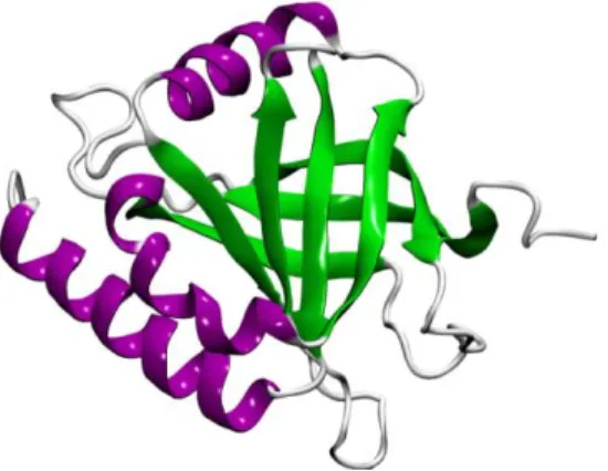
Figure 6. NMR structure of the human Med25-ACID (PDB 2XNF)156. Human MED25 (hMed25) is also a 90 kDa protein (747 residues) and its ACID comprises residues 394 – 543129. It was first described as the human activator-recruited cofactor 92 (ARC92) identified in interaction studies with VP16129. hMed25 has been more extensively studied compared to aMed25. It has been shown to interact with several TFs involved in different cellular processes, including retinoid activation by RARα, chondrogenesis by SOX9, insulin secretion in pancreatic cells by HNF4α, cellular growth and differentiation by PEA3 subfamily members, and the endoplasmic reticulum stress response by ATF6α131-134,157. In addition, hMed25 interacts with Varicella-zoster virus protein IE62, which activate transcription of viral genes129,130,158,159. The structure of the hMed25-ACID (residues 394-543) has been solved by NMR and it comprises seven β-strands forming a β-barrel flanked by three helices156,160-162 (Figure 6). The interaction between hMed25 and VP16 has been studied in detail and the VP16 binding site on hMed25-ACID has been defined129,156,158,162. Moreover, mutations in hMed25 have been related to Charcot-Marie-Tooth neuropathy, an inherited neurological disease which results in muscle degeneration in the limbs163.
1.4.2 The plant-specific transcription factor Dreb2a
Dreb2a belongs to the large AP2/ERBP (Ethylene Responsive Element Binding Protein) TF family which is unique to plants164-166. These TFs interact with cis-acting dehydration-responsive promoter elements and regulate genes involved in drought-, salt-, and cold stress response pathways167,168. Dreb2a functions in response to drought stress and high salinity. The protein is around 38 kDa, its TAD is located in the C-terminal
within residues 254-335 and it also contains a negative regulatory domain (NRD) in the middle region within residues 135 – 165169. The NRD might explain the regulatory effects of Dreb2a which have been found to be both positive and negative155,169. aMed25 was reported to interact with Dreb2a 168-335 in vitro and yeast-2-hybrid assays showed that the minimal domain required for interaction was located within residues 169-254 in Dreb2a155,168. Phenotypes of a series of mutants, i.e. med25 mutants were drought resistant and dreb2a mutants showed early flowering time, suggested that the interaction of aMed25/Dreb2a had a negative regulatory effect through the NRD of Dreb2a155.
1.4.3 The human herpes simplex virus 1 VP16 protein
VP16 is a 54 kDa protein (490 amino acids) and it belongs to the Herpesviridae tegument protein family; proteins that form the capsid layer which encloses the viral genome and function as transcriptional activators during viral infection of viral immediate early genes170. Transcriptional activation by VP16 has been reported to operate through interaction with different general TFs, including TFIIA, TFIIB, TFIIF, TFIIH, TBP, hTAFII31 and hTAFII32129,139,171-176. Thus, the VP16-TAD has been extensively studied and in addition to its interaction with the human Med25-ACID, it has also been reported to interact with another Mediator subunit, Med17177. The VP16-TAD can be divided in two subdomains, the N-terminal (residues 412-452) and the C-terminal (452-490), which function independently and complementary to each other175,178,179. The VP16-TAD is unstructured in free state as many other TADs, however it adopts an α-helical conformation upon binding to its target proteins139,141,144, a characteristic also observed in other TADs e.g. p53 141-143. Within the two subdomains, the formation of α-helical segments has been located around residues 429-450 and 465-488141. A nine amino acid sequence (DFDLDMLGD) that play a key role for VP16-TAD transcription activation has been located to the first segment (429-450)137,178. In addition, the DFDLDMLGD motif has also been identified in a range of TFs and is proposed to be a conserved domain which can be recognized by the same regulatory protein. Such is the case for VP16, p53, HSF1, NF-kB and NFAT1 which all contain the DFDLDMLGD motif within their TADs and share the ability to interact with TAF9 (TAFII31)136,139,140.
2. Aims
The focus of my thesis included two major subjects. The first subject was to investigate how folding and stability of a heptameric protein was affected by biologically relevant environments such as macromolecular crowding. The second subject was to study the interaction of Mediator subunit 25 with specific TFs, and the structural shifts occurring upon binding. Understanding these processes in more detail will improve our knowledge of protein folding in vivo and will contribute to learn more about the molecular recognition between proteins that regulate gene expression, which is one of the most central cellular processes that occur in crowded environments.
Effects of macromolecular crowding on a heptameric protein:
The first specific aim was to assess the effects of macromolecular crowding, or excluded volume effects, on unfolding and dissociation of the heptameric human protein, cpn10. This protein represents an excellent model system for studies of oligomeric proteins since both intra- and intermolecular interactions are involved in its folding process. Chemical and thermal denaturation were used to induce unfolding/dissociation in a crowded condition. In addition, the overall stability and monomer-heptamer equilibrium were also assessed in such conditions. To create a crowded environment in the in vitro system, I used the inert synthetic polymer Ficoll 70 at specific concentrations. This crowding agent is usually used for macromolecular crowding studies since it does not interact with proteins and does not interfere with different spectroscopic measurements.
Interaction studies between transcriptional regulatory proteins:
The second specific aim was to characterize the structure, stability and interaction between Med25 and the TF Dreb2a from A. thaliana in the presence and absence of an oligonucleotide that contains a consensus Dreb2a binding site. The main purpose was to study in more detail the potential structural shifts that occur upon interaction between Dreb2a, the Dreb2a consensus sequence and Med25 in different combinations. Many TFs are unstructured in dilute buffers, but might gain a more ordered structure upon binding to their targets. In addition, structural shifts might underlie the mechanisms of action by which the Mediator complex functions as a co-regulator of transcription. An additional task was to investigate the interactions of the unrelated TFs Dreb2a and VP16 with Med25 proteins from human and A. thaliana. The Med25 homologs share low sequence similarity, yet their folds might offer similar surfaces for ligand recognition. This study helps to understand the molecular recognition between
transcriptional regulatory proteins and thereby the conserved function and structure of the ancient Mediator complex.
A combination of biophysical- and biochemical methods (fluorescence, circular dichroism, surface plasmon resonance, calorimetry, NMR, size exclusion chromatography, cross-linking, GST pull-downs) were used in the two sub projects.
3. Methods
3.1 Protein stability, folding and binding
Protein folding and binding can be studied by applying the laws of thermodynamics180. Equation (1) shows the thermodynamic principle of Gibbs free energy which can be used to describe the favorable state of protein folding and binding reactions. The folding stability of a protein can be obtained by measuring the difference in free energy between the folded and unfolded states.
∆G = ∆H - T∆S (1)
Where ∆G is the change of free energy, ∆H is the change of enthalpy, T is the absolute temperature and ∆S is the change in entropy. Since every system strives for minimum energy, proteins will fold or bind into a conformation with the most favorable ∆H and T∆S. Then, the net protein stability is marginal and can be easily disturbed by changing its environment, e.g. by changing the pH, the salt concentration, the temperature or by adding chemical denaturants. The relative contributions from the enthalpy can be gained by chemical bonding within the protein structure (hydrogen bonding and van der Waals interactions) and the entropy corresponds to the conformational freedom and also to hydrophobic effects.
∆G of any given reaction can also be related to the equilibrium constant Keq in equation (2), where R is the general gas constant.
∆G = -RT lnKeq (2)
For protein folding studies, a two-state model can be assumed where the protein is in equilibrium between the folded (F) and unfolded (U) states. The rate constants are defined as the ratio of the population of the unfolded and folded states in terms of the protein fraction present (f) in those states (3). To obtain information about Keq, the system has to be disturbed, which can be achieved by inducing unfolding, as mentioned above, with heat or with chemical denaturants such as urea or GuHCl.
Keq = kU/kF = [fU]/[fF] (3)
In paper I, the thermal induced unfolding was used to extract Tm values which are the melting temperature at the denaturation midpoint. Then, the thermal data was fitted to a modified expression of the van’t Hoff equation using equations (4) and (5), where ∆Hm is the enthalpy of denaturation at
Tm, y(T) is the CD or the fluorescence signal at specific T’s, yF and yU are the measured signals, and mF and mU are the slopes181,182.
Kobs(T) = exp[(∆Hm/R)((1/Tm)-(1/T))] (4)
y(T) = [yF + mFT + Kobs(yU + mUT)]/(1 + Kobs) (5) For chemical induced unfolding, ∆GU can be extracted using the Linear Extrapolation Method (LEM) which describes the linear relationship between ∆GU and the denaturant concentration [D]183. Then, the denaturant dependence of ∆GU can be described as in equation (6) where ∆GH2O can be calculated by extrapolating back to zero denaturant concentration. This yields the slope or m-value which is the dependence of free energy on denaturant concentration.
∆GU = ∆GH2O - m [D] (6)
In paper I, ∆GU,diss was calculated by using an apparent two-state model coupled to dissociation (diss). Then, the Keq for a heptamer was defined as in equation (7).
KU,diss = [Umonomer]7 / [Fheptamer] (7)
For protein-ligand binding, an equilibrium expression can also be used that describes a two-state process (8) where L is the ligand binding to protein P and forming the complex (PL)54.
P + L PL (8)
The chemical equilibrium is the ratio of the rate of association Kon (P+L PL) and the rate of dissociation Koff (PL P+L). The constants for association (KA) and dissociation (KD) can be described as in equation (9) and (10).
KA = [PL] / [P] [L] (9)
KD = [P] [L] / [PL] (10) The KD value is generally used to characterize the strength of a binding reaction. The KD corresponds to the concentration of the ligand at which half of the protein molecules are bound or at which the protein is half saturated. Consequently, the binding free energy can be calculated using KD and eq. (2). In a saturated reaction, a binding curve can be plotted using equation (11), where Bmax is the maximum specific binding value and Y is the degree of saturation.
3.2 CD spectroscopy
Circular dichroism (CD) is a technique conventionally used to investigate the structure and stability of proteins. Some advantages of CD measurements include their simplicity and short recording times. A typical measurement to assess the folded state of a protein can be done in some minutes using low protein concentrations and small volumes (e.g. 5 µM pure protein, 200 µL for a 1 mm cuvette). The instrument measures the difference in absorbance between left and right circularly polarized light. Proteins are optically active because of their chiral nature and they will respond differently to the left and right circularly polarized light. Therefore, a particular protein will have a particular CD spectrum that provides information about its secondary (far-UV below 250 nm) and its tertiary structure (near-(far-UV above 250 nm)184. The secondary structural information obtained from the far-UV CD spectrum of a protein corresponds to the backbone conformation and the contribution from secondary structure components such as α-helical, β-sheet and random coil. Some known spectral features can be used to characterize the overall contribution from these secondary structure elements. For instance, the content of α-helixes gives a positive signal maximum around 190-195 nm and negative signals at 208 and 222 nm. The β-sheet content gives a positive signal around 195-200 nm and a negative signal at 215-220 nm. Random coil gives a positive signal around 220 nm and a negative signal around 200 nm185. The amount of secondary structure elements can be estimated by using the CD spectrum of the protein and available deconvolution softwares such as CDNN186.
Protein stability can be assessed by monitoring CD signal changes at a specific wavelength as a function of temperature or chemical denaturants. Then, thermodynamic data such as the midpoint of the unfolding transition (Tm), the van’t Hoff enthalpy (∆HU) of unfolding and the free energy (∆GU) can be calculated. Generally the Gibbs-Helmholtz equation is used to fit the data for a two-state transition. In paper I only Tm values were extracted from the thermal denaturation fit. In paper II CD was used mostly to monitor secondary structure content.
3.3 Fluorescence spectroscopy
Fluorescence spectroscopy is a very sensitive method to assess protein folding and kinetics at very low protein concentrations (e.g. 1 µM pure protein, 100 µL, 3 mm cuvette). It measures the intensity of light emitted by a molecule as it returns to its ground state after excitation. Fluorescence signal from proteins are contributed from aromatic residues such as
tryptophans and tyrosines. These chromophores can be excited between 260 to 295 nm wavelengths and after the system goes back to equilibrium, the emission of the light have a different intensity or have shifted position to another wavelength. The emission changes can be monitored from the folded to the unfolded state since in a folded state the protein aromatic residues are normally located in a hydrophobic environment. Then, upon unfolding the environment will change and those residues will give lower intensities as the signals are quenched by the solvent187,188.
Tryptophan fluorescence was used in paper II to assess the integrity of proteins at an excitation of 290 nm with an emission maximum around 350 nm. In paper I, tyrosine fluorescence was used (cpn10 lacks tryptophan) at an excitation of 280 nm with an emission maximum around 304 nm.
3.4 Nuclear Magnetic Resonance
Solution-state nuclear magnetic resonance (NMR) spectroscopy is a powerful technique to study protein structures, dynamics and interactions at atomic resolution. It provides residue-specific information and can capture events down to the picosecond time scale. Some disadvantages are the need of concentrated samples and the limitation when it comes to the protein size. It is generally difficult to obtain proteins at high concentrations and some proteins that are unstable start to precipitate above certain concentrations. Large proteins are difficult to study by NMR either due to the number of resonances that might complicate the spectra or just because the size of the protein affects the tumbling rate and thereby the signal line-width and amplitude. To be able to study proteins using NMR, the protein sample has to be isotopically labeled and present at a concentration of at least 50 µM and volumes around 300 µL. However, the required protein concentration depends on the type of experiment, the spectrometer magnetic field strength and the running time, which can help to increase the signal-to-noise ratio. NMR measurements are based on quantum mechanical properties of the elemental isotopes nuclei (1H, 13C, 15N, 19F and 31P) which have a characteristic spin (I = 1/2), and a nuclear magnetic moment. When an external magnetic field is applied, the spin will have two energy states (+1/2 and -1/2), one aligning with the external field and the other one with opposite direction. The electromagnetic radiation is therefore used to stimulate transitions and coherences between the energy states of the spins in a sample where specific nucleus are being studied. The difference in energy between the two spin states is generally small and proportional to the external magnetic field strength. Generally, the energy difference is given in frequency units (MHz) and the chemical shifts are referred in units of parts
per million (ppm). In a typical one-pulse experiment the magnetization is flipped to 90 degrees to be able to record the resonance signals. Then the NMR signal will be proportional to the amount of magnetization in a xy-plane which oscillates with a frequency that induces current. The time dependent current is recorded as a so called free induction decay (FID) and Fourier transformed to a 1D spectrum that represent the intensity as function of frequency189.
A typical protein NMR experiment is to acquire 2D heteronuclear single quantum coherence (HSQC) spectrum, and as the name suggests more than one nucleus is involved in the measurement. A 1H,-15N HSQC gives rise to one cross-peak from each amino group in the backbone corresponding to one residue in the protein. Since the nucleus in the amino acids within the protein structure experience a distinct chemical environment, then each of them will have a distinct chemical shift in the spectrum.
3.4.1 Chemical shift perturbation
Chemical shift perturbation (CSP) experiments can be used to study interaction between proteins and other biomolecules. In a typical CSP experiment, a series of 1H,-15N HSQC spectra monitors the chemical shifts from a 15N-labelled protein upon titration with an unlabeled protein interacting partner (ligand). From CSP data several features about the interaction can be obtained, such as the rate and binding affinity, and in addition the binding sites can be mapped. The binding sites can be localized by identification of the residues that change their chemical shift position in the spectrum upon addition of ligand. Protein-ligand complexes are very dynamic systems and the rate at which the components of the complex exchange between the free and bound states affects the spectra and gives rise to specific features that can be analyzed. If the exchange rate is slow in the chemical shift time scale, two sets of signals will be observed for the free and bound state. If the exchange rate is fast, only one signal will be observed as the average of the two states. Finally if the exchange rate is intermediate, only one signal is observed but with distinctly line-broadened peaks. To trace the peaks from free to bound state, increasing ligand concentrations can be added to reach saturation and to facilitate the analysis of the residues involved in interaction190-192. This method was used in paper III to map the binding-site of Dreb2a in Med25 proteins.
3.5 Surface plasmon resonance
Surface plasmon resonance (SPR) is a technique that enables the detection of interactions between proteins or other biomolecules in real-time. The instrument measures the changes of refraction index between two interfaces after polarized light irradiation. A typical experiment requires low protein concentrations and small volumes (<5 µM). The ligand is immobilized on a sensor surface or a chip, which is a conducting film composed by a thin layer of gold. The analyte which will be the potential interacting partner is in free solution and is injected over the chip in a continuous flow. The interaction is recorded due to the increase of molecule density upon association, which results in a different reflection of light at a certain angle. The SPR angle of reflectivity at a certain resonance condition will change while molecules associate until saturation is reached. Then, dissociation will make the angle to return back to baseline once all analyte is completely removed from the sensing surface. The change of the SPR angle will be proportional to the mass of the bound molecules and the result of interaction can be displayed as a sensorgram against time where the y axis is the binding response in resonance units (RU). From the binding curve the rate of association and dissociation, and the affinity can be obtained193. This method was used in paper II and III to probe binding between Med25 proteins and the Dreb2a and VP16 TFs, and to obtain the KDs of their binding.
3.6 Isothermal titration calorimetry
Binding processes can be fully thermodynamically characterized by using isothermal titration calorimetry (ITC). This technique is sensitive and detects the heat generated or absorbed when proteins or other molecules interact. During a typical experiment one protein sample is placed in a sample cell (~20 µM) and the protein ligand (present in at least a 10-fold excess in concentration) to be titrated is placed in a syringe compartment. Then the ligand is injected into the cell repeatedly at certain injection volumes until the system reaches saturation. The peaks obtained from the raw data and the heat flow as a function of time can be integrated and then fitted to an appropriate binding model (e.g. one site-binding model) to yield the binding enthalpy (ΔH) and the binding affinity (KA) from which the dissociation constant KD can be derived. Also, the free energy of binding can be calculated using eq. (2) and the entropy of binding by using eq. (1). The thermodynamic data gives more detailed information about the physical basis of molecular interactions. The favorable or unfavorable changes in entropy and enthalpy can describe the formation of a complex. A negative enthalpy change for instance, reports favorable interaction due to hydrogen
and van der Waals bonds at the proteins interface. Hydrophobic effects will also favor association in a process that can be viewed as a transfer of the ligand from solvent to the protein environment which increases the entropy194. ITC was used in paper III to characterize the interaction between Dreb2a and Med25 proteins.
3.7 Cross-linking
Chemical cross-linking is a method that can be used to probe transient and stable interactions between proteins. It involves the formation of covalent bonds between proteins by using reagents that contain reactive groups which will react with the functional groups of proteins. The principal idea to probe interaction is that if two proteins are physically interacting with each other, then they can also be covalently cross-linked to each other by the reagent. There is a range of crosslinking reagents available. One which is often used is glutaraldehyde which can react with any two amino groups that are close to each other in space. Because of its promiscuous nature, glutaraldehyde has low specificity but can be used at mild conditions as a convenient initial tool to investigate protein interactions. The reagent is also used to characterize oligomerization states195-197. This method was used in paper I in combination with denaturation of cpn10. The heptamer crosslinks efficiently with glutaraldehyde and can be visualized using SDS-PAGE. Upon addition of chemical denaturants at concentrations that favor unfolding, the monomers will not crosslink and will appear as monomers in SDS-PAGE. By performing these experiments an apparent two-state unfolding reaction for cpn10 was probed.
3.8 GST pull-downs
This is a relatively easy and straightforward method to probe potential protein-protein interactions using cell lysates or pure proteins at relatively low concentrations. One of the interacting proteins has to be GST-tagged (the bait) and can be purified in one step to further be mixed with putative binding partners (the prey). Prey proteins can be either purified proteins or come directly from cell lysates. The assay is based on the immobilization of the bait protein on glutathione sepharose beads. The GST protein pre-bounded to beads is then incubated with bait proteins. The non-bound material is washed off with an adequate buffer and the proteins that interact with the bait will remain on the beads. The interacting proteins can be eluted and resolved in a SDS-PAGE, and analyzed by Coomassie-, silver staining or Western blotting using appropriate antibodies198. GST pull-downs experiments were used in paper II and III.
4. Results
4.1 Summary of papers
4.1.1 Macromolecular crowding stabilizes cpn10 monomers
Paper I: Macromolecular crowding extended to a heptameric system: The
co-chaperonin protein 10
In vivo, proteins fold and associate in highly crowded environments. Previous experiments have shown that macromolecular crowding stabilizes monomeric proteins towards heat perturbation and also can modulate their structure40-42,199. Cpn10 is a ring-shaped oligomer consisting of seven identical β-barrel monomers and it is an important human protein that functions to help other proteins to fold. The stability of the heptamer is dominated by subunit-subunit interactions and the individual monomers have low stability on their own73-75. To assess the effects of macromolecular crowding on the cpn10 protein, where denaturation involves unfolding and dissociation, the thermodynamic and kinetic properties of the cpn10 were investigated both in diluted buffer solution and in an in vitro crowded system. Ficoll 70, a sucrose-based polymer, was used to create a crowded environment in vitro at concentrations up to 300 mg/ml.
Increased stability in presence of Ficoll 70:
The results of a set of spectroscopic experiments showed that cpn10 is stabilized by both, thermal and chemical perturbation in presence of 300 mg/ml Ficoll 70. The thermal stability of cpn10 increased by 4°C in the presence of Ficoll 70 (Figure 7A). The stabilizing effects toward chemical perturbations were reflected in the transition midpoint position, which increased by 0.5 M GuHCl in Ficoll 70 (Figure 7B). Cross-linking experiments supported the denaturant midpoints shifts in the presence of Ficoll 70 and the apparent two-state transition from folded heptamer to unfolded monomers.
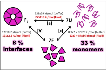
Free energy of unfolding/dissociation increased in presence of Ficoll 70: The coupled unfolding/dissociation free energy for the heptamer increased by about 36 kJ/mol (average free energy in buffer 239 kJ/mol and in Ficoll 275 kJ/mol) (Figure 7C and 8 [a]). The spectroscopic data was analyzed by a new approach using the complete spectra (fluorescence) at each condition and global analysis of unfolding curves at every wavelength in the span of 294 to 396 nm. The data was fitted to an apparent two-state reaction coupled to dissociation going from a folded heptamer to unfolded monomers
(F7↔7U). In order to probe the unfolding mechanisms in chemical denaturants, NMR diffusion experiments were performed and the results indicated that the diffusion coefficient of unfolded cpn10 was approximately the same in both denaturants indicating a monomeric unfolded state. Further, the unfolding kinetics of the heptamer was investigated and it was found that the unfolding rate constants were slower in crowded conditions, which might in part explain the higher equilibrium stability found for cpn10 in the presence of Ficoll.
Figure 7. Increased stability of cpn10 in presence of Ficoll 70. A) Thermal midpoint as function of cpn10 concentration. Inset: thermal unfolding curves for cpn10 monitored by CD 230 nm changes. B) Transition midpoints from the GuHCl induced unfolding as function of cpn10 concentration. C) Free energy for coupled unfolding/dissociation from GuHCl induced unfolding. D) Heptamer-monomer transition as a function of cpn10 concentration monitored by tyrosine fluorescence changes. F7 indicates folded heptamer, F folded monomer, U unfolded monomer. Cpn10 in absence (blue circles) and presence of 300 mg/ml Ficoll 70 (red squares).
Heptamer dissociation is affected in the presence of Ficoll 70:
The macromolecular crowding impact on the heptamer-monomer dissociation constant was assessed by measuring fluorescence changes. The full spectra were also analyzed as above. The heptamer-monomer transition midpoint was lower in Ficoll 70 (3.1 µM) compared to in buffer (8.1 µM) indicating tighter binding in crowded conditions (Figure 7D). The free energy values for heptamer-monomer dissociation were calculated to 191 and 177 kJ/mol in Ficoll and buffer, respectively (Figure 8 [b]).
Figure 8. Thermodynamic cycle of the cpn10 heptamer unfolding and dissociation. Free energy values are shown for the reactions both in buffer and in the presence of 300 mg/ml Ficoll 70. [a] Data from coupled unfolding/dissociation going from a folded heptamer to unfolded monomers. [b] Heptamer-monomer dissociation assessed by fluorescence changes. [c] Monomer stability calculated from [a] and [b]. The stability increased by 8% on the protein-protein interfaces and by 33 % on the individual monomers in presence of Ficoll 70.
Finally, using the free energy values in [a] and [b], energy values for [c] were calculated. Based on these results, a thermodynamic cycle was constructed (Figure 8) which reveals that macromolecular crowding increases stability by 8% on the protein-protein interfaces and by 33 % on the individual monomers. This means that macromolecular crowding exerts larger effects on the individual monomers.
4.1.2 Conformational changes upon interaction between Med25 and Dreb2a
Paper II: Interactions between DNA, transcriptional regulator Dreb2a and
the Med25 mediator subunit from Arabidopsis thaliana involve conformational changes.
Mediator is a multi-protein complex that transmits regulatory signals from DNA-bound TFs to the Pol II transcription machinery109,111,117. How exactly Mediator accomplish this function is not entirely understood. Several studies indicate that interaction between TFs and Mediator subunits triggers conformational changes which might be the way that regulatory signals are propagated through Mediator to the Pol II transcription machinery112,114,115. In this study, the interaction between the activator interaction domain of A. thaliana Med25 (aMed25-ACID) and Dreb2a were investigated in absence and presence of an oligonucleotide containing the canonical Dreb2a DNA-binding site. The main purpose was to explore the potential structural changes that might occur upon complex formation.
The C-terminal of Dreb2a is unstructured in the free state:
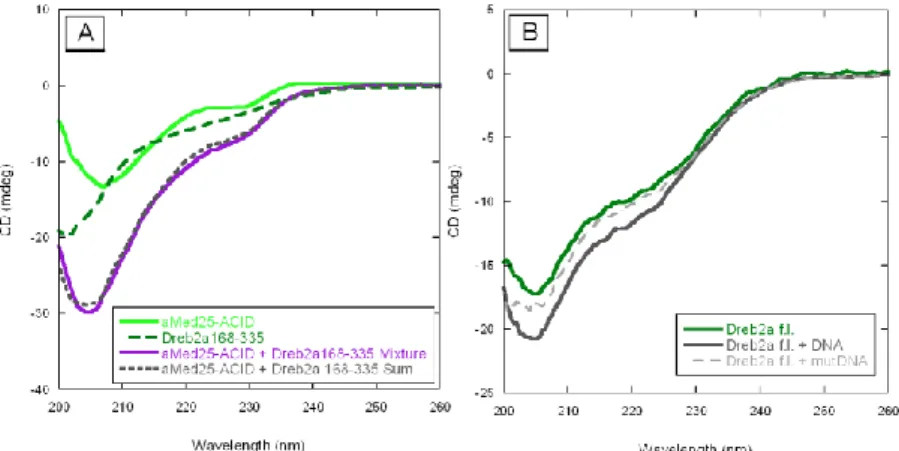
The secondary structures of each protein individually and the mixture of proteins were monitored by CD spectroscopy. The ACID of Med25 (Med25551-680) showed ordered structures with negative peaks at 205 and 230 nm while the C-terminal of Dreb2a (Dreb2a168-335) was unstructured, when they were analyzed in their free state (Figure 9A). However, full-length Dreb2a (Dreb2af.l.) fused to a GB1-tag showed some ordered conformations with negative peaks around 205 and 224 nm, thus indicating the presence of some α-helical content (Figure 9B).
Gain of structure upon complex formation:
The interaction between Dreb2a168-335 and aMed25-ACID was studied by SPR. In these experiments we determined the KD for the binding between the proteins to 1 µM. We were unable to detect any interaction-induced structural changes in these proteins in the CD measurements (Figure 9A). Our SPR experiments also showed interaction of aMed25-ACID and Dreb2af.l.. However, in this case the kinetics was complex and displayed a fast association and a slow dissociation rate. The KD was approximated to 0.1 µM. Regarding this interaction, the mixture of Dreb2af.l. and aMed25-ACID at a one-to-one ratio resulted in an increased negative CD signal which indicated that structural shifts occurred upon interaction. The stoichiometry of binding between aMed25-ACID and Dreb2af.l. was determined to 1:1 using size exclusion chromatography.