Doctoral Thesis in Biological Physics
The Biophysics of Na
+
,K
+
-ATPase in
neuronal health and disease
EVGENY AKKURATOV
Stockholm, Sweden 2020
kth royal institute of technology
The Biophysics of Na
+
,K
+
-ATPase in
neuronal health and disease
EVGENY AKKURATOV
Doctoral Thesis in Biological Physics KTH Royal Institute of Technology Stockholm, Sweden 2020
Academic Dissertation which, with due permission of the KTH Royal Institute of Technology, is submitted for public defence for the Degree of Doctor of Philosophy on Friday the 18h December 2020, at 1:00 p.m. in Fire, Science for Life Laboratory, Tomtebodavägen 23A, Solna.
© Evgeny Akkuratov ISBN 978-91-7873-738-3 TRITA-SCI-FOU 2020:47
iii
Abstract
Na+,K+-ATPase is one of the most important proteins in the mammalian
cell. It creates sodium and potassium gradients which are fundamental for the membrane potential and sodium-dependent secondary active transport. It has a second role in the cell as a receptor that by binding chemicals from the cardiotonic steroids family, the most knowledgeable of them is ouabain, triggers various signaling pathways in the cell which regulate gene activation, proliferation, apoptosis, etc. It has been shown that several severe neurological diseases are associated with mutations in
the Na+,K+-ATPase encoding genes. Although Na+,K+-ATPase was
discovered already in 1957 by the Danish scientist Jens Skou, the knowledge about the function of this enzyme is still not complete.
In the studies included in the thesis, we have learned more about the
function of Na+,K+-ATPase in different aspects of health and disease. In
study I we showed a mechanism of ouabain-dependent regulation of the NMDA receptor, one of the most important receptors in the nervous
system, via binding with Na+,K+-ATPase. This allows us to look at the
Na+,K+-ATPase as regulator via protein-protein interaction. In study II we
investigated a different aspect of Na+,K+-ATPase functioning – to look at
how binding of ouabain to Na+,K+-ATPase activates a number of signaling
cascades by looking at the phosphoproteome status of the cells. This allows us to see the whole picture of ouabain-mediated cascades and further
characterize them. In study III we focused on the role of Na+,K+-ATPase in
severe epileptic encephalopathy caused by a mutation in the ATP1A1 gene. We performed a molecular and cellular study to describe how mutations affects protein structure and function and found that this mutation
iv
converts the ion pump to a nonspecific leak channel. In study IV we performed a translational study of the most common mutation for rapid-onset dystonia-parkinsonism. We studied how this mutation affects the nervous system on the protein-, cellular-, and organism level and found that the complete absence of ultraslow afterhyperpolarization (usAHP) could explain gait disturbances found in patients. In the on-going study we
showed that Na+,K+-ATPase can oligomerize and that this effect is
triggered by ouabain binding to the Na+,K+-ATPase. In this study, we
utilized a novel fluorescence labelling approach and used biophysical
techniques with single molecule sensitivity to track Na+,K+-ATPase
interactions.
In summary, we applied biophysical and molecular methods to study
different aspects of the function of Na+,K+-ATPase, and gained insights
that could be helpful not only for answering fundamental questions about
Na+,K+-ATPase but also to find a treatment for patients with diseases
associated with mutations in this protein.
Keywords
Na+,K+-ATPase, NMDA receptor, ouabain, phosphoproteome,
v
Sammanfattning
Na+,K+-ATPas är ett av de viktigaste proteinerna i däggdjurscellen. Det
skapar natrium- och kaliumgradienter som är grundläggande för den elektriska potentialen över cellmembranet och för natriumberoende sekundär aktiv transport. Det har dessutom en roll som receptor som genom att binda hjärtstimulerande steroider, varav den mest kända är ouabain, startar olika signalvägar i cellen som bl.a. reglerar genaktivering, prolifiering och apoptos. Det har visats att flera allvarliga neurologiska
sjukdomar är kopplade till mutationer i Na+,K+-ATPas gener. Trots att
Na+,K+-ATPas upptäcktes redan 1957, av Dansken Jens Skou, är vår
kunskap om enzymets funktion ännu inte komplett.
I studierna i denna avhandling har vi lärt oss mer om Na+,K+-ATPas
funktion inom hälsa och sjukdomar. I studie I påvisade vi en ouabain-beroende reglering av NMDA-receptorn – en grundläggande receptor i
nervsystemet – via bindning till Na+,K+-ATPas. Detta visar att Na+,K+
-ATPas fungerar som en regulator genom att direkt interagera med andra
proteiner. I studie II undersökte vi en annan sida av Na+,K+-ATPas
funktion – hur bindning av ouabain till Na+,K+-ATPas aktiverar flera
signal-kaskader – genom att titta på cellens fosfoproteoms-status. Vi kunde på så sätt få en mer heltäckande bild av ouabain-styrda kaskader,
och karakterisera dem. I studie III fokuserade vi på Na+,K+-ATPas roll i
svårartad epileptisk encefalopati orsakad av en mutation i ATP1A1-genen. Vi utförde en molekylär och cellulär studie för att beskriva hur en mutation påverkar proteinets struktur och funktion, och fann att mutationen omvandlar jonpumpen till en ospecifik läckkanal. I studie IV genomförde vi en translationell studie för den vanligaste mutationen vid dystoni parkinsonism med snabb debut. Vi studerade hur mutationen påverkar
vi
nervsystemet på protein-, cell-, och organismnivå och fann att frånvaro av ultralångsam efterhyperpolarisering skulle kunna förklara patienters
problem med gången. I pågående studie visade vi att Na+,K+-ATPas kan
oligomerisera och att detta startas av bindning till ouabain. I denna studie
utvecklade vi en fluorescensmärkning av Na+,K+-ATPas, och
oligomeriseringen studerades med fluorescenstekniker med en-molekyl-känslighet.
Sammanfattningsvis har vi använt biofysikaliska och molekylära metoder
för att studera olika aspekter av Na+,K+-ATPas funktion och nått insikter
som kan vara till hjälp, inte bara för att beskriva grundläggande molekylära funktioner men även för att hitta botemedel mot sjukdommar kopplade till
mutationer i Na+,K+-ATPas.
Sökord
Na+,K+-ATPas, NMDA-receptorn, ouabain, fosfoproteom, oligomerisering,
vii
List of publications
This thesis is included in the following articles and manuscripts:
I. Akkuratov EE*, Westin L*, Vazquez-Juarez E, de Marothy M,
Melnikova AK, Blom H, Lindskog M, Brismar H, Aperia A. “Ouabain Modulates the Functional Interaction Between Na,K-ATPase and NMDA Receptor”. Mol Neurobiol. 2020 Oct;57(10):4018-4030.
II. Panizza E, Zhang L, Fontana JM, Hamada K, Svensson D,
Akkuratov EE, Scott L, Mikoshiba K, Brismar H, Lehtiö J, Aperia
A. ”Ouabain-regulated phosphoproteome reveals molecular
mechanisms for Na+,K+-ATPase control of cell adhesion,
proliferation, and survival”. FASEB J. 2019 Sep;33(9):10193-10206.
III. Ygberg S*, Akkuratov EE*, Howard RJ, Taylan F, Jans DC,
Mahato DR, Kinoshita PF, Nennesmo I, Lindskog M, Andersson M, Lindstrand A, Brismar B, Aperia A. ”Severe epileptic encephalopathy caused by a de novo germline Trp931Arg mutation in ATP1A1 that converts Na,K-ATPase into an ion channel” (Manuscript, submitted).
IV. Akkuratov EE*, Sorrell F*, Sousa V, Paukar M, Picton L, Jans
DC, Andersson M, Fritz N, Zhang X, Liebmann T, Lindskog M, Svenningsson P, Miles G, Brismar H, Aperia A. “Mechanisms by which the T613M mutation causes mobility and gait disturbances in Rapid-Onset Dystonia-Parkinsonism” (Manuscript).
* Equal contribution
The author contribution to the publications and manuscripts in this thesis:
I. Conception, experimental design, performed part of experiments,
analyzed data, prepared figures, and wrote manuscript.
II. Performed and analyzed data for sodium imaging part, prepared
figure, and relevant text for this part of the study.
III. Experimental design, performed part of experiments, analyzed
data, prepared several figures, and contributed to the manuscript.
IV. Conception, experimental design, performed almost all
experiments, analyzed data, prepared figures, and wrote the manuscript.
viii
Ongoing project
The following project is on-going, and some preliminary results are presented in the thesis
Akkuratov EE1, Schach KL1, Heimgärtner J2, Meineke B2, Elsässer SJ2,
Wennmalm S1, Sezgin E3, Brismar H1,3. ”Analysis of Na,K-ATPase
oligomerization in plasma membrane”
1 – Science for Life Laboratory, Department of Applied Physics, Royal
Institute of Technology, Stockholm, Sweden
2 – Science for Life Laboratory, Department of Medical Biochemistry and
Biophysics, Division of Genome Biology, Karolinska Institutet, Stockholm, Sweden
3 – Science for Life Laboratory, Department of Women's and Children's
ix
List of abbreviations
aaRS aminoacyl-tRNA synthetase
ADP adenosine diphosphate
AMPAR α-amino-3-hydroxy-5-methyl-4-isoxazolepropionic acid
receptor
AHC alternating hemiplegia of childhood
ATP adenosine triphosphate
Cas9 CRISPR associated protein 9
CD cytoplasmic domain
CRISPR clustered regularly interspaced short palindromic repeats
CTS cardiotonic steroids
D-AP5 D-2-Amino-5-phosphonovaleric acid
EGFR epidermal growth factor receptor
ERK extracellular signal-regulated kinase
FCCS fluorescence cross-correlation spectroscopy
FCS fluorescence correlation spectroscopy
FHM familial hemiplegic migraine
FRET Förster resonance energy transfer
FXYD Phe-Xaa-Tyr-Asp (FXYD) motif
GABA γ-aminobutyric acid
GFP green fluorescence protein
GPCR G protein-coupled receptor
GST glutathione S-transferase
HA hemagglutinin
HEK human embryonic kidney cell line
IP3R inositol trisphosphate receptor
ncAA non-canonical amino acid
x
NKA Na+,K+-ATPase
NMDAR N-methyl-D-aspartate receptor
PCR polymerase chain reaction
PDB protein data bank
pHluorin pH-sensitive green fluorescent protein
PI3K phosphoinositide 3-kinase
PLA proximity ligation assay
PylT pyrrolysine-tRNA
RDP rapid-onset dystonia-parkinsonism
SPIEDAC strain-promoted inverse electron-demand Diels-Alder
cycloaddition
STORM stochastic optical reconstruction microscopy
TCOK trans-cyclooct-2-ene-L-lysine
TM transmembrane domain
xi
Table of contents
ABSTRACT ... III SAMMANFATTNING ... V LIST OF PUBLICATIONS ... VII
ONGOING PROJECT ... VIII LIST OF ABBREVIATIONS ... IX TABLE OF CONTENTS ... XI
1 INTRODUCTION ... 1
1.1 NA+,K+-ATPASE: STRUCTURE AND FUNCTION ... 1
1.2 CATALYTICAL CYCLE OF NA+,K+-ATPASE ... 3
1.3 CARDIOTONIC STEROID FAMILY ... 5
1.4 NA+,K+-ATPASE AND SIGNALING PATHWAYS ... 9
1.5 NA+,K+-ATPASE AND PROTEIN-PROTEIN INTERACTIONS ... 11
1.6 NA+,K+-ATPASE, AND CELLULAR ION HOMEOSTASIS ... 13
1.7 MUTATIONS IN NA+,K+-ATPASE’S SUBUNITS ... 14
1.8 INVOLVEMENT OF NA+,K+-ATPASE IN THE PROGRESSION OF NEURODEGENERATIVE AND PSYCHIATRIC DISEASES ... 17
2 MATERIALS AND METHODS ... 19
2.1 MODEL SELECTION ... 19
2.1.1 Mouse model ... 19
2.1.2 Primary culture of neurons ... 20
2.1.3 Immortalized cell lines ... 21
2.2 CELL BIOLOGY METHODS ... 21
xii
2.2.2 Cell survival assay ... 22
2.2.3 Electrophysiology ... 23
2.3 FLUORESCENCE MICROSCOPY STUDIES ... 23
2.3.1 Live widefield imaging ... 24
2.3.2 Confocal microscopy ... 25
2.3.3 Stochastic optical reconstruction microscopy (STORM) .. 26
2.3.4 Fluorescence correlation spectroscopy ... 27
2.4 ANIMAL STUDIES ... 28
2.4.1 Genotyping of animals ... 28
2.4.2 Behavioral tests ... 29
3 SUMMARY AND DISCUSSION ... 31
3.1 PAPER I.OUABAIN MODULATES THE FUNCTIONAL INTERACTION BETWEEN NA+,K+-ATPASE AND NMDARECEPTOR ... 31
3.2 PAPER II.OUABAIN-REGULATED PHOSPHOPROTEOME REVEALS MOLECULAR MECHANISMS FOR NA+,K+-ATPASE CONTROL OF CELL ADHESION, PROLIFERATION, AND SURVIVAL ... 34
3.3 PAPER III.SEVERE EPILEPTIC ENCEPHALOPATHY CAUSED BY A DE NOVO GERMLINE TRP931ARG MUTATION IN ATP1A1 THAT CONVERTS NA,K-ATPASE INTO AN ION CHANNEL ... 36
3.4 PAPER IV.MECHANISMS BY WHICH THE T613M MUTATION CAUSES MOBILITY AND GAIT DISTURBANCES IN RAPID-ONSET DYSTONIA -PARKINSONISM ... 39
4 ONGOING STUDY ... 43
5 CONCLUSIONS AND FUTURE PERSPECTIVE ... 53
5.1 NEUROLOGICAL DISEASES AND NA+,K+-ATPASE ... 53
xiii
5.3 NA+,K+-ATPASE AND ITS INTERACTOME ... 56 6 ACKNOWLEDGMENTS ... 59 7 REFERENCES ... 63
1
1 Introduction
1.1 Na+,K+-ATPase: structure and function
The Na+,K+-ATPase (NKA) or sodium pump is an integral membrane protein of all animal cells. It drives three sodium ions out of the cell and
two potassium ions into the cell and uses the energy of one ATP molecule1.
It creates an electrochemical gradient of sodium and potassium ions which is used to set the membrane potential and is the driving force for secondary active transport across the plasma membrane. NKA was discovered in 1957
by Jens Skou who received the Nobel prize for this discovery in 19972. The
first crystal structure was published in 20073 and additional
conformational states were published in the years following4–6.
A functionally active NKA protein contains 2 subunits: catalytical α-subunit and regulatory β-α-subunit. Usually, it contains a third α-subunit
which belongs to the FXYD family of proteins7 (Fig 1.1A). Previously, it was
shown that NKA can form a dimer or even tetramer of α-β complexes. Dimerization has been shown by crystallography using two-dimensional
membrane crystals8, high-performance gel chromatography with purified
protein9, and a cross-linking approach10. However, there have not been
many recent papers about this topic. This dimerization might occur due to
an interaction between two α subunits9 but a β-β interaction has also been
2
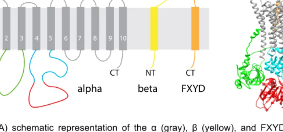
Figure 1.1. A) schematic representation of the α (gray), β (yellow), and FXYD (orange)
subunits of NKA. B) 3D structure of NKA based on 4HQJ PDB structure. Α subunit domains A- (green), N- (cyan), and P- (red) domains highlighted.
The α-subunit weighs around 110 kDa, contains 10 transmembrane domains (TM1-TM10) and the N-terminus and C-terminus are located in the cytoplasm. The N-terminus together with cytoplasmic domain 2 (CD2) between TM2 and TM3 form an actuator domain (A-domain). The cytoplasmic domain 3 (CD3) between TM4 and TM5 forms two domains – the nucleotide-binding domain (N-domain) and the phosphorylation domain (P-domain) (Fig. 1.1B). The transmembrane domains are α-helixes
and they form binding sites for the transported ions3,5. There is a binding
site on the α-subunit for specific inhibitors belonging to the class of cardiotonic steroids (CTS). In mammals, there are four isoforms of NKA – α1 to α4 each of which has different affinities to sodium, potassium, ATP, and CTS. The α1-subunit is expressed by all cell types whereas the α2-subunit is mostly expressed in glia, heart, muscles, and adipocytes. The α3-subunit is primarily expressed in neurons and the α4-α3-subunit is expressed
in the testis11. The sequence identity between human α1-, α2- and
α3-subunits is 87% while the α4-subunit has a sequence identity of only 78% outside inside 1 2 3 4 5 6 7 8 9 10 CT NT NT CT CT NT
alpha beta FXYD
3
with the other subunits. The sequence identity between α-isoforms from
different species is 85% or higher7.
The β-subunit is highly glycosylated extracellularly and its weight can range from 35 to 55 kDa. It contains just 1 transmembrane domain and the N-terminus is located in the cytoplasm and the C-terminus is extracellular.
The β-subunit is essential for the occlusion of potassium ions12, for
trafficking to the membrane13, and participates in cell-cell adhesion14,15.
There are three isoforms β1-β3, which are much less identical to each other
than α-isoforms – 35-47%, and between species – 50% or higher7. The
β1-subunit is expressed by all cells whereas the β2-β1-subunit was found in skeletal muscle, pineal gland, and nervous tissues, and β3-subunit was
found in testis, retina, liver, and lung11.
FXYD proteins are the smallest subunit in NKA – only around 10 kDa. They are all characterized by the Phe-Xaa-Tyr-Asp (FXYD) motif and contain just 1 transmembrane domain. The N-terminus is located in the extracellular space and the C-terminus is in the cytoplasm. There are 7 known isoforms of FXYD proteins in mammals although this family is
much larger in other organisms16,17. This subunit regulates the affinity of
NKA to sodium and potassium18, however, the presence of FXYD proteins
is not required for functional expression of NKA19.
1.2 Catalytical cycle of Na+,K+-ATPase
There are two conformational states of NKA during the catalytic cycle – E1-state which has a high affinity to sodium and E2-E1-state which has a high
affinity to potassium. They were crystallized in 20135,6 and 20073
respectively and the exact positions of sodium and potassium binding sites
4
partially substitute Na+6. The E2-state allows the binding of not only K+ but
also other cations such as Li+, Cs+, Tl+, NH4+, and finally Rb+20, which can
be used to measure NKA pump activity by Rb+ uptake. During the catalytic
cycle, NKA is phosphorylated at aspartate-369 and there are two
phospho-states of the NKA: E1-P and E2-P1. The total reaction can be summarized
as:
ATP + 3Na+in + 2K+out => ADP + Pi + 3Na+out + 2K+in
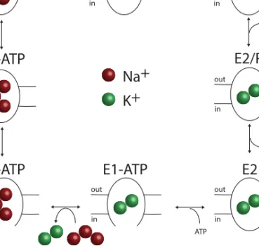
A schematic description of the NKA catalytic cycle was proposed by Post
and Alberts following some adjustments5,21 (Fig. 1.2). NKA in the E1-state
has a high affinity to Na+. Na+ binding to NKA drives phosphorylation of
the enzyme, converting it to the E1-P and then, via an occluded state where sodium ions are not accessible from either side, NKA advances to the E2-P
state which has a low affinity for Na+ and high affinity for K+. Sodium ions
are released to the extracellular space and potassium ions bind to NKA. This drives dephosphorylation of the enzyme and it is converted to the E2 state where it can bind ATP again and, via an occluded state where potassium sodium ions are not accessible from either side, NKA progresses to the E1 state with bound ATP and the cycle is repeated. Each reaction has been reproduced in experimental studies and many laboratories have described the effect of the mutations using this approach. Small molecules from the class of cardiotonic steroids can only bind NKA in the E2 conformation and this binding blocks the catalytical cycle.
5
Figure 1.2. Post-Alberts scheme of the NKA catalytical cycle5.
1.3 Cardiotonic steroid family
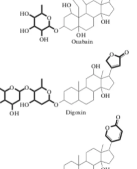
Cardiotonic steroids (also known as cardiac glycosides) are cyclopentane perhydro phenanthrene derivatives that contain an unsaturated lactone ring at C-17. There are two subclasses of cardiotonic steroids (CTS): cardenolides, which contain a 5-membered lactone ring, and bufadienolides, which contain a 6-membered lactone ring. Some of the CTS can be glycosylated at C-3 with one or more sugar residues (Fig. 1.3). Ouabain, derived from the plant Acokanthera schimperi is widely used in experimental studies due to its high-water solubility among CTS.
Ouabain binds NKA in the E2-state and enters a pocket which is formed by transmembrane domains 1 and 6 of the α-subunit, which is followed by a
conformation rearrangement of the cytoplasmic part of the α-subunit22,23.
out in out in out in out in out in out in out in out in +ADP H2O Pi ATP
Na+
K+
E1P-ADP
E2P
E2P
E2/P
iE2
E1-ATP
E1-ATP
E1-ATP
6
The position of the N- and P-domain in ouabain-bound NKA is almost identical to NKA without ouabain. The A-domain rotates about 10° with
transmembrane domains 1 and 2 moving towards the ouabain molecule24.
The crystallization of the NKA with other CTS such as digoxin and bufalin shows that the level of CTS glycosylation affects the strength of CTS binding. Substituents of the CTS steroid core affects the position of
transmembrane domains 1 and 225.
Figure 1.3. Chemical structures of ouabain, digoxin (both - cardenolides), and bufalin
(bufadienolide). Sugar motifs (to the left of the molecule) and lactone rings (to the right of the molecule) are highlighted.
Chemicals from the cardenolides subfamily have been isolated from plants while chemicals from the bufadienolides were found in amphibians. These chemicals have been widely used to treat a cardiac failure for several
centuries26. Recently it was shown that a few of the CTS might be produced
in mammals and, in particular, in humans. The first evidence of the existence of endogenous ouabain was discovered in the 1980s. Hamlyn and
7
colleagues showed by mass spectrometry the presence of an endogenous
cardiotonic steroid in blood plasma which they identified as ouabain27.
They also found ouabain in the adrenals of humans, cows, and rats as well as in cultures of adrenal cells. Ouabain is associated with many diseases
including hypertension28, congestive heart failure29, primary
aldosteronism, and essential hypertension30. Most of the studies above
have been performed by using the immunoreactivity method which was
described before31. An isomer of ouabain has been found in the bovine
hypothalamus32 and in 2012, ouabain was found in the cerebrospinal fluid
of mammals including humans33.
In addition to ouabain, other endogenous cardiotonic steroids could also
be hormones. Digoxin from cardenolides has been found in urine34 and
bovine adrenal glands35. There are specific antibodies against digoxin
called DigiBand36 which are being tested for medical application37. Digoxin
has been shown to prevent ouabain-induced hypertension38. Although the
results were observed repeatedly39, the mechanism of this antagonism
action is not understood.
Marinobufagenin (MBG), which belongs to another subclass of CTS
(bufadienolides) has also been proposed as a hormone26. Bufalin-like
immunoreactivity has been detected in human bile40, urine41, and blood
plasma42. Bufadienolide compounds have been detected by
mass-spectrometry in human cataractous lenses43 and placentae44. Using both
mass-spectrometry and nuclear magnetic resonance, bufalin-like compounds were identified as MBG. Evaluation of MBG is associated with chronic renal failure, idiopathic hyperaldosteronism, essential
hypertension, acute congestive heart failure45, and preeclampsia46.
8
“endogenous ouabain” stimulates the adrenocortical production of
marinobufagenin47.
There is also research that does not support the hypothesis of endogenous
ouabain48–50. Most of the studies of endogenous CTS have used an
immunochemical method to measure CTS concentrations. The method is based on the binding of antibodies to a specific CTS which leads to a high risk of cross-reactivity with closely related compounds. The pathways of ouabain, digoxin, and marinobufagenin synthesis in mammals are not well understood. There is some evidence that ouabain could be synthesized in
the adrenal glands51,52 or the hippocampus53 but exact enzymes are
unknown. Despite these uncertainties, there is an abundance of other
articles that show data about the existence of endogenous ouabain26,54–57
which is strong evidence that ouabain is produced in mammals.
The binding site of CTS consists of amino acids Q111, Q119, G120, and N122 in the first extracellular loop of the α subunit and they play an important role in the sensitivity of the enzyme to CTS. These amino acids are highly conserved in all animals but mutations in them do not affect enzymatic
function58. This conservation supports the idea that the site for CTS
binding is required for other functions of the NKA besides its enzymatic function. In animals who feed on organisms with high CTS concentrations, these amino acids have been mutated to make the α-subunit CTS-resistant but this has only happened in one isoform of the α subunit: α3 in snakes
and α1 in butterflies and rodents59, while the other subunits in these
animals remain sensitive to CTS. It has also been shown that ouabain is a signaling molecule, activating several signaling processes in diverse cell
types7,60–62. This has led to the hypothesis that CTS is a hormone with the
9
1.4
Na+,K+-ATPase and signaling pathwaysThe first evidence that the binding of ouabain to NKA drives signaling cascades came from a study from Askari and Xie groups in which the exposure of myocytes to ouabain resulted in a calcium-dependent
activation of c-fos and c-jun mRNA63. This paper started a new research
era regarding NKA's role as a receptor. During the following years, the same group reported that ouabain stimulated the Ras-Raf-MEK-ERK
pathway64 and activated tyrosine Src kinase and epidermal growth factor
receptor65. Later it was proposed that NKA and Src can form a complex and
the binding of ouabain to NKA freed the kinase domain of Src resulting in
its activation66. An NKA-derived peptide Src inhibitor was developed to
prevent ouabain-triggered signaling67. However, a few recent studies
question the existence of an NKA/Src complex68,69 (Fig. 1.4).
In parallel to these studies, the Aperia group showed that ouabain induces
regular low-frequency Ca2+i oscillations which leads to activation of
Nf-kB70. These effects are realized through activation of the IP3R which is in
this case InsP3 independent71. Activation of IP3R occurs due to an
interaction of the N-terminus of NKA and the IP3R with ankyrin B acting
as an adapter molecule72,73. Digoxin and marinobufagenin can also drive
this signaling pathway and the presence of Src inhibitors abolish calcium
oscillations62.
Ouabain application has also been reported to activate
phosphatidylinositide 3-kinase (PI3K) and Akt74. The PI3K-Akt pathway is
involved in the regulation of NKA function75 and PI3K binds to a
proline-rich motif of the α-subunit of NKA76. Interestingly, the Ras-Raf-MEK-ERK
signaling pathway is not PI3K-dependent74 and PI3K-Akt signaling is not
10
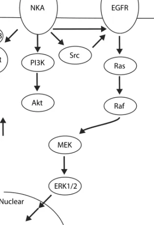
Figure 1.4. The three most studied ouabain-triggered signaling cascades
Interestingly, multiple groups have shown that ouabain’s signaling effect is observed not only at concentrations where it significantly blocks NKA activity but also at lower concentrations than do not block NKA activity.
Ouabain has also been shown to trigger various additional pathways 77–83
which seem to be not only isoform-specific but also cell type-specific. These processes alter gene expression, cell proliferation, and anti-apoptotic processes but the whole picture of ouabain- or other CTS-driven signaling is still unknown. out in Ouabain NKA Src EGFR Ras Raf MEK ERK1/2 Nuclear PI3K Akt AnkB IP3R ER Ca2+
11
1.5 Na+,K+-ATPase and protein-protein interactions
NKA is a well-known partner in many different protein-protein
interactions7. One of the first pieces of evidence that NKA interacts with
other proteins came from signaling studies where multiple groups showed
interactions with Src kinase66, IP3R72, PI3K76, and more. It has been shown
that NKA is involved in protein-protein interactions with caveolin-1 which
is a scaffolding protein participating in the formation of caveolae84. It is
hypothesized that caveolin-bound NKA forms a signaling, non-pumping pool of NKA since its interaction with caveolin might affect its enzymatic
function85. However, a separate group showed that the activity of caveolar
and non-caveolar bound NKA are similar86 and that caveolin-1, annexin-2,
and NKA form a large multiprotein complex.
Usually, the N-terminus of NKA is involved in protein-protein interactions. This may be because the end part of the N-terminus is not crucial for pump function and truncation of the N-terminus does not affect enzymatic
function87. Moreover, crystallography studies showed that the N-terminus
tail has a high degree of disorder23 and binding of ouabain leads to
conformational changes of the A-domain which includes the N-terminus7.
This suggests that the mechanism of ouabain-dependent regulation of protein-protein interactions might involve an N-terminus conformational change. NKA interacts with aquaporin 4 (AQP4), a water channel expressed in astrocytes, via the N-terminus as well. AQP4 interacts with both the α1 and α2 isoforms of NKA and at the same time interacts with
the metabotropic glutamate receptor mGluR588. This macrocomplex might
be important for the regulation of water and potassium homeostasis in astrocytes.
NKA interacts with other glutamate transporters and receptors which indicates the involvement of NKA in the functional regulation of
12
glutamatergic neurons. It has been shown that NKA co-immunoprecipitates with the glutamate transporter 1 (GLT-1 or EAAT2) and glutamate aspartate transporter (GLAST or EAAT2) which are expressed mostly in astrocytes and participate in the re-uptake of glutamate into the cell. Interestingly, in synaptosomes, ouabain showed dose-dependent inhibition of the glutamate transporter while in astrocytes, ouabain concentrations lower than 1uM increased glutamate transporter activity whereas higher concentrations lead to inhibition of
glutamate transporter activity89. In neurons, NKA co-precipitates with the
ionotropic glutamate receptors – NMDAR and AMPAR 90,91, and in both
cases ouabain leads to a reduction in total protein levels of both the receptors. Taken together, there is evidence of deep involvement of NKA and ouabain in glutamatergic synapses. Interestingly, the gene encoding the neuron-specific α3 isoform of NKA is a neighbor of the gene encoding the kainite receptor subunit GRIK5 (5000 bp between them) which belongs to the third ionotropic glutamate receptor – kainite receptor. This may predict the co-regulation of these genes and possible functional and protein-protein interactions between them, but it has not been studied so far.
NKA interacts and colocalizes with other receptors, channels, and
transporters – with dopamine receptors D1 and D292,93, with glycine
transporter GlyT294, sodium-calcium exchanger NCX95, and Nax
channels96. Since NKA interacts with the scaffolding protein PSD-9597
which is involved in the interaction between many different channels and
receptors98, it can be predicted that NKA can form a macromolecular
complex with Na-dependent and independent receptors and channels in synapses. The variety of such interactions should be studied systematically.
13
1.6 Na+,K+-ATPase, and cellular ion homeostasis
NKA is a primary-active transport protein which means that it uses energy from ATP to make an electrochemical gradient of sodium and potassium ions. The sodium gradient is the driving-force for secondary-active transport in animals. Sodium goes from the extracellular side of the plasma membrane to the cytoplasm and other molecules can be co-transported with sodium (symport) or be transported in opposite direction (antiport).
The sodium/calcium exchanger (NCX) is an example of Na+-Ca2+
counter-transport where the gradient of sodium is used to remove calcium from the cytoplasm. Interestingly, NKA and NCX can colocalize with each other
which probably allows Ca2+ regulation in a local microenvironment95. This
may also be regulated by endogenous ouabain in the brain99.
Another example of sodium-dependent antiport is the sodium/proton exchanger. The SLC9A1 gene encodes a Na+/H+ antiporter that is a
member of the solute carrier family 9 and is involved in pH regulation100.
Mutations in this gene lead to Lichtenstein-Knorr syndrome characterized
by cerebellar ataxia and sensorineural hearing loss101,102. The
sodium/potassium/chloride co-transporter is an example of the symport
of three ions in the stoichiometry 1Na+:1K+:2Cl- which makes it
electroneutral. This protein regulates ion balance and cell volume and it
participates in the reabsorption of these electrolytes from urine103.
Antagonists of the sodium/potassium/chloride co-transporter, encoded by the NKCC1 gene, leads to the restoration of low chloride levels in neurons which might be useful for the treatment of neurological and psychiatric
disorders104. Also, the sodium gradient is used for the co-transport of many
metabolites including glucose105, amino acids106, and more. NKA plays an
important role in the nervous system for the re-uptake
14
The formation of a gradient of potassium and sodium ions is important to maintain the resting membrane potential in cells. Due to the high permeability of the membrane for potassium, it has the most impact on
setting the membrane potential108. Resting membrane potentials vary in
different cell types. Astrocytes, myocytes, and neurons have the lowest
resting membrane potentials109,110 of all cells. During the action potential,
cation gradients are disturbed and the NKA is involved in the restoration
of these gradients to the resting state levels111. NKA mediates
afterhyperpolarization in neurons112 which is affects the excitability of
neurons113.
Taken together, it is clear that the NKA is essential for all organs in animals, especially the brain, and it is not surprising that NKA can use up
to 50% of the ATP produced in the brain114.
1.7
Mutations in Na+,K+-ATPase’s subunitsIt is known that a homozygous knockout of the α1, α2, or α3 subunit is lethal whereas heterozygous knockouts are viable and have small abnormalities. These include higher muscle and heart contraction for α2 knockouts, a lower cardiac contraction for α1 knockouts, and a few
behavior abnormalities for α2 and α3 knockouts115–117. However, humans
with genetic pathogenic variations in these subunits, even in the heterozygous state, can result in several different diseases and syndromes. Pathogenic variants in the ATP1A2 gene encoding the α2 subunit lead to familial hemiplegic migraine type 2 (FHM2) and the link between genetic
variations in ATP1A2 and the disease was first found in 2003118. FHM2 is
an autosomal dominant inherited form of migraine with aura119. There are
4 different types of FHM characterized by pathogenic variants in different genes. Most of the pathogenic variants in ATP1A2 are single nucleotide variants which lead to amino acid changes. Mutations causing FHM2 has
15
a scattered distribution along the protein molecule and there are no
preferable positions120.
Pathogenic variants in ATP1A3 give rise to several diseases. Mutations in this gene leading to rapid-onset dystonia-parkinsonism (RDP) were first
described in 2004121. RDP was described in 1993 and it is an
autosomal-dominant disease characterized by the sudden onset of dystonia and
parkinsonism122. Most of the RDP mutations are single nucleotide variants.
They are located in transmembrane domains and cytoplasmic and extracellular loops. The most common mutation, T613M, is located in the
P-domain123,124. The link between pathogenic variants in ATP1A3 and other
diseases, such as alternating hemiplegia of childhood (AHC), was found in 2012125,126. AHC is a rare neurological disorder characterized by early-onset episodes of hemiplegia, dystonia, various paroxysmal symptoms, and developmental impairment. Most of the mutations causing this disease are located in transmembrane regions and the phenotype of this disease is often more severe but there are phenotypes which are in between RDP and AHC120,123,127. Most of these mutations lead to reduced NKA activity and some of them affect protein expression. There is a third disease associated with ATP1A3 which is CAPOS (cerebellar ataxia, areflexia, pes cavus, optic nerve atrophy, and sensorineural deafness) syndrome, which is
characterized by one mutation, E818K128. There are several other
ATP1A3-related disorders: early infantile epilepsy and encephalopathy (EIEE)129,
fever-induced paroxysmal weakness, and encephalopathy (FIPWE)130, and
relapsing encephalopathy with cerebellar ataxia (RECA)131.
It has been shown that the ATP1A1 gene is intolerant to missense and
loss-of-function variants132. Somatic mutations in the ATP1A1 gene has been
shown for adrenal aldosterone-producing adenomas133. However, recently
Charcot-16
Marie-Tooth Type 2 which is characterized by distal weakness and atrophy,
sensory loss, and absence of reflexes132,134. Another syndrome, hereditary
spastic paraplegia (HSP), was shown to be associated with a mutation in
ATP1A1 as well135. Most of these mutations were located in the cytoplasmic
regions of the protein. Also, mutations in ATP1A1 lead to
hypermagnesemia, refractory seizures, and intellectual disability136. The
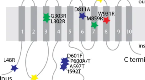
phenotype of these patients was more severe, and these three mutations were located in transmembrane domains (Fig. 1.5).
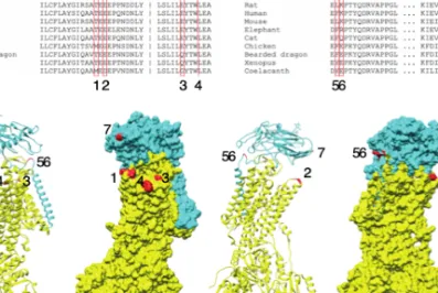
Figure 1.5. Distribution of pathogenic variants in ATP1A1. Mutations labeled by blue and
yellow stars are associated with Charcot-Marie-Tooth132,134. Mutations labeled by green stars
are associated with hypermagnesemia, refractory seizures, and intellectual disability136.
Mutation labeled by red star is associated with severe epileptic encephalopathy described in paper III from this thesis.
Taken together, pathogenic variations in the three genes encoding α1-3 lead to diseases in the nervous system which also indicates the importance of NKA for brain functioning.
17
1.8 Involvement of Na+,K+-ATPase in the progression of
neurodegenerative and psychiatric diseases
NKA is involved in the progression of multiple diseases. Parkinson’s disease belongs to a group of neurodegenerative diseases called synucleinopathies. α-synuclein is a protein that aggregates in Lewy bodies which contributes to Parkinson’s disease. It has been shown that α-synuclein interacts with the α3 subunit of NKA and this interaction leads
to cluster formation and decreases stimulus-triggered efficiency of Na+
extrusion137. The authors found that two amino acids which are α3-specific,
Leu878, and Asn879, in the extracellular loop between transmembrane domains 7 and 8 are responsible for this interaction. Interestingly, another group independently showed that almost the same amino acids from the α3 subunit of NKA, Asn879, and Trp880, play a key role in the interaction
between NKA and β-amyloid aggregates, amylospheroids138. These
aggregates are formed during the progression of a second neurodegenerative disease, Alzheimer’s disease. Also, it has been shown that the α3 subunit of NKA can interact with tau assemblies which have
prion-like properties139 and with misfolded SOD1 during the progression
of amyotrophic lateral sclerosis140. Interestingly, in the last case, this
interaction is mediated by intracellular domains of the α3 subunit. As these studies have shown that these interactions are unique for α3, there is good evidence that the neuronal-specific α3 subunit of NKA is involved in the pathogenic progression of prion-like diseases.
The α3 subunit of NKA is expressed mostly in GABAergic interneurons in
the brain141. Loss of GABAergic interneurons was found in patients with
psychiatric diseases such as bipolar disorder and schizophrenia142.
Interestingly, there is an ouabain-triggered model of bipolar disorder143
and there are studies that endogenous ouabain might be involved in the
18
protein level of the α3 subunit was lower in GABAergic neurons in the frontal cortex and hippocampus in bipolar disorder and schizophrenia as compared with the controls. However, in the temporal cortex, the level of
α3 subunit expression was higher145. This data opens up a discussion about
the involvement of the α3 subunit of NKA not only in prion-like neurodegenerative diseases but also in the progression of psychiatric diseases.
19
2 Materials and Methods
2.1 Model selection
We need to select an experimental model that is appropriate for the particular research question. This is an important step in the design of a study because every model has advantages and limitations. To answer biomedical questions, the best model would be humans, but this is impossible due to ethical dilemmas. Researchers have developed several different alternative models, and in this thesis, I have used several of them.
2.1.1 Mouse model
To study processes on the organism level we would like to use a model that is as close to humans as possible regarding anatomy and physiology. The model should reproduce relatively fast and be amenable to genetic modification to mimic human diseases. Mice are one of the most popular choices due to their fast reproductive cycle (3 months from birth to be sexually mature), ease of genetic modification (many well-established techniques available) and it is relatively close to humans (it belongs to the class Mammalia and suborder Euarchontoglires which includes rodents and primates). Their genome is sequenced and, using the CRISPR-Cas9 technique it is possible to get knockout or knock-in models in six months. Using a model organism, we can study how genetic modifications and/or treatments affect animal behavior, biochemical processes in different tissues, and, using microscopy, study neuronal circuits in the central nervous system. However, the use of model organisms does have a few limitations. Firstly, for many research questions that can be addressed on a single-cell level, it is better to use cell models that require the use of much fewer animals. Also, it can be important to use a more robust model that
20
gives better reproducibility without the individual variability observed between animals.
2.1.2 Primary culture of neurons
To study processes that do not require the original anatomical structure and specific connections between neurons we can use a primary culture of rat neurons. This model allows us to study processes on the single-cell level and cells maintain neuronal characteristics such as physiology, morphology, and synaptic connections. Neurons are isolated from the rat hippocampus at embryonic day 18.5 and after 2-3 weeks in vitro culture, they form dendrites and axons. Primary neuron cell culture is a very robust model for studying neuronal metabolism. It requires the use of fewer animals which is preferred according to the 3R principles. Primary neuron cell cultures can be transiently transfected and used to study how protein modifications affect neuronal metabolism. Cell cultures are beneficial as they are much easier to transfect in comparison to tissue slices. This model is commonly used for live imaging and measuring changes in intracellular sodium and calcium concentration.
Neurons from rat hippocampus were prepared from embryonic day 18.5 embryos of both sexes. The hippocampus was removed, washed in Hank’s balanced salt solution (HBSS) containing 20 mM HEPES, then incubated for 10 min at 37 °C in HBSS with 20 mM HEPES and 0.25% trypsin and dissociated in minimum essential media (MEM) by pipetting using a
fire-polished Pasteur pipette. Cells were plated at a density of 4*104/cm2 for
imaging or 10*104/cm2 for immunocytochemistry on glass coverslips
previously coated overnight with poly-ornithine (80 µg/ml) in MEM containing 10% horse serum, 2 mM L-glutamine, and 1 mM sodium pyruvate for three hours. Then the medium was replaced with Neurobasal medium containing 2% B27, 0.5 mM L-glutamine, and 1%
21
penicillin/streptomycin. Half of the medium volume was replaced with Neurobasal medium containing 2% B27, 0.125 mM L-glutamine, and 1% penicillin/streptomycin twice per week.
Although this model is robust and requires fewer laboratory animals, sometimes we want to study individual proteins that do not have a neuronal origin. Also, the primary culture of neurons is characterized by a high level of heterogeneity and can only be used for up to 3-4 weeks. Immortalized cell lines can instead be used as a more robust model with less variability.
2.1.3 Immortalized cell lines
To study protein function or protein-protein interactions, immortalized cell lines can be used. They are easy to work with, do not require any use of laboratory animals, and grow fast. There many cell lines available and in the presented studies we have used HEK293 and COS-7 cell lines which are derived from human embryonic kidney and monkey kidney tissue respectively. They were grown in DMEM supplemented with 10% FBS and 1% penicillin/streptomycin. To maintain them, the cells were passaged every 3-4 days. Cells at early passages were frozen in FBS/DMSO for further studies. This model in combination with studies in silico on structural data could shed light on protein function and be used to study protein conformational changes, activity, and membrane expression. Also, these cell lines are a general model for all mammalian cells, and we have used this concept in our phosphoproteomic study.
2.2 Cell biology methods
To study different aspects of cell metabolism we applied different cell biology techniques. To express a protein of interest we transfected a
22
primary cell culture or cell line with a plasmid containing the sequence of the protein of interest. For testing, whether a mutation affects protein function, we used a survival assay. For the analysis of membrane potential, we used a whole-cell electrophysiological approach.
2.2.1 Transfection
We transfected cells to make them express a protein of interest. We transfected them using lipofectamine – a commercial reagent that creates liposomes in which plasmids carrying the coding sequence of the protein of interest are transferred to the cell. These plasmids usually have a CMV (cytomegalovirus) promoter which leads to overexpression in all mammalian cells. The efficiency of transfection depends on the cell type – in primary cultures of neurons the efficiency is quite low and does not exceed 5-10% while the efficiency of transfection of a cell line can be up to 100%. Cultures were transfected 24-48 h before experiments using Lipofectamine 2000 (Thermo Fisher Scientific) or Lipofectamine LTX (Thermo Fisher Scientific) for neurons and cell lines respectively according to the manufacturer's protocol. Transfected cells were identified by the presence of a fluorescent protein-tagged to the protein of interest or the presence of a separate fluorescent protein on the different plasmid. This approach is used in all studies in this thesis.
2.2.2 Cell survival assay
NKA maintains the sodium and potassium gradient across the plasma membrane and a cell will not survive if the NKA is not functional. This fact makes cell lines a good model for screening whether mutations in NKA affect its function. To test this, we first needed to eliminate the endogenous NKA function which we did use ouabain-selection. HEK293 and COS-7 cells only express the α1 subunit of NKA which, in primates, is
ouabain-23
sensitive with a dissociation constant (Kd) of around 10 nM. We transfected
a cell line with a plasmid carrying the α1 coding sequence containing
mutations that lead to ouabain-resistance. After transfection, 10 µM
ouabain was added to the cell media for 24-48 hours to block endogenous NKA activity without affecting exogenous NKA activity. This approach allowed us to specifically select transfected cells that express ouabain-resistant NKA. By comparing wild-type and mutant NKA, we could test whether the mutation affects NKA function and thereby cell viability.
2.2.3 Electrophysiology
To measure the resting membrane potential and the rate of spontaneous activity of primary neurons in cell culture, we used a whole-cell patch-clamp setup. Using a micropipette of 3-7 megaohm resistance filled with an intracellular mimicking solution (120 mM potassium gluconate, 24 mM
KCl, 4 mM NaCl, 4 mM MgCl2, 0.16 mM EGTA, 10 mM HEPES, 4 mM K2
-ATP, pH 7.2 adjusted with KOH) attached to neurons. We broke the membrane and connected the solution in the pipette with the cell cytosol. A second electrode was located in the bath solution where neurons were
maintained (110 mM NaCl, 4 mM KCl, 1 mM NaH2PO4, 25 mM NaHCO3,
1.5 mM CaCl2, 1.2 mM MgCl2, 10 mM glucose and 20 mM HEPES at pH 7.4
at 37°C). When working with GFP-transfected cells, we found the cells using a fluorescence microscope. Recordings were performed using an Axopatch 200B amplifier (Molecular Devices) and pClamp software (version 8.2). In the current-clamp mode, we recorded resting membrane potential and spontaneous activity.
2.3 Fluorescence microscopy studies
Different fluorescence microscopy modalities have been key to the research in this thesis. In our studies, proteins were either fused to various
24
fluorescent proteins or labeled by fluorescent antibodies. Also, cells were loaded with dyes to measure the concentration of intracellular inorganic ions. The sample is illuminated at a specific wavelength and the emission light – which is shifted to higher wavelengths – is collected using a sensitive camera or photomultiplier tube (PMT). To test how mutations and/or ligands affect sodium and calcium homeostasis in neurons we performed live imaging on a widefield microscope. To look for protein distribution we used confocal microscopy. To search for co-localization, we used stochastic optical reconstruction microscopy (STORM) which belongs to a group of super-resolution microscopy techniques. An additional approach to search for protein-protein interactions is fluorescence correlation spectroscopy (FCS), which we used in our study.
2.3.1 Live widefield imaging
We used wide-field microscopy to collect light from the entire field of view to perform fast recordings of cellular activity. We used this technique for sodium and calcium imaging with a frequency of 0.4-1 Hz. The sample was either loaded with a dye 30-60 min before the experiment or cells were transfected with a genetically encoded calcium indicator GCaMP 24-48 h before the experiment. We used Asante Natrium Green-2 AM for measuring sodium and Fura-2 AM for measuring calcium ions. These dyes contain an AM (acetoxymethyl) ester which makes the dyes electrically neutral, allowing them to easily cross the plasma membrane. When inside the cell, the AM ester is cleaved by esterase, and the dye becomes negatively charged and unable to leave the cytosol. After loading the cells at 37C, they were rinsed 3 times in media to remove excess dye and then mounted onto the microscope. Asante Natrium Green-2 has an excitation maximum at
517 nm and an emission maximum at 542 nm146. Fura-2 AM belongs to a
group of ratiometric dyes that show a shift of their excitation peak when
25
signal with an emission maximum of 510 nm at two different excitation wavelengths (340 and 380 nm) cancels out differences in experimental parameters such as cell thickness and dye loading. While widefield microscopy usually uses a mercury lamp with a combination of filters, we used a monochromator which gives precise excitation wavelengths. Since
Fura-2 AM can be toxic for cells and inhibit NKA147 we also used a
genetically encoded calcium indicator GCaMP6f with an excitation maximum at 496 nm and an emission maximum at 513 nm. The low transfection efficiency allowed us to record from individual neurons as we often had just one transfected neuron in the field of view. We mounted a perfusion system to the widefield microscope to allow the rapid addition or removal of chemicals in the perfusate. Usually, we used a pump speed of 2 mL/min which does not stress neurons and gave a fast exchange rate in the perfusion chamber.
2.3.2 Confocal microscopy
We used confocal microscopy in situations where we needed to collect signals only from one focal plane. In confocal microscopy, a focused laser beam is scanned across the sample and the signal at each point is collected and used to reconstruct an image of the scanned area. Variation of the size of the pinhole allowed us to collect more or less out-of-focus signal, balancing between the amount of signal collected and the axial resolution. In our projects, this technique was used to determine whether a protein was localized at the plasma membrane. To do so, we collect light only from a plane at the half-height of the cell where we could estimate the localization of the protein. Also, to make a 3D image we scanned plane by plane to form an image stack. We then performed maximum intensity projections which represent the 3D structure of the cell better than a widefield microscopy image.
26
2.3.3 Stochastic optical reconstruction microscopy (STORM)
The resolution of images produced by both widefield and confocal microscopy is limited by diffraction and is dependent on the wavelength used and the numerical aperture of the objective. Even in the best scenario, the resolution cannot be lower than 150-250 nm. Different techniques have been proposed to improve the resolution of light microscopes. Some of them involve specimen preparation such as expansion microscopy. Another approach is to use superresolution microscopy for which the Nobel Prize in Chemistry was awarded in 2014 to Eric Betzig, Stefan W. Hell, and William E. Moerner. We have performed superresolution imaging using STORM in our studies.
STORM is based on the principle that the position of a molecule can be determined with a precision of a few nanometers by fitting the recorded signal from that molecule to a Gaussian distribution model. To do so, we need to make sure that we are fitting the signal from a single molecule since fitting the signal from two or more molecules with a single Gaussian would give the wrong position. To be able to use STORM, photoactivatable fluorescent molecules are used, of which only a few are activated during each frame. By repeatedly activating a subset of different molecules and fitting the emission (in our experiments we recorded 20000-25000 frames per image) we could reconstruct an image with a very high resolution down
to 10-20 nm148. In our experiments, we used a Zeiss Elyra (Carl Zeiss)
microscope equipped with a Plan-Apochromat 100x/1.46 Oil objective and an Andor iXon EM-CCD camera. Two laser lines (488 nm and 642 nm) were used to activate, excite and deactivate Atto-488 and Alexa-647 respectively. A low-wavelength 405 nm laser is used to continuously push molecules into an activated state. We used STORM to judge whether two proteins co-localized on the plasma membrane of neurons or not.
27
2.3.4 Fluorescence correlation spectroscopy
To look for oligomerization of the NKA we used applications of fluorescence correlation spectroscopy (FCS) such as combination of Förster Resonance Energy Transfer and Fluorescence Correlation
Spectroscopy (FRET-FCS) and fluorescence cross-correlation
spectroscopy (FCCS). FCS detects fluorescence fluctuations from molecules diffusing in a solution or a lipid membrane through a sub femtoliter detection volume and can be used to measure concentrations and diffusion coefficients of molecules as well as molecular interactions. From single color FCS, it is possible to extract not only the average transit time and the average number of molecules in the area but also the brightness of molecules which might indicate an oligomerization state. FRET-FCS detects the acceptor emission from FRET-active oligomers containing both donor and acceptor. In FRET measurements in general, the sensitivity is limited by the amount of background-signal. Background occurs when some of the donor emission is detected in the red channel, i.e. so called cross-talk, and when acceptors to a small extent are excited directly by the 488 nm laser. By using donors and acceptors with an unusually large spectral shift – such as Alexa 488 and Alexa 647 – the background signal from such non-interacting, monomeric donors and acceptors is made very small. Thereby interactions can be detected even in cases where only a small fraction of the available donor and acceptor molecules form FRET-active oligomers. In our ongoing study, we use FRET-FCS and FCCS and two-color labeled α and/or β subunit of NKA to see whether we have a fraction of interacting molecules or not. Since none of the studies of NKA oligomerization have been performed on living cells, our ongoing study will give us the possibility to solve the question of whether two or more NKA interact in real-time and if so, how we can modulate this oligomerization.
28
2.4 Animal studies
To study how a mutation leads to a disease on the organism level, we established a mouse model for one of the studied diseases – rapid-onset dystonia-parkinsonism (RDP). This is a knock-in mouse model, T613M ATP1A3, created by using the CRISPR/Cas9 technology. Since all humans with RDP have this mutation in the heterozygous state, we used heterozygous animals as a model of the disease and their wild-type littermates as control animals. To do this we crossed wildtype male and heterozygous female or vice versa to get a 1:1 ratio between wildtype and heterozygous animals. To see whether the offspring inherit the mutation we performed genotyping of the animals. To characterize abnormalities in animals we used a set of behavior studies that indicate motor, anxiety, or cognitive abnormalities compared to wild-type animals.
2.4.1 Genotyping of animals
There are just 2 basepairs (bp) that differ between the sequence of transgenic mutants and wildtype. To test whether an animal carries the mutation, we isolated DNA from tissue (ear clipping) and performed a PCR with the specific primers to amplify a region of the ATP1A3 sequence containing the mutation, giving a 418 bp product size. Then, we cut the product of the PCR using a MlsI restriction enzyme which recognizes the TGGCCA sequence. In the wildtype sequence, there is just one restriction site and, after running the DNA on a gel, we should get bands at 341 and 77 bp. In the mutant sequence, there are two restriction sites and we should get bands at 230, 111, and 77 bp. Since heterozygous animals have one wild-type allele and one mutant allele we should get 4 bands: 341, 230, 111, and 77 bp.
29
2.4.2 Behavioral tests
To fully characterize the phenotype of our mouse model we applied different tests that characterize different aspects of its behavior.
The open-field test was used to analyze the explorative behavior of the animals. Mice were placed in the center of an open square area and their locomotion activity was recorded by a video camera mounted on the top of the apparatus. Then we extracted the total distance traveled by the animal and its velocity. Here we could judge whether the animal was hyperactive or hypoactive.
The elevated plus-maze test was used to measure the anxiety level of the animals. Mice were placed in the center of a four-arm apparatus were two arms were walled and two were without walls. As in the previous test, a video camera recorded the animal movement from the top and we extracted the total time which mice spent in the open and in close arms. Mice spending more time in the open arms indicated lower levels of anxiety.
A forced swim test was used to analyze depression-like behavior. Mice were placed in a plastic cylinder filled with warm water. The time spent passively floating and actively floating was recorded. Mice spending more time actively floating showed a lower level of depressive-like behavior.
A rotarod test was used to analyze the motor coordination, balance, and grip strength. Mice were placed on a rotating rod at low speed (5 rpm) and after 30 seconds it was accelerated to high speed (40 rpm) for 3 minutes. The test was repeated 3 times per day during 2 consecutive days and we calculated the latency to fall during each round.
30
A vertical pole test was used to analyze whether the nigrostriatal pathway is affected in mutant animals. Mice were placed on the top of a vertical pole with their heads towards the top. Then the mouse will turn and climb back, returning to the home cage. These times were calculated and presented as an average of five trials after one training trial.
A catwalk test was used to analyze gait, coordination, and motor abnormalities. The mice were placed in the apparatus which is a dark elongated tunnel. The mice were allowed to explore and walk freely in this tunnel, and a video camera recorded them in the center of the tunnel through the bottom glass floor. Three consecutive days of training were required and on the next day, the test was performed.
A passive avoidance test was used to analyze long-time memory. The mice were placed in the light section of the apparatus which contained two sections: a dark and a light section with a closed-door in between. After one minute the door was opened and when the mouse entered the dark section the door was closed, and an electrical current was applied to the floor of the dark section. The next day the mouse was placed again in the light section and the latency to go to the dark section was measured.
31
3 Summary and Discussion
3.1 Paper I. Ouabain Modulates the Functional Interaction Between Na+,K+-ATPase and NMDA Receptor
In this paper, our goal was to study how ouabain regulates structural and functional interaction between NKA and NMDA receptor (NMDAR). The NMDAR is one of the most popular proteins to study in the neuroscience field. It is characterized by high complexity and it plays an important role in synaptic plasticity and molecular memory. It belongs to the class of ionotropic glutamate receptors. There are two types of glutamate receptors – ionotropic and metabotropic which work as ion channels and GPCRs proteins respectively. The NMDAR is permeable to sodium and calcium ions and the flux of calcium ions plays a role in the regulation of synaptic plasticity. A large influx of calcium via the NMDAR often leads to excitotoxicity which can happen following a stroke.
Previously, my former colleagues and I published a paper describing a
functional interaction between the NKA and the NMDAR91. We showed
that they co-precipitate using the co-immunoprecipitation method and that the application of ouabain leads to a reduction of the amount of NMDAR subunit protein after one hour. These results together with the
results from other groups99,149,150 inspired us to study this interaction in live
conditions by using various light microscopy approaches. First, we showed that ouabain administration to a primary culture of hippocampal neurons leads to attenuated NMDAR-dependent calcium influx. We showed that this effect is observed regardless of the method, whether we apply it to the entire coverslip with neurons or locally to an individual neuron. Application of ouabain together with glutamate which activates all types of