ZDENKA PRGOMET
THE ROLE OF WNT5A IN
ORAL SQUAMOUS CELL
CARCINOMA
ZDENKA PR GOMET MALMÖ UNIVERSIT Y THE R OLE OF WNT5A IN OR AL SQU AMOUS CELL C AR CIN OMA DOCT OR AL DISSERT A TION IN ODONT OL OG YT H E R O L E O F W N T 5 A I N O R A L S Q U A M O U S C E L L C A R C I N O M A
Malmö University, Faculty of Odontology
Doctoral Dissertations 2017
© Zdenka Prgomet 2017 Illustrations: Zdenka Prgomet ISBN 978-91-7104-754-0 (print)
ZDENKA PRGOMET
THE ROLE OF WNT5A IN
ORAL SQUAMOUS CELL
CARCINOMA
Malmö University, 2017
Department of Oral Pathology
Faculty of Odontology
Malmö, Sweden
This publication is also available at: http://dspace.mah.se/handle/2043/21509
“A dream doesn´t become reality through magic; it takes sweat, determination and hard work” Colin Powell
CONTENTS
ABSTRACT ... 11
LIST OF PAPERS ... 13
ABBREVATIONS AND DEFINITIONS ... 15
INTRODUCTION ... 19
Oral squamous cell carcinoma ...20
Oral mucosa ...21
Risk factors for OSCC...21
Clinical features and histology ...21
Prognostic factors ...23
Treatment and prognosis ...25
The WNT signaling ...25
WNT proteins: production, secretion, classification ...26
WNT receptors ...28
WNT signaling modulation ...28
WNT/β-catenin signaling pathway ...29
β-catenin-independent WNT signaling pathway ...30
WNT5A ...31
WNT5A signaling and function ...32
WNT5A interaction with WNT/β-catenin signaling pathway .32 WNT5A regulation ...33
WNT5A signaling in odontogenesis and oral mucosa ...34
WNT5A in OSCC ...34
WNT5A and other cancers ...34
AIMS ... 37
MATERIAL AND METHODS ... 39
Cell lines and treatments...39
Tissue samples ...40
Ethical aspects ...40
Paper II ...40
Paper III and IV ...40
Cell based experiments (Paper I) ...41
Measurements of Ca2+ signaling ...41
Wound healing assay ...41
Measurements of BrdU positive cells ...41
Cell invasion assay ...42
Analysis of PKC activity ...42
Validation of antibodies (Paper II) ...42
WNT5A antibodies ...42
Optimization of IHC protocol for WNT5A antibodies ...43
Pre-absorption test of the primary antibodies ...44
Protein expression ...44
Western blot analysis (papers I - IV) ...44
Immunohistochemistry ...46
Gelatin zymography - Paper IV ...47
Statistical analysis ...47
Paper I ...47
Paper III ...47
Paper IV ...48
RESULTS ... 49
Paper I - Migration and invasion of oral squamous carcinoma cells is promoted by WNT5A, a regulator of cancer progression .. 49
Paper II - Optimization, validation and identification of two reliable antibodies for immunodetection of WNT5A ...53
Paper III - Higher expression of WNT5A protein in oral squamous cell carcinoma compared to normal oral epithelium and dysplasia ...57
Paper IV - WNT5A activates MMP2 in oral squamous cell carcinoma ...61
DISCUSSION ... 65 CONCLUSIONS ... 73 Future research...74 POPULÄRVETENSKAPLIG SAMMANFATTNING ... 75 ACKNOWLEDGEMENTS ... 77 REFERENCES ... 81 PAPERS I – IV ... 99
ABSTRACT
Cancer is one of the leading causes of death worldwide and further research into the cancer biology is required to improve treatment and survival. Cancer can start in all sites of the body and is characterized by uncontrolled cell growth and capability of invading surrounding tissue and spreading to other sites of the body. The most common type of cancer occurring in the oral cavity is oral squamous cell carcinoma (OSCC). Tobacco, including the betel quid and other types of smokeless tobacco, is the main risk factor for development of OSCC. Even though therapeutic treatment of OSCC has been improved, the prognosis is still poor, and the 5-year survival remains around 50%. The ongoing research have shown that proteins involved in signaling pathways play an important role in the progression of cancer, however, no reliable biomarkers have yet been identified as predictors of OSCC progression.
Over the past years, a protein named WNT5A, has been shown to be involved in different types of cancer, either by promoting or suppressing cancer progression. However, its role in OSCC is still ambiguous. This thesis aimed to provide an insight into the role of WNT5A signaling in OSCC.
We started by investigating the functional role of WNT5A in OSCC cells. Due to the lack of the endogenous WNT5A expression, we treated the cells with recombinant WNT5A (rWNT5A) and observed activation of the non-canonical WNT/Ca2+/PKC signaling in OSCC
cells. This rWNT5A-induced signaling enhanced migration and invasion of OSCC cells, which was ascertained by different WNT5A inhibitors. These inhibitors eliminated the rWNT5A-induced WNT/
Ca2+/PKC signaling and migration of OSCC cells indicating a
promoting role of WNT5A in OSCC cells.
Before investigating the expression of WNT5A in human OSCC tissues, we evaluated four commercially available WNT5A antibodies for use in immunohistochemistry (IHC) and western blot analysis (WB). Cytoplasmic WNT5A immunostaining pattern was observed with all four antibodies but only the polyclonal AF645 antibody was able to detect WNT5A protein in WNT5A-positive cell lysates. Pre-absorption tests revealed that the polyclonal AF645 antibody is the best antibody for detection of WNT5A by WB while the monoclonal 3A4 antibody is the most suitable for use in IHC.
Using the monoclonal 3A4 antibody, we investigated the expression of WNT5A in human OSCC tissues. No expression of WNT5A was observed in normal oral epithelium or in mild grade of dysplasia. However, cytoplasmic WNT5A immunostaining was found in 38% of dysplastic tissues and in 81% of the OSCCs. We also noticed that WNT5A was more expressed in advanced OSCCs than in early invasive OSCCs. Furthermore, we did not observe any correlation between expression of WNT5A and two adhesion-proteins, β-catenin and E-cadherin, either in OSCC tissues or in OSCC cells. These findings suggest that WNT5A does not affect the canonical WNT/ β-catenin pathway or downregulation of E-cadherin in OSCC.
At last, we investigated if the rWNT5A-induced WNT/Ca2+/PKC
signaling affected secretion and activation of matrix metalloproteinases (MMPs) in OSCC cells. We found that rWNT5A-induced activation of MMP2 is not dependent on activation of either β-catenin, ERK1/2, or p38-MAPK, but could instead be induced by WNT5A/Ca2+/PKC
pathway.
In conclusion, these findings suggest that WNT5A acts as a cancer promoter in OSCC by facilitating cell migration and invasion via the WNT5A/Ca2+/PKC signaling and activation of MMP2.
LIST OF PAPERS
This thesis is based on the following papers, referred to in the text by their Roman numerals:
I. Migration and invasion of oral squamous carcinoma cells is promoted by WNT5A, a regulator of cancer progression
Prgomet Z., Axelsson L., Lindberg P., Andersson T.
J Oral Pathol Med. 2015 Nov; 44 (10) 776-84.
II. Optimization, validation and identification of two reliable antibodies for immunodetection of WNT5A
Prgomet Z., Andersson T., Lindberg P.
Biotech & Histochem.
In press. DOI: 10.1080/10520295.2016.1255995
III. Higher expression of WNT5A protein in oral squamous cell carcinoma compared to normal oral epithelium and dysplasia.
Prgomet Z., Andersson T., Lindberg P.
Submitted to European Journal of Oral Sciences.
IV. WNT5A activates MMP2 in oral squamous cell carcinoma.
Prgomet Z., Andersson T., Lindberg P.
Papers I and II are Open Access Article available under the terms of the Creative Commons Attribution Non-Commercial License CC BY-NC which permits non-commercial use, distribution and reproduction in any medium, provided the original work is properly cited.
The author, Zdenka Prgomet (ZP), was the main contributor to the work process, from performing the experimental work, evaluating the results to writing the manuscripts.
ABBREVATIONS AND DEFINITIONS
AKT v-akt murine thymoma viral oncogene homolog ANOVA Analysis of variance
APC Adenomatous polyposis coli ARF6 ADP-ribosylation factor 6
BrdU Bromodeoxyuridine
CAMKII Ca2+/calmodulin-dependent protein kinase II
Cdc42 Cell division control protein 42 homolog CK-1 Casein kinase 1
CRD Cysteine-rich domain
DAB Diaminobensidine
DAG Diacylglycerol
Dkk Dickkopf
DNA Deoxyribonucleic acid
DVL Dishevelled
ECM Extracellular matrix
EGFR Epidermal growth factor receptor EMT Epithelial-mesenchymal transition ERK Extracellular-signal-regulated kinase FAK Focal adhesion kinase
FFPE Formalin-fixed paraffin-embedded
FZD Frizzled
GF109203X Inhibitor of PKC α, β1, β2 and γ Go 6983 Inhibitor of PKC α, β, γ, δ and ζ GPCR G protein-coupled receptor GSK3 Glycogen synthase kinase 3 HIAR Heat-induced antigen retrieval
HPBK Peroxidase-blocking solution
HPV Human papilloma virus
HRP Horseradish peroxidase
HSPG Heparan sulphate proteoglycan
HuR Human antigen R
IHC Immunohistochemistry
IL-6 Interleukin-6 Int 1 Integration 1
JNK c-Jun N-terminal kinase LEF Lymphoid enhancer factor
LRP Lipopotein receptor-related protein MAPK Mitogen-activated protein kinase MAPT-AM Ca2+ chelator
MARCKS Myrisotylated alanine-rich C kinase substrate MEK Mitogen-activated protein kinase kinase MMP Matrix metalloproteinase
MMTV Mouse mammary tumor virus
mRNA Messenger ribonucleic acid MUSK Muscle-specific receptor kinase NFAT Nuclear factor of activated T-cells NFκB Nuclear factor κB
NLK Nemo-like kinase
OSCC Oral squamous cell carcinoma p38-MAPK p38 mitogen activated protein kinase PCP Planar cell polarity
PI3K Phosphoinositide 3-kinase
PKC Protein kinase C
PLC Phospholipase C
PMA Phorbol 12-myristate 13-acetate PMD Potentially malignant disorder PP2A Protein phosphatase 2A PTK7 Protein tyrosine kinase 7 PVDF Polyvinylidene fluorid
qPCR Quantitative polymerase chain reaction Rac RAS-related c3 botulinum toxin substrate
Rb Retinoblastoma
Rho RAS homolog gene family member
rWNT3A Recombinant wingless-type MMTV integration site family member 3A
rWNT5A Recombinant wingless-type MMTV integration site family member 5A
RYK Receptor related to tyrosine kinase SCC Squamous cell carcinoma
SDS-PAGE Sodium dodecyl sulphate polyacrylamide gel electrophoresis
SEM Standard error of the means SFRP Secreted Frizzled-related protein
TCF T-cell factor
TGFβ Transforming growth fator β TNM Tumor, lymph node, metastasis TSG Tumor suppressor gene
uPA Urokinase plasminogen activator VEGF Vascular endothelial growth factor WB Western blot analysis
Wg Wingless
WIF-1 WNT-inhibitory factor 1
WNT Wingless-type MMTV integration site β-TrCP β-transducin repeat containing protein
INTRODUCTION
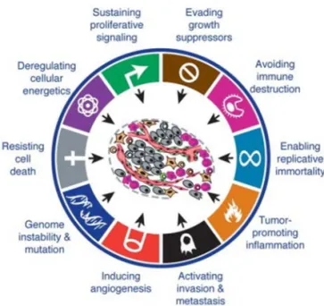
Cancer includes 100 different diseases, all of which share the features of uncontrolled cell growth and capability of invading surrounding tissue and spreading to other sites of the body (1). In 2012, there were 14.1 million new cancer cases and 8.2 million deaths caused by cancer worldwide (2). An accumulation of mutations and epigenetic alterations in oncogenes and tumor suppressor genes (TSGs) leads to the transformation of normal cells into cancer (3,4). During the multistep of carcinogenesis, the cancer cells require functional capabilities in order to survive, grow and spread. These are referred to as “hallmarks of cancer” and include sustained proliferation, the evasion of growth suppression, the escape of death, limitless replication, the evasion of immune destruction, the induction of angiogenesis, promoted genomic instability, the employment of tumor-associated inflammation, the reprogramming of energy metabolism and induced invasion and metastasis (Figure 1) (5). One of the signaling pathways that is involved in cancer progression and associated with several of these hallmarks is WNT5A signaling. This thesis is an attempt to provide an insight into the role of WNT5A in oral squamous cell carcinoma (OSCC).
Figure 1. Hallmarks of cancer. Reprinted and modified from Cell; Vol 144, Issue 5, 646-674, 2011; Douglas Hanahan, Robert A. Weinberg; Hall-marks of Cancer: The Next Generation; with permission from Elsevier (5).
Oral squamous cell carcinoma
The most common subtype of head and neck squamous cell carcinoma (HNSCC) found in the oral cavity is oral squamous cell carcinoma. It differs from other squamous cell carcinomas (SCC) of the head and neck region in the molecular aspect, but also due to the risk factors specific to the oral cavity (6). The oral cavity comprises the tongue, floor of the mouth, buccal mucosa, palate, and gingiva. The cancer of oral cavity and lip was the 11th most commonly occurring cancer
worldwide, with an incidental rate of 300,400 cases in 2012 and with a mortality rate of approximately 50%. It was found mostly among males in Melanesia, South-Central Asia, and Central and Eastern Europe (2).
Oral mucosa
Mucosa of the oral cavity consists of stratified squamous epithelium and underlying connective tissue called lamina propria. These are separated by a layer of extracellular matrix (ECM) called basal lamina or basement membrane. The stratified squamous epithelium consists of different layers and protects the underlying connective tissue from external pathogens and chemicals. The oral epithelium can be either keratinized or non-keratinized depending on the location and function in the oral cavity. The basement membrane controls the molecular passage and possesses signaling functions that control epithelial cell differentiation and polarity. The basement membrane is 50-100 nm thick and consists of type III, IV, and VII collagen, laminin, heparan sulphate proteoglycan (HSPG), and fibronectin (7).
Risk factors for OSCC
Several risk factors for the development of OSCC have been recognized, among which tobacco is the main factor, including the betel quid and other types of smokeless tobacco (8-10). Studies regarding alcohol as a risk factor for the development of OSCC have shown different results (11,12) but the use of alcohol in combination with tobacco increases the risk for OSCC (11). Another important etiological factor is high-risk human papilloma virus (HPV) 16 and 18 that has been more associated with the development of SCC in oropharynx (tonsils and base of the tongue) rather than the oral cavity (13-15).
Clinical features and histology
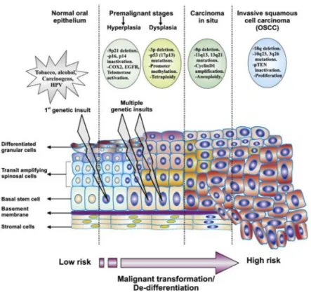
The genetic and epigenetic alterations in normal epithelial cells caused by tobacco, alcohol, or viruses give rise to precancerous keratinocytes and are the starting point for the malignant transformation (Figure 2). The additional alterations affecting cellular functions, such as growth, differentiation, DNA repair, and apoptosis, result in precancerous epithelium. The precancerous epithelium may at first appear clinically normal, but in time it may develop into potentially malignant disorder (PMD) (16-18). The PMDs are clinically diagnosed as leukoplakia, erythroplakia, lichenoid lesion, and oral submucous fibrosis (19), while histologically these can be diagnosed as dysplastic or non-dysplastic. Dysplasia is graded as mild, moderate, and severe,
and malignant transformation is more often seen in the moderate and severe grades of dysplasia than in the mild grade (19,20). The precancerous epithelium may eventually transform into oral cancer (16,17,21). However, the biggest challenge is to predict which PMDs are at risk of transformation. The PMD is histologically considered as cancer when abnormal epithelium overgrowth invades the underlying connective tissue (20). The malignant transformation of dysplasia into OSCC has been shown to vary between 5% and 36% (22).
Figure 2. Malignant transformation of OSCC. Reprinted from Biochimica et Biophysica Acta (BBA); Vol 1806, Issue 2, 146-162, 2010; Vinitha Ric-hard, M. Radhakrishna Pillai; The stem cell code in oral epithelial tumori-genesis: The cancer stem cell shift hypothesis; with permission from Elsevier (23).
Prognostic factors
Clinical and histopathological grading systems are used as prognostic tools for OSCC. Clinical staging includes TNM classification and the location of the primary tumor. Over the years, different histopathological grading systems have been developed. The World Health organization (WHO) grading system, adapted from Broders et al. (24), was the first described grading system and is based on the subjective valuation of, e.g., the keratinization of the tumor, cellular and nuclear size and shape, and mitotic activity. This system divides tumor differentiation into three grades: grade 1 - well-differentiated, grade 2 - moderately-differentiated, and grade 3 - poorly-differentiated tumors (25,26). Another grading system called malignancy grading (MG), was presented by Bryne et al., that modified the WHO grading system by the addition of pattern of invasion (27). Later on, two other grading systems were developed: the histological risk (HR) model and BD model. In addition to WHO grading system, the HR model includes assessment of the worst pattern of invasion, lymphocytic host response, and perineural invasion, while BD model describes tumor budding and the depth of tumor invasion in millimeters as important factors for the prediction of lymph node metastasis (28,29). Over the years, the WHO and MG grading systems have been criticized due to the subjectivity and lack of correlation with the outcome for OSCC patients. However, a study that evaluated these four grading systems showed that BD grading system correlates with the OSCC outcome and the authors suggested BD system as a possible prognostic tool (30).
Prognostic biomarkers
Califano et al. found loss of heterozygosity at chromosomes 3p, 9p, and 17p in early events of HNSCC malignant transformation (31). Later on, another study showed that these genetic markers, along with mutations in p53 gene (TP53), were found in the epithelium that surrounds the carcinoma, and that this altered mucosa remained after surgical treatment. These alterations were suggested to cause local recurrence and a second primary tumor (32). Despite the extensive research, no reliable biomarkers have so far been identified as predictors of the malignant transformation of precancerous keratinocytes. However, a variety of proteins involved in signaling
pathways were shown to be involved in the progression of OSCC. One of them is epidermal growth factor receptor (EGFR), which has been shown to be overexpressed in OSCC and suggested to be a marker for the malignant transformation of precancerous lesions (33). Other proteins, among many, that were shown to be overexpressed in OSCC are cyclin D1, NFκB, and VEGF (34,35). The most common altered TSG in OSCC are p53 and p16 (34).
OSCC cells move collectively, and for this they need adherence junctions which consist of transmembrane proteins E-cadherin, VE-cadherin, and N-cadherin. The intracellular domain of cadherins interacts with β-catenin present in the cytoplasm and together these control tissue polarity. Besides the role in cell adhesion, β-catenin acts as a signaling factor in WNT signaling pathway (36). E-cadherin was shown to be downregulated in OSCC and is considered to be an important epithelial-mesenchymal transition (EMT) marker in OSCC (37,38).
In order to invade the underlying connective tissue and metastasize, cancer cells need to pass through the basement membrane and make their way through the ECM. This process is in part mediated by proteases such as urokinase plasminogen activator (uPA) and matrix metalloproteinases (MMPs) (39,40). uPA and its receptor uPAR have been shown to be overexpressed in OSCC (41,42). Several studies have shown the increased activity and expression of MMP2 and MMP9 in OSCC (43-45). The signaling pathways that have been shown to be involved in MMP regulation are EGFR/MEK/ERK, EGFR/FAK/AKT, PKC/RAF/ERK, and p38-MAPK (46,47).
Moreover, the early HPV oncogenes E6 and E7 were shown to contribute to carcinogenesis by affecting TSGs p53 and retinoblastoma (Rb) (48). Epigenetic modifications in oncogenes and TSGs have also been associated with early events in oral carcinogenesis (6,49,50). Among the epigenetically silenced genes in OSCC are p16, E-cadherin, but also the components of WNT signaling, such as secreted Frizzled-related proteins (SFRPs) and WNT-inhibitory factor 1 (WIF-1) (50-52).
Treatment and prognosis
The treatment and prognosis for OSCC are determined by clinical and histopathological grading systems. Surgical treatment still remains the main treatment of OSCC, with adjuvant radiotherapy. In early stage OSCC, only radiotherapy is applied, while in advanced stages or when surgery is not applicable, the main treatment is adjuvant chemoradiotherapy (53-55). Research into the biology of OSCC has led to the development of the approved targeted therapy, cetuximab, which is an antibody directed to EGFR (56). Cetuximab was shown to be efficient as treatment for unresectable advanced and recurrent/ metastatic OSCCs (57).
Despite the improvement in the therapeutic treatment of OSCC, many patients have recurrence. The recurrence could be due to the insufficient removal of the primary tumor or the formation of a second primary tumor at the same location. A meta-analysis showed that local recurrence occurs more often if the surgical margins were <5 mm (58). It was also shown that recurrence occurs more often in younger patients, without affecting the overall 5-year survival rate (59). The reports on 5-year survival differ due to the number of patients and the part of the world referred to, and rates vary between 28% and 80% (60).
The WNT signaling
WNT signaling has been studied since the late 1980s and has been proven to be essential for embryonic development, stem cell control, and tissue homeostasis by regulating cell fate, proliferation, movement, adhesion, invasion, and polarity (61-65). Moreover, abnormal WNT signaling can contribute to the progression of different diseases, including cancer (66,67). WNT signaling was first reported in Drosophila melanogaster, where mutations in gene Wingless (Wg) caused failure in the development of the wings (68). A few years later, Integration I (Int1) gene was identified as an oncogene which caused mammary carcinomas in mice after activation by the mouse mammary tumor virus (MMTV) (69). Later on, Wg gene was shown to be a homolog to Int1 gene (70-72), and in 1991 the name “WNT” was derived from the combination of the names of these two genes (73).
WNT proteins: production, secretion, classification
At present, there are 19 members of the WNT family. The first one was named WNT1 after Wg/Int1 gene (73,74). The WNT proteins share 27-83% homology in the amino acid sequence (75), they all have 22-24 conserved cystein residues, and one or more sites for N-linked glycosylation (76,77).
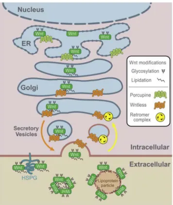
Once WNTs are synthesized, the membrane bound O-acetyltransferase called Porcupine attaches a palmitate to an N-terminal conserved cysteine residue and a palmitoleic acid to a serine residue (78-80). These post-translational lipid modifications make the WNTs more hydrophobic, a property that is required for their secretion as well as for their ability of binding the receptor and signal triggering (61,80). The lipid-modified WNTs are then coupled with cargo-adaptor proteins p24 that transport WNTs from the endoplasmic reticulum to the Golgi apparatus (81,82), where the transmembrane protein named WNTless interacts with the lipid group of the modified WNTs (83). The WNTless transports WNTs in the secretory vesicles to the plasma membrane and it also aids the binding of WNTs to their receptors (Figure 3) (61). Another post-translational modification of WNTs is glycosylation, which occurs at several aspargine residues at N-terminal (80). Even though it is suggested that glycosylation is involved in WNT folding, activation, and secretion (79,84), its role and how and where this takes place remains unclear.
Following secretion, WNTs need to be distributed in order to trigger the signaling. They can act both in autocrine and paracrine signaling which are in part facilitated by the ability of WNTs to bind to HSPGs. The HSPGs are located on the surface of the cells and in the ECM, and have been shown to maintain the activity of WNTs and to keep them at the cell surface. The lipid modifications enable WNTs to be secreted and distributed by exosomes or carrier proteins, and to trigger signaling on distant cells (80,85-87).
Figure 3. WNT secretion. Reprinted from Developmental Cell; Vol 17, Issue 1, 9-26, 2009; Bryan T. MacDonald, Keiko Tamai, Xi He; Wnt/β-Catenin Signaling: Components, Mechanisms, and Diseases; with permission from Elsevier (88).
Wong et al. showed that WNTs can be divided into three categories based on their ability to cause morphological transformation and alternations in the growth of C57MG mammary epithelial cells (89). WNTs were divided into highly transforming, moderately transforming, or those that were not able to induce changes in morphology or cell growth (89). Later on, the same WNT proteins were shown to have different effects on β-catenin where the transforming WNTs induced accumulation of β-catenin in the cytosol and the non-transforming did not (90). Based on these studies, WNTs and the signaling pathways that they activate are divided into “canonical” (β-catenin-dependent) and “non-canonical” (β-catenin-independent), where the most common “canonical” WNTs are WNT1, WNT3A and WNT8 and “non-canonical” are WNT5A and WNT11. Nevertheless, the combination of the WNT
receptor, co-receptor, cellular context, and the given WNT will determine which signaling pathway will be activated, meaning that canonical WNTs will activate the non-canonical signaling and vice versa (64,91,92).
WNT receptors
In order to induce intracellular signaling cascades, WNTs need to interact with their cell surface receptors. The main receptor that WNTs interact with is a family of a seven-transmembrane-spanning G-protein-coupled receptors (GPCR) named Frizzled (FZD) (93,94). There are 10 human FZD receptors that can transmit both β-catenin-dependent and β-catenin-independent signaling (92). FZDs contain a soluble, extracellular cysteine-rich domain (CRD) that serves as the binding site for WNTs. The WNT/FZD interactions are not completely understood but first insights by crystallography studies have revealed that WNTs bind to FZD/CRD at two sites. At one binding site, the palmitoileic acid on the WNT interacts with the CRD using conserved amino acid residues, while the second binding site involves other hydrophobic amino acid contacts between CRD and WNT (95). The interaction of WNT/FZD often causes the phosphorylation of the intracellular protein named Dishevelled (DVL) that further transmits WNT signaling (93).
The other receptors involved in WNT signaling are low-density lipoprotein receptor-related protein 5 and 6 (LRP5/6), receptor tyrosine kinase-like orphan receptor 1 and 2 (ROR1/2), protein tyrosine kinase 7 (PTK7), receptor tyrosine kinase (RYK), and muscle skeletal receptor tyrosine kinase (MUSK) (91).
WNT signaling modulation
The WNT signaling requires strict regulation, which is mediated by various endogenous proteins such as the Dickkopf proteins (Dkks), SFRPs, and WIF-1 (96). Dkks bind to LRP5/6 and inhibit the LRP5/6-dependent WNT signaling (97). WIF-1, on the other hand, inhibits both canonical and non-canonical WNT signaling pathways by binding to WNTs and preventing their interaction with the receptors (98). Like WIF-1, SFRPs inhibit both canonical and non-canonical WNT signaling pathways by binding directly to WNTs or to FZDs
WNT/β-catenin signaling pathway
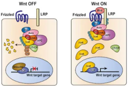
The first discovered WNT signaling pathway involved β-catenin (100). This pathway involves two states, one when the WNT is present, and the other one which occurs without WNT (Figure 4). In the absence of WNT, the ubiquitination and proteosomal degradation of β-catenin is facilitated by the destruction complex which involves proteins GSK3, CK-1, Axin, APC, PP2A, and E3-ubiquitin ligase β-TrCP (101). On the other hand, in the presence of WNT, the destruction complex gets inactivated leading to the accumulation of β-catenin in the cytoplasm and translocation to the nucleus. WNT binding to FZD in the presence of LRP5/6 recruits DVL, which in turn recruits GSK3, CK-1, and Axin to the plasma membrane. The GSK3 then phosphorylates LRP5/6 which leads to accumulation of β-catenin in the cytoplasm (88). It has been suggested that recruitment of the destruction complex disrupts its interaction with β-TrCP, which means that β-catenin is not being degraded but instead accumulates in the cytoplasm (102). This leads to translocation of β-catenin into the nucleus where it interacts with the TCF/LEF transcription factors to induce the transcription of target genes e.g. MYC, SNAIL, CCND1 (88,93).
Figure 4. WNT/β-catenin signaling. Reprinted from Cell; Vol 149, Issue 6, 1245-1256, 2012; Vivian S.W. Li et al.; Wnt Signaling through Inhibition of β-Catenin Degradation in an Intact Axin1 Complex; with permission from
β-catenin-independent WNT signaling pathway
The β-catenin-independent signaling pathway is more complex than the one depending on β-catenin. It involves many different signaling pathways which are classified by the receptor, co-receptor or downstream effector that WNT uses for its signal transduction. The two most studied β-catenin-independent signaling pathways are WNT/Ca2+ and WNT/Planar Cell Polarity (PCP) (Figure 5). Due to
such a variety in signaling pathways, there is quite a good possibility that these pathways overlap (91).
Figure 5. β-catenin-independent WNT signaling. Taken from Linnskog R (2014) (103).
The WNT/Ca2+ signaling pathway was first described in zebrafish
embryos (104) and was suggested to stimulate Ca2+ release in
into activation of phospholipase C (PLC), that stimulates production of diacylglycerol (DAG) and inositol-1,4,5-trisphosphate (IP3) which in turn releases Ca2+ from intracellular stores. Increased free Ca2+
activates three effectors: protein kinase C (PKC; in the presence of DAG) (106), calcineurin (107), or Ca2+/calmodulin-dependent kinase
II (CAMKII) (108). Calcineurin activates the transcriptional factor nuclear factor associated with T cells (NFAT) that translocates to the nucleus and affects genes involved in cell fate and cell migration (107).
The WNT/PCP signaling pathway involves WNT binding to FZD and co-receptors ROR1, ROR2, or PTK7 which leads to activation of DVL. This triggers the activation of small GTPases Rac1, RhoA, and JUN-N-terminal kinase (JNK) which in turn affect actin cytoskeleton and cell polarity (91). The planar cell polarity signaling regulates the cellular orientation according to body axis and was first discovered in Drosophila (109).
WNT5A
Early studies of WNT5A showed that human WNT5A gene was localized to chromosome 3p14-p21 and had the highest homology with mouse WNT5A. Moreover, the mature human WNT5A protein was shown to contain 343 aa residues, giving it a theoretical molecular weight of 38 kDa (110). As with other WNT proteins, WNT5A is post-translationally modified. It has a palmitate attached to one conserved cysteine residue (Cys104) and glycans attached to
four aspargine residues (Asn114, Asn120, Asn311, and Asn325). The
palmitoylation was shown to be important for WNT5A interaction with the CRD of FZD5 and signal transduction whereas glycosylation is important for the secretion of active WNT5A (79). These modifications contribute to a functional WNT5A molecular weight of approximately 43 kDa. Moreover, WNT5A gene was shown to produce two protein isoforms, a short WNT5A-S isoform and a long WNT5A-L isoform. These were shown to differ in their effect on cell proliferation, WNT5A-S promoted whereas WNT5A-L inhibited cell proliferation; however, further studies are recommended to determine their roles in oncogenesis (111).
WNT5A signaling and function
Upon binding to the receptor, WNT5A elicits various downstream signaling pathways, among which, WNT/Ca2+ and WNT/PCP are
the best described. WNT5A was first discovered to be essential for embryonic development (112,113) but later on it was shown to be involved in different diseases, including cancer, by affecting cell proliferation, differentiation, migration, invasion, adhesion, and polarity (114). WNT5A can trigger its signaling through several receptors of the Frizzled family (FZD2, FZD3, FZD4, FZD5, FZD6, FZD7, and FZD8), ROR1, ROR2, PTK7, and RYK (115).
For instance, the interaction of WNT5A with FZD5 was shown to activate PKC and increase the invasion of melanoma cells (116). By inducing Ca2+ release, WNT5A impaired breast cancer cell migration
by activating small GTPase Cdc42 which in turn suppressed the activation of ERK1/2 and MMP9 (117). Similarly, through the activation of Ca2+ and CAMKII, WNT5A was shown to enhance
Notch1 signaling in HEK293 cells (118). Moreover, WNT5A binding to FZD2 and RYK suppressed the activity of MMP2 and invasiveness in human glioma (119), whereas binding to ROR2 led to the activation of DVL that induced MMP13 transcription via the activation of Rac/JNK/c-JUN and ATF in osteosarcoma cells (120). By binding to FZD7 in presence of RYK, WNT5A induced PI3K/AKT signaling that promoted melanoma cell migration and therapeutic resistance (121). Similarly, WNT5A induced the migration of gastric cancer cells by the inactivation of GSK3β and activation of RhoA via PI3K/AKT signaling (122). Moreover, WNT5A was shown to increase the migration of various cell types by the activation of adhesion-dependent FAK (79).
WNT5A interaction with WNT/β-catenin signaling pathway
WNT5A plays a dual role in its signaling since it can both inhibit and activate the WNT/β-catenin pathway. In Xenopus, WNT5A was shown to inhibit the WNT/β-catenin pathway in two ways, by phosphorylation of GSK3 following activation of WNT5A/Ca2+/
PKC, and by interference of the WNT5A/Ca2+/CAMKII pathway
with the transcription factor complex. Thus, inactivation of the WNT/β-catenin pathway by WNT5A leads to the inhibition of
was shown to inhibit the WNT/β-catenin pathway in autocrine manner. Thus, stimulation of the cells with extracellular Ca2+ induced
WNT5A secretion, which in turn triggered signaling through ROR2 that enhanced the degradation of β-catenin by increased SHIAH expression (124). Another study showed that WNT5A could inhibit the WNT/β-catenin pathway through activation of Ca2+/CAMKII and
NLK that interfered with β-catenin-induced gene transcription (125). By interacting with FZD2, WNT5A can inhibit WNT3A-dependent phosphorylation of LRP6 and thus promote the degradation of β-catenin (126), whereas interacting with FZD4 and LRP5 instead leads to the activation of the WNT/β-catenin pathway by stabilization of β-catenin (127).
WNT5A regulation
Early research on WNT5A signaling and expression showed that gene rearrangement or amplification did not cause the increased expression of WNT5A in malignant melanoma and prostate cancer (128). Since then, the research on aberrant WNT5A signaling and function has revealed that the expression of WNT5A can be misregulated in different ways. For instance, IL-6 was shown to upregulate WNT5A expression in malignant melanoma cells by activation of p38-MAPK (129). A recent study on idiopathic pulmonary fibrosis showed that WNT5A expression is upregulated by TGFβ and WNT7B (130) while extracellular Ca2+ upregulated WNT5A in colon cancer (124).
Activation of the NFκB, Hedgehog, TGFβ, and Notch signaling pathways were shown to upregulate WNT5A expression due to the presence of binding sites of their downstream effectors on WNT5A gene promoter (131). In pancreatic cancer, TGFβ was shown to induce WNT5A expression through transcriptional factor CUTL1 (132). In malignant melanoma, the RNA-binding protein HuR was shown to positively regulate WNT5A expression (133) whereas in breast cancer WNT5A expression was suppressed by the same protein (134). Moreover, activation of ARF6 and ERK by EGF was shown to negatively regulate the expression of WNT5A in order to induce EMT in gastric cancer (135). WNT5A expression was also shown to be regulated by epigenetic silencing of WNT5A gene. In esophageal squamous cell carcinoma, WNT5A was silenced by promoter CpG methylation and in that way lost its tumor suppressor property (136).
WNT5A signaling in odontogenesis and oral mucosa
A few studies have reported the involvement of WNT5A signaling pathway in odontogenesis and oral mucosa development. For instance, during formation of dentin and pulp, the overexpression of WNT5A promotes cell adhesion and inhibits cell migration of human dental papilla cells through the activation of RhoA-dependent or RhoA-independent JNK signal (137). WNT5A was shown to regulate odontoblast growth, differentiation and pattering as well as tongue and papilla development through ROR2-mediated inhibition of WNT/β-catenin signaling pathway (138,139).
WNT5A in OSCC
Although many studies have investigated WNT5A signaling and function in different types of cancer, the expression and function of WNT5A in oral cancer is still unclear. Early study on the expression of the WNT family in HNSCC showed that increased WNT5A mRNA expression was found in 4 out of 10 HNSCC cell lines, but not in normal oral squamous epithelial cells (140). In contrast, Uraguchi et al. showed that WNT5A mRNA expression was found in 2 out of 11 HNSCC cell lines, but also in normal keratinocytes and fibroblasts (141). Furthermore, Taki et al. showed that SNAIL-induced EMT upregulated WNT5A gene expression in OSCC cells (142). In vivo studies showed that increased WNT5A mRNA and protein expression was found in human OSCC tissues as well as in rat tongue SCC, whereas its expression was lacking in normal oral epithelium (143-145). Furthermore, Liu et al. showed that interaction of CTHRC1 with WNT5A promotes the migration of OSCC cells through the WNT/PCP pathway by activating RhoA, Rac and JNK (145). In addition, WNT5A was shown to be related to malignant transformation of actinic cheilitis (AC) to lip squamous cell carcinoma (LSCC) (146).
WNT5A and other cancers
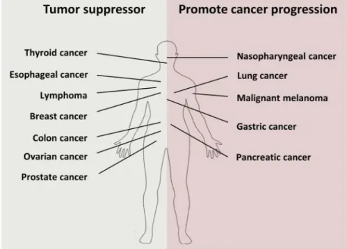
The WNT5A research have revealed that WNT5A is involved in variety of diseases and it has been shown to act both as a suppressor (in colon, breast, prostate, and thyroid cancer) and a promoter (in malignant melanoma, gastric, and pancreatic cancer) of carcinogenesis, depending on the cellular context (Figure 6). For
and can induce migration and invasion by promoting EMT (147). WNT5A acts as a cancer promoter even in malignant melanoma where it induces cell migration through Ca2+/PKC signaling (116,148).
Moreover, WNT5A is overexpressed in malignant melanoma tissues and overexpression correlates with poor outcome (149). In gastric cancer, WNT5A enhances cancer progression by the up-regulation of laminin γ2 (150) and GSK3β phosphorylation (122). WNT5A also promotes the migration and invasion of nasopharyngeal carcinoma cells via PKC (151). Numerous studies have shown the cancer suppressor role of WNT5A in colon and breast cancer. For instance, it has been shown that overexpression of WNT5A induces Ca2+ release and activation of CAMKII in colon cancer cells, which
in turn activates apoptosis and inhibits EMT and proliferation via activation of Bax (152). In breast cancer, on the other hand, WNT5A can suppress the migration of cancer cells by inactivating ERK1/2 and MMP9 via Cdc42 (117). Furthermore, WNT5A suppresses thyroid carcinoma by inducing phosphorylation of β-catenin and suppression of C-MYC via Ca2+/CAMKII (153).
Figure 6. WNT5A as a cancer suppressor and a cancer promoter. Taken from Linnskog R (2014) (103).
WNT5A and cancer therapy
The ongoing research on the functional role of WNT5A during carcinogenesis has made WNT5A a potential target for cancer therapy. A few studies have proposed that WNT5A signaling could be modulated by peptides. Hence, based on the fact that loss of WNT5A expression leads to the increased migration and invasion of breast cancer cells, Safholm et al. developed a small hexapeptide named Foxy5 that possesses anti-migratory effects on breast cancer cells (154). This WNT5A-derived peptide consisting of 6 aa (Met-Asp-Gly-Cys-Glu-Leu) and a formyl group at N-terminal was shown to suppress the invasion of breast cancer cells and to prevent the formation of lung and liver metastases in vivo (154,155). Foxy5 is currently in a Phase 1b study that is currently recruiting participants (www.ClinicalTrials.gov: NCT02655952). The opposite role of WNT5A found in other types of cancer has evoked research into molecules that could antagonize the cancer-promoting role of WNT5A. One such molecule was developed by the same group that developed Foxy5 (156). By replacing the formyl group of Foxy5 with a t-butoxycarbonyl group, a new hexapeptide named Box5 was shown to inhibit WNT5A-induced migration and invasion of melanoma cells (156). However, Box5 as an anti-metastatic target requires further in vitro and in vivo studies.
AIMS
Given the fact that WNT5A can both promote and suppress cancer progression depending on the cellular context, the overall aim of this thesis was to investigate the role of WNT5A in OSCC.
The specific aims were to:
• Investigate the effect of non-canonical WNT5A signaling on OSCC cell migration and invasion.
• Find a reliable commercially available antibody for the detection of WNT5A protein by immunohistochemistry or western blot analysis.
• Investigate if the expression of WNT5A protein is greater in OSCC compared to the adjacent normal epithelium, and dysplasia. • Investigate if WNT5A regulates the expression of the
cell-adhesion proteins E-cadherin and β-catenin.
• Investigate if WNT5A increases the invasion of OSCC by activating MMP2 and MMP9.
MATERIAL AND METHODS
The material and methods used in this thesis are described in detail in the “Material and methods” sections of each individual paper.
Cell lines and treatments
Two oral tongue carcinoma cell lines, SCC9 (CRL-1629) and SCC25 (CRL-1628), used in papers I, III, and IV, were purchased from American Type Culture Collection (ATCC). The molecular characterization of these two cell lines was performed by ATCC in 2008 (SCC9) and 2009 (SCC25). A 1:1 mixture of Dulbecco’s modified Eagle’s medium and Ham’s F12 medium supplemented with 10% fetal bovine serum, 400 ng/ml hydrocortisone, 5 U/ml penicillin, 0.5 U/ml streptomycin and 2 mM L-glutamine was used for the routine maintenance of the cells. Cell culturing and all cell treatments were performed at 37°C and 5% CO2. Prior to experiments in papers III and IV, the cells were cultured in serum-free medium overnight and treated either with 0.1% BSA in phosphate-buffered saline (PBS) as the control or with 0.4 µg/mL recombinant WNT5A (rWNT5A) in serum-free medium. After 48 h treatment, the experiments were implemented with western blot (WB) analysis (papers III and IV), and gelatin zymography (paper IV).
The human mammary carcinoma cell line, MDA-MB468, used in paper II was purchased from ATCC. It was transfected either with an empty vector (468-EV) or a WNT5A-containing vector (468-5A) and maintained as described previously (117).
Tissue samples
Ethical aspects
The experimental design of papers III and IV was approved by The Ethics Committee of the Swedish Southern Health Care Region. Paper II is a quality assurance study that was approved by the Faculty of Odontology, Malmö University, Malmö, Sweden.
Paper II
Based on previous reports on WNT5A expression (157-162), tissues of normal breast, breast cancer, and placenta served as positive controls, while normal liver tissue served as the negative control.
Paper III and IV
Out of 207 patient samples histologically diagnosed as OSCC at the Department of Oral Pathology, Faculty of Odontology, Malmö University, Malmö, Sweden, from 2003 to 2010, 21 unidentified samples were selected for experimental analysis in paper III, and 25 samples for experimental analysis in paper IV. The inclusion criteria for paper III were: the presence of normal epithelium adjacent to the affected tissue (referred to here simply as normal epithelium), dysplasia, and OSCC in the same sample tissue, sufficient patient data, and tissue material. The patient cohort consisted of 12 females and 9 males at a median age of 69. The biopsies were extracted from different locations of the mouth: tongue (n = 10), mandible (n = 4), buccal mucosa (n = 4), trigonum retromolare (n = 1), alveolar ridge (n = 1), and the floor of the mouth (n = 1).
The inclusion criteria for paper IV were: histological diagnosis “oral squamous cell carcinoma”, available TNM data, and technically sufficient sample material. The patient cohort in paper IV consisted of 13 females and 12 males at a median age of 70. The analyzed biopsies were extracted from different locations of the mouth: tongue (n = 10), mandible (n = 7), buccal mucosa (n = 3), trigonum retromolare (n = 1), and alveolar ridge (n = 4).
Cell based experiments (Paper I)
All cell treatments and functional experiments were performed at 37°C and 5% CO2.
Measurements of Ca
2+signaling
SCC9 and SCC25 cells were cultured on glass coverslips in cell culture medium overnight and incubated with the fluorescent calcium indicator, Fura-2-AM (4 µM), for 30 min. The cells were excited at 340 and 380 nm and the fluorescent Fura-2 signal was recorded at 510 nm. The levels of cytosolic free Ca2+ were measured before
and after treatment with 0.4 µg/ml rWNT5A and were expressed as the ratio 340/380 nm. The specificity experiments were performed by pretreatment of the cells with 100 µM Box5 for 40 min before stimulation with rWNT5A in the presence of either of these.
Wound healing assay
Cell migration was analyzed using a wound healing assay since OSCC cells migrate collectively, in sheets (163). SCC9 and SCC25 cells were cultured in 48-well plates in complete cell culture medium until 100% confluence. The cell layers were scratched with a pipette tip, rinsed with PBS to remove detached cells, and incubated with serum-free medium supplemented with or without rWNT5A, Box5, Foxy5, MAPT-AM, GF109203X, or Go 6983 for 48 h. Prior to experiments with MAPT-AM, GF109203X, and Go 6983, the cells were pre-incubated with these for 30 min before scratching. The effect on cell migration was calculated as a percentage of the scratched area at 0 h and after 48 h using the Image-J software.
Measurements of BrdU positive cells
To analyze the possible effects of rWNT5A on cell proliferation during wound healing assay, 10 µM BrdU was added to the cells for the last 24 h of a 48 h incubation with rWNT5A. After 48 h, the experiment proceeded according to the manufacturer’s instructions for Cell Proliferation ELISA, BrdU kit. In brief, the cells were fixed with FixDenat for 30 min, incubated with anti-BrdU-POD for 90 min, and after the addition of a specific substrate, the absorbance was measured.
Cell invasion assay
After culturing, SCC9 and SCC25 cells were re-suspended in serum-free medium and mixed either with 0.4 µg/ml rWNT5A or with the control (0.1 mM EDTA, 0.5% CHAPS in PBS pH 6.8). The mixtures of single cell suspensions were added to the upper chamber of the BD BioCoat Matrigel Invasion Chambers kit, while a complete cell culture medium was added to the lower chamber. After 48 h of cell invasion, the cells were fixed in 4% paraformaldehyde and stained with crystal violet, followed by the removal of the Matrigel and mounting of the membranes on glass slides. All of the intact cells that had invaded the membrane were counted.
Analysis of PKC activity
SCC9 and SCC25 cells were cultured in 6-well plates, and after reaching 80% confluence, the cells were rinsed with PBS and incubated for 2 h with serum-free medium supplemented either with 0.4 µg/ml rWNT5A or with 0.4 µg/ml rWNT5A and 100 µM Box5 together. For the experiments with Box5, the cells were pre-incubated with 100 µM Box5 in serum-free medium overnight. As the control for PKC activation, 10-20 nM of PMA was added to the cells 90 sec prior the end of the 2 h incubation. The experiments were carried out by WB.
Validation of antibodies (Paper II)
WNT5A antibodies
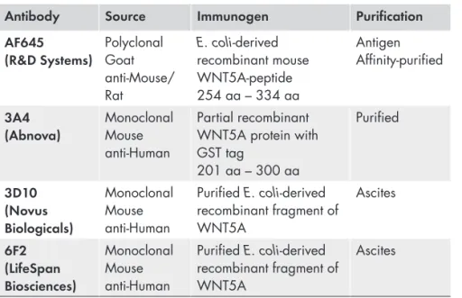
Four different antibodies were evaluated for the detection of WNT5A protein: polyclonal goat anti-mouse/rat WNT5A (AF645) from R & D Systems, monoclonal mouse anti-human WNT5A clone 3A4 (H00007474-M04) from Abnova Corp., monoclonal mouse anti-human WNT5A clone 3D10 (NBP1-47438) from Novus Biologicals and monoclonal mouse anti-human WNT5A clone 6F2 (LS-B3859) from LifeSpan BioSciences. These antibodies are referred to here as AF645, 3A4, 3D10 and 6F2, respectively (Table 1).
Table 1. The characteristics of WNT5A antibodies.
Antibody Source Immunogen Purification
AF645
(R&D Systems) Polyclonal Goat anti-Mouse/ Rat E. coli-derived recombinant mouse WNT5A-peptide 254 aa – 334 aa Antigen Affinity-purified 3A4 (Abnova) Monoclonal Mouse anti-Human Partial recombinant WNT5A protein with GST tag 201 aa – 300 aa Purified 3D10 (Novus Biologicals) Monoclonal Mouse anti-Human Purified E. coli-derived recombinant fragment of WNT5A Ascites 6F2 (LifeSpan Biosciences) Monoclonal Mouse anti-Human Purified E. coli-derived recombinant fragment of WNT5A Ascites
Optimization of IHC protocol for WNT5A antibodies
Three µm thick sections of formalin fixed, paraffin embedded tissues (FFPE) were deparaffinized, rehydrated, and exposed to heat-induced antigen retrieval (HIAR) at 95° C in a Decloaking ChamberTM. Two
buffers, 10 mM citrate buffer, pH 6.0, and TRIS-EGTA buffer (10 mM TRIS, 0.5 mM EGTA), pH 9.0, were evaluated for HIAR. After cooling and washing with TBS-T, the sections were incubated either with background sniper for 10 min, 3% BSA in TBS-T for 30 min, or 2.5% normal horse serum for 30 min at room temperature (RT) prior to addition of primary antibody. The optimal dilution of WNT5A antibodies was obtained by serial dilutions of the antibodies either with 2.5% normal horse serum, 1.5% BSA in TBS-T, or antibody diluent. Following incubation with the primary antibodies for 20 min at RT or overnight at 4° C, sections were incubated with HPBK for 10 min prior to addition of secondary antibodies: ENVISION, horse anti-goat, or rabbit anti-goat for 20 or 30 min at RT. The immunoreaction was visualized using diaminobenzidine (DAB), followed by counterstaining with hematoxylin, dehydration, and mounting. After each step, the slides were washed with TBS-T. The omission of the primary antibody was used as an additional negative control for the secondary antibody. The images were obtained using a Nikon Eclipse 80i light microscope at 400 x.
Pre-absorption test of the primary antibodies
The diluted antibodies were incubated overnight at 4° C with 10 x molar excess of rWNT5A or rWNT3A before application to the tissues and blots. For western blot, the pre-absorption test was performed on two blots loaded with aliquots from the same samples. One blot was probed with the antibody-antigen complex and the other with the primary antibody alone. MDA-MB468 cell lysate supplemented with 4 ng/µl rWNT3A was used as a specificity test for the polyclonal AF645 antibody for WB.
The evaluation of the pre-absorption test for IHC was performed in two steps. First, it was ascertained that identical areas were immunostained in the two consecutive sections used for the test. The results revealed that the stained area of each pair of tissue sections, calculated as IHC stained area (DAB)/total area, were identical. Thereafter, the staining intensities were compared for each pair of tissue sections stained with primary antibody alone or with primary antibody that had been pre-incubated with rWNT5A or rWNT3A. The stained areas and the staining intensities were evaluated using ImageJ software.
Protein expression
Western blot analysis (papers I - IV)
Cell lysis and SDS-PAGE (Papers I, III and IV)
After cell culturing and/or treatments, cells were lysed in ice-cold lysis buffer (20 mM Tris-HCl, pH 7.5, 150 mM NaCl, 30 mM sodium pyrophosphate, 1 mM EDTA, 1.5 mM MgCl2, 0.1 mM sodium orthovandate, 10% glycerol and 1% Triton X-100) supplemented with protease inhibitors (1 tablet Complete mini EDTA-free and 1 tablet PhosSTOP) for 30 min on ice and centrifuged at 14,000 rpm for 30 min at 4°C. After protein estimation, samples containing equal amounts of protein were suspended in 4X Laemmli sample buffer, boiled and loaded on an 8% or 10% sodium dodecyl sulfate polyacrylamide electrophoresis gel (SDS-PAGE). After electrophoresis, the proteins were transferred onto PVDF membranes by semi-dry WB for 1 h.
Paper I
To investigate the expression of WNT5A protein in the two OSCC cell lines, the blots were incubated with polyclonal goat anti-mouse/ rat WNT5A (AF645) antibody diluted 1/200 in 5% (w/v) non-fat skimmed milk powder in TBS-T overnight at 4°C. After incubation with the secondary antibody, the blots were developed with a chemiluminescence HRP substrate. β-actin was used as the loading control.
Paper I - Analysis of PKC activity
To analyze the activation of PKC we investigated phosphorylation of the endogenous PKC substrate named myristoylated alanine-rich protein kinase C substrate (MARCKS). The blots were incubated with P-MARCKS primary antibody diluted 1/500 in 5% BSA in TBS-T overnight at 4°C. The blots were developed with a chemiluminescence HRP substrate. β-actin was used as the loading control.
Paper II
For native WB, cell lysates of MDA-MB468 and 468-EV (negative controls) and MDA-MB468 supplemented with 4 ng/µl rWNT5A and 468-5A (positive controls) were prepared in 2x Tris-glycine native sample buffer (200 mM Tris, 20% glycerol, 0.005% bromophenol blue), pH 8.6, and 30 µg of total protein was loaded on a 10% PAGE without SDS. After electrophoresis, the native blots were incubated in 0.1% SDS for 15 min before semi-dry blotting onto a PVDF membrane for 1 h, excluding SDS. For reducing WB, lysates of the same cell lines as for native WB were prepared in the same lysis buffer as for paper I. After protein estimation, samples were suspended in 4x NuPAGE LDS sample buffer supplemented with 200 mM dithiothreitol (DTT), boiled, and loaded onto a 10% SDS-PAGE. Following semi-dry WB, both native and reduced blots were incubated with four different WNT5A antibodies diluted 1/100 in 3% (w/v) non-fat skimmed milk powder in TBS-T overnight at 4° C. The blots were developed with a chemiluminescence HRP substrate. α-Tubulin antibody was used as the loading control.
Paper III
Following the transfer, the blots were incubated overnight at 4°C with monoclonal primary antibodies: E-cadherin diluted 1/1000 in 3% (w/v) non-fat skimmed milk powder in TBS-T, non-phosphorylated (active) β-catenin diluted 1/1000, or total β-catenin diluted 1/2000 in 3% (w/v) BSA in TBS-T. The blots were developed with a chemiluminescence HRP substrate. α-tubulin was used as the loading control.
Paper IV
After semi-dry blotting, the blots were incubated overnight at 4°C with primary antibodies against p-ERK, ERK, p-p38, and p38 diluted 1/1000 in 3% (w/v) BSA in TBS-T. The blots were developed with a chemiluminescence HRP substrate. The visualized bands were quantified by integrated densitometric analyses using ChemiImager 4400 software. α-tubulin was used as the loading control.
Immunohistochemistry
Paper III and IV
Three µm thick serial sections of FFPE tissues were deparaffinized and rehydrated prior to HIAR with 10 mM citrate buffer, pH 6.0 at 95°C for 20 min (for β-catenin and E-cadherin) or 40 min (for WNT5A) in a Decloaking ChamberTM. The sections were then incubated with
background sniper for 10 min followed by incubation with primary antibodies overnight at 4°C; WNT5A at a dilution of 1/75, β-catenin 1/1000, and E-cadherin 1/400. After incubation with HPBK for 10 min and ENVISION for 20 min, the immunoreaction was visualized with DAB.
Paper III and IV - Evaluation of immunohistochemistry
The protein expression of WNT5A, β-catenin, and E-cadherin was evaluated semi-quantitatively in serial tissue sections of normal epithelium, dysplasia, and OSCC. The expression was assessed with a Nikon Eclipse 80i light microscope with 40× objective magnification in at least two visual fields in two to four cell layers of normal epithelium and dysplasia, and in the periphery of OSCC islands located at the invasive front. The periphery was considered the first or second cancer-cell layer closest to the connective tissue. OSCCs
were categorized as early invasive OSCC (infiltrating islands, clusters, or bands of cancer cells in the lamina propria) or advanced OSCC (islands, clusters, or bands of cancer cells that invaded the underlying muscle layer) (20). Immunostaining for WNT5A, β-catenin, or E-cadherin was defined as positive if >50% of cells were stained (150,164). The intensity of the positive immunostaining was graded as weak (1) or strong (2), and was assessed in cytoplasm and nucleus for WNT5A; in membrane, cytoplasm and nucleus for β-catenin; and in membrane for E-cadherin.
Gelatin zymography - Paper IV
The conditioned cell-culture media from treated cells were collected, gently centrifuged, and kept in -80°C prior to further examination. The cell-culture media from SCC9 cells were concentrated with Amicon Ultra-2 ml centrifugal filter with Ultracel-10 membrane prior to analysis. The conditioned media were analyzed using 10% Zymogram gel with 0.1% gelatin. Following protein separation by electrophoresis, the gels were renatured by removing SDS, incubated in developing buffer overnight and stained with Coomassie Brilliant Blue. The clear bands representing the gelatin degradation by MMPs were quantified by integrated densitometric analysis using the ChemiImager 4400 software.
Statistical analysis
Paper I
The data are presented as means ± standard error of the means (SEM), and statistical analysis was performed using either a two-tailed Student´s t-test or analysis of variance (ANOVA; with Dunnett´s multiple comparison test for post analysis) using GraphPad Prism 5.0 software. All experiments were repeated at least three times.
Paper III
The immunohistochemical data was statistically analyzed with SPSS Statistics 23.0 software. The differences in the expression of WNT5A,
β-catenin, and E-cadherin between normal epithelium, dysplasia, and OSCC within a single patient tissue sample were estimated by two-tailed Wilcoxon signed ranks test. The differences in the expression of WNT5A, β-catenin, or E-cadherin in early and advanced OSCCs were assessed by the Mann-Whitney test. The correlation between the expression of WNT5A and β-catenin or E-cadherin, as well as the correlation between WNT5A protein expression and tumor size (T), were determined by two-tailed Spearman rank correlations. Statistical analysis of the western blot data was performed using the GraphPad Prism 5.0 software. The experiments were performed at least three times and are presented as means ± standard error of the means. The differences in the protein expression between the two groups were determined by the paired, two-tailed Student’s t-test.
Paper IV
Statistical analysis of the zymography and western blot data was done using the GraphPad Prism 5.0 software. The experimental data are presented as means ± standard of means and were determined by a paired, two-tailed Student´s t-test, and were performed at least three times. The differences in the protein expression between two groups were determined by a paired, one-tailed Student´s t-test. The immunohistochemical data was statistically analyzed using the SPSS Statistics 23.0 software. The correlation between WNT5A protein expression and tumor size (T) and between WNT5A expression and differentiation grade were determined by the two-tailed Spearman rank correlation.
RESULTS
Paper I - Migration and invasion of oral squamous
carcinoma cells is promoted by WNT5A, a regulator of
cancer progression
Key findings:
• rWNT5A triggers release of cytosolic free Ca2+ in OSCC
cells
• rWNT5A activates PKC in OSCC cells
• rWNT5A induces migration and invasion of OSCC cells without affecting cell proliferation
• Box5 inhibits rWNT5A-mediated migration of OSCC cells
Activation of WNT/Ca
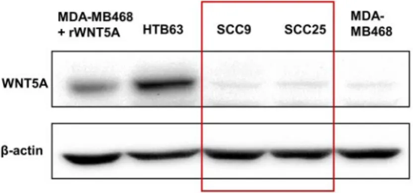
2+signaling
In order to study the functional role of WNT5A in OSCC, we first examined the endogenous expression of WNT5A protein in SCC9 and SCC25 by WB. Because we could not detect any expression of WNT5A in these two cell lines (Figure 7), we stimulated the cells with rWNT5A, which elicited an immediate increase in the cytosolic free Ca2+ levels (Figure 8A). This rWNT5A-induced Ca2+ response was
suppressed by the WNT5A inhibitor, Box5 (t-boc-Met-Asp-Gly-Cys-Glu-Leu hexapeptide) (Figure 8B).
Figure 7. Loss of WNT5A protein expression in OSCC cells, SCC9 and SCC25.
Figure 8. A) Increase of cytosolic free Ca2+ levels after stimulation of OSCC
cells with 0.4 µg/ml rWNT5A presented as the ratio of the Fura-2 signals generated by excitation at 340 and 380 nm. B) Pre-incubation of OSCC cells with 100 µM Box5 for 40 min prior to stimulation with 0.4 µg/ml rWNT5A led to a significant decrease in cytosolic free Ca2+ response.
Influence of rWNT5A on cell proliferation, migration,
and invasion
Next, we examined the effect of the rWNT5A-induced non-canonical WNT/Ca2+ signal on cell migration and proliferation in a wound
healing assay. rWNT5A had no effect on the number of BrdU positive cells in either of the cell lines. However, we noticed a substantial increase in cell migration after 48 h stimulation with rWNT5A. This rWNT5A-induced effect on cell migration was confirmed by
Instead, when we stimulated the cells with rWNT5A in the presence of Box5, we noticed that the rWNT5A-induced migration was abolished in both cell lines (Figure 9). Moreover, rWNT5A elicited cell invasion only in SCC25 (Figure 10).
Figure 9. Box5 (100 µM) significantly decreased rWNT5A-induced migration of OSCC cells.
Figure 10. Invasion of SCC9 (A) and SCC25 (B) cells in the absence or presence of rWNT5A (0.4 µg/ml).
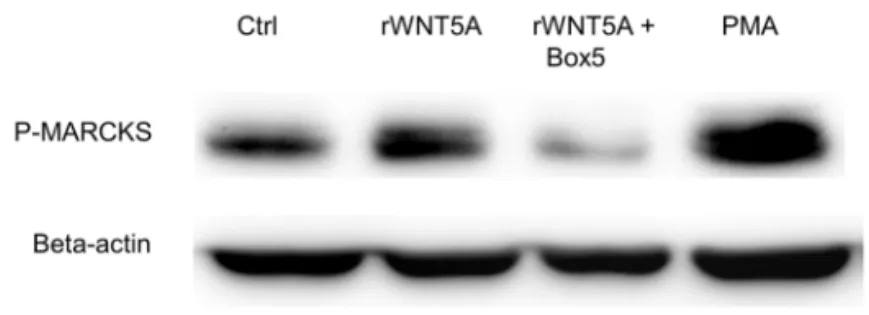
Influence of rWNT5A on PKC activation
To examine the activation of PKC in OSCC cells, we measured the phosphorylation of the endogenous PKC substrate, MARCKS, after treating the cells with rWNT5A alone or in the presence of Box5. The phosphorylation of MARCKS was induced by rWNT5A but suppressed by Box5 (Figure 11). WNT5A/Ca2+/PKC-mediated cell migration was
eliminated when we treated the cells with rWNT5A in the presence of MAPT-AM and two PKC inhibitors, GF109203X which inhibits PKC α, β1, β2 and γ, and Go 6983 which inhibits α, β, γ, δ and ζ the main PKC isoforms activated by WNT5A (116,148) (Figure 12).
Figure 11. rWNT5A (0.4 µg/ml) increased the activation of PKC (phosphory-lation of MARCKS) in OSCC cells which was inhibited by 100 µM Box5.
Paper II - Optimization, validation and identification of two
reliable antibodies for immunodetection of WNT5A
Key findings:
• IHC protocol should be optimized for each antibody to avoid unspecific background staining
• Polyclonal AF645 antibody detected WNT5A in WB • Pre-absorption test with AF645 did not alter any change in
cytoplasmic immunostaining but it reduced the appearance of WNT5A protein band
• Pre-absorption test with 3A4 reduced cytoplasmic immunostaining
• WB is not always adequate for the validation of antibodies intended for use in IHC
Immunostaining patterns for WNT5A expression
At first, we evaluated and optimized IHC protocols for each of the four investigated WNT5A antibodies by examining different HIAR methods with regards to buffer, incubation time, and temperature, different nonspecific background blocking solutions, and secondary antibodies (Table 1).
Applying the optimized IHC protocol on WNT5A-positive and WNT5A-negative tissues revealed a distinct variation of WNT5A immunostaining patterns with the investigated antibodies. The polyclonal AF645 antibody displayed mostly perinuclear and cytoplasmic but also some nuclear WNT5A immunostaining in positive controls. In the negative control tissue, considerably less cytoplasmic, perinuclear, and nuclear immunostaining was observed with AF645 antibody than in positive tissues. The three monoclonal antibodies, 3A4, 3D10, and 6F2, detected exclusively cytoplasmic WNT5A immunostaining in the positive control tissues. There was
no presence of WNT5A immunostaining in the negative control with the 3A4 antibody, while clear cytoplasmic staining was observed with 3D10 and 6F2 antibodies (Figure 13).
Figure 13. WNT5A immunostaining pattern of normal breast tissue with four antibodies.
Validation of IHC by WB
Next, we evaluated the antibodies by WB using WNT5A-positive and WNT5A-negative cell lysates. The WNT5A protein band at 43 kDa was exclusively identified in the WNT5A-positive cell lysates by the polyclonal AF645 antibody. Multiple protein bands were detected by the other three antibodies but none at 43 kDa. The WNT5A protein band was not detected by any of the antibodies under nonreducing conditions.
Specificity of immunoreactivity
To investigate the specificity of the antibodies, we performed pre-absorption tests. Pre-incubation of the AF645 antibody with
perinuclear and nuclear immunostaining in positive control tissues (Figure 14). However, we observed a reduced appearance of WNT5A protein band in WNT5A-positive cell lysates after pre-incubation with rWNT5A (Figure 15). Moreover, WNT3A protein band was not detected by AF645 antibody in MDA468-rWNT3A cell lysate. When we pre-incubated the 3A4 antibody with rWNT5A and rWNT3A, the intensity of cytoplasmic immunostaining was reduced by 89% and 39%, respectively (Figure 16).
Figure 14. The intensity of the cytoplasmic, perinuclear and nuclear immu-nostaining was not reduced after pre-incubation of AF645 antibody with rWNT5A.
AF645 AF645 1:10 rWNT5A
Figure 15. Reduced intensity of WNT5A protein band in WNT5A-positive cell lysates after pre-incubation of AF645 antibody with rWNT5A.
Figure 16. The intensity of cytoplasmic immunostaining was reduced after pre-incubation of 3A4 antibody with rWNT5A.
Paper III - Higher expression of WNT5A protein in oral
squamous cell carcinoma compared to normal oral
epithelium and dysplasia
Key findings:
• WNT5A is not expressed in normal oral epithelium or in mild grade of dysplasia
• WNT5A expression was detected in moderate and severe grades of dysplasia, and was even higher in OSCC • WNT5A did not cause any change in β-catenin or
E-cadherin expression in OSCC cells
Histopathological findings
The clinicopathological characteristics of our patient cohort are presented in Table 2. Within the group, 23.8% had mild grade of dysplasia, 38.1% moderate, and 38.1% severe. The TNM staging at the primary surgery revealed that most of the OSCCs were at stage T1 (52.4%) and stage T2 (28.6%). Within the group, 81% of the patients had well-differentiated OSCCs, 9.5% had moderately-differentiated and 9.5% had poorly-differentiated OSCCs. We classified 12 samples as early invasive OSCCs and 9 samples as advanced OSCCs.